Review

Management of Pheochromocytomas and Paragangliomas: A Case-Based Review of Clinical Aspects and Perspectives

1
Student Scientific Circle of Oncology and Radiotherapy, Medical University of Gdansk, 80-214 Gdansk, Poland
2
International Research Agenda 3P Medicine Laboratory, Medical University of Gdansk, 80-210 Gdansk, Poland
3
Department of Oncology and Radiotherapy, Medical University of Gdansk, 80-214 Gdansk, Poland
*
Authors to whom correspondence should be addressed.
J. Clin. Med. 2022, 11(9), 2591; https://doi.org/10.3390/jcm11092591
Submission received: 17 March 2022 / Revised: 30 April 2022 / Accepted: 1 May 2022 / Published: 5 May 2022
(This article belongs to the Special Issue Neuroendocrine Tumors: Challenges and Future Perspectives)

Abstract

:
Paraganglioma and pheochromocytoma are rare medical conditions. Thus, there are still a small number of studies, clinical trials, and evidence-based data in this field. This makes clinical decisions more difficult. In this study, we present a case report enriched with a short review of available essential clinical data, indicating the need for constant metoxycatecholamine level observation and a proper diagnostic imaging approach, especially in terms of ongoing pandemics. Our research also provides a summary of the molecular background of these diseases, indicating their future role in clinical management. We analyzed the ClinicalTrials.gov dataset in order to show future perspectives. In this paper, the use of the PET-CT before MRI or CT is proposed in specific cases during diagnosis processes contrary to the guidelines. PET-CT may be as effective as standard procedures and may provide a faster diagnosis, which is important in periods with more difficult access to health care, such as during the COVID-19 pandemic.

Graphical Abstract

1. Introduction

Pheochromocytoma (PCh) is a tumor located in an adrenal gland, whereas paraganglioma (PGL) is an extra-adrenal tumor that is commonly formed near blood vessels and nerves [1]. The nomenclature of these rare neuroendocrine tumors (NETs) depends on primary location, as they lack relevant differences in their histology [1,2]. Both PCh and PGL have similar prognoses—their five-year overall survival (OS) is evaluated to be approximately 95% in nonmetastatic disease and 34–60% in recurrent or metastatic neoplasms [3]. At the same time, the incidence rate of PCh and PGL is rising [4,5,6]. This may be associated with better access to cross-sectional imaging; hence, a 28.1% greater detection rate of PCh and PGL was observed between the periods of 1995–2004 and 2005–2016 [7,8].
The main risk factors include genetic predisposition, age between 30 and 50, race/ethnicity (the tumors are less common in African Americans), and Carney triad (a nonhereditary multitumoral syndrome that affects young women) [9,10]. Hypoxia may also play a crucial role in PCh and PGL tumorigenesis, and every 500 m increase in elevation contributes to an 0.31/100,000 increase in the prevalence of these tumors.
As PGL may present similar symptoms to PCh when secreting catecholamines, the differentiation of the tumors can be challenging. However, if it is hormonally inactive, it might be asymptomatic, even in advanced stages. The most common symptoms are tachycardia, hypertension, headache, paleness, excessive sweating, and anxiety (Figure 1) [1]. The most common location of catecholamine secreting PGL is the abdomen and pelvis [1]. Head-and-neck PGL tumors are rarely hormonally active; thus, symptoms are associated with their location [11]. PGL may cause a mass effect in the neck, and if more advanced, it may lead to dysphonia, dysphagia, or neck pain [11]. Moreover, glomus tympanicum tumor and temporal paragangliomas manifest through hearing impairment, tinnitus, and balance disturbance [11]. Glomus jugulare, glomus vagale, and carotid body tumors can present with cranial nerve (IX-XII; X-XII; X, XII) palsies, respectively [11]. The purpose of this review was to provide a relevant summary of PCh and PGL management, especially in the clinical setting, and to discuss the current guidelines in the context of the presented case report.

2. Case Report

In January 2018, a 38-year-old woman was admitted to the surgical department for open resection of a solid-cystic lesion (94 × 80 × 70 mm), discovered in regular ultrasound examination and further revealed in computed tomography (CT). The tumor was localized in the tail of the pancreas with potential involvement of surrounding tissues, including the adrenal glands. CA125, CEA, and Ca19.9 were within normal ranges. On her first admission to the clinic (a day before planned surgery), she presented with heart palpitations, headache, blood hypertension, and petulance. The patient’s only comorbidity was Hashimoto’s disease (HD). Her family history was positive for HD but negative for cancer. The tumor was completely removed, along with its capsule and the whole left adrenal gland. The remaining part of the pancreas was surgically released and secured. The histopathological examination confirmed the PCh diagnosis. The tumor was characterized by the presence of highly atypical cells, division figures (5/50 high power fields), positive chromogranin A (CgA) and synaptophysin (Syn), and partially positive S-100 protein, as well as a Ki67 index of 1%.
After the surgery, our patient was initially followed up by the surgical outpatient clinic. Unfortunately, the next follow-up at an endocrinological outpatient clinic was canceled, due to the lack of emerging symptoms and prolonged waiting time for an endocrinological consultation (which, in Poland, is 1–2 years) [12]. In January 2020, due to the appearance of similar symptoms as those before the surgery (such as heart palpitations and anxiety), the patient ended up in a private health care clinic. After endocrinological consultation and laboratory tests, further care was recommended in an oncological outpatient clinic. At that time, levels of metoxycatecholamines in the patient’s 24 h urine test were as follows: metanephrine—550 µg/24 h (reference limit: 74–297 µg/24 h), 3-methoxytyramine—1047 µg/24 h (reference limit: 94–400 µg/24 h), and normetanephrine—6902 µg/24 h (reference limit: 105–354 µg/24 h). These results, together with the patient’s clinical symptoms, raised suspicion of recurrence. Somatostatin receptor scintigraphy and computed tomography (SPECT) focused on the head, chest, abdomen, and pelvis were performed and showed no signs of local or distant relapse. Moreover, positron emission tomography (PET) was ordered in search of a secondary lesion. However, due to the COVID-19 pandemic and national lockdown in Poland, our patient refused to perform PET as soon as possible; hence, the procedure was postponed until August 2020. In June 2020, the patient performed another full-body Tc-99-octreotide somatostatin receptor scintigraphy and CT, but the radiological picture was stationary (no sign of relapse). Finally, 68GA-DOTATE PET (combining somatostatin analog tyrosine-3 octreotate with the PET tracer gallium Ga68) was performed, and the new highly metabolically active lesion of the neck and the skull base was detected (standardized uptake value: 15.1) (Figure 2A,B). Further, the patient was admitted to the Department of Oncology and Radiotherapy in September 2020. In order to precisely assess local advancement before the planned treatment, a magnetic resonance (MR) of the head and neck with intravenous contrast was performed. A polycystic tumor putting pressure on the left tonsil of the cerebellum (without infiltration or edema) was observed. The tumor infiltrated the left sublingual nerve area and reached the left internal carotid artery canal. Moreover, it closed the left internal jugular vein in the inferior bulb region (Figure 2C). Based on clinical and radiological images, unresectable PGL (Fishers’ D1) was diagnosed. Hence, from October to November 2020, radical volumetric arc radiotherapy (RT) was undertaken within the area of the tumor and the base of the skull, with adequate margins (Varian, TrueBeam linac; 6 MV photons; D = 54 Gy/27 fr). The treatment tolerance was good; the patient suffered from grade 1 mucositis (Common Toxicity Criteria version 5) and pain for 4 weeks after RT. Four months after RT, in March 2021, significant biochemical regression in the 24 h urine test was observed (metanephrine—88.9 µg/24 h; 3-methoxytyramine—356.5 µg/24 h; normetanephrine—1858.2 µg/24 h), together with a reduction in the initial symptoms (palpitation, petulance). Until now, the patient’s general condition is good and is monitored in our clinic every 3 months. The levels of metoxycatecholamines are slowly decreasing, and in August 2021, the lab tests revealed the following results: metanephrine—65.1 µg/24 h, 3-methoxytyramine—224.8 µg/24 h, and normetanephrine—1180.5 µg/24 h. We summarized the changes in clinical findings in Table 1.

3. Genetic Background

Genetics plays a major role in the characteristics of PCh and PGL and has an impact on tumor location, clinical symptoms, hormonal activity, and characteristic imaging phenotype [13,14]. Currently, there are numerous PCh- and PGL-associated genetic syndromes reported in the literature such as MEN-2 (germline mutation in proto-oncogene RET), von Hippel–Lindau (mutation in suppressor gene VHL), neurofibromatosis type 1 (mutation in suppressor gene NF1), PGL syndromes 1–5 (mutations in succinate dehydrogenase subunits SDHD, SDHAF2, SDHC, SDHB, and SDHA, respectively), and hereditary syndromes (mutation in transmembrane protein 127 and MYC-associated factor X genes) [1]. Jochmanova et al. divided all these mutations in PCh and PGL into three clusters according to functional phenotypes, i.e., pseudohypoxic signaling, kinase signaling, and wingless and Int-1 (Wnt) signaling clusters [14]. Interestingly, the expressions of genes belonging to each group had similar clinical images. We divided the genes into three groups and summarized all clinically important data found in the literature in Table 2.

4. Diagnosis

4.1. Measurement of Metanephrines
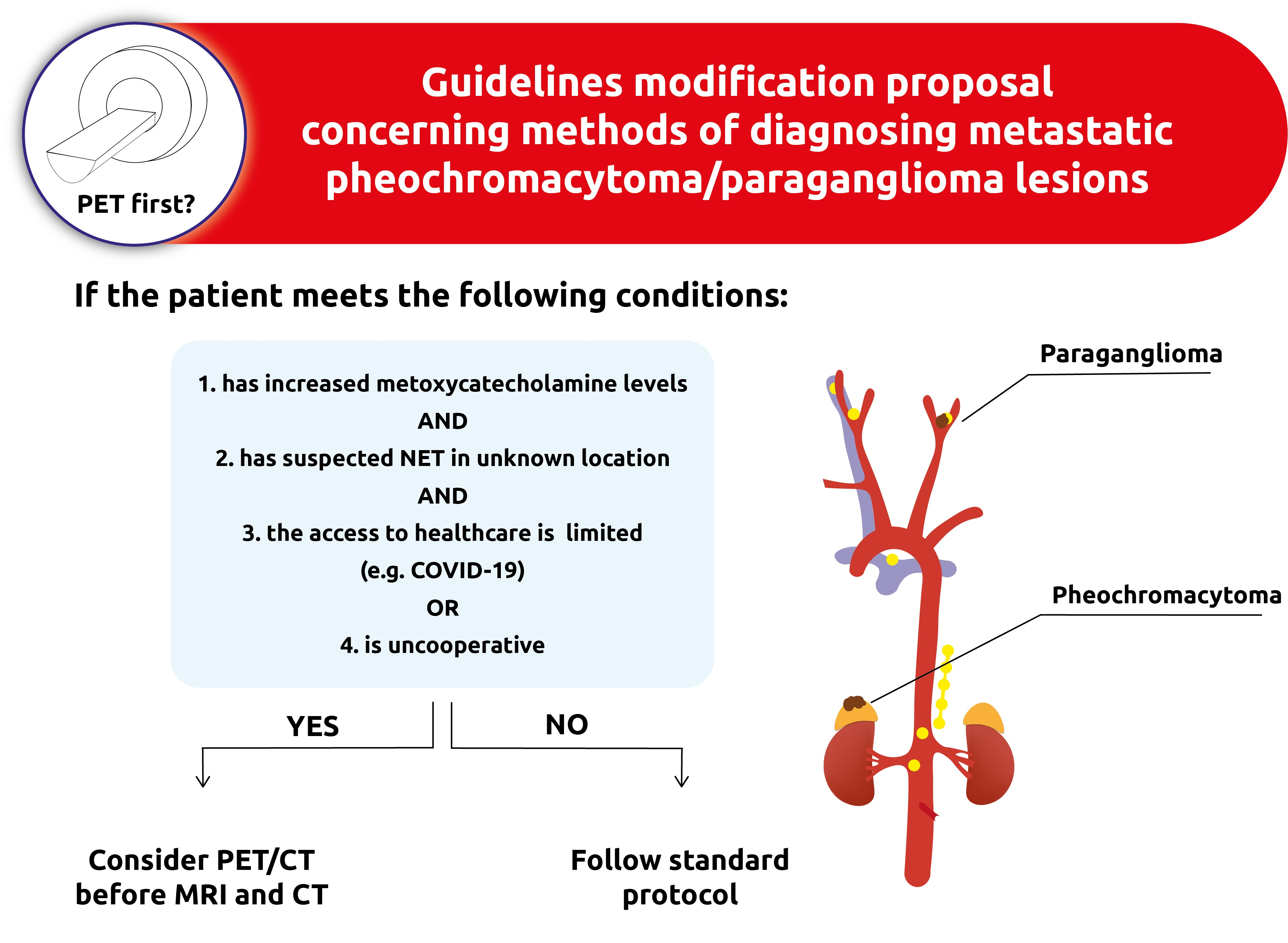
The diagnosis of PGL tumors should be based on a plasma or urinary metanephrines analysis, as it is a quick and noninvasive approach. The patients can be divided into two groups based on the risk of PCh or PGL development, i.e., a high-risk group (harboring paternal mutation) and a low-risk group (those who inherited a pathogenic variant influencing the maternal gene). Although, in general, there is no significant difference in the diagnostic value between plasma and urine, plasma is the preferred material among high-risk patients, since it shows higher specificity and sensitivity in comparison to urine analysis [32]. In dopamine-secreting PGL, plasma 3-methoxytyramine level should also be measured, as it is a specific marker of this type of tumor [32]. We summarized the information about diagnosis with metanephrine detection from Szosland et al. study and updated the recommended method with the results of Därr et al., as shown in Table 3 [33,34].
Usually, in clinical practice, a 24 h urine test for deconjugated metanephrines (though it gives numerous false-positive results) and measurement of free metanephrines in plasma are performed [32]. Därr et al. reported that the most accurate method of measurement for metanephrines is high-performance liquid chromatography (HPLC) with both coulometric/electrochemical or tandem mass-spectrometric methods, as it provides a sensitivity of 94% and specificity of 93% [34]. Interestingly, a supine position during blood sample collection provided significantly higher sensitivity in comparison to a seated position (not observed for 24 h urine test), whereas specificity was higher in a group with samples collected in the sitting position than in a group with performed 24 h urine test (without relevance of position during sample collection) [34]. To summarize, the best method seems to be the measurement of plasma concentration collected in the supine position. The level of catecholamines can be affected by different drugs, e.g., tricyclic antidepressants, serotonin, norepinephrine reuptake inhibitors, and levodopa. When the increase in metanephrines level is less than fourfold above the upper reference limit, it is beneficial to confirm the diagnosis with a clonidine suppression test, which showed a sensitivity of 100% and specificity of 96% [35].

4.2. Imaging

Regarding imaging methods, ultrasonography may play an essential role as the preliminary tool that detects up to 80–90% of PGL tumors [35]. However, despite its high sensitivity, it provides low specificity, which makes it impossible to profile the tumor and plan the proper treatment. According to the Endocrine Society Clinical Practice Guidelines, 2014 CT ought to be performed on the majority of PGL patients to provide the highest detection rate and images that are appropriate for treatment planning. On the other hand, in the case of the skull base and neck PGL, magnetic resonance imaging (MRI) is recommended. This statement is also supported by the Working Group of the European Society of Hypertension guidelines, which confirm that an MRI is a first-line imaging method for head-and-neck tumors, whereas CT is recommended for tumors located in the rest of the body. When disseminated PGL is suspected, it is advised to use 123I-metaiodobenzylguanidine (MIBG) scintigraphy. However, 18 F-FDG PET/CT should be performed in patients with certain types of metastatic PCh and PGL [36]. In the 2014 guidelines, 123I- MIBG was recommended, whereas, in 2020, its value was considered limited. Nowadays, 18F-FDOPA PET and 68Ga-DOTA-somatostatin analog (SSA) are the most preferable techniques [32,36]. Another available method is somatostatin receptor scintigraphy, known as octreotide scan (111In-pentetreotide). However, due to high cost, high radiation exposure, and lower sensitivity and specificity in comparison with other methods, its use is rather limited [37].

4.3. Clinical Presentation Associated with Genetic Background

In general, clinical presentations of PCh and PGL are comparable and depend on hormonal activity and primary localization. However, the presence of each or both tumor types may be a part of multiple hereditary genetic syndromes, which makes diagnosis easier. The most frequent mutations are SDHB—10%, SDHD—9%, VHL—7%, RET—6%, and NF1—5–7% (Table 2) [24,25,26,27,28,29,30,31]. Familial PGL (FPGL) is divided into five subtypes, with impaired tumor suppressor function from which FPGL-4 and FPGL-1 are most frequent. PGL tumors are localized in the abdomen, pelvis, mediastinum, and head-and-neck region. In the second type of von Hipple–Lindau syndrome, PCh is present alongside hemangioblastomas in the retina or central nervous system, clear-cell renal-cell carcinoma, endocrine neoplasia, and pancreatic cancer in different combinations. Proto-oncogene mutation in RET gene is associated with three subtypes of multiple endocrine neoplasia 2 (MEN2) syndromes. In both MEN2A and MEN2B subtypes, PCh may occur with medullary thyroid carcinoma and hyperthyroidism. Additionally, amyloidosis of the skin is present in MEN2A, whereas multiple mucosal neuromas and Marfan-like syndrome are common for MEN2B syndrome. Finally, in NF-1 cafe-au-lait macules, neurofibromas around the head and neck, optic glioma, lish nodules, osseous lesion, and PCh can be observed frequently [16].

4.4. Histopathology

Histologically, PCh and PGL are very similar and often indistinguishable. However, some minor differences in cellular structure can be highlighted. PCh cells are characterized by higher nuclear atypia and cytoplasmic basophilia in comparison to eosinophilic PGL cells. Frequently use immunohistochemistry markers are neuron-specific enolase (NSE), S-100 protein, Syn, CgA, cytokeratin, n (CK), and vimentin. Nonetheless, their utility is questionable. S-100 protein may be used to identify Zellballen nests; however, its occurrence was reported in both PCh and PGL [38,39]. Furthermore, in PGL, all the above markers were tested to distinguish benign and malignant lesions. Among others, CK and vimentin seemed to be more frequently expressed in benign tumors (22.2% versus 5.3% and 76.9% versus 12.5%, respectively) [39]. With recent advances in targeted sequencing, the role of immunohistochemistry staining seems to be outdated. Currently, genetic profiling of these tumors with the new generation-sequencing (NGS) method should be performed, as it provides detection of all mutations at the same time, with higher specificity and sensitivity [38].

4.5. Differential Diagnosis

PCh and PGL—despite their molecular subtypes—need to be distinguished from other tumors such as subclinical PCh, lipid-poor adenoma, and adrenocortical carcinoma. The first refers to a disease that is not advanced enough to be present with severe clinical symptoms; however, subtle hormonal activity can be identified [40]. Adenomas are benign tumors without the need for special management when detected. To distinguish them from PCh with high specificity, a biphasic, contrast-enhanced CT scan should be performed. PCh, when compared with lipid-poor adenomas, is larger, shows cystic degeneration, and presents higher values in unenhanced, arterial, and venous phases, while the enhancement ratio in arterial and venous phases is lower [41]. Finally, the enhancement loss between PCh and adrenocortical carcinoma is similar; thus, clinical–non-radiological features are key to distinguishing them properly [42]. Most adrenocortical malignant tumors are diagnosed with Cushing’s syndrome and signs of virilization. Additionally, they tend to be much more aggressive, with rapidly increasing clinical symptoms [43,44].

5. Treatment

5.1. Local Treatment of Early Disease

The first step in catecholamine secreting PCh and PGL management is the administration of α-adrenergic blockers to minimize adverse cardiovascular effects before surgery (nonselective α-blocker phenoxybenzamine is the drug of choice) [45]. For laparoscopic resection of PCh, there is no significant difference in intraoperative hemodynamics, complication rates, and the length of stay in a hospital between groups treated with nonselective and selective α-blockers. However, patients with α-1 selective drugs have a higher risk of transient hypertension during surgery and a greater need for postoperative support [46,47]. To avoid reactive tachycardia caused by α-blockers, the use of β-blockers is recommended (except labetalol) after administering the α-blocker. Prolonged oversecretion of catecholamines reduces blood volume; hence, it is advised to raise sodium and fluid intake perioperatively [36]. The minimally invasive laparoscopic approach is preferable in the surgical treatment of unilateral PCh (≤6 cm) and PGL, with no infiltration of adjacent tissues or metastasis. In cases of bilateral PCh (e.g., in MEN2, VHL), cortical sparing adrenalectomy of one of the glands should be performed [48]. Open surgery should be performed in the case of PCh > 6 cm and PGL located near the aortocaval region [49]. Cervical and intracranial PGL may require more complex techniques due to unfavorable location [50]; therefore, surgery remains the gold standard, and available data show that only complete resection can be potentially curative. Unfortunately, this procedure may be complicated and burdened with a high risk of complications. Embolization may be used as a preoperative therapy to reduce the size of the tumor and minimize the bleeding during an operation—it is indicated in large PGL and PGL of the jugular vein and should be performed 2–3 days before the surgery [51]. If there are technical or medical contraindications for surgery, radiotherapy remains the standard of care, with high local control. Stereotactic radiosurgery is recommended for skull-based tumors smaller than 3 cm [32]. For larger lesions, fractionated and highly conformal techniques are preferable [52]. If the resection was partial, radiotherapy may also be used as a postoperative therapy [53].

5.2. Treatment of Metastatic or Relapsed Disease

Unfortunately, approximately 10% of the patients develop metastases, and 6.5–16.5% experience local relapse [50]. In such cases, a 131I-MIBG therapy may be considered to control tumor growth and the patient’s symptoms. Notably, 131-MIBG is a form of therapy that depends on specific receptors. MIBG, which is an analog of guanethidine, shares structural similarity with norepinephrine and can bind to a receptor for this hormone [54]. Then, MIBG is transported to secretory granules or remains in the cytoplasm. In therapy, MIGB is conjugated with I-131, a radionuclide with meaningful β-particles emission. This emission accounts for cell damage due to their high mean energy and energy deposition [54]. A meta-analysis of 17 studies including 243 patients showed that stable disease (SD), and partial hormonal response may be obtained in over 50 % of patients with PGL and over 40 % with PCh [55]. In another study, on 131I-MIBG, Pryma et al. showed that it enables sustained blood pressure control and high 12-month tumor response rates (92% partial response (PR) or stable disease (SD); 68% PR or SD with a decrease in serum chromogranin levels) [56]. A study of long-term outcomes (median survival time from diagnosis was 11.5 years), conducted by Thorpe et al., reported that imaging, laboratory, and symptomatic response on treatment were obtained for both PCh and PGL [57]. Another potential option is a peptide receptor radionuclide therapy (PRRT), with lutetium-177 (177Lu)-labeled DOTA-Tyr3-octreotate (DOTATATE; oxodotreotide), yttrium-90 (90Y), or 177Lu-labeled somatostatin analogs (SSAs), which is intended for patients with metastases or patients disqualified for surgery [50,58]. Patients with metastatic PCh and PGL may receive chemotherapy (a combination of cyclophosphamide, vincristine, and dacarbazine is recommended) to alleviate the symptoms, although with poor effectiveness [45]. Another way of palliative treatment is an ablation of the metastatic tumors—patients with bone, chest-wall, and retroperitoneal metastases can undergo radiofrequency ablation or cryoablation, whereas patients with liver metastases should be treated with radiofrequency ablation or ethanol injection [48]. RT is also an option for symptomatic metastases regardless of the location of the tumor.

6. Future Directions in PGL Treatment

Treatment for malignant PCh and PGL tumors has still limited value. Thus, many clinical trials seeking a more targeted approach are being conducted [59]. Currently, 65 clinical trials are registered on ClinicalTrials.gov. In this section, we describe molecularly targeted agents and immunotherapy that could be relevant, as well as future therapeutic options for PCh and PGL management.

6.1. Molecular Targeted Agents

6.1.1. Everolimus

Everolimus is a inhibitor of the mTOR pathway – a promising target in future therapies. To maximize its efficiency, the combination treatment was proposed, and subsequent studies on cell lines were conducted. Nölting et al. showed that everolimus in combination with lovastatin was significantly more effective (additive effect on inhibition) than everolimus alone [60]. In addition, in mouse PCh MPC and MTT cell lines, this combination inhibited EGFR and AKT signaling in the most prominent way [60]. Another study confirmed these results, revealing that treatment with everolimus and lovastatin significantly inhibits AKT and mTORC1/p70S6K signaling, without ERK upregulation [61]. Moreover, Fankhauser et al. sought to combine everolimus with BYL719 (PI3Ka inhibitor, alpelisib), earlier evaluated in breast cancer [62,63]. Although both inhibitors decreased cell viability in a relevant way, the combination was more effective (additive effect) [62].
The efficacy of mTOR inhibitors monotherapy was reported in the literature. Do-Youn et al. conducted a phase II study of everolimus in monotherapy in a heterogeneous group of patients with nonfunctioning NETs (n = 27) in which five had PCh, and two had PGL tumors. In this group of seven patients, five achieved SD, and two developed progressions (PDs). The median progression-free survival (PFS) was 3.8 months, and in four patients, radiological regression was observed [64]. Although the response was high in NETs, it was limited in both PCh and PGL [64]. Nevertheless, the data from such a small number of subjects are inconclusive and should be treated carefully. Furthermore, it is proved that, in both primary and metastatic PCh, the mTOR pathway is significantly dysregulated, with a preferential overactivation in head-and-neck PGL or in the group of patients with SDHX mutations (suggested by Oudijk et al.) [65,66,67,68,69,70]. However, inhibition of mTORC1 alone may not be enough because of compensatory activation of PIK3K/AKT and EGFR-related pathways, or RAS/RAF/ERK signaling [60,71,72,73,74,75].

6.1.2. Axitinib

Axitinib, which is a VEGF receptor tyrosine kinase inhibitor previously investigated in renal-cell carcinoma, was assessed in metastatic, recurrent, or primary unresectable PCh or PGL tumors [76]. In the group of 12 patients, 5 achieved PR, 5 had SD, and 2 developed PD. The median of PFS was 7.7 (3.3–16.8) months.

6.1.3. Cabozantinib

Cabozantinib is a multiple tyrosine kinases inhibitor of MET, RET, VEGFR2, and AXL [77]. One of the registered studies focuses on cabozantinib s-malate alone, whereas another study, on cabozantinib in combination with atezolizumab (CABATEN). Until now, the results of a phase I study on the use of cabozantinib in children and adolescents have been published. However, only one patient with PGL was included, achieving SD [77].

6.1.4. Sunitinib and Sorafenib

Another promising drug is sunitinib, which is currently under investigation, with many studies supporting its role in gastrointestinal stromal tumors, advanced renal-cell carcinoma, and pancreatic NETs. Sunitinib is an orally administered drug and multitargeted inhibitor of receptor tyrosine kinases (RTKs) that mainly affects VEGFRs, PDGF-Rs, FLT3, RET, and mTOR signaling [78,79]. However, Saito et al. hypothesized that the effect of sunitinib is associated with angiogenesis inhibition as well as with direct antitumor effects [79].
Surprisingly, this drug-induced apoptosis of PC12 cell lines reduced the expression of antiapoptotic protein Bcl-2 activation of proapoptotic BAD and, crucially, led to an inhibition of the above-mentioned Akt and mTOR- related pathways, followed by a reduction in S6K1, the target of mTOR signaling [79]. Additionally, studying a xenograft model in mice, Denorme et al. confirmed the antiangiogenic activity of sunitinib and sorafenib. In addition, the numbers of apoptotic cells in vitro and in vivo in the treated tumors were higher, for both sunitinib and sorafenib, than those in control tumors, suggesting the direct effect on cells of these drugs [80].
Although results from basic models are promising, a phase II clinical trial (SNIPP) did not show considerable changes. In a group of 25 patients, the disease control rate was 83%, and the median PFS was 13.4 (range—5.3–24.6) months. Moreover, 3 patients achieved PR (all these patients had confirmed germline mutation), 16 achieved SD (>12 months), 4 developed PD, and the status of 2 patients is unknown [81].

6.1.5. HIF Inhibitors

One of the promising but still weakly investigated drugs in PCh and PGL are hypoxia-inducible factor inhibitors (HIFs). Recent studies proved the potential feasibility and efficacy of HIF-2a inhibitors in the treatment of patients with clear-cell renal-cell carcinoma (RCC) (phase I, III, III of clinical trials) [18]. Due to similar molecular drivers present in RCC and both PCh and PGL, HIFs seem to be reasonable targets in these neoplasms [82].
Until now, available data indicate that, in both PGL and PCH, HIF-2a is overexpressed, while HIF1-a is not [83,84]. Therefore, currently, two clinical trials targeting HIF-2a are registered on ClinicaTrials.gov, and both have a “recruiting” status; the first is a phase II study of Belzutifan/MK-6482 monotherapy in advanced pheochromocytoma/paraganglioma, while the other is a phase I study of DFF332 in combination with everolimus, spartalizumab plus taminadenant in clear-cell renal-cell carcinoma and advanced hereditary PCh and PGL.

6.2. Anti-PD-1 Immunotherapy

Pembrolizumab, a checkpoint inhibitor of programmed cell death 1 (PD-1) used in the treatment of many types of cancer, was also investigated in relation to PCh and PGL. A phase 2 open-label trial on nine patients with these tumors showed that the nonprogression rate (NPR) assessed within 27 weeks was 43%, objective response rate (ORR) was 0%, and clinical benefit rate (CBR) was 75% [85]. Jimenez et al. revealed that, in a population of 11 patients treated with pembrolizumab, 40% achieved NPR (at 27 weeks), 9% ORR, and 73% CBR. PFS was 5.7 months (range: 4.37–unreached) [86].

6.3. Cancer Vaccines

Interestingly, one of the trials (1/2 phase) will investigate a novel therapeutic vaccine (EO2401) in combination with nivolumab, a PD-1 inhibitor [87]. Currently, some studies seek to use vaccines against PCh and PGL. For example, the attenuated Newcastle Disease Virus vaccine showed a cytotoxic effect on the PC12 rat pheochromocytoma cell line in two independent studies [88,89]. Another study focused on chromogranin A-based vaccine. In vitro analyses showed that, in vaccinated mice, a large increase in CgA-specific cytotoxic cells was noted. PCh tumors (exogenously applied) were significantly infiltrated by CD8+ cells. The lysis of the tumor and a reduction in its size were confirmed in an experimental model of PCh [90].
Although numerous promising drugs have been investigated, the future of PCh and PGL treatment remains uncertain. Clinical trials related to these tumors are hard to execute due to relatively small groups of patients diagnosed with progressive PCh and PGL and difficulties with recruitment. Multicenter and international collaborations are needed to make significant progress in the treatment of PCh and PGL tumors.

7. Discussion

Our case report emphasizes the role of metoxycatecholamines in the observation of patients after an incidence of PCh and PGL. Despite the relatively low cost and noninvasiveness of metoxycatecholamines measurement, it enables early detection of local and distant relapse or a second primary tumor in another location. Taking mentioned data into consideration, metoxycatechelomines can be treated as useful parameters that allow an appropriate level of oncological vigilance to be maintained and should be measured up to even 10 years after diagnosis at follow-up visits [36]. In our center, we usually recommend the measurement of metoxycatecholamines every 3 months for hormonally active tumors, whereas for inactive tumors, the follow-up is not conducted. In the case of PGL tumors, fast diagnosis may be crucial for survival. Metoxycatecholamines are also extremely useful in monitoring the course of the disease and the efficiency of the treatment [91,92,93].
As we also described, in our case study, the abdomen, chest, and head-and-neck CT, as well as somatostatin receptor scintigraphy, did not reveal the presence of PGL early after biochemical relapse. The decision of conducting CT as a first-line method is based on guidelines and the evidence that head-and-neck PGL cause catecholamine hypersecretion significantly less frequently than PGL tumor located in other parts of the body [93]. In addition, the first PGL tumor of our patient was PCh, which made recurrence in the abdomen region more probable. We started to consider different imaging methods to provide a faster diagnosis of a tumor in an unknown location. We finally chose [68] Ga-DODATE PET and subsequently MRI with intravenous contrast, which, under these circumstances (negative CT and SRS), seemed to offer the highest sensitivity. Both techniques confirmed considerable metabolically active PGL in the neck region (infiltrating the sublingual nerve, closing the internal jugular vein, and reaching the left internal carotid artery canal). Contrary to the guidelines [32,36], we performed first [68] Ga-DODATE PET, before MRI imaging. The decision was taken with a consideration of patients’ exhaustion triggered by prolonged diagnostic procedures associated with the peculiarity of the case and the unfavorable COVID-19 outbreak period. PET provided rapid information about the location of the tumor, which was subsequently confirmed and evaluated by MRI. We started to consider whether it is advisable to perform PET before CT or MRI, specifically in the case of PGL tumors in unknown locations or with suspected metastatic characteristics. Han et al. conducted a meta-analysis of 13 studies, which proved that pooled detection rate of PET is approximately 93%, indicating its high diagnostic value, comparable to or even more relevant than other imaging methods [94]. Following the guidelines [32,36], in our case, we had to perform three imaging steps. First, CT had to be performed to confirm or reject the diagnosis of the most probable PGL tumor located in the abdomen or chest. Second, after exclusion of the first clinical hypothesis, MRI had to be performed to find potential PGL located in the head and neck region. Then, in the end, PET had to be performed, as a tumor is known for its high probability of metastasis. Performing PET before CT and MRI may be more beneficial, enabling the elimination of unnecessary diagnostic steps, which may reduce costs and risk of exposure for patients. It seems to be valuable, especially in the case of uncooperative patients. It is often observed in daily practice that prolonged diagnosis prompts patients to therapy refusal. In our case, the patient was not determined to complete the diagnostic process. In combination with limited access to imaging during the COVID-19 pandemic, it prolonged the period when the patient received the first line of treatment up to several months.
Our case supports an observation that the COVID-19 pandemic has influenced the health care system and has disrupted the management of various diseases [95,96]. The patient was afraid of visits to the hospital due to the risk of COVID-19 infection, which was the main reason for the interruption of medical procedures. Lazzerini et al. also observed that effect during the pandemic in Italy [97]. Additionally, some studies report that waiting time significantly influences patients’ satisfaction and engagement with therapy and may lead to more intensive pain or anxiety [98,99,100,101,102]. Hence, the need to limit the number of necessary procedures, which may be beneficial to final patient outcomes. It may also significantly decrease costs, without a lack of related decrease in specificity and sensitivity. Moreover, it may also reduce the potential COVID-19 exposure in medical centers during these procedures. According to the Working Group on Endocrine Hypertension of the European Society of Hypertension, we have two available options, i.e., 18F-FDOPA positron emission tomography (18-FD) and 68Ga-DOTA-somatostatin analog (Ga-68). We finally used the Ga-68, which provided us with a fast diagnosis. Our approach seems to be relevant to the recently published umbrella meta-analysis conducted by Treglia et al. That study showed that, in the case of PCh and PGL, the diagnostic value (measured by lesion-based pool detection rate) of SSA is higher than that of 18-FD or 18F-FDG (respectively, 93%, 80%, 74%) [103].
Genetics seems to be crucial in PCh and PGL tumor development. As we reported in Section 3. Genetic Background, genetics may influence the diagnosis, treatment, and prognosis of patients. A molecularly targeted approach may be the future of diagnosis and treatment of PCh and PGL. Although current knowledge of a clinical outlook associated with specific mutations is still insufficient, we can make effort to adjust our diagnosis and treatment approaches to a specific gene, especially if clinical symptoms strongly suggest one of the mutations or syndromes. In order to make gene-based decisions easier, we summarized the data available in the literature and associated them with selected mutations, indicating also missing parts in the research. Although there are still some missing parts in the literature, considerable progress has been made since the first study revealed RET proto-oncogene as the risk factor for PGL in 1993 [93,104]. There is a need for more studies that will fill the gaps and make the management of PCh and PGL tumors more personalized and effective.

Author Contributions

Writing, review and editing: B.K.S., A.P., O.S. and J.R. All authors have read and agreed to the published version of the manuscript.

Funding

This research received no external funding.

Institutional Review Board Statement

Not applicable.

Informed Consent Statement

Not applicable.

Acknowledgments

Medical writing and/or editorial assistance was provided by Aleksandra Olsson.

Conflicts of Interest

The authors have no conflict of interest to declare that are relevant to the content of this article.

References

  1. Neumann, H.P.H.; Young, W.F.J.; Eng, C. Pheochromocytoma and Paraganglioma. N. Engl. J. Med. 2019, 0381, 552–565. [Google Scholar] [CrossRef]
  2. International Agency for Research on Cancer. WHO Classification of Tumours of Endocrine Organs, 4th ed.; Lloyd, R., Osamura, R., Klöppel, G., Rosai, J., Eds.; WHO: Geneva, Switzerland, 2017; Volume 10.
  3. National Cancer Institute. Rare Endocrine Tumors: Paraganglioma. Available online: https://www.cancer.gov/pediatric-adult-rare-tumor/rare-tumors/rare-endocrine-tumor/paraganglioma (accessed on 1 February 2022).
  4. Stenström, G.; Svärdsudd, K. Pheochromocytoma in Sweden 1958-1981. An analysis of the National Cancer Registry Data. Acta Med. Scand. 1986, 220, 225–232. [Google Scholar] [CrossRef]
  5. Ebbehoj, A.; Stochholm, K.; Jacobsen, S.F.; Trolle, C.; Jepsen, P.; Robaczyk, M.G.; Rasmussen, Å.K.; Feldt-Rasmussen, U.; Thomsen, R.W.; Søndergaard, E.; et al. Incidence and Clinical Presentation of Pheochromocytoma and Sympathetic Paraganglioma: A Population-based Study. J. Clin. Endocrinol. Metab. 2021, 106, e2251–e2261. [Google Scholar] [CrossRef] [PubMed]
  6. Severi, S.; Bongiovanni, A.; Ferrara, M.; Nicolini, S.; Di Mauro, F.; Sansovini, M.; Lolli, I.; Tardelli, E.; Cittanti, C.; Di Iorio, V.; et al. Peptide receptor radionuclide therapy in patients with metastatic progressive pheochromocytoma and paraganglioma: Long-term toxicity, efficacy and prognostic biomarker data of phase II clinical trials. ESMO Open 2021, 6, 100171. [Google Scholar] [CrossRef]
  7. Leung, A.A.; Pasieka, J.L.; Hyrcza, M.D.; Pacaud, D.; Dong, Y.; Boyd, J.M.; Sadrzadeh, H.; Kline, G.A. Epidemiology of pheochromocytoma and paraganglioma: Population-based cohort study. Eur. J. Endocrinol. 2021, 184, 19–28. [Google Scholar] [CrossRef]
  8. Gruber, L.M.; Hartman, R.P.; Thompson, G.B.; McKenzie, T.J.; Lyden, M.L.; Dy, B.M.; Young, W.F.; Bancos, I. Pheochromocytoma Characteristics and Behavior Differ Depending on Method of Discovery. J. Clin. Endocrinol. Metab. 2019, 104, 1386–1393. [Google Scholar] [CrossRef]
  9. Cancer.Net. Pheochromocytoma and Paraganglioma Guide: Risk Factors. Available online: https://www.cancer.net/cancer-types/pheochromocytoma-and-paraganglioma/risk-factors (accessed on 1 February 2022).
  10. Carney, J.A. Carney triad. Front. Horm. Res. 2013, 41, 92–110. [Google Scholar] [CrossRef]
  11. Gilbo, P.; Morris, C.G.; Amdur, R.J.; Werning, J.W.; Dziegielewski, P.T.; Ma, J.K.; Mendenhall, W.M. Radiotherapy for benign head and neck paragangliomas: A 45-year experience. Cancer 2014, 120, 3738–3743. [Google Scholar] [CrossRef] [PubMed]
  12. Przychodni, Ś. Endokrynolog. Available online: https://swiatprzychodni.pl/specjalnosci/endokrynolog/ (accessed on 1 February 2022).
  13. Taïeb, D.; Pacak, K. Genetic Determinants of Pheochromocytoma and Paraganglioma Imaging Phenotypes. J. Nucl. Med. 2020, 61, 643–645. [Google Scholar] [CrossRef] [PubMed]
  14. Jochmanova, I.; Pacak, K. Genomic Landscape of Pheochromocytoma and Paraganglioma. Trends Cancer 2018, 4, 6–9. [Google Scholar] [CrossRef] [PubMed]
  15. Gutmann, D.H.; Ferner, R.E.; Listernick, R.H.; Korf, B.R.; Wolters, P.L.; Johnson, K.J. Neurofibromatosis type 1. Nat. Rev. Dis. Prim. 2017, 3, 1–18. [Google Scholar] [CrossRef] [PubMed]
  16. Liu, P.; Li, M.; Guan, X.; Yu, A.; Xiao, Q.; Wang, C.; Hu, Y.; Zhu, F.; Yin, H.; Yi, X.; et al. Clinical Syndromes and Genetic Screening Strategies of Pheochromocytoma and Paraganglioma. J. Kidney Cancer VHL 2018, 5, 14–22. [Google Scholar] [CrossRef] [PubMed]
  17. Astuti, D.; Ricketts, C.J.; Chowdhury, R.; McDonough, M.A.; Gentle, D.; Kirby, G.; Schlisio, S.; Kenchappa, R.S.; Carter, B.D.; Kaelin, W.G.; et al. Mutation analysis of HIF prolyl hydroxylases (PHD/EGLN) in individuals with features of phaeochromocytoma and renal cell carcinoma susceptibility. Endocr. Relat. Cancer 2011, 18, 73–83. [Google Scholar] [CrossRef] [PubMed] [Green Version]
  18. Peng, S.; Zhang, J.; Tan, X.; Huang, Y.; Xu, J.; Silk, N.; Zhang, D.; Liu, Q.; Jiang, J. The VHL/HIF Axis in the Development and Treatment of Pheochromocytoma/Paraganglioma. Front. Endocrinol. 2020, 11, 586857. [Google Scholar] [CrossRef] [PubMed]
  19. Muth, A.; Crona, J.; Gimm, O.; Elmgren, A.; Filipsson, K.; Askmalm, M.S.; Sandstedt, J.; Tengvar, M.; Tham, E. Genetic testing and surveillance guidelines in hereditary pheochromocytoma and paraganglioma. J. Intern. Med. 2019, 285, 187–204. [Google Scholar] [CrossRef]
  20. Crona, J.; Taïeb, D.; Pacak, K. New perspectives on pheochromocytoma and paraganglioma: Toward a molecular classification. Endocr. Rev. 2017, 38, 489–515. [Google Scholar] [CrossRef] [PubMed]
  21. Guo, A.X.; Cui, J.J.; Wang, L.Y.; Yin, J.Y. The role of CSDE1 in translational reprogramming and human diseases. Cell Commun. Signal. 2020, 18, 14. [Google Scholar] [CrossRef] [Green Version]
  22. Abdallah, A.; Pappo, A.; Reiss, U.; Shulkin, B.L.; Zhuang, Z.; Pacak, K.; Bahrami, A. Clinical manifestations of Pacak-Zhuang syndrome in a male pediatric patient. Pediatr Blood Cancer. 2020, 67, e28096. [Google Scholar] [CrossRef]
  23. Pillai, S.; Gopalan, V.; Smith, R.A.; Lam, A.K.Y. Updates on the genetics and the clinical impacts on phaeochromocytoma and paraganglioma in the new era. Crit. Rev. Oncol. Hematol. 2016, 100, 190–208. [Google Scholar] [CrossRef] [Green Version]
  24. Fishbein, L.; Nathanson, K.L. Pheochromocytoma and paraganglioma: Understanding the complexities of the genetic background. Cancer Genet. 2012, 205, 1–11. [Google Scholar] [CrossRef] [Green Version]
  25. Buffet, A.; Burnichon, N.; Favier, J.; Gimenez-Roqueplo, A.P. An overview of 20 years of genetic studies in pheochromocytoma and paraganglioma. Best Pract. Res. Clin. Endocrinol. Metab. 2020, 34, 101416. [Google Scholar] [CrossRef] [PubMed]
  26. Lefebvre, M.; Foulkes, W.D. Pheochromocytoma and paraganglioma syndromes: Genetics and management update. Curr. Oncol. 2014, 21, e8–e17. [Google Scholar] [CrossRef] [PubMed]
  27. Korpershoek, E.; Favier, J.; Gaal, J.; Burnichon, N.; Van Gessel, B.; Oudijk, L.; Badoual, C.; Gadessaud, N.; Venisse, A.; Bayley, J.-P.; et al. SDHA Immunohistochemistry Detects Germline SDHA Gene Mutations in Apparently Sporadic Paragangliomas and Pheochromocytomas. J. Clin. Endocrinol. Metab. 2011, 96, E1472–E1476. [Google Scholar] [CrossRef] [PubMed]
  28. Oudijk, L.; de Krijger, R.R.; Rapa, I.; Beuschlein, F.; De Cubas, A.A.; Dei Tos, A.P.; Dinjens, W.N.M.; Korpershoek, E.; Mančíková, V.; Mannelli, M.; et al. H-RAS Mutations Are Restricted to Sporadic Pheochromocytomas Lacking Specific Clinical or Pathological Features: Data From a Multi-Institutional Series. J. Clin. Endocrinol. Metab. 2014, 99, E1376–E1380. [Google Scholar] [CrossRef] [PubMed] [Green Version]
  29. Burnichon, N.; Cascón, A.; Schiavi, F.; Morales, N.P.; Comino-Mendez, I.; Abermil, N.; Inglada, L.; de Cubas, A.A.; Amar, L.; Barontini, M.; et al. MAX Mutations Cause Hereditary and Sporadic Pheochromocytoma and Paraganglioma. Clin. Cancer Res. 2012, 18, 2828–2837. [Google Scholar] [CrossRef] [Green Version]
  30. Gaal, J.; Burnichon, N.; Korpershoek, E.; Roncelin, I.; Bertherat, J.; Plouin, P.-F.; De Krijger, R.R.; Gimenez-Roqueplo, A.-P.; Dinjens, W.N.M. Isocitrate Dehydrogenase Mutations Are Rare in Pheochromocytomas and Paragangliomas. J. Clin. Endocrinol. Metab. 2010, 95, 1274–1278. [Google Scholar] [CrossRef]
  31. Lang, F.; Jha, A.; Meuter, L.; Pacak, K.; Yang, C. Identification of Isocitrate Dehydrogenase 2 (IDH2) Mutation in Carotid Body Paraganglioma. Front. Endocrinol. 2021, 12, 731096. Available online: https://www.frontiersin.org/article/10.3389/fendo.2021.731096 (accessed on 10 January 2022). [CrossRef]
  32. Lenders, J.W.M.; Kerstens, M.N.; Amar, L.; Prejbisz, A.; Robledo, M.; Taieb, D.; Pacak, K.; Crona, J.; Zelinka, T.; Mannelli, M.; et al. Genetics, diagnosis, management and future directions of research of phaeochromocytoma and paraganglioma: A position statement and consensus of the Working Group on Endocrine Hypertension of the European Society of Hypertension. J. Hypertens. 2020, 38, 1443–1456. [Google Scholar] [CrossRef]
  33. Szosland, K.; Kopff, B.; Lewiński, A. Pheochromocytoma-chromaffin cell tumor. Endokrynol. Pol. 2006, 57, 54–62. Available online: https://journals.viamedica.pl/endokrynologia_polska/article/view/25774 (accessed on 10 January 2022).
  34. Därr, R.; Kuhn, M.; Bode, C.; Bornstein, S.R.; Pacak, K.; Lenders, J.W.; Eisenhofer, G. Accuracy of recommended sampling and assay methods for the determination of plasma-free and urinary fractionated metanephrines in the diagnosis of pheochromocytoma and paraganglioma: A systematic review. Endocrine 2017, 56, 495–503. [Google Scholar] [CrossRef]
  35. Farrugia, F.A.; Martikos, G.; Tzanetis, P.; Charalampopoulos, A.; Misiakos, E.; Zavras, N.; Sotiropoulos, D. Pheochromocytoma, diagnosis and treatment: Review of the literature. Endocr. Regul. 2017, 51, 168–181. [Google Scholar] [CrossRef] [PubMed] [Green Version]
  36. Lenders, J.W.M.; Duh, Q.Y.; Eisenhofer, G.; Gimenez-Roqueplo, A.-P.; Grebe, S.K.G.; Murad, M.H.; Naruse, M.; Pacak, K.; Young, W.F., Jr. Pheochromocytoma and paraganglioma: An endocrine society clinical practice guideline. J. Clin. Endocrinol. Metab. 2014, 99, 1915–1942. [Google Scholar] [CrossRef] [PubMed]
  37. Jain, A.; Baracco, R.; Kapur, G. Pheochromocytoma and paraganglioma-an update on diagnosis, evaluation, and management. Pediatr. Nephrol. 2020, 35, 581–594. [Google Scholar] [CrossRef] [PubMed]
  38. Oudijk, L.; Gaal, J.; Koopman, K.; de Krijger, R.R. An Update on the Histology of Pheochromocytomas: How Does it Relate to Genetics? Horm. Metab. Res. 2019, 51, 403–413. [Google Scholar] [CrossRef] [PubMed] [Green Version]
  39. Feng, N.; Zhang, W.Y.; Wu, X.T. Clinicopathological analysis of paraganglioma with literature review. World J. Gastroenterol. 2009, 15, 3003–3008. [Google Scholar] [CrossRef]
  40. Mannelli, M.; Lenders, J.W.M.; Pacak, K.; Parenti, G.; Eisenhofer, G. Subclinical phaeochromocytoma. Best Pract. Res. Clin. Endocrinol. Metab. 2012, 26, 507–515. [Google Scholar] [CrossRef] [Green Version]
  41. An, Y.-Y.; Yang, G.Z.; Lin, B.; Zhang, N.; Hou, H.-T.; Zhu, F.-M.; Tian, F.-J.; Wang, J. Differentiation of lipid-poor adenoma from pheochromocytoma on biphasic contrast-enhanced, CT. Abdom. Radiol. 2021, 46, 4353–4361. [Google Scholar] [CrossRef]
  42. Szolar, D.H.; Korobkin, M.; Reittner, P.; Berghold, A.; Bauernhofer, T.; Trummer, H.; Schoellnast, H.; Preidler, K.W.; Samonigg, H. Adrenocortical Carcinomas and Adrenal Pheochromocytomas: Mass and Enhancement Loss Evaluation at Delayed Contrast-enhanced CT. Radiology 2005, 234, 479–485. [Google Scholar] [CrossRef]
  43. Jain, S.; Agarwal, L.; Nadkarni, S.; Ameta, A.; Goyal, A.; Kumar, R.; Rao, A.; Gupta, K. Adrenocortical carcinoma posing as a pheochromocytoma: A diagnostic dilemma. J. Surg. Case Rep. 2014, 2014, rju030. [Google Scholar] [CrossRef] [Green Version]
  44. Ni, H.; Htet, A. Adrenal cortical carcinoma masquerading as pheochromocytoma: A case report. Ecancermedicalscience 2012, 6, 277. [Google Scholar] [CrossRef]
  45. Shah, U.; Giubellino, A.; Pacak, K. Pheochromocytoma: Implications in tumorigenesis and the actual management. Minerva Endocrinol. 2012, 37, 141–156. [Google Scholar] [PubMed]
  46. Weingarten, T.N.; Cata, J.P.; O’Hara, J.F.; Prybilla, D.J.; Pike, T.L.; Thompson, G.B.; Grant, C.S.; Warner, D.O.; Bravo, E.; Sprung, J. Comparison of two preoperative medical management strategies for laparoscopic resection of pheochromocytoma. Urology 2010, 76, 508.e6–508.e11. [Google Scholar] [CrossRef] [PubMed]
  47. Randle, R.W.; Balentine, C.J.; Pitt, S.C.; Schneider, D.F.; Sippel, R.S. Selective Versus Non-selective α-Blockade Prior to Laparoscopic Adrenalectomy for Pheochromocytoma. Ann. Surg. Oncol. 2017, 24, 244–250. [Google Scholar] [CrossRef] [PubMed] [Green Version]
  48. National Cancer Institute. Pheochromocytoma and Paraganglioma Treatment: Health Professional Treatment. Available online: https://www.cancer.gov/types/pheochromocytoma/hp/pheochromocytoma-treatment-pdq#_9 (accessed on 14 February 2022).
  49. Prakash, P.; Ramachandran, R.; Tandon, N.; Kumar, R. Open surgery for pheochromocytoma: Current indications and outcomes from a retrospective cohort. Indian J. Urol. 2020, 36, 21–25. [Google Scholar] [CrossRef]
  50. Garcia-Carbonero, R.; Matute Teresa, F.; Mercader-Cidoncha, E.; Mitjavila-Casanovas, M.; Robledo, M.; Tena, I.; Alvarez-Escola, C.; Arístegui, M.; Bella-Cueto, M.R.; Ferrer-Albiach, C.; et al. Multidisciplinary practice guidelines for the diagnosis, genetic counseling and treatment of pheochromocytomas and paragangliomas. Clin. Transl. Oncol. 2021, 23, 1995–2019. [Google Scholar] [CrossRef]
  51. Makeieff, M.; Thariat, J.; Reyt, E.; Righini, C.A. Treatment of cervical paragangliomas. Eur. Ann. Otorhinolaryngol. Head Neck Dis. 2012, 129, 308–314. [Google Scholar] [CrossRef] [Green Version]
  52. González, I.D.; Vallejo, A.; Guerrero Lizcano, E.; Pabón Girón, A. Adrenal Pheochromocytoma Treated With Stereotactic Body Radiation Therapy. Cureus 2021, 13, e12456. [Google Scholar] [CrossRef]
  53. Merzouqi, B.; el Bouhmadi, K.; Oukesou, Y.; Rouadi, S.; Abada, R.L.; Roubal, M.; Mahtar, M. Head and neck paragangliomas: Ten years of experience in a third health center. A cohort study. Ann. Med. Surg 2021, 66, 102412. [Google Scholar] [CrossRef]
  54. Carrasquillo, J.A.; Pandit-Taskar, N.; Chen, C.C. I-131 Metaiodobenzylguanidine Therapy of Pheochromocytoma and Paraganglioma. Semin. Nucl. Med. 2016, 46, 203–214. [Google Scholar] [CrossRef] [PubMed]
  55. Van Hulsteijn, L.T.; Niemeijer, N.D.; Dekkers, O.M.; Corssmit, E.P.M. (131)I-MIBG therapy for malignant paraganglioma and phaeochromocytoma: Systematic review and meta-analysis. Clin. Endocrinol. 2014, 80, 487–501. [Google Scholar] [CrossRef]
  56. Pryma, D.A.; Chin, B.B.; Noto, R.B.; Dillon, J.S.; Perkins, S.; Solnes, L.; Kostakoglu, L.; Serafini, A.N.; Pampaloni, M.H.; Jensen, J.; et al. Efficacy and Safety of High-Specific-Activity (131)I-MIBG Therapy in Patients with Advanced Pheochromocytoma or Paraganglioma. J. Nucl. Med. 2019, 60, 623–630. [Google Scholar] [CrossRef] [PubMed] [Green Version]
  57. Thorpe, M.P.; Kane, A.; Zhu, J.; Morse, M.A.; Wong, T.; Borges-Neto, S. Long-Term Outcomes of 125 Patients With Metastatic Pheochromocytoma or Paraganglioma Treated With 131-I MIBG. J. Clin. Endocrinol. Metab. 2020, 105, e494–e501. [Google Scholar] [CrossRef] [PubMed]
  58. Taïeb, D.; Hicks, R.J.; Hindié, E.; Guillet, B.A.; Avram, A.; Ghedini, P.; Timmers, H.J.; Scott, A.T.; Elojeimy, S.; Rubello, D.; et al. European Association of Nuclear Medicine Practice Guideline/Society of Nuclear Medicine and Molecular Imaging Procedure Standard 2019 for radionuclide imaging of phaeochromocytoma and paraganglioma. Eur. J. Nucl. Med. Mol. Imaging. 2019, 46, 2112–2137. [Google Scholar] [CrossRef] [PubMed]
  59. Tanabe, A.; Naruse, M. Recent advances in the management of pheochromocytoma and paraganglioma. Hypertens. Res. 2020, 43, 1141–1151. [Google Scholar] [CrossRef]
  60. Nölting, S.; Maurer, J.; Spöttl, G.; Prada, E.T.A.; Reuther, C.; Young, K.; Korbonits, M.; Göke, B.; Grossman, A.; Auernhammer, C.J. Additive Anti-Tumor Effects of Lovastatin and Everolimus In Vitro through Simultaneous Inhibition of Signaling Pathways. PLoS ONE 2015, 10, e0143830. [Google Scholar] [CrossRef] [Green Version]
  61. Nölting, S.; Garcia, E.; Alusi, G.; Giubellino, A.; Pacak, K.; Korbonits, M.; Grossman, A.B. Combined blockade of signalling pathways shows marked anti-tumour potential in phaeochromocytoma cell lines. J. Mol. Endocrinol. 2012, 49, 79–96. [Google Scholar] [CrossRef] [Green Version]
  62. Fankhauser, M.; Bechmann, N.; Lauseker, M.; Goncalves, J.; Favier, J.; Klink, B.; William, D.; Gieldon, L.; Maurer, J.; Spöttl, G.; et al. Synergistic Highly Potent Targeted Drug Combinations in Different Pheochromocytoma Models Including Human Tumor Cultures. Endocrinology 2019, 160, 2600–2617. [Google Scholar] [CrossRef] [Green Version]
  63. André, F.; Ciruelos, E.; Rubovszky, G.; Campone, M.; Loibl, S.; Rugo, H.S.; Iwata, H.; Conte, P.; Mayer, I.A.; Kaufman, B.; et al. Alpelisib for PIK3CA-Mutated, Hormone Receptor–Positive Advanced Breast Cancer. N. Engl. J. Med. 2019, 380, 1929–1940. [Google Scholar] [CrossRef]
  64. Oh, D.Y.; Kim, T.W.; Park, Y.S.; Shin, S.J.; Shin, S.H.; Song, E.-K.; Lee, H.J.; Lee, K.-W.; Bang, Y.-J. Phase 2 study of everolimus monotherapy in patients with nonfunctioning neuroendocrine tumors or pheochromocytomas/paragangliomas. Cancer 2012, 118, 6162–6170. [Google Scholar] [CrossRef]
  65. Chaux, A.; Brimo, F.; Gonzalez-Roibon, N.; Shah, S.; Schultz, L.; Rizk, J.-M.; Argani, P.; Hicks, J.; Netto, G.J. Immunohistochemical Evidence of Dysregulation of the Mammalian Target of Rapamycin Pathway in Primary and Metastatic Pheochromocytomas. Urology 2012, 3952, A1 YP-A18. [Google Scholar] [CrossRef]
  66. Oudijk, L.; Papathomas, T.; de Krijger, R.; Korpershoek, E.; Gimenez-Roqueplo, A.-P.; Favier, J.; Canu, L.; Mannelli, M.; Rapa, I.; Currás-Freixes, M.; et al. The mTORC1 Complex Is Significantly Overactivated in SDHX-Mutated Paragangliomas. Neuroendocrinology 2017, 105, 384–393. [Google Scholar] [CrossRef] [PubMed]
  67. Du, J.; Tong, A.; Wang, F.; Cui, Y.; Li, C.; Zhang, Y.; Yan, Z. The Roles of PI3K/AKT/mTOR and MAPK/ERK Signaling Pathways in Human Pheochromocytomas. Int. J. Endocrinol. 2016, 2016, 5286972. [Google Scholar] [CrossRef] [PubMed]
  68. Giubellino, A.; Bullova, P.; Nölting, S.; Turkova, H.; Powers, J.F.; Liu, Q.; Guichard, S.; Tischler, A.S.; Grossman, A.B.; Pacak, K. Combined inhibition of mTORC1 and mTORC2 signaling pathways is a promising therapeutic option in inhibiting pheochromocytoma tumor growth: In vitro and in vivo studies in female athymic nude mice. Endocrinology 2013, 154, 646–655. [Google Scholar] [CrossRef] [PubMed] [Green Version]
  69. Zhang, X.; Wang, X.; Xu, T.; Zhong, S.; Shen, Z. Targeting of mTORC2 may have advantages over selective targeting of mTORC1 in the treatment of malignant pheochromocytoma. Tumor Biol. 2015, 36, 5273–5281. [Google Scholar] [CrossRef]
  70. Zhang, X.; Wang, X.; Qin, L.; Xu, T.; Zhu, Z.; Zhong, S.; Zhang, M.; Shen, Z. The Dual mTORC1 and mTORC2 Inhibitor PP242 Shows Strong Antitumor Activity in a Pheochromocytoma PC12 Cell Tumor Model. Urology 2015, 85, e1–e273. [Google Scholar] [CrossRef]
  71. Carracedo, A.; Ma, L.; Teruya-Feldstein, J.; Rojo, F.; Salmena, L.; Alimonti, A.; Egia, A.; Sasaki, A.T.; Thomas, G.; Kozma, S.C.; et al. Inhibition of mTORC1 leads to MAPK pathway activation through a PI3K-dependent feedback loop in human cancer. J. Clin. Investig. 2008, 118, 3065–3074. [Google Scholar] [CrossRef]
  72. O’Reilly, K.E.; Rojo, F.; She, Q.B.; Solit, D.; Mills, G.B.; Smith, D.; Lane, H.; Hofmann, F.; Hicklin, D.J.; Ludwig, D.L.; et al. mTOR inhibition induces upstream receptor tyrosine kinase signaling and activates Akt. Cancer Res. 2006, 66, 1500–1508. [Google Scholar] [CrossRef] [Green Version]
  73. Zitzmann, K.; von Rüden, J.; Brand, S.; Göke, B.; Lichtl, J.; Spöttl, G.; Auernhammer, C.J. Compensatory activation of Akt in response to mTOR and Raf inhibitors—a rationale for dual-targeted therapy approaches in neuroendocrine tumor disease. Cancer Lett. 2010, 295, 100–109. [Google Scholar] [CrossRef]
  74. Nölting, S.; Grossman, A.B. Signaling pathways in pheochromocytomas and paragangliomas: Prospects for future therapies. Endocr. Pathol. 2012, 23, 21–33. [Google Scholar] [CrossRef]
  75. Saddozai, U.A.K.; Wang, F.; Akbar, M.U.; Zhang, L.; An, Y.; Zhu, W.; Xie, L.; Li, Y.; Ji, X.; Guo, X. Identification of Clinical Relevant Molecular Subtypes of Pheochromocytoma. Front. Endocrinol. 2021, 12, 605797. [Google Scholar] [CrossRef]
  76. Rini, B.I.; Plimack, E.R.; Stus, V.; Gafanov, R.; Hawkins, R.; Nosov, D.; Pouliot, F.; Alekseev, B.; Soulières, D.; Melichar, B.; et al. Pembrolizumab plus Axitinib versus Sunitinib for Advanced Renal-Cell Carcinoma. N. Engl. J. Med. 2019, 380, 1116–1127. [Google Scholar] [CrossRef] [PubMed]
  77. Chuk, M.K.; Widemann, B.C.; Minard, C.G.; Liu, X.; Kim, A.; Bernhardt, M.B.; Kudgus, R.A.; Reid, J.M.; Voss, S.D.; Blaney, S.; et al. A phase 1 study of cabozantinib in children and adolescents with recurrent or refractory solid tumors, including CNS tumors: Trial ADVL1211, a report from the Children’s Oncology Group. Pediatr. Blood Cancer. 2018, 65, e27077. [Google Scholar] [CrossRef] [PubMed]
  78. Ferrari, S.M.; Centanni, M.; Virili, C.; Miccoli, M.; Ferrari, P.; Ruffilli, I.; Ragusa, F.; Antonelli, A.; Fallahi, P. Sunitinib in the Treatment of Thyroid Cancer. Curr. Med. Chem. 2019, 26, 963–972. [Google Scholar] [CrossRef] [PubMed]
  79. Saito, Y.; Tanaka, Y.; Aita, Y.; Ishii, K.-A.; Ikeda, T.; Isobe, K.; Kawakami, Y.; Shimano, H.; Hara, H.; Takekoshi, K. Sunitinib induces apoptosis in pheochromocytoma tumor cells by inhibiting VEGFR2/Akt/mTOR/S6K1 pathways through modulation of Bcl-2 and BAD. Am. J. Physiol. Endocrinol. Metab. 2012, 302, E615–E625. [Google Scholar] [CrossRef] [Green Version]
  80. Denorme, M.; Yon, L.; Roux, C.; Gonzalez, B.; Baudin, E.; Anouar, Y.; Dubessy, C. Both sunitinib and sorafenib are effective treatments for pheochromocytoma in a xenograft model. Cancer Lett. 2014, 352, 236–244. [Google Scholar] [CrossRef]
  81. O’Kane, G.M.; Ezzat, S.; Joshua, A.M.; Bourdeau, I.; Leibowitz-Amit, R.; Olney, H.J.; Krzyzanowska, M.; Reuther, D.; Chin, S.; Wang, L.; et al. A phase 2 trial of sunitinib in patients with progressive paraganglioma or pheochromocytoma: The SNIPP trial. Br. J. Cancer 2019, 120, 1113–1119. [Google Scholar] [CrossRef] [PubMed]
  82. Toledo, R.A. New HIF2α inhibitors: Potential implications as therapeutics for advanced pheochromocytomas and paragangliomas. Endocr. Relat. Cancer 2017, 24, C9–C19. [Google Scholar] [CrossRef] [PubMed] [Green Version]
  83. Favier, J.; Brière, J.J.; Burnichon, N.; Rivière, J.; Vescovo, L.; Benit, P.; Giscos-Douriez, I.; De Reyniès, A.; Bertherat, J.; Badoual, C.; et al. The Warburg Effect Is Genetically Determined in Inherited Pheochromocytomas. PLoS ONE 2009, 4, e7094. [Google Scholar] [CrossRef] [Green Version]
  84. Morin, A.; Goncalves, J.; Moog, S.; Castro-Vega, L.-J.; Job, S.; Buffet, A.; Fontenille, M.-J.; Woszczyk, J.; Gimenez-Roqueplo, A.-P.; Letouzé, E.; et al. TET-Mediated Hypermethylation Primes SDH-Deficient Cells for HIF2α-Driven Mesenchymal Transition. Cell Rep. 2020, 30, 4551–4566.e7. [Google Scholar] [CrossRef]
  85. Naing, A.; Meric-Bernstam, F.; Stephen, B.; Karp, D.D.; Hajjar, J.; Ahnert, J.R.; Piha-Paul, S.A.; Colen, R.R.; Jimenez, C.; Raghav, K.P.; et al. Phase 2 study of pembrolizumab in patients with advanced rare cancers. J. Immunother. Cancer 2020, 8, e000347. [Google Scholar] [CrossRef] [Green Version]
  86. Jimenez, C.; Subbiah, V.; Stephen, B.; Ma, J.; Milton, D.; Xu, M.; Zarifa, A.; Akhmedzhanov, F.O.; Tsimberidou, A.; Habra, M.A.; et al. Phase II Clinical Trial of Pembrolizumab in Patients with Progressive Metastatic Pheochromocytomas and Paragangliomas. Cancers 2020, 12, 2307. [Google Scholar] [CrossRef] [PubMed]
  87. Woon, L.C.; Xin, L.J.J.; Pin, C.S. Nivolumab for the treatment of hepatocellular carcinoma. Expert Opin. Biol. Ther. 2020, 20, 687–693. [Google Scholar] [CrossRef] [PubMed]
  88. Fábián, Z.; Töröcsik, B.; Kiss, K.; Csatary, L.K.; Bodey, B.; Tigyi, J.; Csatary, C.; Szeberényi, J. Induction of apoptosis by a Newcastle disease virus vaccine (MTH-68/H) in PC12 rat phaeochromocytoma cells. Anticancer Res. 2001, 21, 125–135. [Google Scholar] [PubMed]
  89. Szeberényi, J.; Fábián, Z.; Töröcsik, B.; Kiss, K.; Csatary, L.K. Newcastle disease virus-induced apoptosis in PC12 pheochromocytoma cells. Am. J. Ther. 2003, 10, 282–288. [Google Scholar] [CrossRef] [PubMed]
  90. Papewalis, C.; Kouatchoua, C.; Ehlers, M.; Jacobs, B.; Porwol, D.; Schinner, S.; Willenberg, H.S.; Anlauf, M.; Raffel, A.; Eisenhofer, G.; et al. Chromogranin A as potential target for immunotherapy of malignant pheochromocytoma. Mol. Cell. Endocrinol. 2011, 335, 69–77. [Google Scholar] [CrossRef]
  91. Gunawardane, P.T.K.; Grossman, A. Phaeochromocytoma and Paraganglioma. Adv. Exp. Med. Biol. 2017, 956, 239–259. [Google Scholar] [CrossRef] [PubMed]
  92. Corssmit, E.P.M.; Snel, M.; Kapiteijn, E. Malignant pheochromocytoma and paraganglioma: Management options. Curr. Opin. Oncol. 2020, 32, 20–26. Available online: https://journals.lww.com/co-oncology/Fulltext/2020/01000/Malignant_pheochromocytoma_and_paraganglioma_.5.aspx (accessed on 15 February 2022). [CrossRef]
  93. Kong, G.; Schenberg, T.; Yates, C.J.; Trainer, A.; Sachithanandan, N.; Iravani, A.; Kumar, A.R.; Hofman, M.S.; Akhurst, T.; Michael, M.; et al. The Role of 68Ga-DOTA-Octreotate PET/CT in Follow-Up of SDH-Associated Pheochromocytoma and Paraganglioma. J. Clin. Endocrinol. Metab. 2019, 104, 5091–5099. [Google Scholar] [CrossRef]
  94. Han, S.; Suh, C.H.; Woo, S.; Kim, Y.J.; Lee, J.J. Performance of (68)Ga-DOTA-Conjugated Somatostatin Receptor-Targeting Peptide PET in Detection of Pheochromocytoma and Paraganglioma: A Systematic Review and Metaanalysis. J. Nucl. Med. 2019, 60, 369–376. [Google Scholar] [CrossRef] [Green Version]
  95. Brown, E.E.; Kumar, S.; Rajji, T.K.; Pollock, B.G.; Mulsant, B.H. Anticipating and Mitigating the Impact of the COVID-19 Pandemic on Alzheimer’s Disease and Related Dementias. Am. J. Geriatr. Psychiatry 2020, 28, 712–721. [Google Scholar] [CrossRef]
  96. Patt, D.; Gordan, L.; Diaz, M.; Okon, T.; Grady, L.; Harmison, M.; Markward, N.; Sullivan, M.; Peng, J.; Zhou, A. Impact of COVID-19 on Cancer Care: How the Pandemic Is Delaying Cancer Diagnosis and Treatment for American Seniors. JCO Clin. Cancer Inform. 2020, 4, 1059–1071. [Google Scholar] [CrossRef] [PubMed]
  97. Lazzerini, M.; Barbi, E.; Apicella, A.; Marchetti, F.; Cardinale, F.; Trobia, G. Delayed access or provision of care in Italy resulting from fear of COVID-19. Lancet Child Adolesc. Health 2020, 4, e10–e11. [Google Scholar] [CrossRef]
  98. Xie, W.; Yang, X.; Cao, X.; Liu, P. Effects of a comprehensive reservation service for non-emergency registration on appointment registration rate, patient waiting time, patient satisfaction and outpatient volume in a tertiary hospital in China. BMC Health Serv. Res. 2019, 19, 782. [Google Scholar] [CrossRef] [PubMed]
  99. Lee, S.; Groß, S.E.; Pfaff, H.; Dresen, A. Waiting time, communication quality, and patient satisfaction: An analysis of moderating influences on the relationship between perceived waiting time and the satisfaction of breast cancer patients during their inpatient stay. Patient Educ. Couns. 2020, 103, 819–825. [Google Scholar] [CrossRef] [PubMed]
  100. Bleustein, C.; Rothschild, D.B.; Valen, A.; Valatis, E.; Schweitzer, L.; Jones, R. Wait times, patient satisfaction scores, and the perception of care. Am. J. Manag. Care 2014, 20, 393–400. [Google Scholar]
  101. Lamba, N.; Niemierko, A.; Martinez, R.; Leland, P.; Shih, H.A. The Interaction of Waiting Time and Patient Experience during Radiation Therapy: A Survey of Patients from a Tertiary Cancer Center. J. Med. Imaging Radiat. Sci. 2020, 51, 40–46. [Google Scholar] [CrossRef] [PubMed]
  102. Westin, A.M.L.; Barksdale, C.L.; Stephan, S.H. The effect of waiting time on youth engagement to evidence based treatments. Community Ment. Health J. 2014, 50, 221–228. [Google Scholar] [CrossRef]
  103. Treglia, G.; Sadeghi, R.; Giovinazzo, F.; Galiandro, F.; Annunziata, S.; Muoio, B.; Kroiss, A.S. PET with Different Radiopharmaceuticals in Neuroendocrine Neoplasms: An Umbrella Review of Published Meta-Analyses. Cancers 2021, 13, 5172. [Google Scholar] [CrossRef]
  104. Neumann, H.P.; Young, W.F.J.; Krauss, T.; Bayley, J.-P.; Schiavi, F.; Opocher, G.; Boedeker, C.C.; Tirosh, A.; Castinetti, F.; Ruf, J.; et al. 65 YEARS OF THE DOUBLE HELIX: Genetics informs precision practice in the diagnosis and management of pheochromocytoma. Endocr. Relat. Cancer 2018, 25, T201–T219. [Google Scholar] [CrossRef]
Figure 1. The clinical symptoms of pheochromocytoma and paraganglioma.
Jcm 11 02591 g001
Figure 2. (A) combined 68GA-DOTATE PET and CT images showing a large, highly metabolically active mass in the skull; (B) CT scan showing a polycystic tumor compressing the left cerebellar tonsil; (C) combined MR and CT scan showing infiltration of the left sublingual nerve extending to the left internal carotid artery canal and obstructing the left internal jugular vein at the inferior bulb region.
Jcm 11 02591 g002
Table 1. Changes in clinical findings with time.
Metoxycatecholamines
Tested
Levels of Metoxycatecholamines in 24 h Urine Test
January 2020
(µg/Ml)
March 2020
(µg/24 h)
August 2021
(µg/24 h)
Metanephrine55088.965.1
3-Methoxytyramine1047356.5224.8
Normetanephrine69021858.21180.5
Table 2. Clinical data according to a mutation. Summarized data available in the literature. Non-specific (NS) [15,16,17,18,19,20,21,22,23,24,25,26,27,28,29,30,31].
ClusterMutated
Gene
Frequency
of Mutation
MechanismHormonal ActivitySyndrome NamePreferred Image DiagnosticMain LocalizationTreatment
Pseudohypoxic clusterVHL7%Accumulation of HIF-2 αNE, NMTVHL18F-FDOPAAbdomenα or β receptor inhibitors,
surgery
SDHD9%Accumulation of fumarate and succinateNE, NMT, DA, 3-MTPGL 11st choice: 68Ga-somatostatin analog PET/CT
2nd: F-FDG avidity PET/CT
Head and neckα or β receptor inhibitors,
surgery
chemotherapy
(cyclophosphamide, vincristine, acarbazine,
temozolomide)
SDHAF2<1%PGL 21st choice: 68Ga-somatostatin analog
2nd: F-FDG avidity
Chest, carotid body
SDHC0–6.6%PGL 31st choice: 68Ga-somatostatin analog
2nd: F-FDG avidity
Chest, head and neck, carotid body
SDHB10%PGL 41st choice: 68Ga-somatostatin analog
2nd: F-FDG avidity
Chest, abdomen
SDHA3% of sporadic PPGLPGL 51st choice: 68Ga-somatostatin analog
2nd: F-FDG avidity
Head and neck, abdomen
EGLN1/2/32 patientsNo regulation of the stability of HIF-α by PDH-1,-2,-3NE, NMT18F-FDOPA PET/CTAbdomenα or β receptor inhibitors,
surgery
HIF2A2 patientsDysregulation of adaptation to hypoxiaNE, NMTPacak-Zhuang syndrome Avid F-FDOPA
and F-FDG uptake
18F-FDOPA
PET/CT
Abdomenα or β receptor inhibitors,
surgery
IDH1 patientAccumulation of 2-hydroxy,
glutarate
NANANANAα or β receptor inhibitors,
surgery
MDH25 patientsTumor suppression gene mutationsNE, NMTNANAChest, abdomenα or β receptor inhibitors,
surgery
Kinase receptor signalingRET6%Activation of Ras/MAPK and PI2K/AKT
signaling
NE, NMT,
EPI, MT, N-methyltransferase
MEN-218F-FDOPAAdrenal medullaα or β receptor inhibitors,
surgery
FHNAAccumulation of fumarate,
succinate
NE, NMTNA68Ga-DOTATATE
PET/CT
NAα or β receptor inhibitors,
surgery
NF15–7%mTOR
signaling
activation
adrenergic phenotypeNF type 118F-FDOPA
PET-CT
Adrenalα or β receptor inhibitors,
surgery
MAX1.1%Myc
signaling
activation
NE, NMTFamilial PHEO1st choice:
18F-FDOPA
PET/CT
Abdomenα or β receptor inhibitors,
surgery
TMEM1272%mTOR
signaling
activation
NMT, MTFamilial PHEO1st choice:
18F-FDOPA
PET/CT
Abdomenα or β receptor inhibitors,
surgery
H-RAS5.2%
(small group
of patients)
Ras mutationAdrenal, adrenergic phenotypeNA1st choice:
18F-FDOPA
PET/CT
Adrenalα or β receptor inhibitors,
surgery
K-RASNANA1st choice:
18F-FDOPA
PET/CT
Adrenalα or β receptor inhibitors,
surgery
ATRX1 patientLoss of
function of ATRX
Noradrenergic phenotypeNANAAdrenalα or β receptor inhibitors,
surgery
Wnt signaling clusterCSDE14 patientsLoss of
function of CSDE1
Adrenal, adrenergic phenotypeNANAAdrenalα or β receptor inhibitors,
surgery
MAML3NAIncreased Wnt and Hedgehog signalingNE, NMT, EPI, MT NANAAdrenalα or β receptor inhibitors,
surgery
Abbreviations: NE—norepinephrine; NMT—normetanephrine; EPI—epinephrine; MT—metanephrine; DA—dopamine; 3-MT—methoxytyramine, NA—not available; VHL—von Hippel–Lindau.
Table 3. Biochemical diagnosis of PGL tumors; HPLC—high-performance liquid chromatography.
Recommended Material and MethodCatecholamineExample of a Reference Norm
24 h urine test, HPLCNoradrenaline15–80 µg/24 h
24 h urine test, HPLCAdrenaline0–20 µg/24 h
Urine, free metanephrines, spectrophotometricallyMetoxyadrenaline0–12 µg/24 h
Urine, spectrophotometricallyVanillinmandelic acid0–7.9 mg/24 h
Plasma, HPLCNoradrenaline80–498 pg/mL
Plasma, HPLCAdrenaline4–83 pg/mL
Publisher’s Note: MDPI stays neutral with regard to jurisdictional claims in published maps and institutional affiliations.

