Article

Prognostic Value of Systemic Immune-Inflammation Index (SII) in Patients with Glioblastoma: A Comprehensive Study Based on Meta-Analysis and Retrospective Single-Center Analysis

1
Department of Neurosurgery, Zhongnan Hospital of Wuhan University, Wuhan 430071, China
2
Department of Physiology, School of Basic Medical Sciences, Wuhan University, Wuhan 430071, China
3
Department of Laboratory Medicine, Zhongnan Hospital of Wuhan University, Wuhan 430071, China
*
Authors to whom correspondence should be addressed.
These authors contributed equally to this work.
J. Clin. Med. 2022, 11(24), 7514; https://doi.org/10.3390/jcm11247514
Submission received: 19 November 2022 / Revised: 14 December 2022 / Accepted: 16 December 2022 / Published: 19 December 2022

Abstract

:
Inflammation is related to cancer. The systemic immune-inflammation index (SII) has been linked to the prognosis of many types of cancer. The present study aimed to determine the prognostic value of the SII in glioblastoma (GBM) patients based on meta-analysis and single-center retrospective analysis. Relevant publications published before 1 October 2022 were identified by searching PubMed, EMBASE, Cochrane Library databases, and Web of Science. Moreover, 208 GBM patients from Zhongnan Hospital were incorporated. Kaplan–Meier and Cox regression analyses determined the prognostic significance of inflammatory markers. By combining these indicators, we developed scoring systems. Nomograms were also built by incorporating independent variables. The accuracies of nomograms were evaluated by Harrell’s concordance index (c-index) and the calibration curve. According to meta-analysis, an elevated SII predicted the worst overall survival (OS) (Hazard ratio [HR] = 1.87, p < 0.001). Furthermore, a higher SII (>510.8) (HR = 1.782, p = 0.007) also predicted a poorer outcome in a retrospective cohort. The scoring systems of SII-NLR (neutrophil-to-lymphocyte ratio) showed the best predictive power for OS. The nomogram without MGMT (c-index = 0.843) exhibited a similar accuracy to that with MGMT (c-index = 0.848). A pre-treatment SII is independently associated with OS in GBM. A nomogram integrating the SII-NLR score may facilitate a comprehensive survival evaluation independent of molecular tests in GBM.

1. Introduction

Glioblastoma (GBM) is the most frequent kind of malignant central nervous system (CNS) tumor [1]. Despite significant advancements in the treatment of GBM in the past few years, such as maximum safe resection, chemoradiotherapy, and tumor-treating fields, the outcome for patients is still dismal [2,3]. Until now, it has remained difficult to predict the therapeutic response and the prognosis of GBM patients because of the limited realization of valuable biomarkers for GBM. Isocitrate dehydrogenase 1 (IDH 1) and O6-methylguanine-DNA methyltransferase (MGMT) were first introduced to the classification and evaluation of prognosis in patients with GBM based on the 2016 revised World Health Organization (WHO) classification of CNS tumors, which deepened our understanding of genome biology in glioma. Its prognostic value for glioma patients has also been shown [4]. However, the disadvantages of defective technology have limited its widespread application. Additionally, these molecular markers can only be obtained postoperatively. Therefore, it is necessary to identify convenient and effective biomarkers, especially preoperative biomarkers for GBM patients, which may help doctors and patients to make clinical decisions.
It has been known for a long time that there is a biological connection between chronic inflammation and cancer risk [5]. Interactions between tumor cells and inflammatory cells either around the tumor or in peripheral blood can result in a supportive or suppressive microenvironment for tumors. This has aroused the interest of many researchers, and numerous studies have been carried out in recent years [6,7,8]. The hematological peripheral inflammatory markers, easily obtained from routine blood tests, have been investigated as a prognostic factor in several malignant tumors [9,10,11]. Neutrophils, lymphocytes, and platelets are the most common cell types in peripheral blood, and their levels are associated with the body’s immune inflammation status. Due to an increased granulocyte colony-stimulating factor (G-CSF) synthesis from tumor cells, patients with cancers, including glioma, frequently experienced severe neutrophilia and lymphopenia. G-CSF had the potential to shift the lymphocyte lineage to the granulocyte lineage in bone marrow hematopoiesis [12]. The prognostic significance of the neutrophil-to-lymphocyte ratio (NLR) [13] and the lymphocyte-to-monocyte ratio (LMR) [14] has been identified in various types of cancers. Our previous study also showed that the high NLR and low LMR were associated with poor overall survival in GBM patients [15]. Moreover, the significant role of platelets in cancer initiation and development has been explored for a long time [16]. Therefore, an integrated indication based on circulating lymphocyte, neutrophil, and platelet counts may better reflect the inflammatory and immunological condition of the host. The systemic immune-inflammation index (SII) [17], a novel inflammatory biomarker calculated by platelet count*neutrophil count/lymphocyte count, has been determined to be related to worse outcomes in many cancer patients, including breast cancer [18], hepatocellular carcinoma [19] and glioma [20]. Two recent studies reported that a higher SII was associated with a worse outcome in the GBM cohort [21,22]. However, the prognostic value of the SII is still controversial. Yilmaz et al. [23] reported that the SII could not be validated as an independent variable to predict survival in patients with GBM.
The present study aimed to comprehensively explore the prognostic value of the SII in patients with GBM by a meta-analysis and a retrospective study from the single-center cohort. Based on the combination of several prognostic indicators, nomogram models were constructed to predict survival probability for each patient.

2. Materials and Methods

2.1. Procedures of Meta-Analysis

The meta-analysis was preregistered (12 April 2022) on the International Platform of Registered Systematic Review and Meta-analysis Protocols (INPLASY202240072). The study was carried out following the recommendations in the Preferred Reporting Items for Systematic Reviews and Meta-Analyses (PRISMA) guidelines [24]. The completed PRISMA 2009 Checklist is displayed in Supplementary File S1.

2.2. Search Strategy

We searched PubMed, EMBASE, the Cochrane Library database, and the Web of Science for articles that investigated the relationship between the SII and survival in patients with GBM. The publications were published before 1 October 2022. The Mesh Terms included in the meta-analysis were: “Glioblastoma” and “systemic immune-inflammation index”. Other free terms are shown in Supplementary File S2. Reviews and references from included studies were also searched to prevent the omission of pertinent papers.

2.3. Eligibility Criteria

Studies were included if they met the following criteria: (1) patients were pathologically diagnosed with GBM; (2) either a preoperative or postoperative peripheral blood test was performed; and (3) the hazard ratio (HR) and 95% confidence interval (CI) for survival were directly accessible via Cox univariate or multivariate analysis in the article. The exclusion criteria were: (1) studies not on humans; (2) not published in English.

2.4. Study Selection and Data Extraction

Two authors (C.Y. and B.-W. H.) separately examined the relevant literature and discussed any disagreements. The included studies were read full-text to extract original data. For analyses evaluating the association between the SII and prognosis in GBM patients, the primary information was the HR and 95% CIs. We also extracted the characteristics of the studies, including the first author, year of publication, the number of patients, country, age, sampling time, cut-off value, and type of analysis (univariate analysis [UVA]/multivariate analysis [MVA]).

2.5. Quality Evaluation

Independent assessments were made by C.Y. and B.-W. H. of the quality of the studies that were included. The Newcastle–Ottawa Scale (NOS) was utilized to evaluate the quality and risk of bias of the included studies investigating the prognostic usefulness of the SII in GBM patients. Three items, including study selection (0–4 scores), comparability (0–2 scores), and study outcomes (0–3 scores), were used in the process of evaluation. High scores indicated a high quality and low risk of bias.

2.6. Inclusion and Exclusion Criteria for GBM Patients

From January 2016 to June 2021, we incorporated 208 adult GBM patients at Zhongnan Hospital of Wuhan University. Inclusion criteria were as follows: (1) age at diagnosis was over 18 years; (2) all the diagnoses were pathologically confirmed based on 2016 WHO classification of CNS tumors; (3) the status of IDH 1 was wild type and (4) data of clinical characteristics, and preoperative peripheral blood routine examination could be obtained. Exclusion criteria were: (1) patients receiving preoperative radiotherapy and/or chemotherapy (including corticosteroids); (2) patients who have had other malignant tumors in the past; (3) recurrent GBM; or (4) patients enduring perioperative death. All procedures followed the Helsinki Declaration [25]. The end date of the follow-up was September 30, 2022. The STROBE checklist was also performed (Supplementary File S3).

2.7. Ethics Approval

The present study gained approval from the Ethics Committee of Zhongnan Hospital of Wuhan University (No. 2019048).

2.8. Data Collection

We collected data concerning demographic, clinical features, and outcome, including sex, age, locations of tumor, the characteristics of MGMT promoter methylation, Karnofsky performance status (KPS), the extent of tumor resection [subtotal resection (STR) < 100%, gross total resection (GTR) = 100%], and complete postoperative Stupp chemoradiotherapy regimen. Preoperative peripheral blood data of white blood cell (WBC), neutrophil, lymphocyte, platelet, and monocyte count were also reviewed. Using these data, the SII (neutrophil count*platelet count/lymphocyte count), NLR (neutrophil count/lymphocyte count), PLR (platelet-lymphocyte ratio, platelet count/lymphocyte count), and LMR (lymphocyte count/monocyte count) were calculated. The time interval from the operation to death due to any reason or the ending of the last follow-up was utilized to calculate overall survival (OS). The follow-up was performed either in an outpatient setting or by telephone.

2.9. Statistical Analysis

The normal distributions of continuous variables were expressed through mean ± standard deviation, and the non-normal distribution is described as the median and interquartile range (IQR) and analyzed by nonparametric tests. Category variables are exhibited as frequency (percentages) and analyzed between groups using chi-square tests. The best cutoffs of inflammatory markers were obtained through X-tile using the minimum p value method. (version 3.6.1). Kaplan–Meier plots were generated and examined with the log-rank test (R survminer package) to evaluate the prognostic significance of hematological markers. The independent prognostic significance of these markers was determined using univariate and multivariate Cox regression analyses (R survival package). Variables with independent values were included to establish nomograms to predict the two-year survival rate using the R rms package. The prediction accuracy of the nomogram was tested using Harrell’s concordance index (c-index), and the calibration plot was utilized to analyze the degree to which the values that were predicted and those that were actually observed matched up. All the statistical analysis was performed with R software (version 4.0.2; Institute for Statistics and Mathematics, Vienna, Austria) and SPSS (version 24.0, IBM Corporation, Armonk, NY, USA). Statistical significance was a two-sided p value < 0.05. The details of the statistical methods of the meta-analysis are shown in Supplementary File S4.

3. Results

3.1. A Meta-Analysis Evaluating the Prognosis of SII in Patients with GBM

Ninety-six studies evaluating the relationship of the SII with the survival of GBM patients were initially identified through database searching. Duplicate studies were excluded, and conference abstracts or studies lacking sufficient data were excluded after full-text evaluation. Five studies that enrolled 788 patients were included in the final meta-analysis [20,21,22,23,26]. The selection process of the meta-analysis is shown in Supplementary Figure S1. The main characteristics of the incorporated studies are shown in Supplementary Table S1. A higher SII predicted a lower OS of GBM patients (HR = 1.87, 95% CI: 1.51–2.32, p < 0.001) (Figure 1A). The NOS were all seven points, exhibiting a relatively high quality (Supplementary Table S2) and the cut-off values differed greatly among them, ranging from 565 to 1200 (109/L). Considering the variables such as sampling time, sample size, and detection method, subgroup analyses were conducted to identify possible sources of heterogeneity (Supplementary Table S3). In studies with the preoperative blood sample, the elevated SII was associated with the worst outcome (HR = 1.81, 95% CI: 1.43–2.29, p < 0.001), while the SII did not show a significant prognostic value in the subgroup using postoperative samples (HR = 1.92, 95% CI: 0.79–4.67, p = 0.150). The SII was also shown to be an important prognostic marker in the present meta-analysis in the subgroups of sample size and detection method. The results were stable according to sensitivity analysis (Supplementary Figure S2).

3.2. Publication Bias

The publication bias of the SII was evaluated using a typical funnel plot, Begg’s funnel plot, and Egger’s linear regression test. (Figure 1B,C). No significant publication bias was detected. (Pr > |z| = 0.806 for Begg’s test and p > |t| = 0.968 for Egger’s test).

3.3. Clinical Characteristics of GBM Patients from the Single-Center

Basing our research on the meta-analysis, we further assessed the prognostic role of the SII in GBM patients in Zhongnan Hospital retrospectively. The demographic characteristics of all the incorporated patients are shown in Table 1. A total of 208 patients were included in this study, with 84 females and 124 males constituting the cohort. The average age was 57.2 years. Among all the patients, 60.1% of patients had GTR performed on them, and 69.7% of them received standard Stupp chemoradiotherapy postoperatively. The number of patients with MGMT methylation was 89 (42.8%). As shown in Table 1, the patients in the group with a higher SII exhibited a shorter OS (p < 0.001).

3.4. Prognostic Significance of Peripheral Inflammatory Markers in Patients with GBM

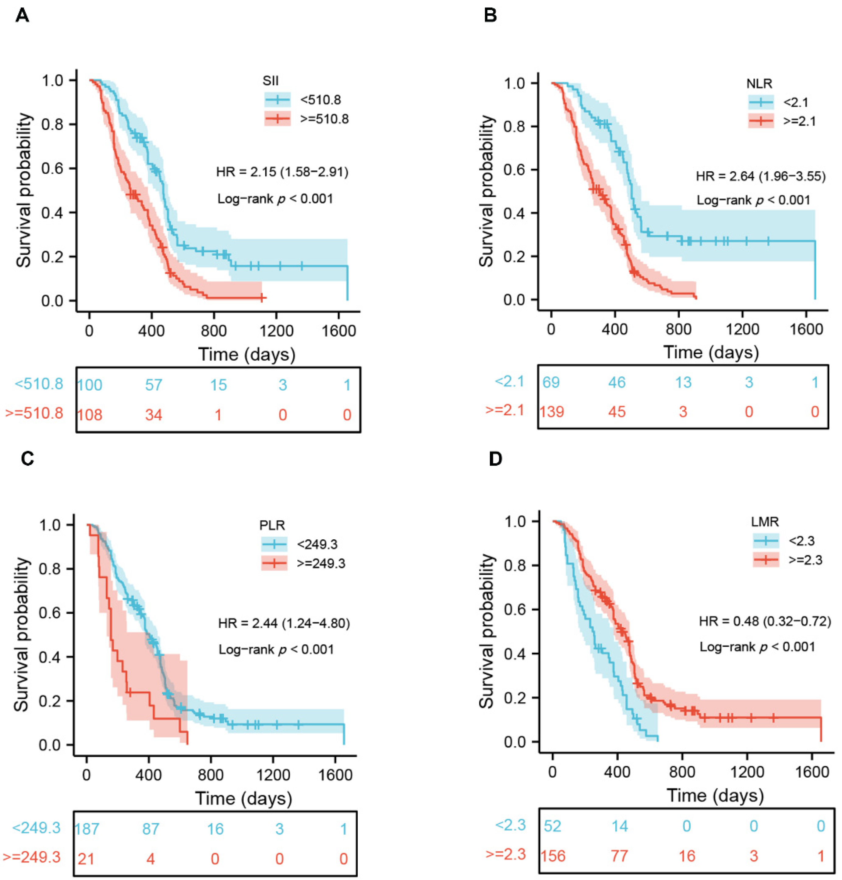
Using X-tile software, we identified the best cutoff of each peripheral indicator. As can be observed in Supplementary Figure S3, the cutoff values of white blood cell (WBC), neutrophil, lymphocyte, platelet, and monocyte were 6.7, 4.9, 1.9, 255, and 0.4 (109 cells/L), respectively. The cutoff values of NLR, LMR, PLR, and SII were 2.1, 2.3, 249.3, and 510.8 (109 cells/L). Subsequently, the patients were divided into two groups based on the cutoff value of every individual marker. Survival analysis exhibited that a higher WBC (p = 0.001), neutrophil (p < 0.001), platelet (p = 0.018), NLR (p < 0.001), PLR (p < 0.001) and SII (p < 0.001) had a worse outcome, whereas those with a higher lymphocyte (p = 0.001) and LMR (p < 0.001) had a better OS (Figure 2 and Supplementary Figure S4). In addition to age, KPS, postoperative standard chemoradiotherapy, the extent of tumor resection, and the status of MGMT, multivariate analysis indicated that the NLR (p = 0.017), PLR (p < 0.001) and SII (p = 0.007) remained independent of prognostic significance (Table 2). Time-dependent ROC analysis demonstrated that the NLR exhibited the best predictive accuracy at one-year and two-year survival rates, followed by the SII and PLR (Supplementary Figure S5). These results indicated the independent prognostic value of the NLR, SII, and PLR, but not the WBC, neutrophil, lymphocyte, platelet counts, and LMR. Moreover, significant correlations were observed between the SII and NLR (r = 0.898), or PLR (r = 0.776); meanwhile, the NLR was also significantly correlated with the PLR (r = 0.622) (Supplementary Figure S6).

3.5. Prognostic Significance of Scoring Systems in Patients with GBM

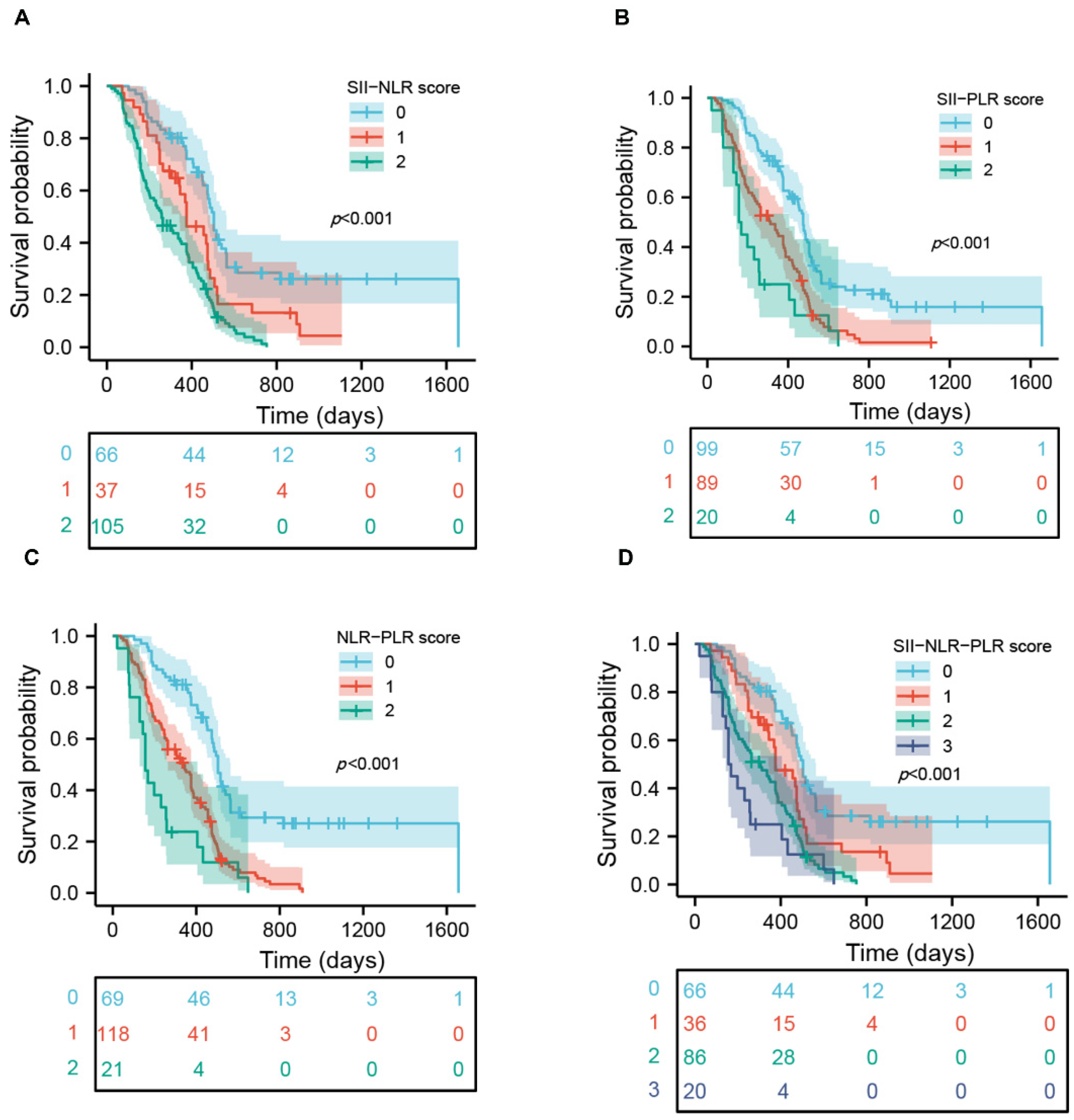
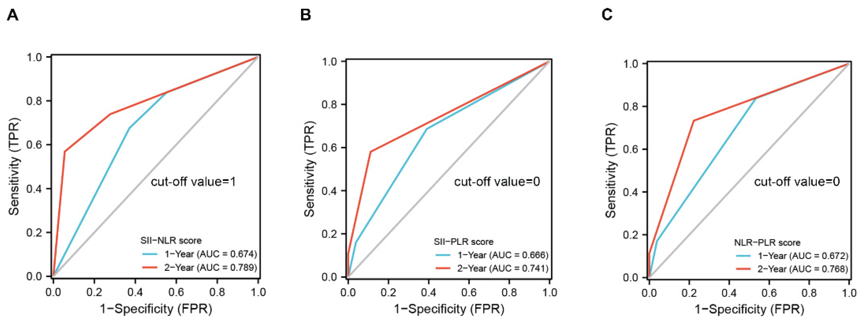
Based on the above results, four different scoring systems, such as the SII-NLR, SII-PLR, NLR-PLR and SII-NLR-PLR score, were developed to investigate whether the combinations of these markers can exhibit a more powerful predicting ability. The scores in the three systems were determined by the presence of each marker’s status associated with a worse outcome (NLR > 2.1, PLR > 249.3, SII > 510.8) (Table 3). For the SII-NLR, SII-PLR and NLR-PLR, there were three categories created for the scoring systems: score 0 (neither variable was present), score 1 (any variable was present), and score 2 (both variables were present). The SII-NLR-PLR score was classified into four groups: score 0 (none of these three variables was present), score 1 (any one of the variables was present), score 2 (any two variables were present), and score 3 (all the variables were present). The OS information of each score system is also shown in Table 3, notably, the score 0 and score 2 group of the NLR-PLR system exhibited the longest or shortest survival time among all the score systems. As shown in Figure 3, the SII-NLR, SII-PLR, NLR-PLR and SII-NLR-PLR were all significantly correlated with OS. All three systems including two variables were independent prognostic variables according to multivariate analysis; however, score 1 group in the SII-NLR-PLR system did not exhibit significant independent prognostic value because, possibly, of a relatively small cohort in each group (Table 4). Therefore, the SII-NLR-PLR may be not a good independent variable for OS in GBM. We further assessed the predictive abilities of the SII-NLR, SII-PLR and NLR-PLR score for OS; time-dependent ROC showed that the SII-NLR scoring system was the best variable for predicting OS at one-year and two-year survival rates compared to two other scoring systems and any individual marker (Figure 4 and Supplementary Figure S5). The details of the cut-off value for the score systems are shown in Supplementary File S5.

3.6. Nomograms for Predicting OS in Patients with GBM

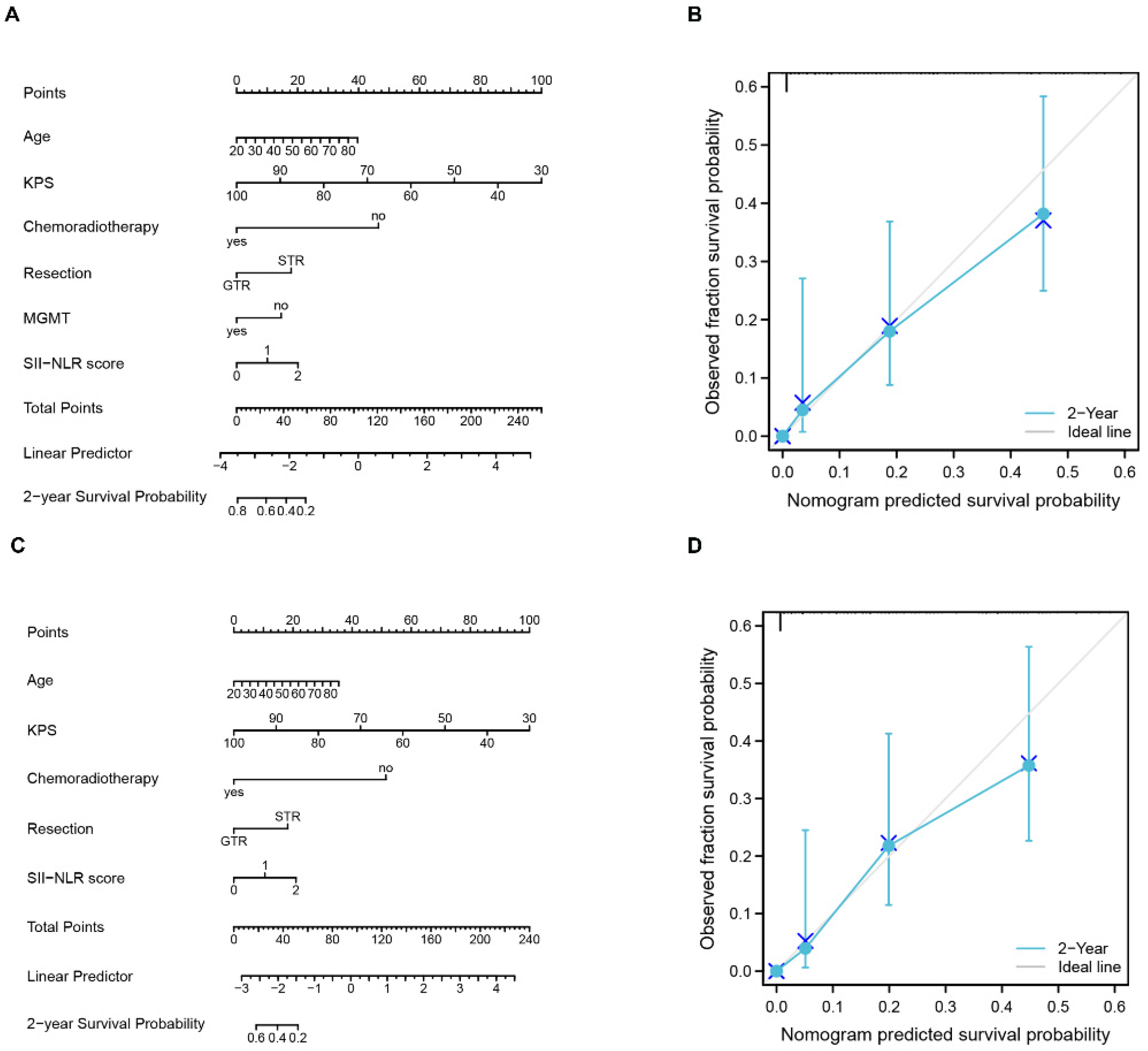
Nomogram, a frequent technique used in oncology research as a visual calculating scale model, delivers an estimated digital prognosis for each patient by effectively integrating several prognostic indicators. Therefore, a nomogram incorporating age, the KPS, chemoradiotherapy, the extent of resection, MGMT, and the SII-NLR score was constructed to assess the ability of these variables to predict the two-year survival in patients with GBM (Figure 5A). In the nomogram, the KPS accounted for the most significant percentage, followed by chemoradiotherapy, age, SII-NLR score, resection extent of the tumor, and MGMT. The c-index of the nomogram was 0.848 (95% CI = 0.836–0.861). The bootstrapped calibration plot of the nomogram performed well with the ideal model (Figure 5B). Considering the disadvantages of the molecular test, another nomogram without MGMT was also constructed (Figure 5C). The c-index of this nomogram was 0.843 (95% CI = 0.830–0.855), which was equal to that of the nomogram incorporating MGMT. The bootstrapped calibration plot of the nomogram without MGMT also functioned well with the ideal model (Figure 5D).

4. Discussion

Many biological systems and disease processes are closely associated with inflammation, including cancer initiation and progression [6]. The correlation between peripheral inflammation status and the prognosis of cancer patients has been widely reported, including in glioma patients. Among those indexes of peripheral inflammation, the SII, a novel inflammatory biomarker, was shown to be an effective predictor of the outcomes in many solid tumors [27,28,29,30]. In this study, the meta-analysis revealed the clinical significance of a preoperative SII in GBM patients, and this was further verified by the retrospective investigation from single-center data. The SII-NLR score showed the best predictive power for OS compared to the SII-PLR score, the NLR-PLR score, and any other single inflammatory marker. Based on the scoring systems, the nomogram with or without MGMT demonstrated a similar equal predictive accuracy and discrimination for estimating OS.
The research on the relationship between inflammation and a tumor has attracted increasing attention. The systemic inflammatory response is considered as an essential feature of the host response to malignant tumors. It has been found that the systemic inflammatory response can affect the prognosis of tumor patients. Our previous study [15] reported that GBM patients exhibited higher neutrophil counts, NLR, and PLR, as well as lower lymphocyte counts and LMR, compared to those in patients with lower-grade glioma. A high NLR and low LMR were associated with unfavorable OS in GBM patients. Some studies have also indicated that an elevated NLR predicts worse outcomes in patients with colorectal cancer [31], breast cancer [32], and other cancers. The SII, calculated using the absolute measures of platelets, neutrophils, and lymphocytes obtained from routine complete blood counts [19], has been suggested as a biomarker superior to both the NLR and PLR in prognosis prediction [33]. Lei et al. [30] demonstrated that the SII could predict higher pathological grades in young premenopausal endometrial cancer patients. The prognostic value of the SII had also been identified in various types of cancers, including hepatocellular carcinoma [19] for the first time, small-cell lung cancer [10], and glioma [20]. However, a study by Yilmaz et al. [23] reported that the SII could not be used as an independent biomarker for progression-free survival (PFS) and OS in patients with GBM. The meta-analysis of this study, including 788 GBM patients from five individual studies, indicated that elevated SII predicted a poorer OS (HR = 1.87, 95% CI: 1.51–2.32). In the subsequent retrospective study, we also found that a higher value of SII significantly correlated with a worse outcome in patients with GBM (HR = 1.782, 95% CI: 1.168–2.719). However, time-dependent ROC analysis demonstrated that the NLR exhibited the best predictive accuracy at one-year and two-year survival rates, followed by the SII and PLR. Moreover, our analysis showed that the combination of these inflammatory markers displayed more predictive power for OS, compared with any single index. The further result of the nomogram model including the most effective SII-NLR scoring system without MGMT provided the evidence that it is reasonable and practical to evaluate outcomes through simply using the periphery inflammatory indicators and relevant therapy information without molecular tests in patients with GBM.
The precise mechanisms through which the SII affected the prognosis of GBM patients remain unclear. The SII is determined by taking into account the peripheral blood’s neutrophil, platelet, and lymphocyte counts. Evidence suggests that it plays a role in the development, progression, and overall evaluation of many diseases, including human bleeding disorders [34], connective tissue diseases [35], and various malignant tumor forms. Cancers were often featured with neutrophilia, and elevated neutrophils helped the construction of a tumor microenvironment through the secretion of some essential cytokines, such as vascular endothelial growth factor (VEGF) [36], interleukin- 6 (IL-6) [37], IL-8 [38], IL-10 [8], prostaglandin [39], matrix metalloproteinases [40], among others. It benefited the progression, angiogenesis, and distant metastasis of tumors. Some crucial cytokines, including G-CSF, IL-1β, VEGF, and IL-6 [41,42,43] from tumor cells, could potentially increase the number of neutrophils in peripheral blood and tumor tissue. The extent of neutrophil infiltration around the tumor was also reported to be significantly associated with the grade of glioma [44].
The levels of platelets are often elevated in patients with malignant tumors, and the elevated platelets may accelerate the proliferation, angiogenesis, and dissemination of tumor cells through some released factors, such as VEGF and platelet-derived growth factor (PDGF) [45,46,47]. Moreover, the high levels of platelets also had the potential to prevent tumor cells from systemic immune attack and to induce invasive mesenchymal-like phenotype and metastasis by the activation of transforming growth factor β (TGF-β) and nuclear factor kappa-B (NF-κB) pathways [48]. As a result, the enhanced platelet counts were combined with neutrophils to construct a microenvironment beneficial to tumor cell survival. Inversely, lymphocyte was the primary type of immune cells playing an essential role in eliminating tumor cells and immune surveillance in the host [7,49]. Unfortunately, increased neutrophil counts can inhibit lymphocyte survival and normal cytotoxic action by producing reactive oxygen species and arginase [41]. Therefore, the SII was a comprehensive reflection of an impaired immunological function and elevated inflammatory status, instrumental in tumor biological behavior. Furthermore, a study by Liang et al. reported that higher levels of SII were significantly correlated with a higher value of Ki-67, a nuclear protein strongly associated with cellular proliferation, tumor grade, and worse survival in patients with glioma [50].
Interestingly, the study by Wang et al. [10] recently reported that the SII was a valuable tool for doctors to choose therapeutic methods in patients with non-small cell lung cancer (NSCLC). The patients with a higher SII may be suitable for immunotherapy compared to those with a lower SII. Meanwhile, SII dynamics during immunology therapy could also effectively predict the survival of patients with metastatic renal cell carcinoma [51]. Diem et al. [52] analyzed the clinical data of 52 patients with metastatic NSCLC treated with nivolumab. They found that elevated pre-treatment NLR and PLR are associated with shorter OS, PFS, and lower response rates in patients with metastatic NSCLC treated with nivolumab independently of other prognostic factors. Recently, Valero et al. [53] performed a retrospective cohort study of 1714 patients with 16 cancer types treated with immune checkpoint inhibitors (ICI), demonstrating a clinical benefit for a wide range of cancer types. Because only a subset of patients experienced a clinical benefit, there is a strong need for biomarkers that are easily accessible across diverse practice settings. Their results showed that a higher NLR was significantly associated with poorer OS and PFS and lower rates of response and clinical benefit after ICI therapy across multiple cancer types. Further analysis indicated that the NLR could be combined with a tumor mutational burden (TMB) for additional predictive capacity. The probability of benefit from an ICI is significantly higher in the NLR low/TMB high group compared to the NLR high/TMB low group. Moreover, Liu et al. [54] reported that the reduction in the NLR and PLR could predict the clinic remission rate and pathological responsiveness rate, respectively, in patients with rectal cancer. These findings suggest that these peripheral inflammatory indicators may be suitable candidates for a cost-effective and widely accessible biomarker.
There are also some limitations to our study. The number of included studies for meta-analysis was relatively small, and more caution should be applied when interpreting results. The nature of our subsequent retrospective research may lead to a selection bias, and we included a relatively small number of patients in this study. Therefore, prospective and well-designed studies with more samples should be performed to clarify the results. Research on systematic inflammatory biomarkers in cancer treatment decision-making is needed, especially in patients with GBM.

5. Conclusions

A pre-treatment SII is a potential prognostic biomarker of OS in patients with GBM. A nomogram that integrates the SII-NLR score may facilitate a comprehensive preoperative survival evaluation independent of molecular tests in patients with GBM.

Supplementary Materials

The following supporting information can be downloaded at: https://www.mdpi.com/article/10.3390/jcm11247514/s1, File S1: PRISMA 2009 Checklist; File S2: Free terms; File S3: STROBE Statement—checklist of items that should be included in reports of observational studies; File S4: Statistical analysis for meta-analysis; File S5: The details of cut-off value; Figure S1: Flow diagram of the study selection process during the meta-analysis; Figure S2: Sensitive analysis of the relationship between SII and OS; Figure S3: Cut-off value of WBC (A), neutrophil (B), lymphocyte (C), platelet (D), monocyte (E), NLR (F), PLR (G), LMR (H) and SII (I) in patients with GBM; Figure S4: Kaplan–Meier survival curves of glioblastoma patients based on the cutoff values of WBC (A), neutrophil (B), lymphocyte (C), platelet (D) and monocyte (E); Figure S5: Time-dependent ROC curve analysis of SII (A), NLR (B) and PLR (C) for predicting OS at one-year and two-year survival rate in patients with GBM; Figure S6: Correlation analysis of SII, NLR and PLR. Table S1: Characteristics of the included prognostic studies; Table S2: Quality evaluation of included studies using the Newcastle Ottawa Scale for cohort studies; Table S3: Subgroup analysis of OS.

Author Contributions

Conceptualization, Z.-Q.L. and Y.-R.L.; methodology, C.Y. and F.T.; software, B.-W.H.; formal analysis, C.Y.; data curation, B.-W.H., J.W., Q.Z. and W.Q.; writing—original draft, C.Y. and B.-W.H.; preparation, Z.-F.W.; writing—review and editing, Z.-Q.L. and Y.-R.L.; visualization, C.Y. and B.-W.H.; supervision, Z.-F.W. and Z.-Q.L. All authors have read and agreed to the published version of the manuscript.

Funding

This research received no external funding.

Institutional Review Board Statement

The study was conducted in accordance with the Declaration of Helsinki, and the present study gained approval from the Ethics Committee of Zhongnan Hospital of Wuhan University (No. 2019048).

Informed Consent Statement

The informed consent from all the included patients was unnecessary due to its retrospective nature. The relevant information was coded and the patients were de-identified.

Data Availability Statement

The original dataset analyzed during the current study is available from the corresponding author upon reasonable request.

Acknowledgments

We thank Yi Guo for his help in statistical analysis.

Conflicts of Interest

The authors declare no conflict of interest.

References

  1. Wesseling, P.; Capper, D. WHO 2016 Classification of gliomas. Neuropathol. Appl. Neurobiol. 2018, 44, 139–150. [Google Scholar] [CrossRef] [PubMed]
  2. Stupp, R.; Mason, W.P.; van den Bent, M.J.; Weller, M.; Fisher, B.; Taphoorn, M.J.B.; Belanger, K.; Brandes, A.A.; Marosi, C.; Bogdahn, U.; et al. Radiotherapy plus concomitant and adjuvant temozolomide for glioblastoma. N. Engl. J. Med. 2005, 352, 987–996. [Google Scholar] [CrossRef] [PubMed] [Green Version]
  3. Stupp, R.; Taillibert, S.; Kanner, A.; Read, W.; Steinberg, D.; Lhermitte, B.; Toms, S.; Idbaih, A.; Ahluwalia, M.S.; Fink, K.; et al. Effect of tumor-treating fields plus maintenance temozolomide vs maintenance temozolomide alone on survival in patients with glioblastoma: A randomized clinical trial. JAMA 2017, 318, 2306–2316. [Google Scholar] [CrossRef] [PubMed] [Green Version]
  4. Zhao, Y.-H.; Wang, X.; Cao, C.-J.; Weng, H.; Xu, C.-S.; Li, K.; Li, J.-L.; Lan, J.; Zeng, X.-T.; Li, Z.-Q. The Clinical Significance of O-Methylguanine-DNA Methyltransferase Promoter Methylation Status in Adult Patients With Glioblastoma: A Meta-analysis. Front. Neurol. 2018, 9, 127. [Google Scholar] [CrossRef] [PubMed] [Green Version]
  5. Hanahan, D.; Weinberg, R.A. Weinberg. Hallmarks of cancer: The next generation. Cell 2011, 144, 646–674. [Google Scholar] [CrossRef] [PubMed] [Green Version]
  6. Coussens, L.M.; Werb, Z. Inflammation and cancer. Nature 2002, 420, 860–867. [Google Scholar] [CrossRef] [PubMed]
  7. Mantovani, A.; Allavena, P.; Sica, A.; Balkwill, F. Cancer-related inflammation. Nature 2008, 454, 436–444. [Google Scholar] [CrossRef]
  8. Albulescu, R.; Codrici, E.; Popescu, I.D.; Mihai, S.; Necula, L.G.; Petrescu, D.; Teodoru, M.; Tanase, C.P. Cytokine patterns in brain tumour progression. Mediat. Inflamm. 2013, 2013, 979748. [Google Scholar] [CrossRef]
  9. Yang, C.; Wen, H.-B.; Zhao, Y.-H.; Huang, W.-H.; Wang, Z.-F.; Li, Z.-Q. Systemic Inflammatory Indicators as Prognosticators in Glioblastoma Patients: A Comprehensive Meta-Analysis. Front. Neurol. 2020, 11, 580101. [Google Scholar] [CrossRef]
  10. Wang, Y.; Li, Y.; Chen, P.; Xu, W.; Wu, Y.; Che, G. Prognostic value of the pretreatment systemic immune-inflammation index (SII) in patients with non-small cell lung cancer: A meta-analysis. Ann. Transl. Med. 2019, 7, 433. [Google Scholar] [CrossRef]
  11. Hu, H.; Yao, X.; Xie, X.; Wu, X.; Zheng, C.; Xia, W.; Ma, S. Prognostic value of preoperative NLR, dNLR, PLR and CRP in surgical renal cell carcinoma patients. World J. Urol. 2017, 35, 261–270. [Google Scholar] [CrossRef]
  12. Massara, M.; Persico, P.; Bonavita, O.; Poeta, V.M.; Locati, M.; Simonelli, M.; Bonecchi, R. Neutrophils in Gliomas. Front. Immunol. 2017, 8, 1349. [Google Scholar] [CrossRef] [PubMed] [Green Version]
  13. Wang, D.; Kang, K.; Lin, Q.; Hai, J. Prognostic Significance of Preoperative Systemic Cellular Inflammatory Markers in Gliomas: A Systematic Review and Meta-Analysis. Clin. Transl. Sci. 2020, 13, 179–188. [Google Scholar] [CrossRef] [PubMed]
  14. Zhu, Y.; Li, M.; Bo, C.; Liu, X.; Zhang, J.; Li, Z.; Zhao, F.; Kong, L.; Yu, J. Prognostic significance of the lymphocyte-to-monocyte ratio and the tumor-infiltrating lymphocyte to tumor-associated macrophage ratio in patients with stage T3N0M0 esophageal squamous cell carcinoma. Cancer Immunol. Immunother. 2017, 66, 343–354. [Google Scholar] [CrossRef]
  15. Yang, C.; Lan, T.; Wang, Y.; Huang, W.-H.; Li, S.-M.; Li, J.; Li, F.-P.; Li, Y.-R.; Wang, Z.-F.; Li, Z.-Q. Cumulative scoring systems and nomograms for predicating survival in patients with glioblastomas: A study based on peripheral inflammatory markers. Front. Oncol. 2022, 12, 716295. [Google Scholar] [CrossRef]
  16. Haemmerle, M.; Stone, R.L.; Menter, D.G.; Afshar-Kharghan, V.; Sood, A.K. The platelet lifeline to cancer: Challenges and opportunities. Cancer Cell 2018, 33, 965–983. [Google Scholar] [CrossRef] [Green Version]
  17. Aziz, M.H.; Sideras, K.; Aziz, N.A.; Mauff, K.; Haen, R.; Roos, D.; Saida, L.; Suker, M.; van der Harst, E.; Mieog, J.S.; et al. The Systemic-immune-inflammation index independently predicts survival and recurrence in resectable pancreatic cancer and its prognostic value depends on bilirubin levels: A retrospective multicenter cohort study. Ann. Surg. 2019, 270, 139–146. [Google Scholar] [CrossRef]
  18. Li, W.; Ma, G.; Deng, Y.; Chen, W.; Liu, Z.; Chen, F.; Wu, Q. Systemic Immune-Inflammation Index Is a Prognostic Factor for Breast Cancer Patients After Curative Resection. Front. Oncol. 2021, 11, 570208. [Google Scholar] [CrossRef]
  19. Hu, B.; Yang, X.-R.; Xu, Y.; Sun, Y.-F.; Sun, C.; Guo, W.; Zhang, X.; Wang, W.-M.; Qiu, S.-J.; Zhou, J.; et al. Systemic immune-inflammation index predicts prognosis of patients after curative resection for hepatocellular carcinoma. Clin. Cancer Res. 2014, 20, 6212–6222. [Google Scholar] [CrossRef] [Green Version]
  20. Topkan, E.; Besen, A.A.; Ozdemir, Y.; Küçük, A.; Mertsoylu, H.; Pehlivan, B.; Selek, U. Prognostic Value of Pretreatment Systemic Immune-Inflammation Index in Glioblastoma Multiforme Patients Undergoing Postneurosurgical Radiotherapy Plus Concurrent and Adjuvant Temozolomide. Mediat. Inflamm. 2020, 2020, 4392189. [Google Scholar] [CrossRef]
  21. Shi, X.; Li, H.; Xu, Y.; Nyalali, A.M.K.; Li, F. The prognostic value of the preoperative inflammatory index on the survival of glioblastoma patients. Neurol. Sci. 2022, 43, 5523–5531. [Google Scholar] [CrossRef]
  22. Pasqualetti, F.; Giampietro, C.; Montemurro, N.; Giannini, N.; Gadducci, G.; Orlandi, P.; Natali, E.; Chiarugi, P.; Gonnelli, A.; Cantarella, M.; et al. Old and new systemic immune-inflammation indexes are associated with overall survival of glioblastoma patients treated with radio-chemotherapy. Genes 2022, 13, 1054. [Google Scholar] [CrossRef] [PubMed]
  23. Yılmaz, H.; Niğdelioğlu, B.; Oktay, E.; Meydan, N. Clinical significance of postoperatif controlling nutritional status (CONUT) score in glioblastoma multiforme. J. Clin. Neurosci. 2021, 86, 260–266. [Google Scholar] [CrossRef] [PubMed]
  24. Moher, D.; Liberati, A.; Tetzlaff, J.; Altman, D.G.; PRISMA Group. Preferred reporting items for systematic reviews and meta-analyses: The PRISMA statement. PLoS Med. 2009, 6, e1000097. [Google Scholar] [CrossRef] [PubMed] [Green Version]
  25. World Medical Association. World Medical Association Declaration of Helsinki: Ethical principles for medical research involving human subjects. JAMA 2013, 310, 2191–2194. [Google Scholar] [CrossRef] [Green Version]
  26. Lv, Y.; Zhang, S.; Liu, Z.; Tian, Y.; Liang, N.; Zhang, J. Prognostic value of preoperative neutrophil to lymphocyte ratio is superior to systemic immune inflammation index for survival in patients with Glioblastoma. Clin. Neurol. Neurosurg. 2019, 181, 24–27. [Google Scholar] [CrossRef]
  27. Wang, D.; Hu, X.; Xiao, L.; Long, G.; Yao, L.; Wang, Z.; Zhou, L. Prognostic Nutritional Index and Systemic Immune-Inflammation Index Predict the Prognosis of Patients with HCC. J. Gastrointest. Surg. 2021, 25, 421–427. [Google Scholar] [CrossRef] [Green Version]
  28. Zhou, Y.; Dai, M.; Zhang, Z. Prognostic significance of the systemic immune-inflammation index (SII) in patients with small cell lung cancer: A meta-analysis. Front. Oncol. 2022, 12, 814727. [Google Scholar] [CrossRef]
  29. Jin, M.; Yuan, S.; Yuan, Y.; Yi, L. Prognostic and clinicopathological significance of the systemic immune-inflammation index in patients with renal cell carcinoma: A meta-analysis. Front. Oncol. 2021, 11, 735803. [Google Scholar] [CrossRef]
  30. Lei, H.; Xu, S.; Mao, X.; Chen, X.; Chen, Y.; Sun, X.; Sun, P. Systemic immune-inflammatory index as a predictor of lymph node metastasis in endometrial cancer. J. Inflamm. Res. 2021, 14, 7131–7142. [Google Scholar] [CrossRef]
  31. Yamamoto, T.; Kawada, K.; Obama, K. Inflammation-related biomarkers for the prediction of prognosis in colorectal cancer patients. Int. J. Mol. Sci. 2021, 22, 8002. [Google Scholar] [CrossRef] [PubMed]
  32. Chen, J.; Deng, Q.; Pan, Y.; He, B.; Ying, H.; Sun, H.; Liu, X.; Wang, S. Prognostic value of neutrophil-to-lymphocyte ratio in breast cancer. FEBS. Open Bio. 2015, 5, 502–507. [Google Scholar] [CrossRef] [PubMed] [Green Version]
  33. Geng, Y.; Shao, Y.; Zhu, D.; Zheng, X.; Zhou, Q.; Zhou, W.; Ni, X.; Wu, C.; Jiang, J. Systemic immune-inflammation index predicts prognosis of patients with esophageal squamous cell carcinoma: A propensity score-matched analysis. Sci. Rep. 2016, 6, 39482. [Google Scholar] [CrossRef] [PubMed]
  34. Geraghty, J.R.; Lung, T.J.; Hirsch, Y.; Katz, E.A.; Cheng, T.; Saini, N.S.; Pandey, D.K.; Testai, F.D. Systemic immune-inflammation index predicts delayed cerebral vasospasm after aneurysmal subarachnoid hemorrhage. Neurosurgery 2021, 89, 1071–1079. [Google Scholar] [CrossRef]
  35. Ruta, V.M.; Man, A.M.; Alexescu, T.G.; Motoc, N.S.; Tarmure, S.; Ungur, R.A.; Todea, D.A.; Coste, S.C.; Valean, D.; Pop, M.C. Neutrophil-To-Lymphocyte ratio and systemic immune-inflammation index-biomarkers in interstitial lung disease. Medicina 2020, 56, 381. [Google Scholar] [CrossRef]
  36. McCourt, M.; Wang, J.H.; Sookhai, S.; Redmond, H.P. Proinflammatory mediators stimulate neutrophil-directed angiogenesis. Arch. Surg. 1999, 134, 1325–1331. [Google Scholar] [CrossRef]
  37. Jabłońska, E.; Kiluk, M.; Markiewicz, W.; Piotrowski, L.; Grabowska, Z.; Jabłoński, J. TNF-alpha, IL-6 and their soluble receptor serum levels and secretion by neutrophils in cancer patients. Arch. Immunol. Ther. Exp. 2001, 49, 63–69. [Google Scholar]
  38. Schaider, H.; Oka, M.; Bogenrieder, T.; Nesbit, M.; Satyamoorthy, K.; Berking, C.; Matsushima, K.; Herlyn, M. Differential response of primary and metastatic melanomas to neutrophils attracted by IL. Int. J. Cancer 2003, 103, 335–343. [Google Scholar] [CrossRef]
  39. Mizuno, R.; Kawada, K.; Sakai, Y. Prostaglandin E2/EP Signaling in the tumor microenvironment of colorectal cancer. Int. J. Mol. Sci. 2019, 20, 6254. [Google Scholar] [CrossRef] [Green Version]
  40. Shamamian, P.; Schwartz, J.D.; Pocock, B.J.; Monea, S.; Whiting, D.; Marcus, S.G.; Mignatti, P. Activation of progelatinase A (MMP-2) by neutrophil elastase, cathepsin G, and proteinase-3: A role for inflammatory cells in tumor invasion and angiogenesis. J. Cell Physiol. 2001, 189, 197–206. [Google Scholar] [CrossRef]
  41. Gregory, A.D.; Houghton, A.M. Tumor-associated neutrophils: New targets for cancer therapy. Cancer Res. 2011, 71, 2411–2416. [Google Scholar] [CrossRef] [PubMed]
  42. Del Prete, A.; Allavena, P.; Santoro, G.; Fumarulo, R.; Corsi, M.M.; Mantovani, A. Molecular pathways in cancer-related inflammation. Biochem. Med. 2011, 21, 264–275. [Google Scholar] [CrossRef] [PubMed]
  43. Lechner, M.; Liebertz, D.J.; Epstein, A.L. Characterization of cytokine-induced myeloid-derived suppressor cells from normal human peripheral blood mononuclear cells. J. Immunol. 2010, 185, 2273–2284. [Google Scholar] [CrossRef] [PubMed] [Green Version]
  44. Han, S.; Liu, Y.; Li, Q.; Li, Z.; Hou, H.; Wu, A. Pre-treatment neutrophil-to-lymphocyte ratio is associated with neutrophil and T-cell infiltration and predicts clinical outcome in patients with glioblastoma. BMC Cancer 2015, 15, 617. [Google Scholar] [CrossRef] [Green Version]
  45. Di Vito, C.; Navone, S.E.; Marfia, G.; Hadi, L.A.; Mancuso, M.E.; Pecci, A.; Crisà, F.M.; Berno, V.; Rampini, P.; Campanella, R.; et al. Platelets from glioblastoma patients promote angiogenesis of tumor endothelial cells and exhibit increased VEGF content and release. Platelets 2017, 28, 585–594. [Google Scholar] [CrossRef]
  46. Westermark, B.; Heldin, C.; Nistér, M. Platelet-derived growth factor in human glioma. Glia 1995, 15, 257–263. [Google Scholar] [CrossRef] [PubMed]
  47. Wojtukiewicz, M.Z.; Sierko, E.; Hempel, D.; Tucker, S.C.; Honn, K.V. Platelets and cancer angiogenesis nexus. Cancer Metastasis Rev. 2017, 36, 249–262. [Google Scholar] [CrossRef] [Green Version]
  48. Labelle, M.; Begum, S.; Hynes, R.O. Direct signaling between platelets and cancer cells induces an epithelial-mesenchymal-like transition and promotes metastasis. Cancer Cell 2011, 20, 576–590. [Google Scholar] [CrossRef] [Green Version]
  49. Gooden, M.J.M.; de Bock, G.H.; Leffers, N.; Daemen, T.; Nijman, H.W. The prognostic influence of tumour-infiltrating lymphocytes in cancer: A systematic review with meta-analysis. Br. J. Cancer 2011, 105, 93–103. [Google Scholar] [CrossRef] [Green Version]
  50. Liang, R.; Chen, N.; Li, M.; Wang, X.; Mao, Q.; Liu, Y. Significance of systemic immune-inflammation index in the differential diagnosis of high. and low-grade gliomas. Clin. Neurol. Neurosurg. 2018, 164, 50–52. [Google Scholar] [CrossRef]
  51. De Giorgi, U.; Procopio, G.; Giannarelli, D.; Sabbatini, R.; Bearz, A.; Buti, S.; Basso, U.; Mitterer, M.; Ortega, C.; Bidoli, P.; et al. Association of systemic inflammation index and body mass index with survival in patients with renal cell cancer treated with nivolumab. Clin. Cancer Res. 2019, 25, 3839–3846. [Google Scholar] [CrossRef]
  52. Diem, S.; Schmid, S.; Krapf, M.; Flatz, L.; Born, D.; Jochum, W.; Templeton, A.J.; Früh, M. Neutrophil-to-Lymphocyte ratio (NLR) and Platelet-to-Lymphocyte ratio (PLR) as prognostic markers in patients with non-small cell lung cancer (NSCLC) treated with nivolumab. Lung Cancer 2017, 111, 176–181. [Google Scholar] [CrossRef] [PubMed]
  53. Valero, C.; Lee, M.; Hoen, D.; Weiss, K.; Kelly, D.W.; Adusumilli, P.S.; Paik, P.K.; Plitas, G.; Ladanyi, M.; Postow, M.A.; et al. Pretreatment neutrophil-to-lymphocyte ratio and mutational burden as biomarkers of tumor response to immune checkpoint inhibitors. Nat. Commun. 2021, 12, 728. [Google Scholar] [CrossRef] [PubMed]
  54. Liu, M.; Feng, Y.; Zhang, Y.; Liu, H. Evaluation of neutrophil-lymphocyte ratio and platelet-lymphocyte ratio on predicting responsiveness to neoadjuvant chemoradiotherapy in locally advanced rectal cancer patients. Biomed. Res. Int. 2022, 2022, 3839670. [Google Scholar] [CrossRef] [PubMed]
Figure 1. Pooled hazard ratio (HR) and 95% confidence intervals (CIs) of SII for OS in glioblastoma patients (A) and detection of publication bias for meta-analysis of survival outcomes based on typical funnel plot (B) and Begg’s funnel plot (C). The references of Lv et al. (2019), Topkan et al. (2020), Yilmaz et al. (2021), Shi et al. (2022) and Pasqualetti et al. (2022) were [20,21,22,23,26].
Jcm 11 07514 g001
Figure 2. Kaplan–Meier survival curves of glioblastoma patients based on the cutoff values of SII (A), NLR (B), PLR (C), and LMR (D).
Jcm 11 07514 g002
Figure 3. Kaplan–Meier survival curves of glioblastoma patients based on SII-NLR score (A), SII-PLR score (B), NLR-PLR score (C) and SII-NLR-PLR score (D).
Jcm 11 07514 g003
Figure 4. Time-dependent ROC curve analysis of SII-NLR score (A), SII-PLR score (B), and NLR-PLR score (C) for predicting OS at one-year and two-year survival rate in patients with GBM.
Jcm 11 07514 g004
Figure 5. Nomogram and calibration curve. In the nomogram (A) and (C) ((A) the nomogram including MGMT; (C) the nomogram without MGMT), each variable was assigned a different score as shown on the largest scale, and the total score for all variables yielded a numerical prediction of two-year survival, with higher scores leading to a worse prognosis. In the calibration curve (B) and (D) ((B) calibration of nomogram A; (D) calibration of nomogram C), the grey line represents the ideal prediction, and the blue line represents the performance of the nomogram.
Jcm 11 07514 g005
Table 1. Clinical and pathological characteristics of patients.
CharacteristicSII < 510.8SII ≥ 510.8p
n100108
Sex, n (%) 0.244
female45 (21.6%)39 (18.8%)
male55 (26.4%)69 (33.2%)
Location, n (%) 0.667
frontal32 (15.4%)31 (14.9%)
temporal27 (13%)26 (12.5%)
parietal12 (5.8%)10 (4.8%)
multiple15 (7.2%)18 (8.7%)
other14 (6.7%)23 (11.1%)
KPS, n (%) <0.001
≤5011 (11.0%)37 (34.2%)
>5089 (89.0%)71 (65.8%)
Chemoradiotherapy, n (%) <0.001
no18 (8.7%)45 (21.6%)
yes82 (39.4%)63 (30.3%)
Resection, n (%) <0.001
STR26 (12.5%)57 (27.4%)
GTR74 (35.6%)51 (24.5%)
MGMT, n (%) 1.000
no57 (27.4%)62 (29.8%)
yes43 (20.7%)46 (22.1%)
Age, median (IQR)58 (51.75, 65)59 (51, 65)0.956
WBC, median (IQR)5.42 (4.62, 6.39)7.05 (5.77, 9.17)<0.001
Neu, median (IQR)3.28 (2.56, 4)5.43 (4.11, 7.49)<0.001
Lym, median (IQR)1.8 (1.38, 2.13)1.3 (0.92, 1.62)<0.001
PLT, median (IQR)178.5 (143.75, 203.5)219 (177.5, 273.5)<0.001
OS, median (IQR)14.95 (9.95, 18.18)8.8 (5.2, 14.9)<0.001
Abbreviations: KPS, Karnofsky performance status; GTR, gross total resection; STR, subtotal resection; IDH, isocitrate dehydrogenase; MGMT, O6-methylguanine-DNA methyltransferase; Neu, neutrophil; Lym, lymphocyte; PLT, platelet; OS, overall survival; SII, systemic immune-inflammation index.
Table 2. Univariate and multivariate analyses of OS in GBM cohorts.
CharacteristicsTotal (N)Univariate AnalysisMultivariate Analysis
Hazard Ratio (95% CI)p ValueHazard Ratio (95% CI)p Value
Sex208
male124Reference
female841.032 (0.761–1.400)0.839
Age2081.025 (1.011–1.040)<0.0011.025 (1.010–1.039)<0.001
Location2081.110 (1.004–1.228)0.0421.047 (0.944–1.162)0.385
KPS208
≤5023Reference
>501850.150 (0.095–0.237)<0.0010.253 (0.148–0.431)<0.001
Chemoradiotherapy208
no63Reference
yes1450.066 (0.043–0.103)<0.0010.136 (0.075–0.245)<0.001
Resection208
STR83Reference
GTR1250.199 (0.145–0.275)<0.0010.433 (0.276–0.680)<0.001
MGMT208
no121Reference
yes870.419 (0.306–0.573)<0.0010.654 (0.452–0.945)0.024
WBC208
≤6.7127Reference
>6.7811.622 (1.198–2.197)0.0020.979 (0.597–1.606)0.934
Neu208
≤4.9134Reference
>4.9742.540 (1.863–3.461)<0.0011.040 (0.588–1.839)0.892
Lym208
≤1.9151Reference
>1.9570.546 (0.382–0.782)<0.0010.830 (0.550–1.251)0.373
PLT208
≤255168Reference
>255401.546 (1.070–2.234)0.0200.710 (0.461–1.096)0.122
Monocyte208
≤0.477Reference
>0.41311.358 (0.990–1.862)0.0581.390 (0.945–2.047)0.095
NLR208
≤2.169Reference
>2.11392.820 (1.992–3.993)<0.0011.769 (1.106–2.829)0.017
PLR208
≤249.3187Reference
>249.3212.478 (1.547–3.969)<0.0012.598 (1.473–4.581)<0.001
LMR208
≤2.352Reference
>2.31560.461 (0.328–0.646)<0.0010.899 (0.590–1.371)0.621
SII208
≤510.8100Reference
>510.81082.226 (1.638–3.025)<0.0011.782 (1.168–2.719)0.007
Abbreviations: KPS, Karnofsky performance status; GTR, gross total resection; STR, subtotal resection; IDH, isocitrate dehydrogenase; MGMT, O6-methylguanine-DNA methyltransferase; WBC, white blood cell; Neu, neutrophil; Lym, lymphocyte; PLT, platelet; SII, systemic immune-inflammation index; HR, hazard ratio; CI, confidence interval.
Table 3. Overall survival based on score system in patients with GBM.
Score N (%)OS (Mean ± SD)p Value
VariablesDefinition (Months)
SII-NLR 208 (100)
Score 0SII < 510.8 and NLR < 2.166 (31.7)17.5 ± 9.9reference
Score 1SII > 510.8 or NLR > 2.137 (17.8)13.4 ± 7.90.012
Score 2SII > 510.8 and NLR > 2.1105 (50.5)9.8 ± 5.80.013
SII-PLR 208 (100)
Score 0SII < 510.8 and PLR < 249.399 (47.6)16.0 ± 9.2reference
Score 1SII > 510.8 or PLR > 249.389 (42.8)10.6 ± 6.4<0.001
Score 2SII > 510.8 and PLR > 249.320 (9.6)7.7 ± 5.7<0.001
NLR-PLR 208 (100)
Score 0NLR < 2.1 and PLR < 249.369 (33.2)17.6 ± 10.0reference
Score 1NLR > 2.1 or PLR > 249.3118 (56.7)11.0 ± 6.2<0.001
Score 2NLR > 2.1 and PLR > 249.321 (10.1)7.4 ± 5.6<0.001
SII-NLR-PLR 208 (100)
Score 0SII < 510.8 and NLR < 2.1 and PLR < 249.366 (31.7)17.5 ± 9.9reference
Score 1SII > 510.8 or NLR > 2.1 or PLR > 249.336 (17.3)13.8 ± 7.80.022
Score 2SII > 510.8 and NLR > 2.1, or SII > 510.8 and PLR > 249.3, or NLR > 2.1 and PLR > 249.386 (41.3)10.2 ± 5.8<0.001
Score 3SII > 510.8 and NLR > 2.1 and PLR > 249.320 (9.6)7.6 ± 5.7<0.001
SII, systemic immune-inflammation index; NLR, neutrophil-to-lymphocyte ratio; PLR, platelet-to-lymphocyte ratio; OS, overall survival; SD, standard deviation.
Table 4. Univariate and multivariate analyses of OS in GBM cohorts based on score systems.
CharacteristicsTotal (N)Univariate AnalysisMultivariate Analysis
Hazard Ratio (95% CI)p ValueHazard Ratio (95% CI)p Value
SII-NLR208 <0.001
Score 066Reference
Score 1371.806 (1.139–2.865)0.0121.679 (1.053–2.678)0.030
Score 21053.043 (2.111–4.385)<0.0012.460 (1.694–3.572)<0.001
NLR-PLR208 <0.001
Score 069Reference
Score 11182.642 (1.852–3.769)<0.0011.844 (1.269–2.680)0.001
Score 2214.557 (2.665–7.792)<0.0013.687 (2.080–6.535)<0.001
SII-PLR208 <0.001
Score 099Reference
Score 1892.126 (1.542–2.931)<0.0011.623 (1.161–2.267)0.005
Score 2203.332 (1.997–5.561)<0.0012.992 (1.740–5.144)<0.001
SII-NLR-PLR208 <0.001
Score 066Reference
Score 1361.751 (1.099–2.790)0.0181.513 (0.939–2.440)0.089
Score 2862.892 (1.983–4.216)<0.0011.961 (1.321–2.910)<0.001
Score 3204.219 (2.441–7.293)<0.0013.543 (1.987–6.317)<0.001
SII, systemic immune-inflammation index; NLR, neutrophil-to-lymphocyte ratio; PLR, platelet-to-lymphocyte ratio; OS, overall survival; HR, hazard ratio; CI, confidence interval.
Publisher’s Note: MDPI stays neutral with regard to jurisdictional claims in published maps and institutional affiliations.