Article

Clinical Characteristics of Patients with Dental Malocclusion: An Otolaryngologic Perspective

Department of Otorhinolaryngology, Dankook University College of Medicine, Cheonan 31116, Korea
*
Author to whom correspondence should be addressed.
J. Clin. Med. 2022, 11(21), 6318; https://doi.org/10.3390/jcm11216318
Submission received: 17 September 2022 / Revised: 18 October 2022 / Accepted: 22 October 2022 / Published: 26 October 2022
(This article belongs to the Special Issue Allergic Rhinitis: Epidemiology, Prevention and Clinical Treatment)

Abstract

:
Purpose: Allergic rhinitis (AR), which is a major cause of upper airway obstruction, may affect the development of the dental malocclusion. This retrospective study was aimed to investigate association between AR and dental malocclusion in otolaryngologic perspectives. Methods: Patients (n = 217) referred to the otolaryngology department before initiating orthodontic treatment were recruited. The frequency and severity of AR symptoms, sinonasal outcome test (SNOT-22) scores, physical examination findings, acoustic rhinometry results, and treatment modalities were retrospectively assessed. Patients with positive skin prick test findings (SPT) (n = 173; orthodontic group) were compared with age- and sex-matched patients being treated for AR (AR group). Results: We found that 76.5% of the enrolled patients had subjective nasal symptoms, and 93.1% patients showed abnormal physical examination findings such as inferior turbinate hypertrophy (82.0%), adenotonsillar hypertrophy (31.8%), or deviated nasal septum (7.4%). The 173 (79.7%) patients with positive SPT results exhibited a significantly higher incidence of rhinorrhoea, sneezing, and inferior turbinate hypertrophy compared to those with negative SPT results. The proportion of patients who underwent pharmacological or surgical treatments was significantly higher among patients with nasal obstruction (92.0%) than among patients without nasal obstruction (36.9%). The frequency and mean visual analogue symptom scores for nasal obstruction, rhinorrhoea, and sneezing, as well as all SNOT-22 domain scores, were significantly higher in the AR group than in the orthodontic group. The minimal cross-sectional area measured with acoustic rhinometry showed no significant difference between groups. Conclusion: Patients with dental malocclusion had a high SPT (+) rate and a high prevalence of structural abnormalities of the upper airway. The early detection and treatment of subclinical AR, other rhinological problems, and structural abnormalities of the upper airway in patients with malocclusion may help us manage malocclusion from an otolaryngologic perspective.

1. Introduction

Craniofacial development is a complex multifactorial phenomenon which can be affected by genetic and environmental factors. As function plays an important role in craniofacial and dental occlusal development [1], previous studies have suggested that prolonged mouth breathing during growth periods of childhood may result in changes in dental and skeletal structures [2,3]. Nasal breathing allows proper growth and development of the craniofacial complex. Mouth breathing is often related to an obstruction in the upper airway, especially in the nasal or pharyngeal regions [4]. Mouth breathing causes postural and structural alterations such as a lower or anterior tongue repositioning of the tongue, open lips, postero-inferior rotation of the mandible, and protrusion of the upper incisors and distal portion of the maxilla [5,6]. Further, changes in the pressure exerted by soft tissues on the teeth and facial bones may also alter these structures. Thus, chronic nasal obstruction is related to craniofacial developmental abnormalities, including temporomandibular joint/dental development disorder, malocclusion, bimaxillary prognathism, and facial asymmetry [7].
The most common cause of mouth breathing is an obstruction of the nasal airway. There are many causes of nasal airway obstruction, including enlarged adenoids and tonsils, deviated nasal septum, sinonasal diseases such as nasal polyps or sinusitis, hypertrophied turbinate, the shape and size of the jaw, and allergic rhinitis (AR).
AR is a major factor affecting the quality of life of children and is a major cause of upper airway obstruction, followed by tonsil and adenoid (T&A) hypertrophy [8,9]. While previous studies have evaluated the effect of AR on the development of dental malocclusion [10,11] and the association between the two diseases from an orthodontic perspective, they have not focused on the otolaryngologic perspective.

2. Methods

2.1. Patient Enrolment

We retrospectively reviewed the medical records of 217 patients who were referred to the otorhinolaryngology department from the dental hospital before initiating orthodontic treatment between January 2010 and June 2018. From the department of orthodontics, patients requiring treatment for malocclusion at this hospital were referred to the otolaryngology department to rule out any structural abnormalities in the upper airway or AR. Of these patients, 102 patients (47.0%) were male, and 115 patients (53.0%) were female. The age distribution varied from 5 to 29 years (mean age: 13.8 years). According to Angle’s classification of dental malocclusion, 125 patients (57.6%) had type I malocclusion, 74 (34.1%) had type II malocclusion, and 18 (8.3%) had type III malocclusion.

2.2. Clinical Course of the Referred Patients

The presence of otolaryngologic symptoms among patients referred from the dental hospital was checked using the Sino-Nasal Outcome Test Score (SNOT-22) questionnaire at the time of the initial visit to the outpatient clinic. Patients were evaluated for abnormalities in the nose and oral cavity through physical and endoscopic examinations by experienced otolaryngologists. The patients also underwent a skin prick test (SPT) and acoustic rhinometry. Patients who complained of otolaryngologic symptoms, as reported in the SNOT-22 questionnaire, were prescribed medications such as antihistamines and decongestants, depending on their symptoms. Patients diagnosed with AR were managed with medications such as antihistamines, intranasal steroid sprays, and leukotriene antagonists in accordance with the Allergic Rhinitis and its Impact on Asthma (ARIA) guidelines [12]. Patients with T&A hypertrophy underwent adenotonsillectomy, and patients with persistent nasal congestion—even those taking appropriate medications—required turbinoplasty. The clinical courses of enrolled patients were investigated through retrospective chart review.

2.3. Outcome Measures

2.3.1. Subjective Findings

Subjective parameters including nasal and allergic symptoms such as nasal obstruction, rhinorrhoea, sneezing, and itching were evaluated. Each patient was asked to express their symptoms using a visual analogue scale (VAS) ranging from 0 to 5 (interpretation: 0 = no problem, 1 = very mild problem, 2 = mild or slight problem, 3 = moderate problem, 4 = severe problem, 5 = very severe problem). In addition, all patients scored their symptoms using the SNOT-22 questionnaire at the first outpatient clinic visit. The SNOT-22 scores were analysed using four domains: rhinological (runny nose, need to blow nose, nasal obstruction, thick nasal discharge, loss of smell/taste, postnasal drip, sneezing, and cough), otofacial (ear fullness, ear pain, facial pain/tenderness, and dizziness), quality of life (reduced concentration, difficulty falling asleep, lack of a good night’s sleep, waking up at night, fatigue, reduced productivity, waking up tired), and psychological (frustrated/restless/irritable, embarrassed, sad) [13].

2.3.2. Objective Findings

Physical examination, SPT, and acoustic rhinometry were the objective parameters of assessment. Patients were evaluated for abnormalities in the nose and oral cavity through physical and endoscopic examinations by experienced otolaryngologists. An evaluation of inferior turbinate hypertrophy was based on the degree of obstruction caused by the anterior aspect of the inferior turbinate relative to the total airway space, and was graded as 0–25% (grade I), 26–50% (grade II), 51–75% (grade III), and 76–100% (grade IV) [14]. Grades III and IV were considered to be indicative of inferior turbinate hypertrophy. SPTs were performed with 50 common aeroallergens, including house dust mites, grass, trees, weeds, feathers, cockroaches, cats, dogs, and moulds. All SPTs and their interpretation were performed by experienced personnel. The largest diameter of the wheal and the diameter orthogonal to it were measured for each allergen, and a mean value was calculated. A positive reaction was defined as a mean wheal diameter equal to or greater than that of the positive control (histamine). All the saline controls were negative. Patients were considered allergic if they had at least one positive SPT for any of the allergens tested [15].
The minimal cross-sectional area (MCA) was measured in both pre- and post-decongestant states using acoustic rhinometry (acoustic rhinometer, ECCO vision 3.72, Hood Instruments, Pembroke, MA, USA).

2.4. Comparative Analysis with An Age- and Sex-Matched Control Group

The “orthodontic group” included 173 patients (79.7%) with positive SPTs, while a group of 173 age- and sex-matched patients who were being treated for AR in our department were randomly assigned to the “AR” group. The subjective and objective parameters of patients in the two groups were then compared.

2.5. Data Analysis, and Statistics

All analyses were performed using the SPSS software (version 22.0; SSPS Inc., IBM Company, Chicago, IL, USA). Pearson’s chi-square test, Fisher’s exact test, and the paired t-test were used for statistical analyses. The statistical significance threshold was set at p < 0.05.

2.6. Ethical Approval and Consent to Participate

This study was approved by the Institutional Review Board of our institution (IRB No. 2021-04-027). Written informed consents were obtained from all participants.

3. Results

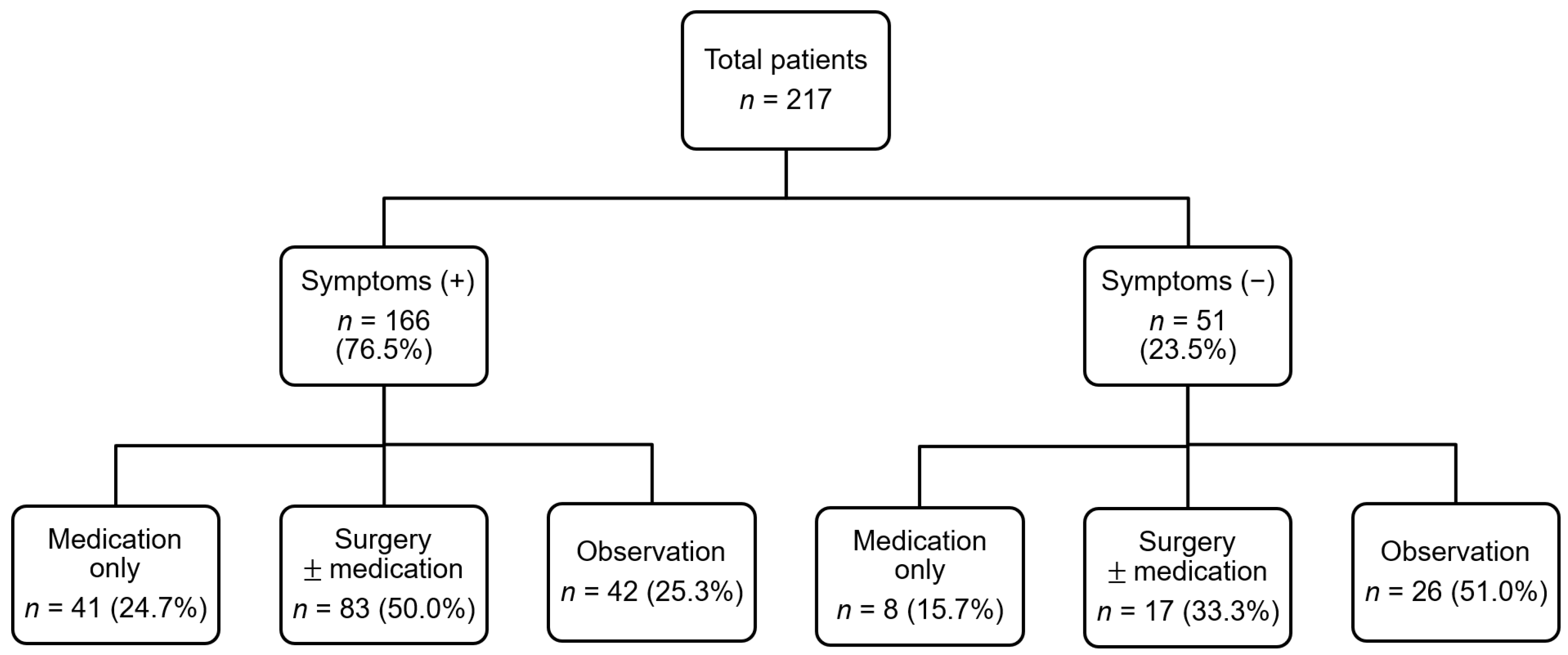
3.1. Demographic and Clinical Characteristics of Patients with Dental Malocclusion

The demographic and clinical characteristics of the enrolled patients are summarised in Table 1. Of the 217 patients, 166 patients (76.5%) had rhinological symptoms: nasal obstruction (n = 125, 57.6%), rhinorrhoea (n = 104, 47.9%), sneezing (n = 82, 37.8%), and itching (n = 60, 27.6%). The mean SNOT-22 scores of the patients were 6.8 ± 6.4, 1.8 ± 2.7, 6.5 ± 6.0, and 1.5 ± 2.5 in the rhinological, otofacial, sleep dysfunction, and psychological dysfunction domains, respectively. Physical examination revealed abnormal findings in 202 (93.1%) patients: inferior turbinate hypertrophy (n = 178, 82.0%), T&A hypertrophy (n = 69, 31.8%), and deviated nasal septum (n = 16, 7.4%). A total of 149 (68.7%) patients underwent medical or surgical treatment for rhinological symptoms. While 49 (22.6%) patients received only medical treatment, 100 (46.1%) patients received surgical treatment: adenotonsillectomy (n = 21, 9.7%), turbinoplasty (n = 30, 13.8%), and adenotonsillectomy with concurrent turbinoplasty (n = 49, 22.6%).
The clinical courses of the study patients are outlined in Figure 1. Of the 166 symptomatic patients, 124 (74.7%) patients underwent medical or surgical treatment, while only 25 (49.0%) patients underwent otolaryngological treatment (Pearson’s chi-square test, p < 0.001). However, there were no significant differences in the incidence of positive SPT between symptomatic patients (n = 136, 81.9%) and asymptomatic patients (n = 37, 72.5%).
As described above, nasal obstruction was the most common symptom of patients with dental malocclusion. Among the 125 patients with nasal obstruction, 115 (92.0%) patients underwent pharmacological or surgical treatment. Of the 92 patients without nasal obstruction, only 34 (36.9%) patients underwent medical or surgical treatment. There was a statistically significant difference in the proportion of patients that received medical or surgical treatment between the two groups (Table 2).

3.2. Comparison between Patients with Positive and Negative SPT

Among the 173 (79.7%) SPT (+) patients, 136 (78.6%) patients had rhinological symptoms: nasal airway obstruction (n = 101), rhinorrhoea (n = 94), sneezing (n = 77), and itching (n = 52). Of the 44 SPT (−) patients, 30 (68.1%) patients had symptoms of nasal obstruction (n = 24), rhinorrhoea (n = 10), sneezing (n = 5), and itching (n = 8).
SPT (+) patients had a significantly higher rate of rhinorrhoea and sneezing than SPT (−) patients (p < 0.001). The prevalence of IT hypertrophy was significantly higher in the SPT (+) group than in the SPT (−) group (p = 0.007) (Table 3). However, there were no significant differences between the two groups in the prevalence of nasal obstruction, itching, T&A hypertrophy, deviated nasal septum, and medical/surgical treatment rates.

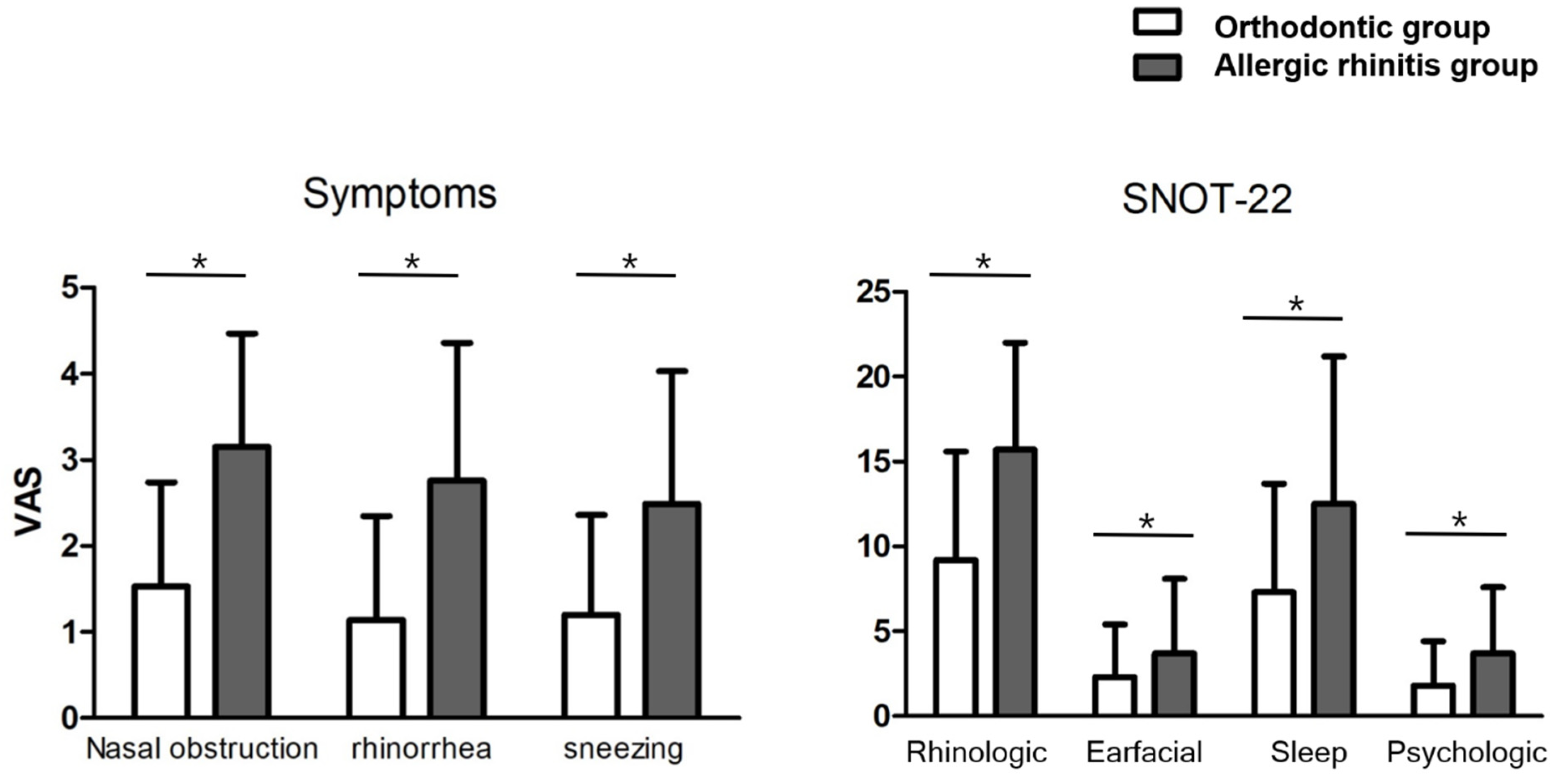
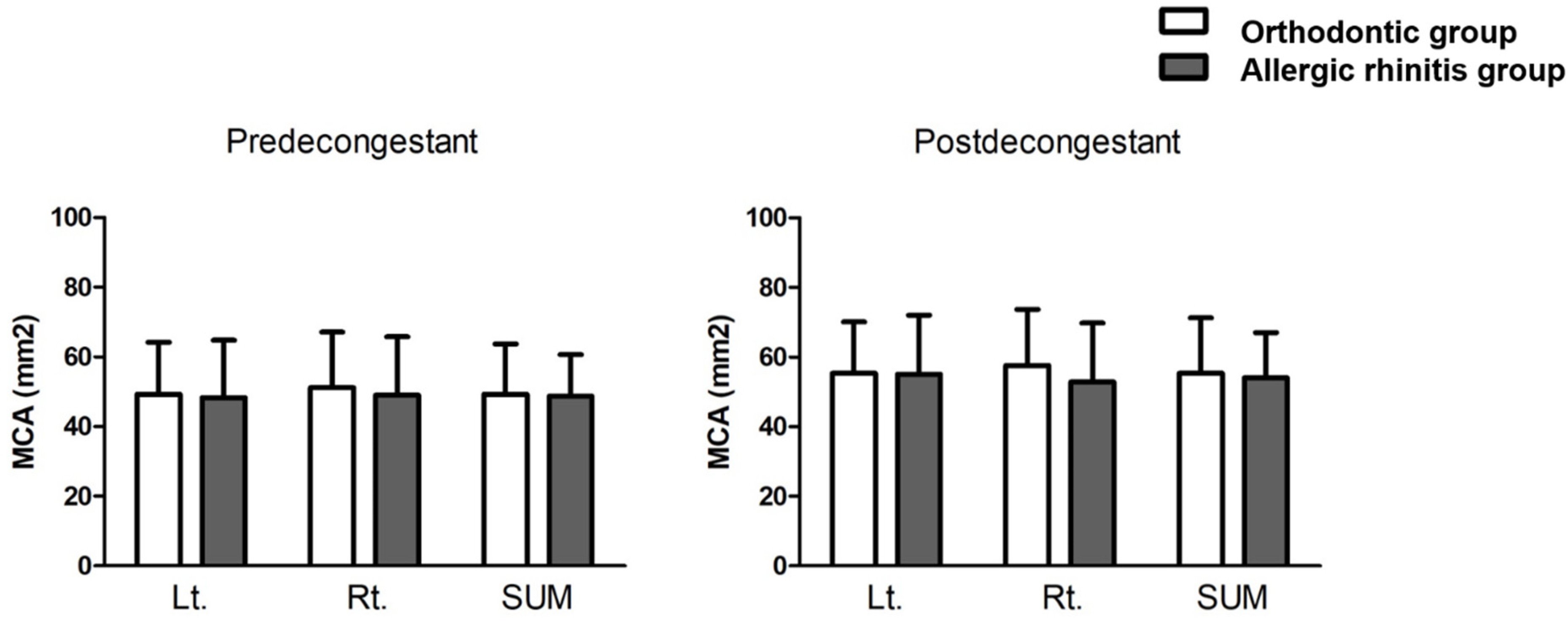
3.3. Comparison between Patients in the Orthodontic and AR Groups

As described above, 173 patients who were being treated for AR in our department were randomly assigned by age and sex matching and defined as the “AR” group. The subjective and objective assessment parameters of patients from the orthodontic and AR groups were compared (Table 4). The frequency of nasal obstruction (p < 0.001), rhinorrhoea (p = 0.002), and sneezing (p < 0.001) was significantly higher in the AR group than in the orthodontic group. On the other hand, the prevalence of abnormal physical examination findings (T&A hypertrophy, IT hypertrophy, and deviated nasal septum) was not significantly different between the two groups. The mean symptom scores of nasal obstruction, rhinorrhoea, and sneezing were significantly higher in the AR group than in the orthodontic group: 3.0 ± 1.4 vs. 2.0 ± 1.4, 2.5 ± 1.6 vs. 1.5 ± 1.5, and 2.3 ± 1.6 vs. 1.5 ± 1.3, respectively (Figure 2, p < 0.001).
The mean SNOT-22 scores of patients in the AR group were significantly higher than those of patients in the orthodontic group in all domains (rhinological: 15.4 ± 6.9 vs. 9.5 ± 6.6, otofacial: 3.4 ± 4.3 vs. 1.9 ± 2.9, sleep: 11.7 ± 8.5 vs. 7.2 ± 6.2, psychological: 3.4 ± 4.0 vs. 1.7 ± 2.7, and total: 39.3 ± 23.6 vs. 23.4 ± 17.0; Figure 2; p < 0.001).
Values related to acoustic rhinometry were also compared between the two groups. The mean MCA values of the right and left nasal cavities in patients from the orthodontic group were 47.9 ± 16.7 mm2 and 46.1 ± 16.5 mm2, respectively. Following mucosal decongestion, these values changed to 54.0 ± 16.4 mm2 and 52.2 ± 16.2 mm2, respectively. The mean MCA values of the right and left nasal cavities in patients from the AR group were 45.7 ± 16.3 mm2 and 46.1 ± 16.0 mm2 in the right and left nasal cavities, respectively. Following mucosal decongestion, these values changed to 51.3 ± 15.8 mm2 and 52.4 ± 16.8 mm2, respectively. We found no significant difference in the mean MCA values between the two groups, suggesting that patients with dental malocclusion might have structural or constitutional problems of the nasal cavity, although the severity of symptoms was slightly less in the orthodontic group than in the AR group (Figure 3).

4. Discussion

In this study, we examined the clinical characteristics of patients with dental malocclusion from an otolaryngologic perspective and evaluated the correlation between AR and dental malocclusion. The incidence of SPT (+) (79.7%), hypertrophied inferior turbinate (82.0%), and T&A hypertrophy (31.8%) in the orthodontic group exceeded the values observed in epidemiological studies in the general population [16,17,18,19]. We found a high prevalence of nasal symptoms and abnormal physical findings even in SPT (−) patients with dental malocclusion. Hypertrophied tonsils, adenoids, and inferior turbinates can cause mouth breathing by obstructing the nasal airway [20,21,22]. These physical findings should be considered as risk factors for dental malocclusion. However, the severity of symptoms was lower among patients in the orthodontic group than those in the AR group. The mean MCA of the nasal cavities measured using acoustic rhinometry was not significantly different between the two groups. These values are similar to those reported in the general population of the same age group in South Korea [23].
Our findings suggest that constitutional or structural problems of upper airway seem to affect the development of dental malocclusions in patients undergoing orthodontic treatment. Upper airway obstruction that induces mouth breathing can lead to the development of malocclusion, by causing an imbalance between the forces from the cheeks and the tongue and an altered position of the mandible [24,25]. Upper airway obstruction and mouth breathing can be induced by many different factors which are responsible for the obstruction of air reflux inside the nasal cavity, deformities of the nasal septum, T&A hypertrophy, nasal polyposis, and AR [26,27].
Asymptomatic AR is believed to lead to persistent inflammation of the nasal mucosa [28]. As this minimal persistent inflammation (MPI) may contribute to co-morbidities of AR such as asthma, it is believed that the treatment strategy may need to include MPI management [29]. For patients with subclinical AR, three major options may be used to treat MPI: antihistamines, anti-leukotrienes, and intranasal corticosteroids [30].
Untreated subclinical AR might contribute to the development of dental malocclusion. Our study suggests that the early detection and treatment of subclinical AR, other rhinological problems, and structural abnormalities of the upper airway in patients with malocclusion may help us manage malocclusion from an otolaryngologic perspective.
Several studies have investigated the association between the type of occlusion and the prevalence of malocclusion in children with nasal obstruction using cephalometric analysis. However, the results of these studies are inconsistent, as the prevalence of malocclusion in children with rhinitis ranges from as high as 78.2% to as low as 3% [31]. Furthermore, few authors have assessed the prevalence of AR and otolaryngologic features in children with dental malocclusion. Luzzi et al. [10] compared 125 patients with malocclusion with 150 healthy patients. They found that children with AR had a three-fold higher risk of developing one or more dento-skeletal alterations. Imbaud et al. [32] evaluated 89 patients who underwent orthodontic treatment and observed a higher prevalence of rhinitis and hypersensitivity to airborne allergens in SPT. However, previous studies, including those described above, did not assess the symptoms or structural abnormalities of AR using quantitative parameters. To our knowledge, this is the first study to compare an orthodontic group with an age- and sex-matched AR group using both subjective and objective quantitative parameters, such as the SNOT-22 questionnaire and acoustic rhinometry. Patients with dental malocclusion in this study had higher prevalences of structural abnormalities on the upper airways and higher rates of SPT (+). Additionally, the results of the acoustic rhinometry showed no differences between the orthodontic group and the AR group. These results imply that structural upper airway abnormalities and subclinical AR may affect dental malocclusion development. Thus, managing dental malocclusions may be assisted by the early detection and treatment of these otolaryngologic problems.
However, the present study has some limitations. Acoustic rhinometry has not yet been accepted as a standard evaluation method because of its unstandardized results [33]. In addition, the results of acoustic rhinometry might be inconsistent due to the nasal cycle, which is a physiological phenomenon that causes a periodic change in nasal airway patency. Thus, future studies must analyse nasal cavity structures using imaging techniques such as computed tomography. In addition, there are various types of malocclusions. Therefore, further research is required to establish the association between AR and the different types of dental malocclusions. Furthermore, a prospective study evaluating the effect of treatment of AR on the development of dental malocclusion is needed to better understand the correlation between AR and dental malocclusion.

5. Conclusions

Our study showed higher prevalence of upper airway structural abnormalities and a higher SPT (+) rate among patients with dental malocclusion. The early detection and treatment of subclinical AR, other rhinological problems, and structural abnormalities of the upper airway in patients with malocclusion may help us manage dental malocclusion.

Author Contributions

J.-H.M. designed the work; S.H.Y. and J.H.C. acquired and analysed data; S.H.Y. and J.-H.M. drafted, revised, and approved the manuscript; J.-H.M. was involved in all aspects of the work. All authors have read and agreed to the published version of the manuscript.

Funding

This research was supported by the Basic Science Research Program through the National Research Foundation of Korea (NRF), funded by the Ministry of Education [2021R1I1A1A01052298]. This research was supported by a grant of the Korea Health Technology R&D Project through the Korea Health Industry Development Institute (KHIDI), funded by the Ministry of Health & Welfare, Republic of Korea (grant number: HI22C0612). This research was supported by the Basic Science Research Program through the National Research Foundation of Korea (NRF), funded by the Ministry of Science and ICT (NRF-2020R1A2C1012105). This research was supported by Basic Science Research Program through the National Research Foundation of Korea (NRF) funded by the Ministry of Education (NRF-2020R1A6A1A03043283). This research was supported by a grant of the Korea Health Technology R&D Project through the Korea Health Industry Development Institute (KHIDI), funded by the Ministry of Health & Welfare, Republic of Korea (grant number: HI20C2088). This work was supported by the Technology Innovation Program (20016884, Development of a multiband Laser Patch devices with micro needle type optical transmission structure for symptom improvement of allergic rhinitis) funded By the Ministry of Trade, Industry & Energy (MOTIE, Korea).

Institutional Review Board Statement

This study was approved by the Institutional Review Board of our institution (IRB No. 2021-04-027).

Informed Consent Statement

Written informed consents were obtained from all participants.

Data Availability Statement

The data that support the findings of this study are available from the corresponding author, upon reasonable request.

Conflicts of Interest

The authors declare no conflict of interest.

Abbreviations

ARallergic rhinitis
ARIAAllergic Rhinitis and its Impact on Asthma
MPIminimal persistent inflammation
SNOT-22sinonasal outcome test-22 questionnaire
SPTskin prick test
T&Atonsil and adenoid
VASvisual analogue scale

References

  1. Moss, M.L. A theoretical analysis of the functional matrix. Acta Biotheor. 1968, 18, 195–202. [Google Scholar] [CrossRef] [PubMed]
  2. Rubin, R.M. Mode of respiration and facial growth. Am. J. Orthod. 1980, 78, 504–510. [Google Scholar] [CrossRef]
  3. Solow, B.; Kreiborg, S. Soft-tissue stretching: A possible control factor in craniofacial morphogenesis. Scand. J. Dent. Res. 1977, 85, 505–507. [Google Scholar] [CrossRef] [PubMed]
  4. Valera, F.C.; Travitzki, L.V.; Mattar, S.E.; Matsumoto, M.A.; Elias, A.M.; Anselmo-Lima, W.T. Muscular, functional and orthodontic changes in pre school children with enlarged adenoids and tonsils. Int. J. Pediatr. Otorhinolaryngol. 2003, 67, 761–770. [Google Scholar] [CrossRef]
  5. Sousa, J.B.; Anselmo-Lima, W.T.; Valera, F.C.; Gallego, A.J.; Matsumoto, M.A. Cephalometric assessment of the mandibular growth pattern in mouth-breathing children. Int. J. Pediatr. Otorhinolaryngol. 2005, 69, 311–317. [Google Scholar] [CrossRef] [PubMed]
  6. Harvold, E.P.; Tomer, B.S.; Vargervik, K.; Chierici, G. Primate experiments on oral respiration. Am. J. Orthod. 1981, 79, 359–372. [Google Scholar] [CrossRef]
  7. Vig, K.W. Nasal obstruction and facial growth: The strength of evidence for clinical assumptions. Am. J. Orthod. Dentofac. Orthop. 1998, 113, 603–611. [Google Scholar] [CrossRef]
  8. Bellanti, J.A.; Wallerstedt, D.B. Allergic rhinitis update: Epidemiology and natural history. Allergy Asthma Proc. 2000, 21, 367–370. [Google Scholar] [CrossRef] [PubMed]
  9. Sadeghi-Shabestari, M.; Jabbari Moghaddam, Y.; Ghaharri, H. Is there any correlation between allergy and adenotonsillar tissue hypertrophy? Int. J. Pediatr. Otorhinolaryngol. 2011, 75, 589–591. [Google Scholar] [CrossRef]
  10. Luzzi, V.; Ierardo, G.; Viscogliosi, A.; Fabbrizi, M.; Consoli, G.; Vozza, I.; Vestri, A.; Polimeni, A. Allergic rhinitis as a possible risk factor for malocclusion: A case-control study in children. Int. J. Paediatr. Dent. 2013, 23, 274–278. [Google Scholar] [CrossRef]
  11. Bresolin, D.; Shapiro, P.A.; Shapiro, G.G.; Chapko, M.K.; Dassel, S. Mouth breathing in allergic children: Its relationship to dentofacial development. Am. J. Orthod. 1983, 83, 334–340. [Google Scholar] [CrossRef]
  12. Brożek, J.L.; Bousquet, J.; Agache, I.; Agarwal, A.; Bachert, C.; Bosnic-Anticevich, S.; Brignardello-Petersen, R.; Canonica, G.W.; Casale, T.; Chavannes, N.H.; et al. Allergic Rhinitis and its Impact on Asthma (ARIA) guidelines-2016 revision. J. Allergy Clin. Immunol. 2017, 140, 950–958. [Google Scholar] [CrossRef] [PubMed] [Green Version]
  13. Kennedy, J.L.; Hubbard, M.A.; Huyett, P.; Patrie, J.T.; Borish, L.; Payne, S.C. Sino-nasal outcome test (SNOT-22): A predictor of postsurgical improvement in patients with chronic sinusitis. Ann. Allergy Asthma Immunol. 2013, 111, 246–251.e2. [Google Scholar] [CrossRef] [PubMed] [Green Version]
  14. Camacho, M.; Zaghi, S.; Certal, V.; Abdullatif, J.; Means, C.; Acevedo, J.; Liu, S.; Brietzke, S.E.; Kushida, C.A.; Capasso, R. Inferior turbinate classification system, grades 1 to 4: Development and validation study. Laryngoscope 2015, 125, 296–302. [Google Scholar] [CrossRef] [PubMed]
  15. Chung, Y.J.; Cho, I.K.; Lee, K.I.; Bae, S.H.; Lee, J.W.; Chung, P.S.; Mo, J.H. Seasonal specificity of seasonal allergens and validation of the ARIA classification in Korea. Allergy Asthma Immunol. Res. 2013, 5, 75–80. [Google Scholar] [CrossRef] [PubMed] [Green Version]
  16. Kuprys-Lipinska, I.; Elgalal, A.; Kuna, P. Skin prick test with inhaled allergens in the general population of Lodz province. Pneumonol. Alergol. Pol. 2009, 77, 229–234. [Google Scholar] [CrossRef] [PubMed]
  17. Kara, C.O.; Ergin, H.; Kocak, G.; Kilic, I.; Yurdakul, M. Prevalence of tonsillar hypertrophy and associated oropharyngeal symptoms in primary school children in Denizli, Turkey. Int. J. Pediatr. Otorhinolaryngol. 2002, 66, 175–179. [Google Scholar] [CrossRef]
  18. Min, Y.G. Pathophysiology, diagnosis, and treatment of allergic rhinitis. Korean J. Otorhinolaryngol.-Head Neck Surg. 2013, 56, 256–265. [Google Scholar] [CrossRef]
  19. Bixler, E.O.; Vgontzas, A.N.; Lin, H.M.; Liao, D.; Calhoun, S.; Vela-Bueno, A.; Fedok, F.; Vlasic, V.; Graff, G. Sleep disordered breathing in children in a general population sample: Prevalence and risk factors. Sleep 2009, 32, 731–736. [Google Scholar] [CrossRef] [Green Version]
  20. Koca, C.F.; Erdem, T.; Bayindir, T. The effect of adenoid hypertrophy on maxillofacial development: An objective photographic analysis. J. Otolaryngol. Head Neck Surg. 2016, 45, 48. [Google Scholar] [CrossRef] [PubMed]
  21. Kallunki, J.; Marcusson, A.; Ericsson, E. Tonsillotomy versus tonsillectomy—A randomized trial regarding dentofacial morphology and post-operative growth in children with tonsillar hypertrophy. Eur. J. Orthod. 2014, 36, 471–478. [Google Scholar] [CrossRef] [PubMed] [Green Version]
  22. Gungor, A.Y.; Turkkahraman, H. Effects of airway problems on maxillary growth: A review. Eur. J. Dent. 2009, 3, 250–254. [Google Scholar] [CrossRef] [PubMed] [Green Version]
  23. Kim, Y.K.; Ro, Y.S.; Kang, J.H.; Kim, J.T.; Kwon, D.H. The change of nasal resistance according to the age and sex in Koreans. Korean J. Otorhinolaryngol.-Head Neck Surg. 1995, 38, 711–717. [Google Scholar]
  24. Cheng, M.C.; Enlow, D.H.; Papsidero, M.; Broadbent, B.H., Jr.; Oyen, O.; Sabat, M. Developmental effects of impaired breathing in the face of the growing child. Angle Orthod. 1988, 58, 309–320. [Google Scholar]
  25. Freng, A. Restricted nasal respiration, influence on facial growth. Int. J. Pediatr. Otorhinolaryngol. 1979, 1, 249–254. [Google Scholar] [CrossRef]
  26. Adamidis, I.P.; Spyropoulos, M.N. The effects of lymphadenoid hypertrophy on the position of the tongue, the mandible and the hyoid bone. Eur. J. Orthod. 1983, 5, 287–294. [Google Scholar] [CrossRef]
  27. Abreu, R.R.; Rocha, R.L.; Lamounier, J.A.; Guerra, A.F. Etiology, clinical manifestations and concurrent findings in mouth-breathing children. J. Pediatr. 2008, 84, 529–535. [Google Scholar] [CrossRef]
  28. Ciprandi, G.; Buscaglia, S.; Pesce, G.; Pronzato, C.; Ricca, V.; Parmiani, S.; Bagnasco, M.; Canonica, G.W. Minimal persistent inflammation is present at mucosal level in patients with asymptomatic rhinitis and mite allergy. J. Allergy Clin. Immunol. 1995, 96 Pt 1, 971–979. [Google Scholar] [CrossRef]
  29. Storms, W.W. Minimal persistent inflammation, an emerging concept in the nature and treatment of allergic rhinitis: The possible role of leukotrienes. Ann. Allergy Asthma Immunol. 2003, 91, 131–140. [Google Scholar] [CrossRef]
  30. Canonica, G.W.; Compalati, E. Minimal persistent inflammation in allergic rhinitis: Implications for current treatment strategies. Clin. Exp. Immunol. 2009, 158, 260–271. [Google Scholar] [CrossRef]
  31. Occasi, F.; Perri, L.; Saccucci, M.; Di Carlo, G.; Ierardo, G.; Luzzi, V.; De Castro, G.; Brindisi, G.; Loffredo, L.; Duse, M.; et al. Malocclusion and rhinitis in children: An easy-going relationship or a yet to be resolved paradox? A systematic literature revision. Ital. J. Pediatr. 2018, 44, 100. [Google Scholar] [CrossRef] [PubMed] [Green Version]
  32. Imbaud, T.C.; Mallozi, M.C.; Domingos, V.B.; Sole, D. Frequency of rhinitis and orofacial disorders in patients with dental malocclusion. Rev. Paul. Pediatr. 2016, 34, 184–188. [Google Scholar] [CrossRef] [PubMed]
  33. Corey, J.P. Acoustic rhinometry: Should we be using it? Curr. Opin. Otolaryngol. Head Neck Surg. 2006, 14, 29–34. [Google Scholar] [CrossRef] [PubMed]
Figure 1. Clinical course of the study participants.
Jcm 11 06318 g001
Figure 2. Comparison of visual analogue score (VAS) for symptoms of allergic rhinitis and sino-nasal outcome test score (SNOT-22) between the orthodontic group and the allergic rhinitis group. A paired t-test was used to compare the two groups. (* p < 0.001).
Jcm 11 06318 g002
Figure 3. Comparison of minimal cross-sectional areas of nasal cavities measured in pre-decongestant and post-decongestant states between the orthodontic and allergic rhinitis groups. Paired t-test was used to compare the two groups. MCA, minimal cross-sectional area; Lt., left; Rt., right; SUM, average value of right and left nasal cavity.
Jcm 11 06318 g003
Table 1. Demographic and clinical characteristics of patients.
VariableTotal Orthodontic Patients (n = 217)
Median age (range), y13.8 (5–29)
Sex, n (%)
Male102 (47.0)
 Female115 (53.0)
Symptoms, n (%)166 (76.5)
 Nasal obstruction125 (57.6)
 Rhinorrhea104 (47.9)
 Sneezing82 (37.8)
 Itching60 (27.6)
 Sleep disturbance15 (6.9)
Abnormal PE findings, n (%)202 (93.1)
 IT hypertrophy178 (82.0)
 T&A hypertrophy69 (31.8)
 Deviated nasal septum16 (7.4)
Treatment, n (%)149 (68.7)
 Medical treatment49 (22.6)
 Surgical treatment100 (46.1)
 Adenotonsillectomy21 (9.7)
 Turbinoplasty30 (13.8)
 Adenotonsillectomy with turbinoplasty49 (22.6)
Abbreviations: PE, physical examination; IT, inferior turbinate; T&A, tonsillar and adenoid.
Table 2. Treatments performed in patients with and without nasal obstruction.
VariableTreatment, n (%)No TreatmentpaOdds Ratio
MedicationSurgery(95% CI)
Nasal obstruction (+), (n = 125)22 (17.6)93 (74.4)10 (8.0)<0.0013.7
(2.7–5.1)
Nasal obstruction (–), (n = 92)27 (29.3)7 (7.6)58 (63.1)
a Compared between nasal obstruction (+) and nasal obstruction (−) patients using Pearson’s chi-square test. Abbreviations: CI, confidence interval.
Table 3. Comparison of demographic and clinical characteristics between skin prick test (+) and skin prick test (−) patients.
VariableTotal Orthodontic Patients (n = 217)paOdds Ratio a (95% CI)
SPT (+)SPT (–)
Total, n (%)173 (79.7)44 (20.3)
Rhinologic Sx, n (%)136 (78.6)30 (68.2)0.1451.7 (0.8–3.6)
 Nasal obstruction101 (58.4)24 (54.5)0.6461.2 (0.6–2.3)
 Rhinorrhea94 (54.3)10 (22.7)<0.0014.0 (1.9–8.7)
 Sneezing77 (44.5)5 (11.4)<0.0016.3 (2.4–16.6)
 Itching52 (30.1)8 (18.2)0.1161.9 (0.8–4.4)
Abnormal PE findings, n (%)
 IT hypertrophy148 (85.5)30 (68.2)0.0072.8 (1.3–5.9)
T&A hypertrophy60 (34.7)9 (20.5)0.072.1 (0.9–4.6)
Deviated nasal septum13 (7.5)3 (6.8)0.8751.1 (0.3–4.1)
Treatment, n (%)123 (71.1)26 (59.1)0.1250.6 (0.3–1.2)
 Medical treatment41 (33.7)8 (18.2)0.4341.4 (0.6–3.2)
 Surgical treatment82 (35.8)18 (40.9)0.4411.3 (0.7–2.5)
 Adenotonsillectomy15 (8.7)6 (13.6)0.3200.6 (0.2–1.7)
 Turbinoplasty26 (15.2)4 (9.1)0.3081.8 (0.6–5.4)
 Adenotonsillectomy with turbinoplasty41 (30.4)8 (18.2)0.4341.4 (0.6–3.2)
a Compared between skin prick test (+) orthodontic patients and control group using Pearson’s chi-square test or Fisher’s exact test. Abbreviations: SPT, skin prick test; Sx, symptoms; PE, physical examination; IT, inferior turbinate; T&A, tonsillar and adenoid; CI, confidence interval.
Table 4. Comparison of subjective/objective findings between the skin prick test (+) orthodontic group and the allergic rhinitis (control) group.
VariableOrthodontic GroupControl Groupp Value aOdds Ratio a (95% CI)
Rhinologic Sx, n (%)136 (78.6)173 (100)
 Nasal obstruction101 (58.4)149 (86.1)<0.0014.4 (2.6–7.5)
 Rhinorrhea94 (54.3)122 (70.5)0.0022.0 (1.3–3.1)
 Sneezing77 (44.5)117 (67.6)<0.001 a2.6 (1.7–4.0)
 Itching52 (30.1)61 (35.3)0.3020.8 (0.5–1.2)
Abnormal PE findings, n (%)
 IT hypertrophy148 (85.5)158 (91.3)0.0930.6 (0.3–1.1)
 T&A hypertrophy60 (34.7)60 (34.7)1.0001.0 (0.6–1.6)
Deviated nasal septum13 (7.5)22 (12.7)0.1090.6 (0.3–1.1)
a Compared between skin prick test (+) orthodontic patients and age- and sex-matched control group using Pearson’s chi-square test. Abbreviations: CI, confidence interval; Sx, symptoms; PE, physical examination; IT, inferior turbinate; T&A, tonsillar and adenoid.
Publisher’s Note: MDPI stays neutral with regard to jurisdictional claims in published maps and institutional affiliations.

