Utilizing Computed Tomography to Analyze the Morphomic Change between Patients with Localized and Metastatic Renal Cell Carcinoma: Body Composition Varies According to Cancer Stage
 , , ,
, , , .jpg) and
and
Abstract
1. Introduction
2. Materials and Methods
3. Results
4. Discussion
5. Conclusions
Author Contributions
Funding
Institutional Review Board Statement
Informed Consent Statement
Data Availability Statement
Conflicts of Interest
Abbreviations
| BMI | Body mass index |
| CT | Computed tomography |
| SAT | Subcutaneous adipose tissue |
| VAT | Visceral adipose tissue |
| SMT | Skeletal muscle tissue |
| RCC | Renal cell carcinoma |
| ccRCC | Clear cell renal cell carcinoma |
| pRCC | Papillary renal cell carcinoma |
| chRCC | Chromophobe renal cell carcinoma |
References
- DeBerardinis, R.J.; Chandel, N.S. Fundamentals of cancer metabolism. Sci. Adv. 2016, 2, e1600200. [Google Scholar] [CrossRef] [Scilit] [PubMed]
- Baracos, V.E.; Martin, L.; Korc, M.; Guttridge, D.C.; Fearon, K.C. Cancer-associated cachexia. Nat. Rev. Dis. Primers 2018, 4, 17105. [Google Scholar] [CrossRef] [Scilit] [PubMed]
- Moch, H.; Cubilla, A.L.; Humphrey, P.A.; Reuter, V.E.; Ulbright, T.M. The 2016 WHO Classification of tumours of the urinary System and male genital organs-Part A: Renal, penile, and testicu- lar tumours. Eur. Urol. 2016, 70, 93–105. [Google Scholar] [CrossRef] [Scilit] [PubMed]
- Bray, F.; Ferlay, J.; Soerjomataram, I.; Siegel, R.L.; Torre, L.A.; Jemal, A. Global cancer statistics 2018: GLOBOCAN estimates of incidence and mortality worldwide for 36 cancers in 185 countries. CA Cancer J. Clin. 2018, 68, 394–424. [Google Scholar] [CrossRef] [Scilit]
- Hsieh, J.J.; Purdue, M.P.; Signoretti, S.; Swanton, C.; Albiges, L.; Schmidinger, M.; Heng, D.Y.; Larkin, J.; Ficarra, V. Renal cell carcinoma. Nat. Rev. Dis. Primers 2017, 3, 17009. [Google Scholar] [CrossRef] [Scilit] [PubMed]
- Petejova, N.; Martinek, A. Renal cell carcinoma: Review of etiology, pathophysiology and risk factors. Biomed. Pap. Med. Fac. Univ. Palacky Olomouc. Czech Repub. 2016, 160, 183–194. [Google Scholar] [CrossRef] [Scilit]
- Otunctemur, A.; Dursun, M.; Ozer, K.; Horsanali, O.; Ozbek, E. Renal cell carcinoma and visceral adipose index: A new risk parameter. Int. Braz. J. Urol. 2016, 42, 955–959. [Google Scholar] [CrossRef] [Scilit] [PubMed]
- Lee, H.W.; Jeong, B.C.; Seo, S.I.; Jeon, S.S.; Lee, H.M.; Choi, H.Y.; Jeon, H.G. Prognostic significance of visceral obesity in patients with advanced renal cell carcinoma undergoing nephrectomy. Int. J. Urol. 2015, 22, 455–461. [Google Scholar] [CrossRef] [Scilit] [PubMed]
- Mizuno, R.; Miyajima, A.; Hibi, T.; Masuda, A.; Shinojima, T.; Kikuchi, E.; Jinzaki, M.; Oya, M. Impact of baseline visceral fat accumulation on prognosis in patients with metastatic renal cell carcinoma treated with systemic therapy. Med. Oncol. 2017, 34, 47. [Google Scholar] [CrossRef] [Scilit]
- Nelke, C.; Dziewas, R.; Minnerup, J.; Meuth, S.G.; Ruck, T. Skeletal muscle as potential central link between sarcopenia and immune senescence. EBioMedicine 2019, 49, 381–388. [Google Scholar] [CrossRef] [Scilit]
- Buchler, T.; Kopecka, M.; Zemankova, A.; Wiesnerová, M.; Streckova, E.; Rozsypalova, A.; Melichar, B.; Poprach, A.; Richter, I. Sarcopenia in metastatic renal cell carcinoma patients treated with cabozantinib. Target. Oncol. 2020, 15, 673–679. [Google Scholar] [CrossRef] [Scilit] [PubMed]
- Darbas, T.; Forestier, G.; Leobon, S.; Pestre, J.; Jesus, P.; Lachatre, D.; Tubiana-Mathieu, N.; Descazeaud, A.; Deluche, E. Impact of body composition in overweight and obese patients with localised renal cell carcinoma. In Vivo 2020, 34, 2873–2881. [Google Scholar] [CrossRef] [Scilit] [PubMed]
- Watanabe, S.; Ishihara, H.; Takagi, T.; Kondo, T.; Ishiyama, R.; Fukuda, H.; Yoshida, K.; Iizuka, J.; Kobayashi, H.; Ishida, H.; et al. Impact of sarcopenia on post-operative outcomes following nephrectomy and tumor thrombectomy for renal cell carci- noma with inferior vena cava thrombus. Jpn. J. Clin. Oncol. 2021, 51, 819–825. [Google Scholar] [CrossRef] [Scilit] [PubMed]
- Fedorov, A.; Beichel, R.; Kalpathy-Cramer, J.; Finet, J.; Fillion-Robin, J.-C.; Pujol, S.; Bauer, C.; Jennings, D.; Fennessy, F.; Sonka, M.; et al. 3D Slicer as an image computing platform for the Quantitative Imaging Network. Magn. Reson. Imaging 2012, 30, 1323–1341. [Google Scholar] [CrossRef] [Scilit]
- Yoshizumi, T.; Nakamura, T.; Yamane, M.; Islam, A.H.M.W.; Menju, M.; Yamasaki, K.; Arai, T.; Kotani, K.; Funahashi, T.; Yamashita, S.; et al. Abdominal fat: Standardized technique for measurement at CT. Radiology 1999, 211, 283–286. [Google Scholar] [CrossRef] [Scilit] [PubMed]
- Batista, M.L., Jr.; Henriques, F.S.; Neves, R.X.; Olivan, M.R.; Matos-Neto, E.M.; Alcântara, P.S.; Maximiano, L.F.; Otoch, J.P.; Alves, M.J.; Seelaender, M. Cachexia-associated adipose tissue morphological rearrangement in gastrointestinal cancer patients. J. Cachexia Sar- Copenia Muscle 2016, 7, 37–47. [Google Scholar] [CrossRef] [Scilit] [PubMed]
- Leibovitz, E.; Ben-David, N.; Shibanov, L.; Elias, S.; Shimonov, M. Visceral adiposity but not subcutaneous fat associated with improved outcome of patients with acute cholecystitis. J. Surg. Res. 2018, 225, 15–20. [Google Scholar] [CrossRef] [Scilit]
- Sato, F.; Maeda, N.; Yamada, T.; Namazui, H.; Fukuda, S.; Natsukawa, T.; Nagao, H.; Murai, J.; Masuda, S.; Tanaka, Y.; et al. Association of epicardial, visceral, and subcutaneous fat with cardiometabolic diseases. Circ. J. 2018, 82, 502–508. [Google Scholar] [CrossRef] [Scilit]
- Merlotti, C.; Ceriani, V.; Morabito, A.; Pontiroli, A.E. Subcutaneous fat loss is greater than visceral fat loss with diet and exercise, weight-loss promoting drugs and bariatric surgery: A critical review and meta-analysis. Int. J. Obes. 2017, 41, 672–682. [Google Scholar] [CrossRef] [Scilit]
- Baracos, V.E.; Mazurak, V.C.; Bhullar, A.S. Cancer cachexia is defined by an ongoing loss of skeletal muscle mass. Ann Palliat. Med. 2019, 8, 3–12. [Google Scholar] [CrossRef] [Scilit] [PubMed]
- Friesen, D.E.; Baracos, V.E.; Tuszynski, J.A. Modeling the energetic cost of cancer as a result of altered energy metabolism: Implications for cachexia. Theor. Biol. Med. Model. 2015, 12, 17. [Google Scholar] [CrossRef] [Scilit] [PubMed]
- Bal, N.C.; Maurya, S.K.; Sopariwala, D.H.; Sahoo, S.K.; Gupta, S.C.; Shaikh, S.A.; Pant, M.; Rowland, L.A.; Bombardier, E.; Goonasekera, S.A.; et al. Sarcolipin is a newly identified regulator of muscle-based thermogenesis in mammals. Nat. Med. 2012, 18, 1575–1579. [Google Scholar] [CrossRef] [Scilit] [PubMed]
- Reig Garcia-Galbis, M.; Rizo Baeza, M.; Cortes Castell, E. [Predictors of weight loss and fat in the dietary management: Sex, age, BMI and consulting assistance]. Nutr. Hosp. 2015, 32, 1028–1035. [Google Scholar] [PubMed]
- Chang, E.; Varghese, M.; Singer, K. Gender and sex differences in adipose tssue. Curr. Diab. Rep. 2018, 18, 69. [Google Scholar] [CrossRef] [Scilit] [PubMed]
- Pillon, N.J.; Krook, A. Innate immune receptors in skeletal muscle metabolism. Exp. Cell Res. 2017, 360, 47–54. [Google Scholar] [CrossRef] [Scilit]
- Ibrahim, M.M. Subcutaneous and visceral adipose tissue: Structural and functional differences. Obes. Rev. 2010, 11, 11–18. [Google Scholar] [CrossRef] [Scilit]
- Schaffler, A.; Scholmerich, J.; Buechler, C. Mechanisms of disease: Adipokines and breast cancer—endocrine and paracrine mechanisms that connect adiposity and breast cancer. Nat. Clin. Pract. Endocrinol. Metab. 2007, 3, 345–354. [Google Scholar] [CrossRef] [Scilit]
- Park, J.; Euhus, D.M.; Scherer, P.E. Paracrine and endocrine effects of adipose tissue on cancer development and progression. Endocr. Rev. 2011, 32, 550–570. [Google Scholar] [CrossRef] [Scilit]
- Greco, F.; Mallio, C.A.; Grippo, R.; Messina, L.; Vallese, S.; Rabitti, C.; Quarta, L.G.; Grasso, R.F.; Beomonte Zobel, B. Increased visceral adipose tissue in male patients with non-clear cell renal cell carcinoma. Radiol. Med. 2020, 125, 538–543. [Google Scholar] [CrossRef] [Scilit]


| Variables | Mean | SD | Range/ Percentage | |||
|---|---|---|---|---|---|---|
| Age | 57.1 | 13.11 | 11–82 | Years old | ||
| Body weight | 67.71 | 12.68 | 29–117 | kg | ||
| Height | 164.1 | 9.02 | 139.0–186.8 | cm | ||
| BMI | 25.17 | 4.01 | 15.01–39.55 | |||
| Tumor-Related Parameters | ||||||
| Clinical T stage | 1a | 109 | 58.0% | |||
| 1b | 33 | 17.6% | ||||
| 2a | 7 | 3.7% | ||||
| 2b | 1 | 0.5% | ||||
| 3a | 35 | 18.6% | ||||
| 3b | 2 | 1.1% | ||||
| 3c | 1 | 0.5% | ||||
| Clinical N stage | 0 | 121 | 64.4% | |||
| 1 | 29 | 15.4% | ||||
| 2 | 1 | 0.5% | ||||
| x | 35 | 18.6% | ||||
| Clinical M stage | 0 | 141 | 75% | |||
| 1 | 47 | 25% | ||||
| Tumor pathology | ccRCC | Stage I | 107 | 72.3% | ||
| Stage II | 2 | 1.4% | ||||
| Stage III | 11 | 7.4% | ||||
| Stage IV | 28 | 18.9% | ||||
| pRCC | Stage I | 13 | 76.5% | |||
| Stage II | 0 | 0% | ||||
| Stage III | 0 | 0% | ||||
| Stage IV | 4 | 23.5% | ||||
| chRCC | Stage I | 12 | 66.7% | |||
| Stage II | 0 | 0% | ||||
| Stage III | 0 | 0% | ||||
| Stage IV | 6 | 33.3% | ||||
| mcRCC | Stage I | 1 | 100% | |||
| Xp11.2 RCC | Stage I | 2 | 100% | |||
| Other | Stage I | 2 | 100% | |||
| Body Composition Parameters | ||||||
| SMT | 1895.19 | 520.83 | 705.69–4148.16 | cm3 | ||
| SAT | 1799.69 | 1070.70 | 5.61–7617.65 | cm3 | ||
| VAT | 1870.86 | 1130.64 | 23.92–5725.61 | cm3 | ||
| SMT at L3 | 140.31 | 31.96 | 57.76–216.15 | cm2 | ||
| SAT at L3 | 138.02 | 70.65 | 38.1–254.4 | cm2 | ||
| VAT at L3 | 139.49 | 79.63 | 1.16–119.6 | cm2 | ||
| Localized RCC (N = 141) | Metastatic RCC (N = 47) | Univariate Analysis | Multi-Variate Analysis | |||||
|---|---|---|---|---|---|---|---|---|
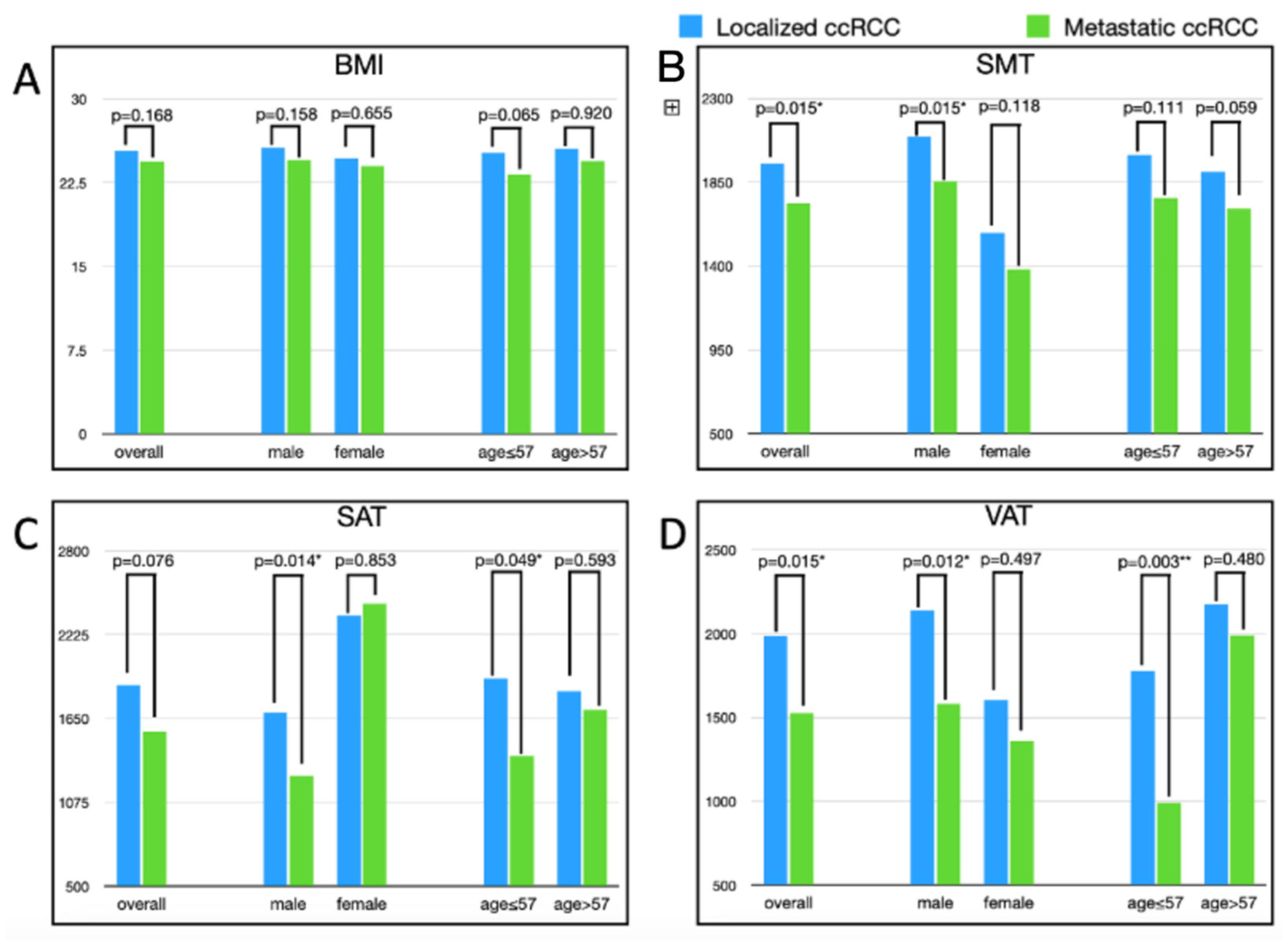
| Mean | SD | Mean | SD | Mean Difference | p-Value | p-Value | ||
| Body weight | kg | 68.7 | 13.14 | 64.96 | 13.43 | −3.74 | 0.096 | 0.799 |
| BMI | kg/m2 | 25.36 | 4.11 | 24.39 | 4.27 | −0.97 | 0.168 | 0.357 |
| SMT | cm3 | 1948.49 | 511.89 | 1735.28 | 520 | −213.21 | 0.015 * | 0.981 |
| SAT | cm3 | 1879.64 | 1038.50 | 1559.81 | 1140 | −319.83 | 0.076 | 0.123 |
| VAT | cm3 | 1986.74 | 1083.09 | 1523.2 | 1209.07 | −463.54 | 0.015 * | 0.020 * |
| SMT-L3 | cm2 | 142.65 | 31.83 | 133.27 | 31.66 | −9.38 | 0.082 | 0.471 |
| SAT-L3 | cm2 | 142.3 | 66.71 | 125.18 | 80.78 | −17.12 | 0.151 | 0.573 |
| VAT-L3 | cm2 | 146.48 | 75.39 | 118.52 | 88.77 | −27.96 | 0.037 * | 0.736 |
| Localized RCC | Metastatic RCC | ||||||
| (Male:Female = 101:40) | (Male:Female = 35:12) | ||||||
| Mean | SD | Mean | SD | Mean Difference | p-Value | ||
| Body weight | Male | 72.29 | 12.18 | 66.65 | 13.16 | −5.64 | 0.023 * |
| Female | 59.82 | 11.17 | 60.05 | 13.56 | 0.23 | 0.953 | |
| BMI | Male | 25.63 | 3.9 | 24.53 | 4.07 | −1.1 | 0.158 |
| Female | 24.69 | 4.58 | 24 | 4.99 | −0.69 | 0.655 | |
| SMT | Male | 2094.8 | 487.26 | 1856.75 | 513 | −238.09 | 0.015 * |
| Female | 1579 | 369.23 | 1381 | 365.33 | −197.98 | 0.118 | |
| SAT | Male | 1690.2 | 924.69 | 1259.32 | 751.6 | −430.87 | 0.014 * |
| Female | 2358 | 1162.48 | 2436.23 | 1601.06 | 78.23 | 0.853 | |
| VAT | Male | 2137.8 | 1070.63 | 1579.8 | 1254.89 | −557.95 | 0.012 * |
| Female | 1605.5 | 1031.41 | 1358.13 | 1098.13 | −247.34 | 0.497 | |
| SMT-L3 | Male | 156.09 | 25.62 | 143.51 | 28.71 | −12.58 | 0.017 * |
| Female | 108.71 | 17.48 | 103.42 | 18.45 | −5.29 | 0.39 | |
| SAT-L3 | Male | 133.75 | 62.58 | 106.88 | 54.8 | −26.87 | 0.026 * |
| Female | 72.55 | 72.56 | 117.6 | 117.61 | 45.05 | 0.6 | |
| VAT-L3 | Male | 158.28 | 74.29 | 123.09 | 92.5 | −35.19 | 0.025 * |
| Female | 70.58 | 70.58 | 79.01 | 79.01 | 8.43 | 0.656 | |
| Age subgroup | |||||||
| Localized RCC | Metastatic RCC | ||||||
| (Young:Old = 67:74) | (Young:Old = 22:25) | ||||||
| Mean | SD | Mean | SD | Mean Difference | p-Value | ||
| Bodyweight | Young | 70.3 | 13.73 | 64.3 | 16.33 | −6 | 0.094 |
| Old | 67.25 | 12.5 | 65.55 | 10.57 | −1.7 | 0.543 | |
| BMI | Young | 25.2 | 4.12 | 23.23 | 4.76 | −1.97 | 0.065 |
| Old | 25.51 | 4.13 | 25.41 | 3.57 | −0.1 | 0.92 | |
| SMT | Young | 1996.5 | 565.92 | 1766.46 | 628.48 | −230.08 | 0.111 |
| Old | 1905 | 457.1 | 1707.85 | 413.5 | −197.15 | 0.059 | |
| SAT | Young | 1925.9 | 1080.34 | 1390.64 | 1142.58 | −535.27 | 0.05 |
| Old | 1837.8 | 1004.68 | 1708.68 | 1139.86 | −129.07 | 0.593 | |
| VAT | Young | 1778.5 | 1091.88 | 991.53 | 800.84 | −786.99 | 0.003 ** |
| Old | 2175.3 | 1046.91 | 1991.07 | 1324.23 | −184.21 | 0.48 | |
| SMT-L3 | Young | 148.34 | 33.05 | 135.88 | 39.37 | −12.46 | 0.147 |
| Old | 137.5 | 29.99 | 130.98 | 23.54 | −6.52 | 0.326 | |
| SAT-L3 | Young | 149.13 | 69.96 | 117.54 | 95.22 | −31.59 | 0.098 |
| Old | 136.1 | 63.46 | 131.9 | 66.88 | −4.2 | 0.778 | |
| VAT-L3 | Young | 131.68 | 77.41 | 75.67 | 60.02 | −56.01 | 0.003 ** |
| Old | 159.88 | 71.41 | 156.22 | 93.74 | −3.66 | 0.839 | |
| Small Tumor Group | Large Tumor Group | Univariate Analysis | Multi-Variate Analysis | |||||
|---|---|---|---|---|---|---|---|---|
| Mean | SD | Mean | SD | Mean Difference | p-Value | p-Value | ||
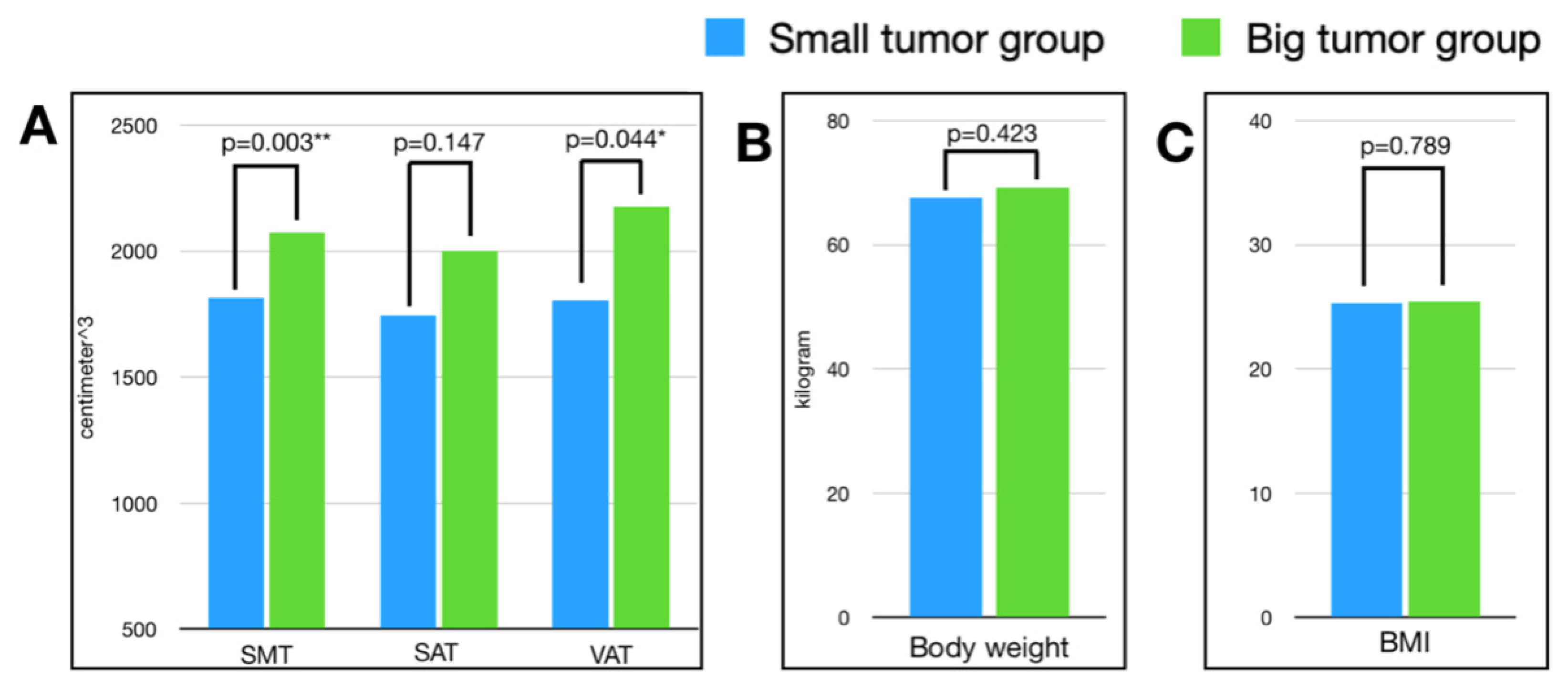
| Body weight | kilogram | 67.59 | 12.35 | 69.18 | 13.05 | −1.59 | 0.423 | 0.802 |
| BMI | kg/cm2 | 25.28 | 4.12 | 25.44 | 4.02 | −0.16 | 0.789 | 0.955 |
| SMT | cm3 | 1815.22 | 478.37 | 2072.38 | 516.07 | −257.16 | 0.003 ** | 0.319 |
| SAT | cm3 | 1744.79 | 826.72 | 2001.81 | 1213.50 | −257.02 | 0.147 | 0.866 |
| VAT | cm3 | 1804.57 | 964.8 | 2176.96 | 1178.50 | −372.39 | 0.044 * | 0.003 ** |
| SMT-L3 | cm2 | 136.79 | 32.14 | 147.44 | 30.6 | −10.65 | 0.047 * | 0.109 |
| SAT-L3 | cm2 | 135.57 | 58.78 | 147.46 | 73.76 | −11.89 | 0.295 | 0.914 |
| VAT-L3 | cm2 | 141.23 | 75.3 | 151.94 | 76.6 | −10.71 | 0.407 | 0.002 ** |
Publisher’s Note: MDPI stays neutral with regard to jurisdictional claims in published maps and institutional affiliations. |
© 2022 by the authors. Licensee MDPI, Basel, Switzerland. This article is an open access article distributed under the terms and conditions of the Creative Commons Attribution (CC BY) license (https://creativecommons.org/licenses/by/4.0/).
Share and Cite
Tan, C.-C.; Sheng, T.-W.; Chang, Y.-H.; Wang, L.-J.; Chuang, C.-K.; Wu, C.-T.; Pang, S.-T.; Shao, I.-H. Utilizing Computed Tomography to Analyze the Morphomic Change between Patients with Localized and Metastatic Renal Cell Carcinoma: Body Composition Varies According to Cancer Stage. J. Clin. Med. 2022, 11, 4444. https://doi.org/10.3390/jcm11154444
Tan C-C, Sheng T-W, Chang Y-H, Wang L-J, Chuang C-K, Wu C-T, Pang S-T, Shao I-H. Utilizing Computed Tomography to Analyze the Morphomic Change between Patients with Localized and Metastatic Renal Cell Carcinoma: Body Composition Varies According to Cancer Stage. Journal of Clinical Medicine. 2022; 11(15):4444. https://doi.org/10.3390/jcm11154444
Chicago/Turabian StyleTan, Chin-Chieh, Ting-Wen Sheng, Ying-Hsu Chang, Li-Jen Wang, Cheng-Keng Chuang, Chun-Te Wu, See-Tong Pang, and I-Hung Shao. 2022. "Utilizing Computed Tomography to Analyze the Morphomic Change between Patients with Localized and Metastatic Renal Cell Carcinoma: Body Composition Varies According to Cancer Stage" Journal of Clinical Medicine 11, no. 15: 4444. https://doi.org/10.3390/jcm11154444
APA StyleTan, C.-C., Sheng, T.-W., Chang, Y.-H., Wang, L.-J., Chuang, C.-K., Wu, C.-T., Pang, S.-T., & Shao, I.-H. (2022). Utilizing Computed Tomography to Analyze the Morphomic Change between Patients with Localized and Metastatic Renal Cell Carcinoma: Body Composition Varies According to Cancer Stage. Journal of Clinical Medicine, 11(15), 4444. https://doi.org/10.3390/jcm11154444