Article

Risk Indicators Affecting the Survival of the Mandibular First Molar Adjacent to an Implant at the Mandibular Second Molar Site: A Retrospective Study

1
Department of Periodontology, School of Dentistry, Kyung Hee University, Seoul 02447, Korea
2
Private Practice in Periodontics and Implant Dentistry, Seoul 02787, Korea
3
Department of Dentistry, Division of Prosthodontics, Hanyang University Medical Center, Seoul 04763, Korea
4
Department of Dentistry, Division of Oral & Maxillofacial Surgery, College of Medicine, Hanyang University, Seoul 04763, Korea
5
Department of Dentistry, Division of Periodontology, College of Medicine, Hanyang University, Seoul 04763, Korea
*
Author to whom correspondence should be addressed.
J. Clin. Med. 2021, 10(12), 2543; https://doi.org/10.3390/jcm10122543
Submission received: 4 May 2021 / Revised: 29 May 2021 / Accepted: 4 June 2021 / Published: 8 June 2021
(This article belongs to the Special Issue Implant Dentistry—Trends, Challenges and Innovations)

Abstract

:
This study aimed to compare the survival of mandibular first molars (MnM1s) adjacent to implants placed in mandibular second molar sites (ImM2s) with MnM1s adjacent to mandibular second molars (MnM2s) and to investigate risk indicators affecting the survival of MnM1s adjacent to ImM2s. A total of 144 patients who had MnM1s adjacent to ImM2s and MnM1s adjacent to MnM2s on the contralateral side were included in this study. Clinical variables and radiographic bone levels were evaluated. The survival of MnM1s adjacent to ImM2s or MnM2s was evaluated using a Kaplan–Meier analysis and Cox proportional hazards model. The 5-year cumulative survival rates of MnM1s adjacent to ImM2s and MnM2s were 85% and 95%, respectively. MnM1s adjacent to ImM2s of the internal implant-abutment connection type had higher multivariate hazard ratios (HR) for loss. MnM1s that had antagonists with implant-supported prostheses also had higher HR for loss. The multivariate HR for the loss of MnM1s adjacent to ImM2s with peri-implant mucositis was 3.74 times higher than MnM1s adjacent to healthy ImM2s. This study demonstrated several risk indicators affecting the survival of MnM1s adjacent to ImM2s. It is suggested that supportive periodontal and peri-implant therapy combined with meticulous occlusal adjustment can prolong the survival of MnM1s and ImM2s.

1. Introduction

Single implant treatment is considered a predictable restorative treatment option [1,2,3]. When a posterior single tooth is missing, single implants are the most biomimetic design.
After the delivery of an implant-supported prosthesis, however, biological and mechanical complications associated with the implant are frequently encountered [4]. Dental/implant occlusion is not a simple contact between opposite surfaces, and includes the friction of multiple inclined planes and different force vectors [5]. Dental implants that do not have periodontal ligament or periodontal mechanoreceptors are more susceptible to bending loads compared to the natural teeth [6,7]. When the occlusal load applied through function or parafunctions exceeds the resistance of the prosthesis, implant components, implant and osseointegrated interface, occlusal overload occurs, causing structural or biological damage [8]. In addition, it is reported that dental implants are thought to be more prone to occlusal overloading than natural teeth because of the loss of the periodontal ligament, which provides shock absorption and periodontal mechanoreceptors, providing tactile sensitivity and proprioceptive motion feedback [9]. Therefore, the occlusal concept applied to the implant-supported prosthesis needs to be different from that of natural teeth because implants react biomechanically in a different fashion to occlusal force [10].
Natural teeth continue to erupt throughout their lives [11,12,13,14]. In contrast, because of the direct contact between the implant and bone, implants do not follow these tooth movements. The continuous eruption and physiological movement of tooth adjacent implants may result in minor occlusal change. Therefore, it may result in traumatic occlusion or overloading in the teeth adjacent implants [15,16,17]. However, the wear on the adjacent teeth and the opposing teeth may compensate for the continuous tooth eruption. The human dentition is a dynamic system being continuously exposed to masticatory and interdental forces [18]. In addition, it is considered that continuous eruption might have evolved as a compensatory mechanism for heavy occlusal wear [18]. The wear of tooth and restoration is influenced by various factors including biological, mechanical, and chemical components [19,20,21]. Therefore, the degree of wear depending on the type of prosthetic material may influence the occlusal change of the tooth adjacent to an implant.
However, to the best of our knowledge, there have been few studies about the survival of the teeth adjacent to the most posterior mandibular implants. Therefore, the purpose of this study was to compare the survival of the mandibular first molars adjacent to implants placed in the mandibular second molar sites with the mandibular first molars adjacent to the mandibular second molars, and to investigate the risk indicators affecting the survival of the mandibular first molars adjacent to implants placed in the mandibular second molar sites.

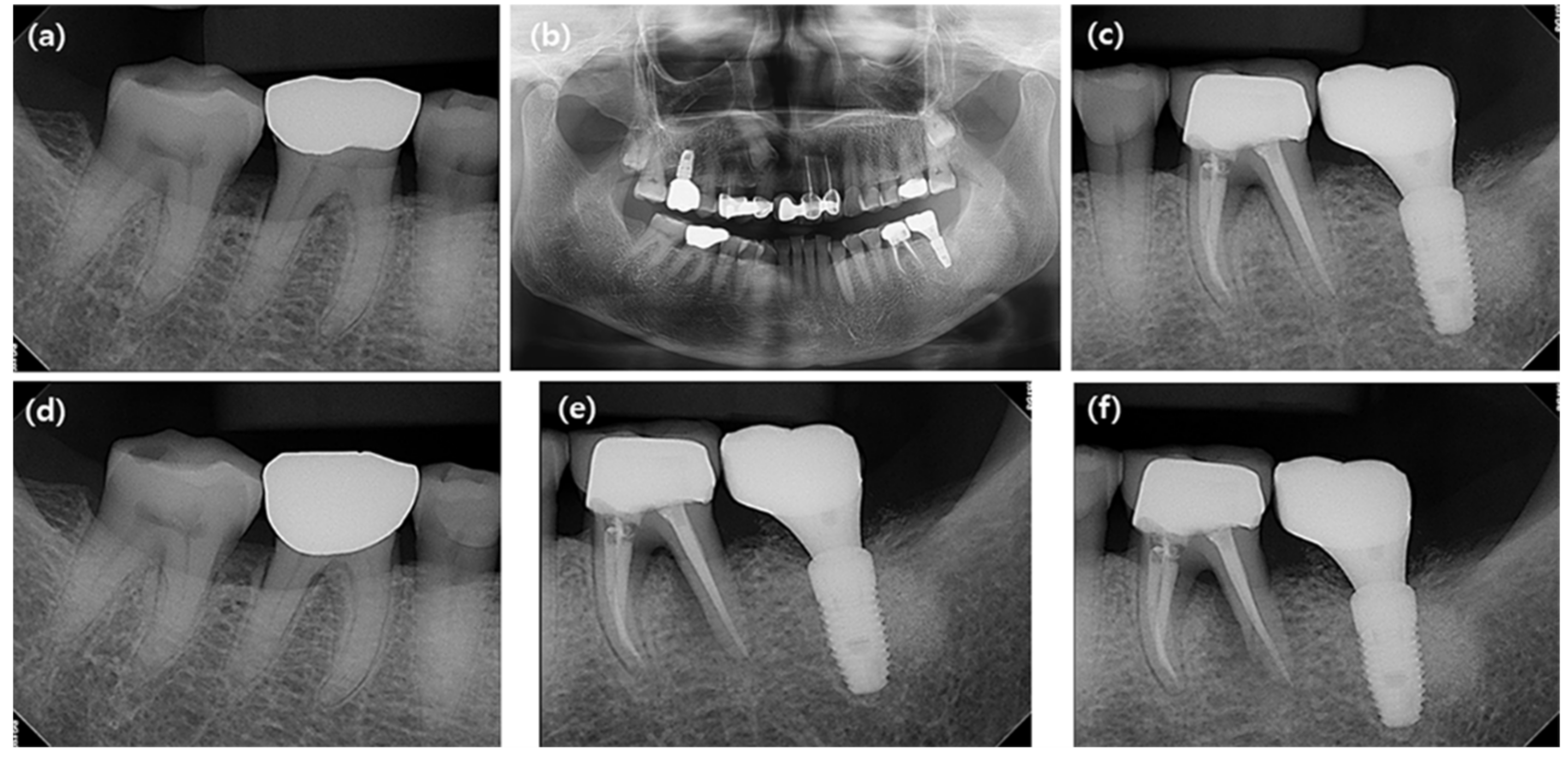
2. Materials and Methods

2.1. Subjects

All patients who underwent single implant placement at the private practice and the Department of Periodontology of Hanyang University Medical Center between March 2000 and August 2019 were screened for eligibility for this study (Figure 1).
Inclusion criteria:
  • Patients who were more than 20 years old
  • Patients who had no relevant systemic conditions or diseases
  • Patients who had the mandibular first molars (MnM1s) adjacent to the single implants placed in the mandibular second molar sites (ImM2s)
  • Patients who had the mandibular first molars (MnM1s) and the mandibular second molars (MnM2s) on the contralateral side
  • Patients who had been followed up at least 2 years after prosthesis delivery of ImM2s
  • Patients who had periapical radiographs after prosthesis delivery and before extraction of MnM1s or the last follow-up visits
Exclusion criteria:
  • Patients who had an active infection or disease affecting bone metabolism and wound healing
  • Patients who had regular use of steroids or other medications affecting bone turnover
This study was approved by the Institutional Review Board of Hanyang University Hospital (HYUH-2020-04-058). STROBE (Strengthening the Reporting of Observational Studies in Epidemiology) guidelines were observed for the preparation of the manuscript.

2.2. Clinical Variables

The following variables were evaluated using patient clinical records. Patient-related variables including age, sex, smoking and bruxism habit were assessed. Patients were considered as presenting “possible” sleep or awake bruxism based on self-reporting using questionnaires and/or the anamnestic part of a clinical examination [22].
Implant-related variables including the implant diameter, implant length, implant-abutment connection type (external vs. internal type) and peri-implant condition of the ImM2 were recorded. The peri-implant condition of ImM2 was classified as healthy, peri-implant mucositis and peri-implantitis [23].
The periodontal status of MnM1 including probing pocket depth (PPD), degree of furcation involvement [24] and endodontic status of MnM1 were assessed using clinical records. Prosthetic material types of the MnM1 (natural tooth, gold crown or porcelain fused metal crown) and the antagonist (natural tooth, gold crown, porcelain fused metal crown or implant-supported prosthesis with porcelain fused metal crown) of the MnM1 adjacent to the ImM2 or the MnM2 were evaluated.

2.3. Radiographic Measurements

The radiographic evaluation was performed using a digital radiography system (CS9300 Select, Carestream Dental LLC, Atlanta, GA, USA). Panoramic and periapical radiographs taken immediately after prostheses delivery of ImM2s (baseline) and at the last follow-up visits (or immediately before the extraction of MnM1s adjacent to ImM2s or MnM2s) were used for radiographic assessment (Figure 2).
Panoramic and periapical radiographs were imported into Analysis Toolkit (Adobe Photoshop CS6, Adobe Systems Inc., San Jose, CA, USA). Radiographic bone levels of the MnM1s adjacent to ImM2s or MnM2s were assessed at the mesial and distal sites using the baseline and the last follow-up (or immediately before the extraction of MnM1s adjacent to ImM2s or MnM2s) periapical radiographs. One blinded examiner (K.-G. H.) twice measured the distance between the cemento-enamel junction and the most coronal level of the alveolar bone of MnM1s for all sets of periapical radiographs at an interval of at least two weeks. The mean of the two measurements was used as the radiographic bone level value. Intra-class correlation coefficients (ICCs) were used to test the reproducibility of the radiographic measurements.

2.4. Survival of Tooth Adjacent to Single Implant

The survival of the MnM1 adjacent to the ImM2 or the MnM2 was evaluated. The follow-up period until the MnM1 adjacent to the ImM2 or the MnM2 had been extracted was recorded. The interval time from the date of the prosthesis delivery of ImM2 to the date of extraction of the MnM1 adjacent to the ImM2 or the MnM2 was assessed in months.

2.5. Statistical Analysis
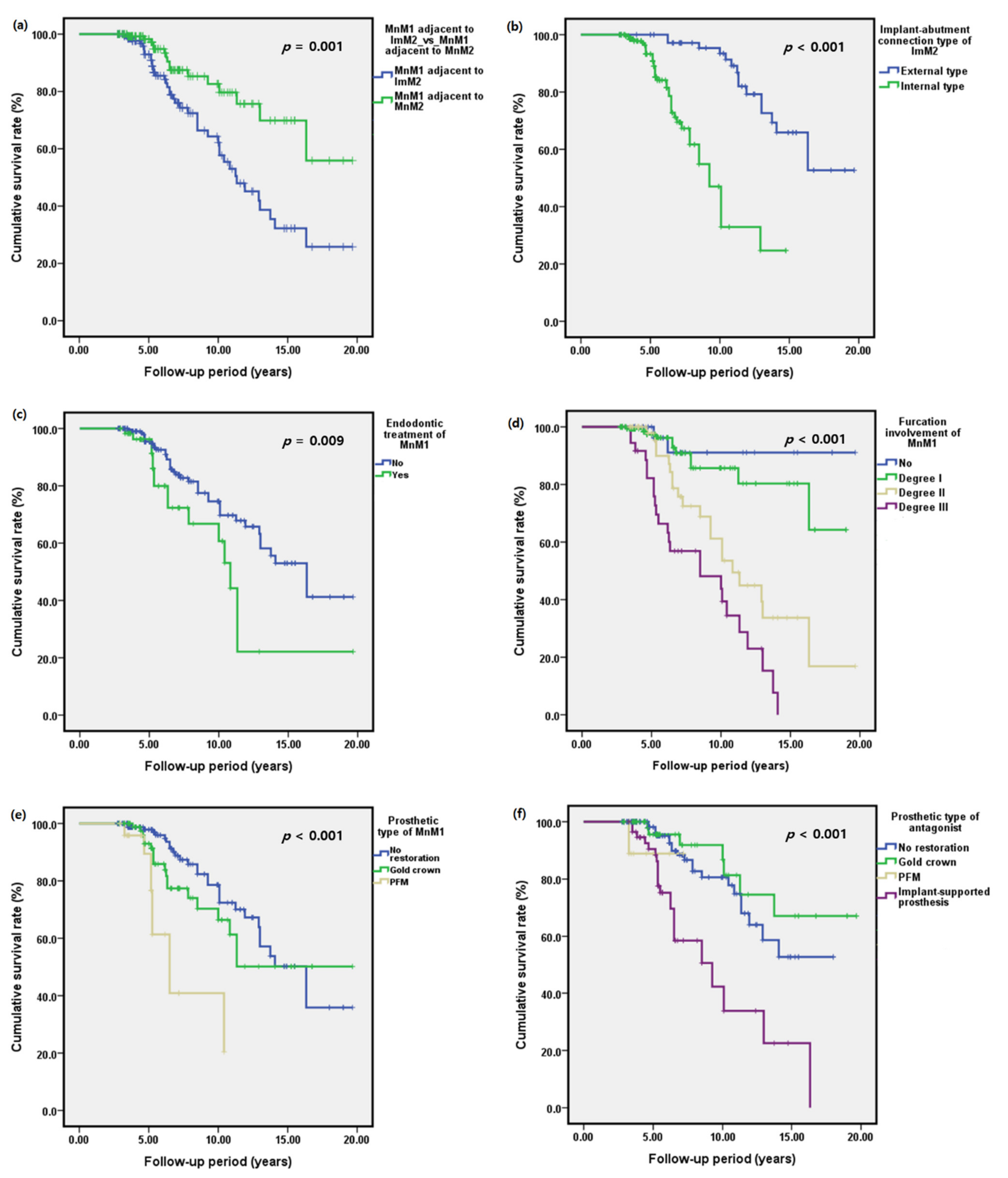
All statistical analyses were performed using SPSS (version 21, IBM Corp., Armonk, NY, USA). Descriptive statistics were performed using means ± standard deviations (SDs) or medians (first quartile, third quartile) for quantitative variables. After normality testing using the Shapiro–Wilk test, the differences in the clinical parameters of MnM1s between baseline and the last follow-up visit were analyzed using the Wilcoxon signed rank test. Differences in probing pocket depths and radiographic bone levels of MnM1s according to the implant-abutment connection type and endodontic treatment status were analyzed using the Mann–Whitney U test. The Kruskal–Wallis test was used to analyze the differences in probing pocket depth and radiographic bone level of MnM1 according to the degree of furcation involvement, endodontic treatment status, the prosthetic material types of MnM1 and the antagonist of MnM1. The cumulative survival rate of MnM1s adjacent to ImM2s or MnM2s was estimated using a Kaplan–Meier analysis. The log-rank test was used to identify the significant differences in the survival functions between the groups. Univariate and multivariate Cox proportional hazards regression models were used to analyze the hazard ratios for the loss of MnM1s adjacent to ImM2s or MnM2s. A p-value less than 0.05 was considered statistically significant.

3. Results

A total of 144 patients (89 men and 55 women) with a mean age of 54.49 ± 9.59 years (range: 27 to 75 years) were included in the present study (Table 1).
Six patients were lost to follow-up. Thirty-eight patients (26.39%) were smokers, and 34 patients (23.61%) had bruxism habits. The mean follow-up period was 6.94 ± 3.76 years (range: 2.75 to 19.67 years). The ICC for the radiographic measurement of the bone level was 0.938. Forty-one MnM1s adjacent to ImM2s were extracted and 16 MnM1s adjacent to MnM2 were extracted (Table 2). The reasons for extraction of MnM1s adjacent to ImM2 were endodontic origin (n = 6), periodontal origin with or without endodontic origin (n = 31) and tooth fracture (n = 4).
There were significant differences in the changes in PPD for MnM1s according to the prosthetic material types of the antagonists in both sides (ImM2s: p < 0.001, MnM2s: p = 0.013; Figure 3).
Regarding the radiographic bone level, MnM1s showed significantly greater radiographic bone loss when the antagonist was an implant-supported prosthesis in both sides (ImM2s: p < 0.001, MnM2s: p = 0.001; Figure 4).
The cumulative survival rate of MnM1s adjacent to ImM2s was 85% (95% confidence interval [CI]: 84.35–85.65%) at 5 years after the implant prosthesis delivery of ImM2s (Figure 5).
The 5-year cumulative survival rate of MnM1s adjacent to MnM2s was 95% (95% CI: 94.67–95.33%). The median survival time of MnM1s adjacent to ImM2s (11.33 years) was significantly shorter than MnM1s adjacent to MnM2s (p = 0.001; log-rank test; Figure 5). The median survival time of MnM1s adjacent to MnM2s or ImM2s after the implant prosthesis delivery of ImM2s with an internal implant-abutment connection type (9.25 years) was significantly shorter than MnM1s adjacent to ImM2s with an external implant-abutment connection type (p < 0.001; log-rank test; Figure 5). The median survival time of MnM1s with furcation involvement of degree III (8.5 years) was significantly shorter than those of MnM1s with no, degree I and degree II furcation involvement (p < 0.001; log-rank test). There were significant differences in the median survival times of MnM1s according to the prosthetic material type of the MnM1s (p < 0.001; log-rank test). When the antagonist of the MnM1 was an implant-supported prosthesis, the median survival time of MnM1s was significantly shorter compared to those with other prosthetic types of antagonists (p < 0.001; log-rank test).
Univariate and multivariate Cox regression models are shown in Table 3.
MnM1s adjacent to MnM2s showed lower multivariate hazard ratios for tooth loss (0.42; 95% CI: 0.23–0.77; p = 0.005) compared to MnM1s adjacent to ImM2. Patients with ImM2s of the internal implant-abutment connection type had a significantly higher multivariate hazard ratio for the loss of MnM1 (4.76; 95% CI: 1.99–11.38; p < 0.001) compared to patients with ImM2s of the external implant-abutment connection type. The multivariate HR for the loss of MnM1s adjacent ImM2s with peri-implant mucositis was 3.74 (95% CI: 1.42–9.89; p = 0.008) times higher than MnM1s adjacent to healthy ImM2s. The MnM1s with degree III furcation involvement showed a significantly higher multivariate hazard ratio for tooth loss (13.23; 95% CI: 2.71–64.59; p = 0.001). MnM1s restored with PFM had a higher multivariate hazard ratio for tooth loss (6.66; 95% CI: 2.17–20.45; p = 0.001). Patients with an antagonist of implant-supported prostheses with ceramo-metal had a higher multivariate hazard ratio for the loss of MnM1 adjacent to ImM2 (5.62; 95% CI: 2.72–11.63; p < 0.001).

4. Discussion

The results of this study showed that MnM1s adjacent to ImM2s exhibited higher hazard ratios for tooth loss compared to MnM1s adjacent to MnM2s. In addition, MnM1s adjacent to ImM2s or MnM2s exhibited higher hazard ratios for tooth loss when the MnM1s had a PFM crown, an antagonist with implant-supported prosthesis, an ImM2 of the internal implant-abutment connection type or an inflammatory peri-implant condition.
Dental implants do not have periodontal ligaments and are osseointegrated by direct bone-to-implant contact, similar to an ankylosed tooth [25,26]. On the other hand, teeth continue to erupt throughout life [11,12,13,14]. The continuous eruption and physiological movement of tooth adjacent implants may result in minor occlusal change. Tooth wear caused by multiple factors including biological, mechanical and chemical components [21] may compensate for the continuous tooth eruption. Wear patterns vary according to occlusion, joint pathology, muscle tone, lubricants, individual dietary habits and prosthetic material type [27,28,29]. Wear properties of restorative dental materials are reported to be different from those of natural teeth [30,31,32]. Additionally, tooth wear can be influenced by the wear rate of antagonists.
In this study, we evaluated the prosthetic material types of MnM1s and antagonist of MnM1s. The MnM1s restored with PFM crowns had a higher multivariate hazard ratio for tooth loss (6.66; 95% CI: 2.17–20.45; p = 0.001) than other restorative materials. Hacker et al. [33] reported that conventional feldspathic porcelain (230 ± 38 μm) caused enamel to wear more than gold did (9 ± 13 μm). The MnM1s had a significantly higher multivariate hazard ratio for tooth loss (5.62; 95% CI: 2.72–11.63; p < 0.001) when antagonists were implant-supported prostheses compared to natural teeth. It has been reported that wear will not occur unless occlusal stress exceeds the strength of the opposing materials, because wear requires the sliding of one surface against the other [19].
The present study showed that the MnM1s with ImM2s of the internal implant-abutment connection type had a significantly higher multivariate hazard ratio for tooth loss (4.76; 95% CI: 1.99–11.38; p < 0.001) compared to the MnM1s with ImM2s of the external implant-abutment connection type. Lee et al. [34] reported that the internal tapered conical connection demonstrated a variable amount of axial displacement with tightening torque and cyclic loading. Furthermore, Ko et al. [35] suggested that the long-term axial displacement of internal conical connection implants should be carefully managed.
Natural teeth have periodontal mechanoreceptors that signal information used for the fine motor control of jaw actions associated with biting, intraoral manipulation and the chewing of food [36]. However, important sensory-motor functions are lost or impaired when periodontal ligament receptors are reduced or eliminated through periodontal breakdown, bruxism, chewing, extraction and anesthesia [36,37]. Thus, a patient’s ability to detect occlusal inaccuracies induced by restorative treatment may be decreased, and inappropriate exteroceptive feedback may thus present a risk for overloading the prosthesis [37]. After the placement of dental implants, it has been reported that the detection thresholds of implant prostheses are increased to a thickness of at least 50–100 µm and 50–100 g upon tooth loading [37]. Thus, several contributing factors including the wear of restoration and the loss of tactile function of an implant may influence the occlusal trauma of an MnM1 adjacent to an ImM2.
An observational study showed that traumatic occlusal forces may be associated with the severity of periodontitis [38]. Additionally, Branschofsky et al. [39] reported that secondary trauma from occlusion is frequently seen in periodontally compromised patients and is positively correlated with the severity of attachment loss. Although a recent consensus reported that there is no evidence that traumatic occlusal forces can accelerate the progression of periodontitis in humans [40], it was reported that occlusal trauma can activate IKK-NF-κB signaling, which may result in the inhibition of osteogenic differentiation in vitro and bone formation in vivo [41].
The MnM1s with grade III furcation involvement showed a significantly greater multivariate hazard ratio for tooth loss (13.23; 95% CI: 2.71–64.59; p = 0.001). This result was similar to a previous study which showed that molars with furcation involvement had an increased risk of tooth loss during supportive periodontal therapy (OR 5.26; 95% CI: 1.46–19.03; p = 0.012) [42]. In addition, we found that the multivariate HR for the loss of MnM1s adjacent to ImM2s with peri-implant mucositis was 3.74 (95% CI: 1.42–9.89; p = 0.008) times higher than MnM1s adjacent to healthy ImM2s.
In the current study, we aimed to investigate the influence of bruxism on occlusal trauma. The univariate hazard ratio for the loss of MnM1s with a bruxism habit was 1.8 (95% CI: 1.02–3.19; p = 0.043). However, we failed to find any significant differences in multivariate hazard ratios for the loss of MnM1s between non-bruxism and bruxism patients.
This study had several strengths including a long-term follow-up period, univariate and multivariate evaluations of risk indicators to minimize confounding factors and tribological evaluation of prostheses. The limitation of the present study was the lack of histological confirmation of occlusal trauma, which is an inherent bias of retrospective studies. In addition, bruxism habits were evaluated by patients’ self-reporting using questionnaires and/or the anamnestic part of a clinical examination without a polysomnographic or an electromyographic evaluation. Therefore, the findings of this study should be interpreted with caution. Future prospective studies with large sample sizes are recommended. Nonetheless, the results of this study can be used as a basis for understanding the occlusal trauma of the MnM1s adjacent to ImM2s and the possible risk indicators for the survival of MnM1s adjacent to ImM2s.

5. Conclusions

This study demonstrated that MnM1s adjacent to ImM2s exhibited higher hazard ratios for tooth loss compared to MnM1s adjacent to MnM2s and identified several risk indicators affecting the survival of MnM1s adjacent to ImM2s. These risk indicators were the internal type of implant-abutment connection, PFM crown restoration of MnM1, antagonists with implant-supported prostheses and inflammatory peri-implant conditions. Therefore, it is suggested that supportive periodontal and peri-implant therapy combined with continuous and meticulous occlusal adjustment can prolong the survival of an implant and the adjacent tooth.

Author Contributions

W.-B.P., K.-H.K. and J.-Y.H. conceived the ideas of study; W.-B.P. and J.-Y.H. carried out the treatment; K.-G.H. collected the data; K.-G.H. and J.-Y.H. analyzed the data; W.-B.P. and K.-H.K. led the writing of the manuscript; J.-Y.H. and K.-G.H. edited and reviewed the manuscript; W.-B.P. and K.-H.K. contributed equally to this study as the first authors. All authors have read and agreed to the published version of the manuscript.

Funding

This research was supported by a grant of the Korea Health Technology R & D Project through the Korea Health Industry Development Institute, funded by the Ministry of Health & Welfare, Republic of Korea (grant number HI20C0013).

Institutional Review Board Statement

The study was conducted according to the guidelines of the Declaration of Helsinki, and approved by the Institutional Review Board of Hanyang University Hospital (HYUH-2020-04-058).

Informed Consent Statement

Patient consent was waived because this was a retrospective observational study.

Data Availability Statement

The data presented in this study are available upon request from the corresponding author. The data are not publicly available because of privacy restrictions.

Acknowledgments

We would like to thank En-Woo Nam of the Biostatistical Consulting and Research Lab, Hanyang University for assistance with the statistical analysis and Seong-Jin Ahn of Hanyang Medical Center for the radiographic evaluation.

Conflicts of Interest

The authors declare no conflict of interest.

References

  1. Zembic, A.; Kim, S.; Zwahlen, M.; Kelly, J.R. Systematic review of the survival rate and incidence of biologic, technical, and esthetic complications of single implant abutments supporting fixed prostheses. Int. J. Oral. Maxillofac. Implant. 2014, 29, 99–116. [Google Scholar] [CrossRef] [Green Version]
  2. Van Velzen, F.J.; Ofec, R.; Schulten, E.A.; Ten Bruggenkate, C.M. 10-year survival rate and the incidence of peri-implant disease of 374 titanium dental implants with a SLA surface: A prospective cohort study in 177 fully and partially edentulous patients. Clin. Oral. Implant. Res. 2015, 26, 1121–1128. [Google Scholar] [CrossRef]
  3. Jemt, T. Single-Implant Survival: More Than 30 Years of Clinical Experience. Int. J. Prosthodont. 2016, 29, 551–558. [Google Scholar] [CrossRef] [PubMed] [Green Version]
  4. Von Stein-Lausnitz, M.; Nickenig, H.J.; Wolfart, S.; Neumann, K.; Von Stein-Lausnitz, A.; Spies, B.C.; Beuer, F. Survival rates and complication behaviour of tooth implant-supported, fixed dental prostheses: A systematic review and meta-analysis. J. Dent. 2019, 88, 103167. [Google Scholar] [CrossRef]
  5. Katona, T.R.; Eckert, G.J. The mechanics of dental occlusion and disclusion. Clin. Biomech. 2017, 50, 84–91. [Google Scholar] [CrossRef]
  6. Yuan, J.C.; Sukotjo, C. Occlusion for implant-supported fixed dental prostheses in partially edentulous patients: A literature review and current concepts. J. Periodontal Implant. Sci. 2013, 43, 51–57. [Google Scholar] [CrossRef] [PubMed]
  7. Rangert, B.R.; Sullivan, R.M.; Jemt, T.M. Load factor control for implants in the posterior partially edentulous segment. Int. J. Oral. Maxillofac. Implant. 1997, 12, 360–370. [Google Scholar]
  8. Laney, W.R. Glossary of Oral and Maxillofacial Implants. Int. J. Oral. Maxillofac. Implant. 2017, 32, Gi-G200. [Google Scholar] [CrossRef] [PubMed]
  9. Koyano, K.; Esaki, D. Occlusion on oral implants: Current clinical guidelines. J. Oral. Rehabil. 2015, 42, 153–161. [Google Scholar] [CrossRef] [PubMed]
  10. Kim, Y.; Oh, T.J.; Misch, C.E.; Wang, H.L. Occlusal considerations in implant therapy: Clinical guidelines with biomechanical rationale. Clin. Oral Implant. Res. 2005, 16, 26–35. [Google Scholar] [CrossRef] [PubMed]
  11. Craddock, H.; Youngson, C. Eruptive tooth movement—the current state of knowledge. Br. Dent. J. 2004, 197, 385–391. [Google Scholar] [CrossRef]
  12. Steedle, J.R.; Proffit, W.R. The pattern and control of eruptive tooth movements. Am. J. Orthod. 1985, 87, 56–66. [Google Scholar] [CrossRef]
  13. Wei, H.; Tomotake, Y.; Nagao, K.; Ichikawa, T. Implant prostheses and adjacent tooth migration: Preliminary retrospective survey using 3-dimensional occlusal analysis. Int. J. Prosthodont 2008, 21, 302–304. [Google Scholar] [PubMed]
  14. Newman, H.N. Attrition, eruption, and the periodontium. J. Dent. Res. 1999, 78, 730–734. [Google Scholar] [CrossRef] [PubMed]
  15. Lee, J.-H.; Kweon, H.H.-I.; Choi, S.-H.; Kim, Y.-T. Association between dental implants in the posterior region and traumatic occlusion in the adjacent premolars: A long-term follow-up clinical and radiographic analysis. J. Periodontal Implant Sci. 2016, 46, 396–404. [Google Scholar] [CrossRef] [Green Version]
  16. Liu, S.; Liu, Y.; Xu, J.; Rong, Q.; Pan, S. Influence of occlusal contact and cusp inclination on the biomechanical character of a maxillary premolar: A finite element analysis. J. Prosthet. Dent. 2014, 112, 1238–1245. [Google Scholar] [CrossRef] [PubMed]
  17. Torbjörner, A.; Fransson, B. Biomechanical aspects of prosthetic treatment of structurally compromised teeth. Int. J. Prosthodont 2004, 17, 135–141. [Google Scholar] [CrossRef] [PubMed]
  18. Wiedemann, C.; Pink, C.; Daboul, A.; Samietz, S.; Volzke, H.; Schulz-Kornas, E.; Krey, K.F.; Holtfreter, B.; Kocher, T. Is Continuous Eruption Related to Periodontal Changes? A 16-Year Follow-up. J. Dent. Res. 2021, 22034521999363. [Google Scholar] [CrossRef]
  19. Oh, W.-S.; DeLong, R.; Anusavice, K.J. Factors affecting enamel and ceramic wear: A literature review. J. Prosthet. Dent. 2002, 87, 451–459. [Google Scholar] [CrossRef] [PubMed]
  20. Ghazal, M.; Kern, M. The influence of antagonistic surface roughness on the wear of human enamel and nanofilled composite resin artificial teeth. J. Prosthet. Dent. 2009, 101, 342–349. [Google Scholar] [CrossRef]
  21. Mair, L.H.; Stolarski, T.A.; Vowles, R.W.; Lloyd, C.H. Wear: Mechanisms, manifestations and measurement. Report of a workshop. J. Dent. 1996, 24, 141–148. [Google Scholar] [CrossRef]
  22. Lobbezoo, F.; Ahlberg, J.; Glaros, A.G.; Kato, T.; Koyano, K.; Lavigne, G.J.; De Leeuw, R.; Manfredini, D.; Svensson, P.; Winocur, E. Bruxism defined and graded: An international consensus. J. Oral. Rehabil. 2013, 40, 2–4. [Google Scholar] [CrossRef]
  23. Berglundh, T.; Armitage, G.; Araujo, M.G.; Avila-Ortiz, G.; Blanco, J.; Camargo, P.M.; Chen, S.; Cochran, D.; Derks, J.; Figuero, E.; et al. Peri-implant diseases and conditions: Consensus report of workgroup 4 of the 2017 World Workshop on the Classification of Periodontal and Peri-Implant Diseases and Conditions. J. Clin. Periodontol. 2018, 45 (Suppl. S20), S286–S291. [Google Scholar] [CrossRef] [PubMed] [Green Version]
  24. Hamp, S.E.; Nyman, S.; Lindhe, J. Periodontal treatment of multirooted teeth. Results after 5 years. J. Clin. Periodontol. 1975, 2, 126–135. [Google Scholar] [CrossRef]
  25. Brånemark, P.I.; Adell, R.; Breine, U.; Hansson, B.O.; Lindstrom, J.; Ohlsson, A. Intra-osseous anchorage of dental prostheses. I. Experimental studies. Scand. J. Plast. Reconstr. Surg. 1969, 3, 81–100. [Google Scholar] [CrossRef] [PubMed]
  26. Schroeder, A.; Van der Zypen, E.; Stich, H.; Sutter, F. The reactions of bone, connective tissue, and epithelium to endosteal implants with titanium-sprayed surfaces. J. Maxillofac. Surg. 1981, 9, 15–25. [Google Scholar] [CrossRef]
  27. Mahalick, J.A.; Knap, F.J.; Weiter, E.J. Occusal wear in prosthodontics. J. Am. Dent. Assoc. 1971, 82, 154–159. [Google Scholar] [CrossRef] [PubMed]
  28. Heintze, S.D.; Cavalleri, A.; Forjanic, M.; Zellweger, G.; Rousson, V. Wear of ceramic and antagonist--a systematic evaluation of influencing factors in vitro. Dent. Mater. 2008, 24, 433–449. [Google Scholar] [CrossRef] [PubMed]
  29. Mundhe, K.; Jain, V.; Pruthi, G.; Shah, N. Clinical study to evaluate the wear of natural enamel antagonist to zirconia and metal ceramic crowns. J. Prosthet. Dent. 2015, 114, 358–363. [Google Scholar] [CrossRef]
  30. Metzler, K.T.; Woody, R.D.; Miller, A.W., 3rd; Miller, B.H. In vitro investigation of the wear of human enamel by dental porcelain. J. Prosthet. Dent. 1999, 81, 356–364. [Google Scholar] [CrossRef]
  31. Sulong, M.Z.; Aziz, R.A. Wear of materials used in dentistry: A review of the literature. J. Prosthet Dent. 1990, 63, 342–349. [Google Scholar] [CrossRef]
  32. Yip, K.H.; Smales, R.J.; Kaidonis, J.A. Differential wear of teeth and restorative materials: Clinical implications. Int. J. Prosthodont. 2004, 17, 350–356. [Google Scholar] [PubMed]
  33. Hacker, C.H.; Wagner, W.C.; Razzoog, M.E. An in vitro investigation of the wear of enamel on porcelain and gold in saliva. J. Prosthet. Dent. 1996, 75, 14–17. [Google Scholar] [CrossRef]
  34. Lee, J.H.; Kim, D.G.; Park, C.J.; Cho, L.R. Axial displacements in external and internal implant-abutment connection. Clin. Oral Implant. Res. 2014, 25, e83–e89. [Google Scholar] [CrossRef]
  35. Ko, K.H.; Huh, Y.H.; Park, C.J.; Cho, L.R. Axial displacement in cement-retained prostheses with different implant-abutment connections. Int. J. Oral. Maxillofac. Implant. 2019, 34, 1098–1104. [Google Scholar] [CrossRef] [PubMed]
  36. Trulsson, M. Sensory-motor function of human periodontal mechanoreceptors. J. Oral. Rehabil. 2006, 33, 262–273. [Google Scholar] [CrossRef] [PubMed]
  37. Jacobs, R.; Van Steenberghe, D. From osseoperception to implant-mediated sensory-motor interactions and related clinical implications. J. Oral. Rehabil. 2006, 33, 282–292. [Google Scholar] [CrossRef] [PubMed]
  38. Ismail, A.I.; Morrison, E.C.; Burt, B.A.; Caffesse, R.G.; Kavanagh, M.T. Natural history of periodontal disease in adults: Findings from the Tecumseh Periodontal Disease Study, 1959–1987. J. Dent. Res. 1990, 69, 430–435. [Google Scholar] [CrossRef] [PubMed]
  39. Branschofsky, M.; Beikler, T.; Schafer, R.; Flemming, T.F.; Lang, H. Secondary trauma from occlusion and periodontitis. Quintessence Int. 2011, 42, 515–522. [Google Scholar]
  40. Jepsen, S.; Caton, J.G.; Albandar, J.M.; Bissada, N.F.; Bouchard, P.; Cortellini, P.; Demirel, K.; De Sanctis, M.; Ercoli, C.; Fan, J.; et al. Periodontal manifestations of systemic diseases and developmental and acquired conditions: Consensus report of workgroup 3 of the 2017 World Workshop on the Classification of Periodontal and Peri-Implant Diseases and Conditions. J. Clin. Periodontol. 2018, 45 (Suppl. S20), S219–S229. [Google Scholar] [CrossRef]
  41. Xu, W.; Lu, Y.; Yue, J.; Lu, W.; Zhou, W.; Zhou, X.; Ye, L.; Zheng, Q.; Zhang, L.; Huang, D. Occlusal trauma inhibits osteoblast differentiation and bone formation through IKK-NF-kappaB signaling. J. Periodontol. 2020, 91, 683–692. [Google Scholar] [CrossRef] [PubMed]
  42. Nibali, L.; Sun, C.; Akcali, A.; Yeh, Y.C.; Tu, Y.K.; Donos, N. The effect of horizontal and vertical furcation involvement on molar survival: A retrospective study. J. Clin. Periodontol. 2018, 45, 373–381. [Google Scholar] [CrossRef] [PubMed]
Figure 1. Flow chart of the study.
Jcm 10 02543 g001
Figure 2. Radiographic measurement. (a) A periapical radiograph of the mandibular first molar (MnM1) adjacent the mandibular second molar (MnM2) at baseline. (b) A panoramic radiograph at baseline. (c) A periapical radiograph of MnM1 adjacent to the single implant placed in the mandibular second molar site (ImM2) at baseline. (d) A periapical radiograph of MnM1 adjacent MnM2 at the last follow-up. (e) A periapical radiograph of MnM1 adjacent to ImM2 at 3-year follow-up after prosthesis delivery of ImM2. (f) A periapical radiograph of MnM1 adjacent to ImM2 at the last follow-up.
Jcm 10 02543 g002
Figure 3. Changes in probing pocket depths of the mandibular first molars (MnM1s) between baseline and the last follow-up according to (a) furcation involvement of MnM1s, (b) prosthetic type of MnM1s, and (c) prosthetic type of antagonists. Abbreviations: ImM2s, implants placed in the mandibular second molar site; MnM2s, the mandibular second molars; PFM, porcelain fused metal crown.
Jcm 10 02543 g003
Figure 4. Changes in radiographic bone levels of the mandibular first molars (MnM1s) between baseline and the last follow-up according to (a) furcation involvement of MnM1s, (b) prosthetic type of MnM1s, and (c) prosthetic type of antagonists. Abbreviations: ImM2s, implants placed in the mandibular second molar site; MnM2s, the mandibular second molars; PFM, porcelain fused metal crown.
Jcm 10 02543 g004
Figure 5. Kaplan–Meier survival curves. (a) The cumulative survival rate of the mandibular first molars (MnM1s) adjacent to implants (ImM2s) was significantly lower than MnM1s adjacent to the mandibular second molars (MnM2s) at the 5-year follow-up. The cumulative survival rate of the mandibular first molars adjacent to ImM2s or MnM2s according to (b) implant-abutment connection type of ImM2s, (c) history of endodontic treatment of MnM1s, (d) degree of furcation involvement of MnM1s, (e) the prosthetic material types of MnM1s and (f) the prosthetic material types of the antagonists of MnM1s.
Jcm 10 02543 g005
Table 1. Patient demographic characteristics.
Variables N
Age (years)<50 years44 (30.56%)
≥50 years100 (69.44%)
(Mean ± SD)(54.49 ± 9.59)
SexMale89 (61.81%)
Female55 (38.19%)
SmokingNon-smoker106 (73.61%)
Smoker38 (26.39%)
BruxismNo110 (76.39%)
Yes34 (23.61%)
Implant-abutment connection typeExternal type39 (27.08%)
Internal type105 (72.92%)
Peri-implant condition of ImM2Healthy68 (47.22%)
Peri-implant mucositis65 (45.14%)
Peri-implantitis11 (7.64%)
Distance between ImM2 and MnM1<3 mm0 (0%)
≥3 mm144 (100%)
Follow-up (years)(Mean ± SD, range)(6.94 ± 3.76, 2.75–19.67)
MnM1 adjacent to ImM2MnM1 adjacent to MnM2
Endodontic treatmentNo114 (79.17%)118 (81.94%)
Yes30 (20.83%)26 (18.06%)
Furcation involvementNo15 (10.42%)19 (13.19%)
Degree I74 (51.39%)85 (59.03%)
Degree II33 (22.92%)26 (18.06%)
Degree III22 (15.28%)14 (9.72%)
Prosthetic typeno restoration84 (58.33%)87 (60.42%)
gold crown47 (32.64%)46 (31.94%)
PFM13 (9.03%)11 (7.64%)
Prosthetic type of antagonistno restoration77 (53.47%)79 (54.86%)
gold crown35 (24.31%)31 (21.53%)
PFM3 (2.08%)6 (4.17%)
Implant-supported prosthesis29 (20.14%)28 (19.44%)
Abbreviations: N, number; SD, standard deviation; MnM1s, the mandibular first molars; ImM2s, implants placed in the mandibular second molar site; MnM2s, the mandibular second molars; PFM, porcelain fused metal crown.
Table 2. The etiology of extraction of the mandibular first molars.
MnM1s Adjacent to ImM2sMnM1s Adjacent to MnM2
ExtractionNo 103128
Yes 4116
Endodontic origin66
Periodontal origin (with or without endodontic origin) 3110
Root fracture40
Abbreviations: MnM1s, the mandibular first molars; ImM2s, implants placed in the mandibular second molar site; MnM2s, the mandibular second molars.
Table 3. Cox proportional hazard ratio for the loss of the mandibular first molar adjacent to an implant placed in the mandibular second molar site.
VariableUnivariate HR95% CIp-ValueMultivariate HR95% CIp-Value
Age<50 years1 1
≥50 years1.640.89–3.020.1151.640.89–3.030.116
SexMale1 1
Female0.760.43–1.360.3580.570.25–1.310.187
SmokingNo1 1
Yes1.210.70–2.090.4891.020.45–2.300.957
BruxismNo1 1
Yes1.81.02–3.190.0431.160.58–2.360.673
Mn. 2nd molar site
Implant supported crown1 1
Natural teeth0.390.22–0.690.0010.420.23–0.770.005
Implant-abutment connection type
External type1 1
Internal type1.490.84–2.660.1734.761.99–11.38<0.001
Implant health
Healthy1 0.4191 0.029
Peri-implant mucositis1.10.51–2.340.8123.741.42–9.890.008
Peri-implantitis1.50.74–3.060.2622.620.98–6.980.054
Endodontic treatment of MnM1
No1 1
Yes2.121.18–3.820.0121.40.50–3.930.518
Furcation involvement of MnM1
01 <0.0011 <0.001
Degree I1.930.43–8.710.3951.820.37–8.920.463
Degree II6.521.52–28.010.0125.231.11–24.630.036
Degree III14.033.28–60.08<0.00113.232.71–64.590.001
Prosthetic type of MnM1
No restoration1 <0.0011 0.004
Gold crown1.450.81–2.590.2072.791.18–6.610.019
PFM5.422.49–11.79<0.0016.662.17–20.450.001
Antagonist prosthetic type of MnM1
No restoration1 <0.0011 <0.001
Gold crown0.640.27–1.510.3060.870.32–2.310.771
PFM2.390.32–18.080.3970.240.02–2.620.242
Implant-supported prosthesis3.391.93–6.00<0.001 5.622.72–11.63<0.001
Note: Bolded font, statistically significant at p-value <0.05. Abbreviations: HR, hazard ratio; CI, confidence interval; MnM1, the mandibular first molar; PFM, porcelain fused metal crown.
Publisher’s Note: MDPI stays neutral with regard to jurisdictional claims in published maps and institutional affiliations.

