Review

Prevention and Management of Complications and Education in Endoscopic Submucosal Dissection

Department of Digestive Endoscopy, Tokyo Women’s Medical University Hospital, 8-1, Tokyo 162-8666, Japan
*
Author to whom correspondence should be addressed.
J. Clin. Med. 2021, 10(11), 2511; https://doi.org/10.3390/jcm10112511
Submission received: 22 May 2021 / Revised: 3 June 2021 / Accepted: 3 June 2021 / Published: 6 June 2021
(This article belongs to the Special Issue Latest Advances in Endoscopic Imaging and Therapy—Part I)

Abstract

:
Endoscopic submucosal dissection (ESD) is considered superior to endoscopic mucosal resection as an endoscopic resection because of its higher en bloc resection rate, but it is more difficult to perform. As ESD techniques have become more common, and the range of treatment by ESD has expanded, the number of possible complications has also increased, and endoscopists need to manage them. In this report, we will review the management of critical complications, such as hemorrhage, perforation, and stenosis, and we will also discuss educational methods for acquiring and improving ESD skills.

1. Introduction

Endoscopic submucosal dissection (ESD) is a form of endoscopic resection that originated in Japan as a treatment for early-stage gastric cancer [1]. Today, ESD has become an accepted treatment not only in Japan but also in other Asian countries and around the world [2]. Its treatment targets are not limited to gastric cancer but also include esophageal cancer, colorectal cancer, pharyngeal cancer, and duodenal cancer. Precancerous lesions and early stage cancers of the gastrointestinal tract that are unlikely to metastasize to the lymph nodes, regardless of the organ, are targeted for ESD. It may also be used to treat residual or recurrent disease. Although ESD is considered superior to conventional endoscopic mucosal resection (EMR) in terms of its high en bloc resection rate, it is more difficult to perform than EMR and requires more delicate endoscopic techniques [3,4]. This inevitably increases the possibility of treatment-related accidents, such as bleeding and perforation. In the case of ESD for esophageal cancer, if the circumference of the mucosal defect after treatment is larger than three fourths of the circumference of the lesion, there is a 90% probability of stenosis during the postoperative healing process [5], which is one of the serious sequelae associated with ESD. The management of these serious complications is important, but prevention is the most important factor. In this article, we review the prevention and management of ESD-related accidents.

Search Strategy

We identified studies through a literature search of the database (PubMed), with the last search performed on 31 May 2021. The key words were “endoscopic submucosal dissection” OR “ESD” AND “complication” OR “postoperative bleeding” OR ”traction” OR “perforation” OR “stenosis” OR “training”. We restricted the studies to those written in English.

2. Critical Complications Associated with ESD

2.1. Bleeding after ESD

Postoperative hemorrhage is one of the most serious complications associated with ESD. Most postoperative hemorrhages can be treated conservatively or with endoscopic hemostasis, but some cases may progress to the point where interventional radiology or surgery is required and may even be fatal, and endoscopists must manage them appropriately. In esophageal and colorectal ESD, the incidence of bleeding after ESD is low, and there are few cases of shock [6]. In the cases of gastric and duodenal ESD, it is not uncommon for shock to occur and for the bleeding site to be filled with bloody components, making hemostasis difficult [7]. The duodenum is known to have a high incidence of postoperative bleeding due to bile exposure [8]. In addition, patients on dialysis and patients taking antithrombotic drugs are known to have an increased risk of postoperative bleeding [9]. Methods to manage bleeding include drug administration, treatment of the ulcer base, and devising a response to antithrombotic agents.

2.1.1. Drugs

As maintaining an intragastric pH higher than 5.4 promotes blood coagulation and platelet aggregation [10], administration of proton pump inhibitors (PPIs) or histamine H2 receptor antagonists (RAs) is expected to prevent postoperative bleeding in gastric ESD, as in cases of peptic ulcer. In fact, PPIs have been shown to be more effective in reducing the risk of post-ESD bleeding than H2 RAs [11,12] and are therefore widely used postoperatively. Furthermore, potassium-competitive acid blockers (P-CAB), which have been introduced recently, inhibit gastric acid secretion more potently and rapidly than PPIs [13], suggesting that they may inhibit postoperative hemorrhage more effectively than PPIs [14]. It has been suggested that PPIs may be useful for prophylaxis in esophageal ESD, although not to the same extent as they are in the stomach, but no significant evidence has been obtained [15].

2.1.2. Treatment of the Ulcer Base

There are many reports that suturing and covering post-ESD ulcers are useful for reducing postoperative bleeding. Suturing has been reported to reduce postoperative bleeding in gastric EMR [16], colorectal ESD [17], and duodenal ESD [18], but there has also been a report that it did not significantly reduce postoperative bleeding in colorectal ESD [13]. Thus, each facility employs it following its own guidelines. In addition to the conventional method using a hemostatic clip [19], there are also methods that use an indwelling snare [20], an overstitch (Apollo Endosurgery., Austin, TX, USA) [21], an over-the-scope clip (OTSC Ovesco Endoscopy GmbH, Tuebingen, Germany) [22], and, more recently, endoscopic hand suturing [23]. Polyglycolic acid (PGA) sheets are often used to cover the ulcer base with a special material after ESD, and they have been reported to be useful even in patients taking antithrombotic drugs [24,25].

2.1.3. Response to Antithrombotic Agents

With the increase in the elderly population worldwide, the number of people taking oral antithrombotic agents is also increasing. While endoscopic bleeding should be avoided as much as possible, thromboembolic events, such as cerebrovascular events, are potentially lethal and require careful handling. The Japan Gastroenterological Endoscopy Society has issued guidelines on antithrombotic agents for endoscopic examination and treatment [26]. These antithrombotic agents include antiplatelet agents, such as aspirin and thienopyridine derivatives, and anticoagulants, such as warfarin, dabigatran, and heparin. The guideline categorizes endoscopic procedures into four categories—conventional endoscopy, endoscopic biopsy, low bleeding risk gastrointestinal endoscopy, and high bleeding risk gastrointestinal endoscopy—and suggests responses on the basis of the risk of thrombosis, such as myocardial infarction and cerebral infarction, and the risk of bleeding after endoscopic treatment. ESD falls under the category of high bleeding risk gastrointestinal endoscopy. In addition, an addendum on anticoagulants, including direct oral anticoagulants, has been published [27], and preoperative measures should be taken, as shown in Table 1 in the case of withdrawal of monotherapy, in Table 2 of dual therapy, and in Table 3 of triple therapy, respectively. As vascular events, such as myocardial infarction or cerebral infarction and postoperative bleeding, can occur even if the guidelines are strictly followed, it is important to obtain sufficient informed consent from patients before treatment.

2.2. Perforation

Perforation in esophageal ESD may lead to mediastinitis, and perforation in gastric and colorectal ESD may lead to peritonitis, which may be fatal in some cases. The incidence of perforation in esophageal, gastric, and colorectal ESD is 3.2% [28], 2.9% [29], and 4.8% [30], respectively. To avoid intraoperative perforation, it is important to obtain a good intraoperative field of view and to reliably visualize the muscularis propria. The traction method has been reported to be useful in many such cases [31]. The clip-with-line and S-O clip methods are the main methods used.
The clip-with-line method is a simple method proposed by Oyama et al. [32]. After the flap is formed, the endoscope is removed, and a silk line or dental floss is tied to the arm of a conventional hemostatic clip outside the body. The scope is reinserted, and the clip is attached to the flap. In this step, it is necessary to avoid accidental entrapment of the muscularis propria, and the thread attached to the flap can be pulled from outside the body with a small amount of force to ensure a good field of view and reliable visualization of the muscularis propria. The proximal side of the colon is generally not a good indication for this method because of interference with the scope. Reinsertion of the scope is necessary in the original method, but a modified method that does not require reinsertion of the scope has also been reported [33].
The S-O clip (Zeon Medical Co., Tokyo, Japan) is a traction device with a spring attached to the clip arm developed by Sakamoto et al. [34]. Similar to the threaded traction method, the clip arm is attached to a spring. As in the threaded traction method, the S-O clip is attached to the flap formed by mucosal incision and dissection. A small rubber ring attached to the tip of the spring is picked up with the hemostatic clip and applied to the opposite-side mucosa. Especially in the case of colorectal ESD, the rubber ring is applied to the opposite side at a site twofold distant from the anal side of the lesion to ensure a good field of view (Figure 1). The traction force can be adjusted by adjusting the air volume in the colon. After the specimen is resected, the rubber band fixed to the intestinal wall can be cut and the lesion retrieved. This method can be used in the whole digestive tract and does not require reinsertion of the scope, but the traction force may weaken as dissection proceeds [33]. However, as S-O clips are not readily available in some countries and regions, the ring-thread counter traction method, in which a loop of thread is attached to the arm of a hemostatic-like clip to obtain traction, is a relatively straightforward method (Figure 2) [35]. In both methods, care must be taken to prevent the clip from moving and accidentally grasping the intrinsic muscle layer. In the event of intraoperative perforation, care should be taken to avoid rushing to suture the perforation. If the patient’s vital signs and abdominal symptoms permit, a mucosal incision or dissection around the perforation should be made to secure space around the perforation, and appropriate closure should be performed. If there is intraoperative abdominal pain or hypotension, which may indicate abdominal compartment syndrome, abdominocentesis should also be considered. The perforation should be sutured using a hemostatic clip or an OTSC, which enables endoscopic suturing of all layers [22,36], and fasting and administration of antibiotics are necessary upon suturing of the perforation. If perforation is refractory to conservative measures, endoscopists should not hesitate to conduct surgical treatment because the perforation may be fatal.

2.3. Stenosis

In esophageal ESD, if the circumference of the postoperative mucosal defect is larger than three fourths of the circumference of the lesion, there is a 90% probability of stenosis during the postoperative healing process [5]. Stenosis is also reported to occur in gastric [37,38] and colorectal ESD [39], although less frequently, when extensive resection is performed. Once stenosis occurs, endoscopic dilatation is frequently required, which drastically lowers the quality of life of the patient [40]. Endoscopists must therefore strive to prevent stenosis. The most important issue in clinical practice is the mechanism involved in post-ESD esophageal stricture, and the existence of a layer of horizontally oriented myofibroblasts directly above the thin intrinsic muscle layer at the site of stricture was first reported in an animal experiment [41]. Thereafter, it has been demonstrated that the same pathological change occurs in the human esophagus where post-ESD esophageal stricture develops and is later removed [42]. Myofibroblasts have been reported to have contractile properties [43], and an increase in the number of contractile myofibroblasts may be the mechanism of stricture. Hence, it is important to suppress the expression of myofibroblasts to prevent stenosis. Local administration of steroids is one of the most widely used methods of prevention. In local administration of steroids, triamcinolone acetonide (TA) is diluted to 5 to 10 mg/mL and injected into the ulcer base immediately after ESD. In general, the needle used for submucosal injection during ESD is often used, but if a needle with a sharp tip is used, TA may be mistakenly injected into the muscular layer, resulting in the risk of esophageal perforation or abscess formation [44]. Therefore, safer injection methods have been reported to avoid mistaken injection into the muscular layer, such as injecting TA with the needle retracted [45] or using a spraying tube [46] (Figure 3). Other stenosis-preventive measures include PGA sheets and cultured autologous oral mucosal epithelial sheets. PGA sheets are materials that have been safely used in the field of surgery to avoid cicatrization and adhesion [47,48]. Iizuka et al. [49] reported that PGA sheets can be applied to the ulcer base after esophageal ESD to prevent stricture formation. Furthermore, Ohki et al. [50] reported that stricture can be avoided by adjusting a sheet of ex vivo cultured autologous oral cells to the ulcer after ESD. These stenosis prevention methods should be used as appropriate, depending on what is available at each facility.

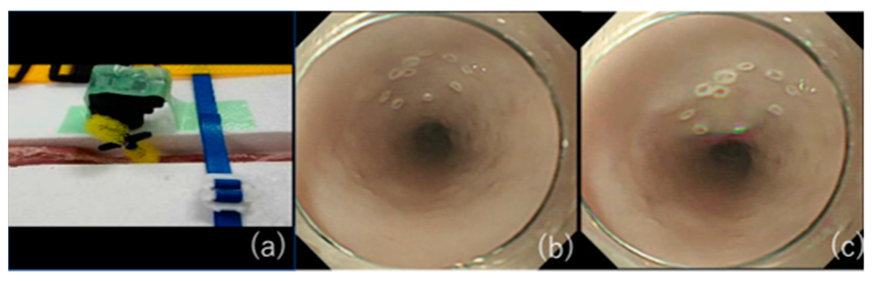
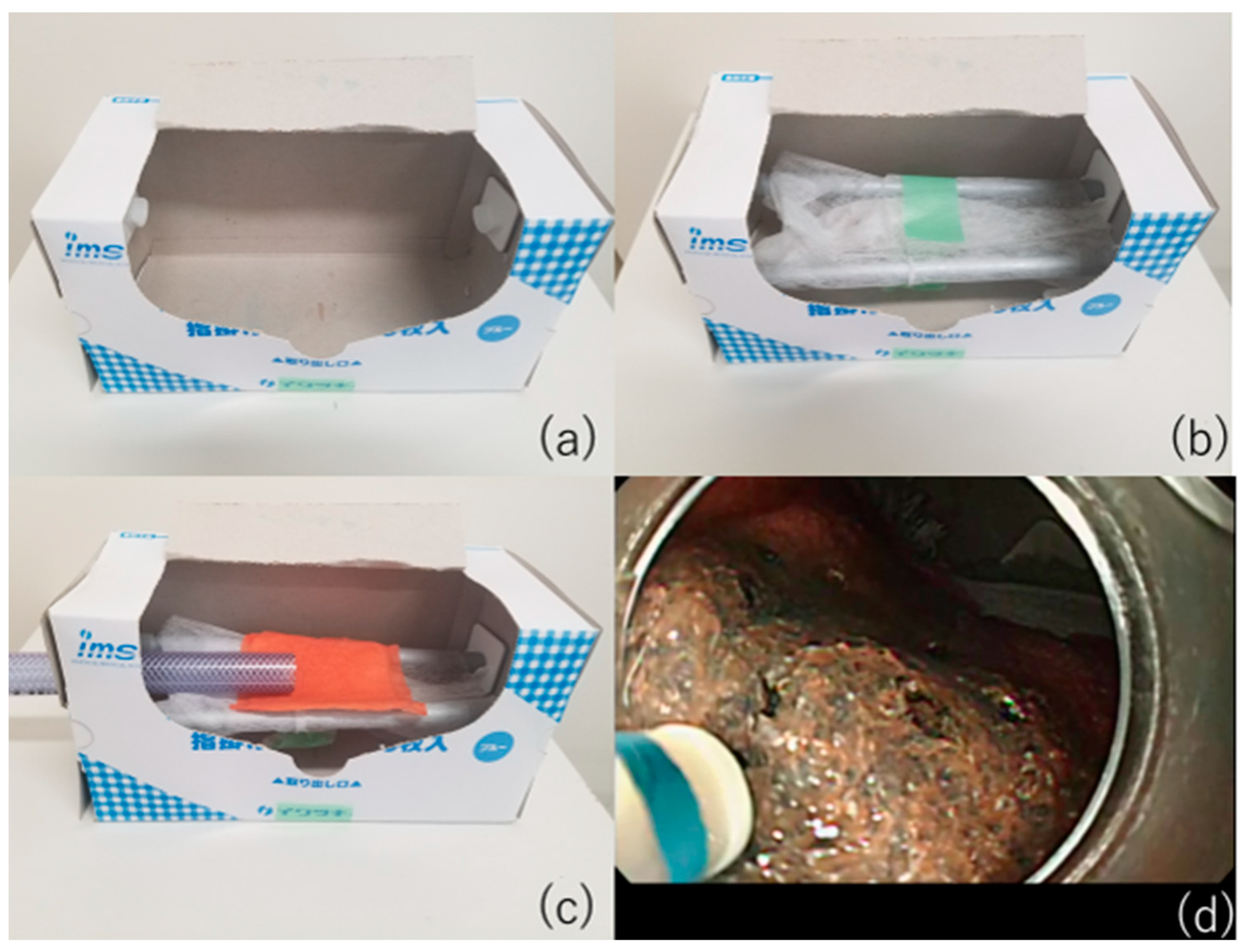
2.4. Education on ESD Techniques

Although various approaches can be used to avoid accidental injury, as described above, it is fundamentally important to acquire a reliable ESD technique that does not cause accidental injury. Although it has been reported that it is generally favorable to start ESD training in the gastric antrum and the rectum because of the thickness of their walls [51], the incidence of gastric cancer is not as high in Europe and the United States as in Asia [52], and it is considered difficult to obtain a chance to improve the technique. The use of pig internal organs as a training model has been reported as a method for the acquisition of techniques before endoscopic treatment in actual clinical practice [53,54,55]. While this model is useful for acquiring basic techniques, it is certainly not realistic because of the absence of factors that make ESD difficult in actual clinical practice, such as heart rate and respiratory variability. Therefore, we have successfully developed an animal model with a heartbeat that is more in line with actual clinical practice [56] and used it to teach future clinicians (Figure 4). As the model has a motor devise that rotates at 80 cycle/min (approximately a normal human heart rate) in approximately 30 cm from the incisors of pig organ, we can perform realistic esophageal ESD training (Figure 5). It is also possible to learn about troubleshooting for intraoperative complications using ex vivo animal models. We can practice suturing a perforation intentionally made by an ESD knife with a hemostatic clip. Intraoperative bleeding, which may seem impossible to reproduce, can be reproduced by injecting red ink into the blood vessels of the organ, allowing us to practice hemostasis [57]. While some facilities and regions may have problems, such as unavailability of pig organs and cleaning of scopes used in animal models, we have also begun to conduct ESD training using Versatile Training Tissue (VTT; KOTOBUKI medical, Yashio, Saitama, Japan) made of “konjac”, a food item, which are readily available and do not require special care to clean the scope [58]. The virtual digestive tract wall is reproduced by placing VTT on a pedestal made of items found in the endoscopy lab, such as an empty plastic apron box, gauze, and rods (Figure 6). As VTT has a three-layer structure, including simulated mucosal, submucosal, and muscle layers, realistic ESD training can be performed by following the normal procedure. A training kit that includes a training model using PVA-H (EndoGel (Sunarrow Kasei Co., LTD, Tokyo, Japan)) [59] and eliminates the hygiene aspect has been reported; however, the konjac model is overwhelmingly cheaper (USD 450 vs. USD 60, respectively). Trainees need to repeat training using these in vitro models to acquire the basic ESD techniques. It is desirable to start the actual treatment from a site in which it is easy to manipulate a scope, such as the antrum or rectum. In addition, it is important for the trainee to be able to complete the ESD procedure in a small number of cases without complications by having one-on-one guidance and techniques and strategies shared with them in real time. In addition, adequate infection control measures, such as full personal protective equipment, should be taken during one-to-one guidance during the COVID-19 pandemic.

3. Conclusions

Intraoperative and postoperative hemorrhage, perforation, and postoperative stenosis are critical sequelae associated with ESD, and their prevention and management were presented in detail in this review. There are multiple methods for each of these, and it is necessary to find an appropriate method for each facility. Furthermore, the establishment of training methods for ESD may be a measure to prevent complications in the long term [60].

Author Contributions

Y.M. was involved in the preparation and submission of this subscript. K.N. was involved in the final correction and proofreading of the manuscript. All authors have read and agreed to the published version of the manuscript.

Funding

This research received no external funding.

Conflicts of Interest

The authors declare no conflict of interest.

References

  1. Gotoda, T.; Yamamoto, H.; Soetikno, R.M. Endoscopic Submucosal Dissection of Early Gastric Cancer. J. Gastroenterol. 2006, 41, 929–942. [Google Scholar] [CrossRef] [PubMed]
  2. Draganov, P.V.; Wang, A.Y.; Othman, M.O.; Fukami, N. AGA Institute Clinical Practice Update: Endoscopic Submucosal Dissection in the United States. Clin. Gastroenterol. Hepatol. 2019, 17, 16–25.e1. [Google Scholar] [CrossRef] [Green Version]
  3. Kobayashi, N.; Yoshitake, N.; Hirahara, Y.; Konishi, J.; Saito, Y.; Matsuda, T.; Ishikawa, T.; Sekiguchi, R.; Fujimori, T. Matched Case-Control Study Comparing Endoscopic Submucosal Dissection and Endoscopic Mucosal Resection for Colorectal Tumors. J. Gastroenterol. Hepatol. 2012, 27, 728–733. [Google Scholar] [CrossRef] [PubMed]
  4. Kim, Y.J.; Kim, E.S.; Cho, K.B.; Park, K.S.; Jang, B.K.; Chung, W.J.; Hwang, J.S. Comparison of Clinical Outcomes among Different Endoscopic Resection Methods for Treating Colorectal Neoplasia. Dig. Dis. Sci. 2013, 58, 1727–1736. [Google Scholar] [CrossRef] [PubMed]
  5. Ono, S.; Fujishiro, M.; Niimi, K.; Goto, O.; Kodashima, S.; Yamamichi, N.; Omata, M. Predictors of Postoperative Stricture after Esophageal Endoscopic Submucosal Dissection for Superficial Squamous Cell Neoplasms. Endoscopy 2009, 41, 661–665. [Google Scholar] [CrossRef]
  6. Kataoka, Y.; Tsuji, Y.; Sakaguchi, Y.; Minatsuki, C.; Asada-Hirayama, I.; Niimi, K.; Ono, S.; Kodashima, S.; Yamamichi, N.; Fujishiro, M.; et al. Bleeding after endoscopic submucosal dissection: Risk factors and preventive methods. World J. Gastroenterol. 2016, 22, 5927–5935. [Google Scholar] [CrossRef]
  7. Yang, C.H.; Qiu, Y.; Li, X.; Shi, R.H. Bleeding after endoscopic submucosal dissection of gastric lesions. J. Dig. Dis. 2020, 21, 139–146. [Google Scholar] [CrossRef]
  8. Shibagaki, K.; Ishimura, N.; Kinoshita, Y. Endoscopic submucosal dissection for duodenal tumors. Ann. Transl. Med. 2017, 5, 188. [Google Scholar] [CrossRef] [PubMed] [Green Version]
  9. Libânio, D.; Costa, M.N.; Pimentel-Nunes, P.; Dinis-Ribeiro, M. Risk Factors for Bleeding after Gastric Endoscopic Submucosal Dissection: A Systematic Review and Meta-Analysis. Gastrointest. Endosc. 2016, 84, 572–586. [Google Scholar] [CrossRef] [Green Version]
  10. Green, F.W., Jr.; Kaplan, M.M.; Curtis, L.E.; Levine, P.H. Effect of Acid and Pepsin on Blood Coagulation and Platelet Aggregation. A Possible Contributor Prolonged Gastroduodenal Mucosal Hemorrhage. Gastroenterol. 1978, 74, 38–43. [Google Scholar] [CrossRef]
  11. Yang, Z.; Wu, Q.; Liu, Z.; Wu, K.; Fan, D. Proton Pump Inhibitors versus histamine-2-Receptor Antagonists for the Management of Iatrogenic Gastric Ulcer after Endoscopic Mucosal Resection or Endoscopic Submucosal Dissection: A Meta-Analysis of Randomized Trials. Digestion 2011, 84, 315–320. [Google Scholar] [CrossRef]
  12. Uedo, N.; Takeuchi, Y.; Yamada, T.; Ishihara, R.; Ogiyama, H.; Yamamoto, S.; Kato, M.; Tatsumi, K.; Masuda, E.; Tamai, C.; et al. Effect of a Proton Pump Inhibitor or an H2-Receptor Antagonist on Prevention of Bleeding from Ulcer after Endoscopic Submucosal Dissection of Early Gastric Cancer: A Prospective Randomized Controlled Trial. Am. J. Gastroenterol. 2007, 102, 1610–1616. [Google Scholar] [CrossRef]
  13. Kondo, M.; Kawamoto, M.; Hasuoka, A.; Kajino, M.; Inatomi, N.; Tarui, N. High-throughput screening of potassium-competitive acid blockers. J. Biomol. Screen. 2012, 17, 177–182. [Google Scholar] [CrossRef]
  14. Gao, H.; Li, L.; Zhang, C.; Tu, J.; Geng, X.; Wang, J.; Zhou, X.; Jing, J.; Pan, W. Comparison of efficacy of pharmacological therapies for gastric endoscopic submucosal dissection-induced ulcers: A systematic review and network meta-analysis. Expert Rev. Gastroenterol. Hepatol. 2020, 14, 207–220. [Google Scholar] [CrossRef] [PubMed]
  15. Kakushima, N.; Hori, K.; Ono, H.; Horimatsu, T.; Uedo, N.; Ohata, K.; Doyama, H.; Kaneko, K.; Oda, I.; Hikichi, T.; et al. Proton Pump Inhibitor after Endoscopic Resection for Esophageal Squamous Cell Cancer: Multicenter Prospective Randomized Controlled Trial. J. Gastroenterol. 2016, 51, 104–111. [Google Scholar] [CrossRef]
  16. Choi, K.D.; Jung, H.Y.; Lee, G.H.; Oh, T.H.; Jo, J.Y.; Song, H.J.; Hong, S.S.; Kim, J.H. Application of Metal Hemoclips for Closure of Endoscopic Mucosal Resection-Induced Ulcers of the Stomach to Prevent Delayed Bleeding. Surg. Endosc. 2008, 22, 1882–1886. [Google Scholar] [CrossRef] [PubMed]
  17. Ogiyama, H.; Tsutsui, S.; Murayama, Y.; Maeda, S.; Satake, S.; Nasu, A.; Umeda, D.; Miura, Y.; Tominaga, K.; Horiki, M.; et al. Prophylactic Clip Closure May Reduce the Risk of Delayed Bleeding after Colorectal Endoscopic Submucosal Dissection. Endosc. Int. Open 2018, 6, E582–E588.e8. [Google Scholar] [CrossRef] [PubMed] [Green Version]
  18. Hoteya, S.; Kaise, M.; Iizuka, T.; Ogawa, O.; Mitani, T.; Matsui, A.; Kikuchi, D.; Furuhata, T.; Yamashita, S.; Yamada, A.; et al. Delayed Bleeding after Endoscopic Submucosal Dissection for Non-Ampullary Superficial Duodenal Neoplasias Might Be Prevented by Prophylactic Endoscopic Closure: Analysis of Risk Factors. Dig. Endosc. 2015, 27, 323–330. [Google Scholar] [CrossRef] [PubMed]
  19. Nishizawa, T.; Suzuki, H.; Goto, O.; Ogata, H.; Kanai, T.; Yahagi, N. Effect of prophylactic clipping in colorectal endoscopic resection: A meta-analysis of randomized controlled studies. United Eur. Gastroenterol. J. 2017, 5, 859–867. [Google Scholar] [CrossRef] [PubMed] [Green Version]
  20. Otake, Y.; Saito, Y.; Sakamoto, T.; Aoki, T.; Nakajima, T.; Toyoshima, N.; Matsuda, T.; Ono, H. New Closure Technique for Large Mucosal Defects after Endoscopic Submucosal Dissection of Colorectal Tumors (with Video). Gastrointest. Endosc. 2012, 75, 663–667. [Google Scholar] [CrossRef]
  21. Abe, S.; Oda, I.; Mori, G.; Nonaka, S.; Suzuki, H.; Yoshianaga, S.; Saito, Y. Complete Endoscopic Closure of a Large Gastric Defect with Endoloop and Endoclips after Complex Endoscopic Submucosal Dissection. Endoscopy 2015, 47 (Suppl. 1), E374–E375. [Google Scholar] [CrossRef]
  22. Kantsevoy, S.V.; Bitner, M.; Mitrakov, A.A.; Thuluvath, P.J. Endoscopic Suturing Closure of Large Mucosal Defects after Endoscopic Submucosal Dissection Is Technically Feasible, Fast, and Eliminates the Need for Hospitalization (with Videos). Gastrointest. Endosc. 2014, 79, 503–507. [Google Scholar] [CrossRef]
  23. Voermans, R.P.; Le Moine, O.; von Renteln, D.; Ponchon, T.; Giovannini, M.; Bruno, M.; Weusten, B.; Seewald, S.; Costamagna, G.; Deprez, P.; et al. Efficacy of Endoscopic Closure of Acute Perforations of the Gastrointestinal Tract. Clin. Gastroenterol. Hepatol. 2012, 10, 603–608. [Google Scholar] [CrossRef]
  24. Akimoto, T.; Goto, O.; Sasaki, M.; Mizutani, M.; Tsutsumi, K.; Kiguchi, Y.; Nakayama, A.; Kato, M.; Fujimoto, A.; Ochiai, Y.; et al. Endoscopic Suturing Promotes Healing of Mucosal Defects after Gastric Endoscopic Submucosal Dissection: Endoscopic and Histologic Analyses in Vivo Porcine Models (with Video). Gastrointest. Endosc. 2020, 91, 1172–1182. [Google Scholar] [CrossRef]
  25. Tsuji, Y.; Fujishiro, M.; Kodashima, S.; Ono, S.; Niimi, K.; Mochizuki, S.; Asada-Hirayama, I.; Matsuda, R.; Minatsuki, C.; Nakayama, C.; et al. Polyglycolic Acid Sheets and Fibrin Glue Decrease the Risk of Bleeding after Endoscopic Submucosal Dissection of Gastric Neoplasms (with Video). Gastrointest. Endosc. 2015, 81, 906–912. [Google Scholar] [CrossRef]
  26. Kawata, N.; Ono, H.; Takizawa, K.; Kakushima, N.; Tanaka, M.; Igarashi, K.; Yoshida, M.; Kishida, Y.; Iwai, T.; Ito, S.; et al. Efficacy of Polyglycolic Acid Sheets and Fibrin Glue for Prevention of Bleeding after Gastric Endoscopic Submucosal Dissection in Patients under Continued Antithrombotic Agents. Gastric Cancer 2018, 21, 696–702. [Google Scholar] [CrossRef] [PubMed] [Green Version]
  27. Fujimoto, K.; Fujishiro, M.; Kato, M.; Higuchi, K.; Iwakiri, R.; Sakamoto, C.; Uchiyama, S.; Kashiwagi, A.; Ogawa, H.; Murakami, K.; et al. Guidelines for Gastroenterological Endoscopy in Patients Undergoing Antithrombotic Treatment. Dig. Endosc. 2014, 26, 1–14. [Google Scholar] [CrossRef] [PubMed]
  28. Kato, M.; Uedo, N.; Hokimoto, S.; Ieko, M.; Higuchi, K.; Murakami, K.; Fujimoto, K. Guidelines for Gastroenterological Endoscopy in Patients Undergoing Antithrombotic Treatment: 2017 Appendix on Anticoagulants Including Direct Oral Anticoagulants. Dig. Endosc. 2018, 30, 433–440. [Google Scholar] [CrossRef] [Green Version]
  29. Odagiri, H.; Yasunaga, H. Complications Following Endoscopic Submucosal Dissection for Gastric, Esophageal, and Colorectal Cancer: A Review of Studies Based on Nationwide Large-Scale Databases. Ann. Transl. Med. 2017, 5, 189. [Google Scholar] [CrossRef] [Green Version]
  30. Hu, J.; Zhao, Y.; Ren, M.; Li, Y.; Lu, X.; Lu, G.; Zhang, D.; Chu, D.; He, S. The Comparison between Endoscopic Submucosal Dissection and Surgery in Gastric Cancer: A Systematic Review and Meta-Analysis. Gastroenterol. Res. Pract. 2018, 2018, 4378945. [Google Scholar] [CrossRef] [PubMed] [Green Version]
  31. De Ceglie, A.; Hassan, C.; Mangiavillano, B.; Matsuda, T.; Saito, Y.; Ridola, L.; Bhandari, P.; Boeri, F.; Conio, M. Endoscopic Mucosal Resection and Endoscopic Submucosal Dissection for Colorectal Lesions: A Systematic Review. Crit. Rev. Oncol. Hematol. 2016, 104, 138–155. [Google Scholar] [CrossRef]
  32. Tsuji, K.; Yoshida, N.; Nakanishi, H.; Takemura, K.; Yamada, S.; Doyama, H. Recent Traction Methods for Endoscopic Submucosal Dissection. World J. Gastroenterol. 2016, 22, 5917–5926. [Google Scholar] [CrossRef]
  33. Oyama, T. Counter Traction Makes Endoscopic Submucosal Dissection Easier. Clin. Endosc. 2012, 45, 375–378. [Google Scholar] [CrossRef] [PubMed]
  34. Yamasaki, Y.; Takeuchi, Y.; Uedo, N.; Kanesaka, T.; Kato, M.; Hamada, K.; Tonai, Y.; Matsuura, N.; Akasaka, T.; Hanaoka, N.; et al. Efficacy of Traction-Assisted Colorectal Endoscopic Submucosal Dissection Using a Clip-and-Thread Technique: A Prospective Randomized Study. Dig. Endosc. 2018, 30, 467–476. [Google Scholar] [CrossRef] [PubMed]
  35. Sakamoto, N.; Osada, T.; Shibuya, T.; Beppu, K.; Matsumoto, K.; Mori, H.; Kawabe, M.; Nagahara, A.; Otaka, M.; Ogihara, T.; et al. Endoscopic Submucosal Dissection of Large Colorectal Tumors by Using a Novel Spring-Action S-O Clip for Traction (with Video). Gastrointest. Endosc. 2009, 69, 1370–1374. [Google Scholar] [CrossRef] [PubMed]
  36. Mori, H.; Kobara, H.; Nishiyama, N.; Fujihara, S.; Matsunaga, T.; Masaki, T. Novel Effective and Repeatedly Available Ring-Thread Counter Traction for Safer Colorectal Endoscopic Submucosal Dissection. Surg. Endosc. 2017, 31, 3040–3047. [Google Scholar] [CrossRef] [PubMed] [Green Version]
  37. Haito-Chavez, Y.; Law, J.K.; Kratt, T.; Arezzo, A.; Verra, M.; Morino, M.; Sharaiha, R.Z.; Poley, J.W.; Kahaleh, M.; Thompson, C.C.; et al. International Multicenter Experience with an Over-the-Scope Clipping Device for Endoscopic Management of GI Defects (with Video). Gastrointest. Endosc. 2014, 80, 610–622. [Google Scholar] [CrossRef] [PubMed]
  38. Tsunada, S.; Ogata, S.; Mannen, K.; Arima, S.; Sakata, Y.; Shiraishi, R.; Shimoda, R.; Ootani, H.; Yamaguchi, K.; Fujise, T.; et al. Case Series of Endoscopic Balloon Dilation to Treat a Stricture Caused by Circumferential Resection of the Gastric Antrum by Endoscopic Submucosal Dissection. Gastrointest. Endosc. 2008, 67, 979–983. [Google Scholar] [CrossRef] [PubMed]
  39. Iizuka, H.; Kakizaki, S.; Sohara, N.; Onozato, Y.; Ishihara, H.; Okamura, S.; Itoh, H.; Mori, M. Stricture after Endoscopic Submucosal Dissection for Early Gastric Cancers and Adenomas. Dig. Endosc. 2010, 22, 282–288. [Google Scholar] [CrossRef]
  40. Hayashi, T.; Kudo, S.E.; Miyachi, H.; Sakurai, T.; Ishigaki, T.; Yagawa, Y.; Toyoshima, N.; Mori, Y.; Misawa, M.; Kudo, T.; et al. Management and Risk Factor of Stenosis after Endoscopic Submucosal Dissection for Colorectal Neoplasms. Gastrointest. Endosc. 2017, 86, 358–369. [Google Scholar] [CrossRef]
  41. Martínek, J.; Juhas, S.; Dolezel, R.; Walterová, B.; Juhasova, J.; Klima, J.; Rabekova, Z.; Vacková, Z. Prevention of Esophageal Strictures after Circumferential Endoscopic Submucosal Dissection. Minerva Chir. 2018, 73, 394–409. [Google Scholar]
  42. Nonaka, K.; Miyazawa, M.; Ban, S.; Aikawa, M.; Akimoto, N.; Koyama, I.; Kita, H. Different Healing Process of Esophageal Large Mucosal Defects by Endoscopic Mucosal Dissection between with and without Steroid Injection in an Animal Model. BMC Gastroenterol. 2013, 13, 72. [Google Scholar] [CrossRef] [PubMed] [Green Version]
  43. Nonaka, K.; Ban, S.; Ryozawa, S. Strictures after Endoscopic Submucosal Dissection of the Esophagus: Are the Histopathological Findings the Same between Human and Porcine Models? Dig. Endosc. 2019, 31, 106. [Google Scholar] [CrossRef] [Green Version]
  44. Hinz, B.; Phan, S.H.; Thannickal, V.J.; Galli, A.; Bochaton-Piallat, M.L.; Gabbiani, G. The Myofibroblast: One Function, Multiple Origins. Am. J. Pathol. 2007, 170, 1807–1816. [Google Scholar] [CrossRef] [PubMed]
  45. Yamashita, S.; Kato, M.; Fujimoto, A.; Maehata, T.; Sasaki, M.; Inoshita, N.; Sato, H.; Suzuki, K.; Yahagi, N. Inadequate Steroid Injection after Esophageal ESD Might Cause Mural Necrosis. Endosc. Int. Open 2019, 7, E115–E121.e21. [Google Scholar] [CrossRef] [Green Version]
  46. Shumeiko, O.; Yoshida, M.; Ono, H. Safe Method of Steroid Injection Following Esophageal Endoscopic Submucosal Dissection for Postoperative Stricture Prevention. Dig. Endosc. 2019, 31, e118–e119. [Google Scholar] [CrossRef] [PubMed] [Green Version]
  47. Misumi, Y.; Nonaka, K.; Kishino, M. Safer Local Injection of Triamcinolone Acetonide for Preventing Post-Endoscopic Submucosal Dissection Esophageal Stenosis. Dig. Endosc. 2021, 33, e77–e78. [Google Scholar] [CrossRef]
  48. Yano, T.; Haro, A.; Shikada, Y.; Okamoto, T.; Maruyama, R.; Maehara, Y. A Unique Method for Repairing Intraoperative Pulmonary Air Leakage with Both Polyglycolic Acid Sheets and Fibrin Glue. World J. Surg. 2012, 36, 463–467. [Google Scholar] [CrossRef]
  49. Maruyama, A.; Tsunoda, A.; Takahashi, M.; Kishimoto, S.; Suzuki, M. Nasopharyngeal Pleomorphic Adenoma Presenting as Otitis Media with Effusion: Case Report and Literature Review. Am. J. Otolaryngol. 2014, 35, 73–76. [Google Scholar] [CrossRef]
  50. Iizuka, T.; Kikuchi, D.; Yamada, A.; Hoteya, S.; Kajiyama, Y.; Kaise, M. Polyglycolic Acid Sheet Application to Prevent Esophageal Stricture after Endoscopic Submucosal Dissection for Esophageal Squamous Cell Carcinoma. Endoscopy 2015, 47, 341–344. [Google Scholar] [CrossRef]
  51. Ohki, T.; Yamato, M.; Ota, M.; Takagi, R.; Murakami, D.; Kondo, M.; Sasaki, R.; Namiki, H.; Okano, T.; Yamamoto, M. Prevention of Esophageal Stricture after Endoscopic Submucosal Dissection Using Tissue-Engineered Cell Sheets. Gastroenterology 2012, 143, 582–588.e2. [Google Scholar] [CrossRef] [PubMed]
  52. Deprez, P.H.; Bergman, J.J.; Meisner, S.; Ponchon, T.; Repici, A.; Dinis-Ribeiro, M.; Haringsma, J. Current Practice with Endoscopic Submucosal Dissection in Europe: Position Statement from a Panel of Experts. Endoscopy 2010, 42, 853–858. [Google Scholar] [CrossRef] [PubMed]
  53. Casamayor, M.; Morlock, R.; Maeda, H.; Ajani, J. Targeted Literature Review of the Global Burden of Gastric Cancer. Ecancermedicalscience 2018, 12, 883. [Google Scholar] [CrossRef] [PubMed] [Green Version]
  54. Ohata, K.; Fu, K.; Sakai, E.; Nonaka, K.; Tashima, T.; Minato, Y.; Ohno, A.; Ito, T.; Tsuji, Y.; Chiba, H.; et al. Esophageal Endoscopic Submucosal Dissection Assisted by an Overtube with a Traction Forceps: An Animal Study. Gastroenterol. Res. Pract. 2016, 2016, 3186168. [Google Scholar] [CrossRef]
  55. Tanimoto, M.A.; Torres-Villalobos, G.; Fujita, R.; Santillan-Doherty, P.; Albores-Saavedra, J.; Chable-Montero, F.; Martin-Del-Campo, L.A.; Vasquez, L.; Bravo-Reyna, C.; Villanueva, O.; et al. Learning Curve in a Western Training Center of the Circumferential en Bloc Esophageal Endoscopic Submucosal Dissection in an In Vivo Animal Model. Diagn. Ther. Endosc. 2011, 2011, 847831. [Google Scholar] [CrossRef] [PubMed]
  56. Ohata, K.; Nonaka, K.; Misumi, Y.; Tsunashima, H.; Takita, M.; Minato, Y.; Tashima, T.; Sakai, E.; Muramoto, T.; Matsuyama, Y.; et al. Usefulness of Training Using Animal Models for Colorectal Endoscopic Submucosal Dissection: Is Experience Performing Gastric ESD Really Needed? Endosc. Int. Open 2016, 4, E333–E339. [Google Scholar] [CrossRef] [PubMed] [Green Version]
  57. Misumi, Y.; Nonaka, K.; Kishino, M. Development of an Animal Model That Reproduces Pulsations for Training in Esophageal Endoscopic Submucosal Dissection. Endoscopy 2020. [Google Scholar] [CrossRef]
  58. Yoshida, N.; Yagi, N.; Inada, Y.; Kugai, M.; Kamada, K.; Katada, K.; Uchiyama, K.; Ishikawa, T.; Takagi, T.; Handa, O.; et al. Possibility of ex vivo animal training model for colorectal endoscopic submucosal dissection. Int. J. Colorectal. Dis. 2013, 28, 49–56. [Google Scholar] [CrossRef]
  59. Shinozaki, K.; Misumi, Y.; Nonaka, K. Digestive Endoscopic Submucosal Dissection Training Using Konjac. Dig. Endosc. 2021. [Google Scholar] [CrossRef] [PubMed]
  60. Mizuno, K.; Sato, H.; Hashimoto, S.; Sato, Y.; Terai, S. A Novel Training Model Composed of Nonbiological Materials for Endoscopic Submucosal Dissection. Gastrointest. Endosc. 2016, 84, 373–374. [Google Scholar] [CrossRef] [PubMed]
Figure 1. Usefullness of a S-O clip. (a) Good traction is obtained by using the S-O clip. (b) (1) Without S-O clip. It is difficult to go into the submucosal layer. (2) The submucosal layer can be easily visualized by applying the S-O clip at a site 2-fold distant from the anal side of the lesion.
Jcm 10 02511 g001
Figure 2. The ring-thread counter traction method. (a) A handmade clip with a ring made of dental floss attached to the arm. (b) Attach the thread ring to the flap. (c) Apply the ring at site 2-fold distant from the anal side of the lesion. (d) Good traction was obtained, as in the case of the S-O clip.
Jcm 10 02511 g002
Figure 3. Safe local injection of TA with a spraying tube. (a) A spraying tube with a dull tip can be used for safe local injection by simply pushing against the submucosal loose tissue. (b) TA is evenly injected into the ulcer bed of esophageal ESD.
Jcm 10 02511 g003
Figure 4. An animal model reproducing pulsations for training in esophageal endoscopic submucosal dissection.
Jcm 10 02511 g004
Figure 5. Pulsation is reproduced by the motor devise. (a) Pulsation is reproduced when the motor device makes contact with the esophageal wall. (b) Noncontact time. (c) Contact time. Dynamic pulsation is reproduced.
Jcm 10 02511 g005
Figure 6. VTT ESD training model without special items. (a) An empty plastic apron box. (b) Place two rods in the box and wrap gauze around them like a stretcher. (c) Penetrate the hose into the box and place the VTT on the gauze. (d) A view during training with the VTT “konjac”, a food item model.
Jcm 10 02511 g006
Table 1. Withdrawal of monotherapy with antiplatelet agents or anticoagulants during gastroenterological endoscopy.
Standard EndoscopyBiopsyLow Risk of BleedingHigh Risk of Bleeding
Aspirin○or withdraw for 3–5 days
ThienopyridineASA/CLZ replacement or withdraw for 5–7 days
Antiplatelet agent other than thienopyridinewithdraw for 1 day
Warfarin
therapeutic range

therapeutic range
○therapeutic range
or
heparin replacement
or
temporary change in DOAC
DOACwithdraw on the day of treatment
or
heparin replacement
ASA, aspirin; CLZ, cilostazole; DOAC, direct oral anticoagulant; ◎ = withdrawal is not required; ○ = withdrawal is required on a case-by-case basis.
Table 2. Withdrawal of dual therapy with antiplatelet agents or anticoagulants during gastroenterological endoscopy.
AspirinThienopyridineAntiplatelet Agent Other than ThienopyridineWarfarinDOAC
○or CLZ replacementwithdraw for 5–7 days
○or CLZ replacement withdraw for 1 day
○or CLZ replacement ○therapeutic range
or
heparin replacement
or
temporary change in DOAC
○or CLZ replacement withdraw on the day of treatment
ASA/CLZ replacementwithdraw for 1 day
ASA/CLZ replacement ○therapeutic range
or
heparin replacement
or
temporary change in DOAC
ASA/CLZ replacement withdraw on the day of treatment
maintain CLZ or withdraw for 1day○therapeutic range
or
heparin replacement
or
temporary change in DOAC
maintain CLZ or withdraw for 1day withdraw on the day of treatment
ASA, aspirin; CLZ, cilostazole; DOAC, direct oral anticoagulant; ○ = withdrawal is required on a case-by-case basis.
Table 3. Withdrawal of triple therapy with antiplatelet agents or anticoagulants during gastroenterological endoscopy.
AspirinThienopyridineAntiplatelet Agent Other than ThienopyridineWarfarinDOAC
○or CLZ replacementwithdraw for 5–7 days ○therapeutic range
or
heparin replacement
or
temporary change in DOAC
○or CLZ replacementwithdraw for 5–7 days withdraw on the day of treatment
○or CLZ replacement Withdraw for 1 day○therapeutic range
or
heparin replacement
or
temporary change in DOAC
○or CLZ replacement Withdraw for 1 day withdraw on the day of treatment
ASA/CLZ replacementwithdraw for 1 day○therapeutic range
or
heparin replacement
or
temporary change in DOAC
ASA/CLZ replacementwithdraw for 1 day withdraw on the day of treatment
ASA, aspirin; CLZ, cilostazole, DOAC, direct oral anticoagulant; ○ = withdrawal is required on a case-by-case basis.
Publisher’s Note: MDPI stays neutral with regard to jurisdictional claims in published maps and institutional affiliations.

