Composite Ferroelectric Membranes Based on Vinylidene Fluoride-Tetrafluoroethylene Copolymer and Polyvinylpyrrolidone for Wound Healing

 , ,
, , 
Abstract

1. Introduction
2. Materials and Methods
2.1. Preparation of VDF–TeFE/PVP Membranes by Electrospinning
2.2. Investigation of Physicochemical Properties of Membranes
2.2.1. Scanning Electron Microscopy (SEM)
2.2.2. X-ray Fluorescence Spectroscopy (XRF)
2.2.3. Gas Chromatography (GC)
2.2.4. Fourier-Transform Infrared Spectroscopy (FTIR)
2.2.5. X-ray Diffraction Analysis (XRD)
2.3. Investigation of Biomedical Properties of Membranes
2.3.1. Adhesion, Viability, and Proliferative Activity of Cells
2.3.2. In Vivo Contaminated Full-Thickness Wound Healing with Composite Membranes
2.4. Statistics
3. Results and Discussion
4. Conclusions
Author Contributions
Funding
Institutional Review Board Statement
Informed Consent Statement
Conflicts of Interest
References
- Chouhan, D.; Mandal, B.B. Silk biomaterials in wound healing and skin regeneration therapeutics: From bench to bedside. Acta Biomater. 2020, 103, 24–51. [Google Scholar] [CrossRef] [PubMed]
- Mayet, N.; Choonara, Y.E.; Kumar, P.; Tomar, L.K.; Tyagi, C.; Du Toit, L.C.; Pillay, V. A Comprehensive Review of Advanced Biopolymeric Wound Healing Systems. J. Pharm. Sci. 2014, 103, 2211–2230. [Google Scholar] [CrossRef] [PubMed]
- Lionelli, G.T.; Lawrence, W.T. Wound dressings. Surg. Clin. N. Am. 2003, 83, 617–638. [Google Scholar] [CrossRef]
- Chouhan, D.; Dey, N.; Bhardwaj, N.; Mandal, B.B. Emerging and innovative approaches for wound healing and skin regeneration: Current status and advances. Biomaterials 2019, 216, 119267. [Google Scholar] [CrossRef] [PubMed]
- Miguel, S.P.; Figueira, D.R.; Simões, D.; Ribeiro, M.P.; Coutinho, P.; Ferreira, P.; Correia, I.J. Electrospun polymeric nanofibres as wound dressings: A review. Colloids Surf. B Biointerfaces 2018, 169, 60–71. [Google Scholar] [CrossRef]
- Juncos Bombin, A.D.; Dunne, N.J.; McCarthy, H.O. Electrospinning of natural polymers for the production of nanofibres for wound healing applications. Mater. Sci. Eng. C 2020, 114, 110994. [Google Scholar] [CrossRef]
- Ambekar, R.S.; Kandasubramanian, B. Advancements in nanofibers for wound dressing: A review. Eur. Polym. J. 2019, 117, 304–336. [Google Scholar] [CrossRef]
- Minary-Jolandan, M.; Yu, M.-F. Nanoscale characterization of isolated individual type I collagen fibrils: Polarization and piezoelectricity. Nanotechnology 2009, 20, 085706. [Google Scholar] [CrossRef]
- Ning, C.; Zhou, Z.; Tan, G.; Zhu, Y.; Mao, C. Electroactive polymers for tissue regeneration: Developments and perspectives. Prog. Polym. Sci. 2018, 81, 144–162. [Google Scholar] [CrossRef]
- Han, M.; Wang, H.; Yang, Y.; Liang, C.; Bai, W.; Yan, Z.; Li, H.; Xue, Y.; Wang, X.; Akar, B.; et al. Three-dimensional piezoelectric polymer microsystems for vibrational energy harvesting, robotic interfaces and biomedical implants. Nat. Electron. 2019, 2, 26–35. [Google Scholar] [CrossRef]
- Ribeiro, C.; Sencadas, V.; Correia, D.M.; Lanceros-Méndez, S. Piezoelectric polymers as biomaterials for tissue engineering applications. Colloids Surf. B Biointerfaces 2015, 136, 46–55. [Google Scholar] [CrossRef] [PubMed]
- Ramadan, K.S.; Sameoto, D.; Evoy, S. A review of piezoelectric polymers as functional materials for electromechanical transducers. Smart Mater. Struct. 2014, 23, 033001. [Google Scholar] [CrossRef]
- Venault, A.; Lin, K.-H.; Tang, S.-H.; Dizon, G.V.; Hsu, C.-H.; Maggay, I.V.B.; Chang, Y. Zwitterionic electrospun PVDF fibrous membranes with a well-controlled hydration for diabetic wound recovery. J. Memb. Sci. 2020, 598, 117648. [Google Scholar] [CrossRef]
- Du, S.; Zhou, N.; Gao, Y.; Xie, G.; Du, H.; Jiang, H.; Zhang, L.; Tao, J.; Zhu, J. Bioinspired hybrid patches with self-adhesive hydrogel and piezoelectric nanogenerator for promoting skin wound healing. Nano Res. 2020. [Google Scholar] [CrossRef]
- He, T.; Wang, J.; Huang, P.; Zeng, B.; Li, H.; Cao, Q.; Zhang, S.; Luo, Z.; Deng, D.Y.B.; Zhang, H.; et al. Electrospinning polyvinylidene fluoride fibrous membranes containing anti-bacterial drugs used as wound dressing. Colloids Surf. B Biointerfaces 2015, 130, 278–286. [Google Scholar] [CrossRef]
- Badaraev, A.D.; Koniaeva, A.; Krikova, S.A.; Shesterikov, E.V.; Bolbasov, E.N.; Nemoykina, A.L.; Bouznik, V.M.; Stankevich, K.S.; Zhukov, Y.M.; Mishin, I.P.; et al. Piezoelectric polymer membranes with thin antibacterial coating for the regeneration of oral mucosa. Appl. Surf. Sci. 2020, 504, 144068. [Google Scholar] [CrossRef]
- Cui, Z.; Drioli, E.; Lee, Y.M. Recent progress in fluoropolymers for membranes. Prog. Polym. Sci. 2014, 39, 164–198. [Google Scholar] [CrossRef]
- Cui, Z.; Hassankiadeh, N.T.; Zhuang, Y.; Drioli, E.; Lee, Y.M. Crystalline polymorphism in poly(vinylidenefluoride) membranes. Prog. Polym. Sci. 2015, 51, 94–126. [Google Scholar] [CrossRef]
- Ameduri, B. From vinylidene fluoride (VDF) to the applications of VDF-containing polymers and copolymers: Recent developments and future trends. Chem. Rev. 2009, 109, 6632–6686. [Google Scholar] [CrossRef]
- Tverdokhlebova, T.T.; Bolbasov, E.N.; Khanova, M.Y.; Antonova, L.V.; Buznik, V.M. Composite fluoropolymer piezoelectric membranes for reconstructive surgery. J. Phys. Conf. Ser. 2020, 1611, 012050. [Google Scholar] [CrossRef]
- Murata, Y.; Koizumi, N. Ferroelectric behavior in vinylidene fluoride-tetrafluoroethylene copolymers. Ferroelectrics 1989, 92, 47–54. [Google Scholar] [CrossRef]
- Feng, C.; Shi, B.; Li, G.; Wu, Y. Preparation and properties of microporous membrane from poly(vinylidene fluoride-co-tetrafluoroethylene) (F2.4) for membrane distillation. J. Memb. Sci. 2004, 237, 15–24. [Google Scholar] [CrossRef]
- Baise, A.I.; Lee, H.; Oh, B.; Salomon, R.E.; Labes, M.M. Enhancement of pyroelectricity in a vinylidene fluoride−tetrafluoroethylene copolymer. Appl. Phys. Lett. 1975, 26, 428. [Google Scholar] [CrossRef]
- Tasaka, S.; Miyata, S. Effects of crystal structure on piezoelectric and ferroelectric properties of copoly(vinylidenefluoride-tetrafluoroethylene). J. Appl. Phys. 1985, 57, 906–910. [Google Scholar] [CrossRef]
- Bolbasov, E.N.; Stankevich, K.S.; Sudarev, E.A.; Bouznik, V.M.; Kudryavtseva, V.L.; Antonova, L.V.; Matveeva, V.G.; Anissimov, Y.G.; Tverdokhlebov, S.I. The investigation of the production method influence on the structure and properties of the ferroelectric nonwoven materials based on vinylidene fluoride—Tetrafluoroethylene copolymer. Mater. Chem. Phys. 2016, 182, 338–346. [Google Scholar] [CrossRef]
- Bolbasov, E.N.; Popkov, D.A.; Kononovich, N.A.; Gorbach, E.N.; Khlusov, I.A.; Golovkin, A.S.; Stankevich, K.S.; Ignatov, V.P.; Bouznik, V.M.; Anissimov, Y.G.; et al. Flexible intramedullary nails for limb lengthening: A comprehensive comparative study of three nails types. Biomed. Mater. 2019, 14, 025005. [Google Scholar] [CrossRef] [PubMed]
- Franco, P.; De Marco, I. The Use of Poly(N-vinyl pyrrolidone) in the Delivery of Drugs: A Review. Polymers 2020, 12, 1114. [Google Scholar] [CrossRef] [PubMed]
- Sengwa, R.J.; Sankhla, S. Dielectric Dispersion Study of Poly(vinyl Pyrrolidone)–Polar Solvent Solutions in the Frequency Range 20 Hz–1 MHz. J. Macromol. Sci. Part B 2007, 46, 717–747. [Google Scholar] [CrossRef]
- Martins, P.; Lopes, A.C.; Lanceros-Mendez, S. Electroactive phases of poly(vinylidene fluoride): Determination, processing and applications. Prog. Polym. Sci. 2014, 39, 683–706. [Google Scholar] [CrossRef]
- Safo, I.A.; Werheid, M.; Dosche, C.; Oezaslan, M. The role of polyvinylpyrrolidone (PVP) as a capping and structure-directing agent in the formation of Pt nanocubes. Nanoscale Adv. 2019, 1, 3095–3106. [Google Scholar] [CrossRef]
- Abdelghany, A.M.; Abdelrazek, E.M.; Badr, S.I.; Morsi, M.A. Effect of gamma-irradiation on (PEO/PVP)/Au nanocomposite: Materials for electrochemical and optical applications. Mater. Des. 2016, 97, 532–543. [Google Scholar] [CrossRef]
- Kobayashi, M.; Tashiro, K.; Tadokoro, H. Molecular Vibrations of Three Crystal Forms of Poly(vinylidene fluoride). Macromolecules 1975, 8, 158–171. [Google Scholar] [CrossRef]
- Tashiro, K.; Abe, Y.; Kobayashi, M. Computer simulation of structure and ferroelectric phase transition of vinylidene fluoride copolymers (1) vdf content dependence of the crystal structure. Ferroelectrics 1995, 171, 281–297. [Google Scholar] [CrossRef]
- Kochervinskii, V.V. The properties and applications of fluorine-containing polymer films with piezo- and pyro-activity. Russ. Chem. Rev. 1994, 63, 367–371. [Google Scholar] [CrossRef]
- Prateek; Bhunia, R.; Garg, A.; Gupta, R.K. Poly(vinylpyrrolidone)/Poly(vinylidene fluoride) as Guest/Host Polymer Blends: Understanding the Role of Compositional Transformation on Nanoscale Dielectric Behavior through a Simple Solution–Process Route. ACS Appl. Energy Mater. 2019, 2, 6146–6152. [Google Scholar] [CrossRef]
- Eisa, W.H.; Al-Ashkar, E.; El-Mossalamy, S.M.; Ali, S.S.M. PVP induce self-seeding process for growth of Au@Ag core@shell nanocomposites. Chem. Phys. Lett. 2016, 651, 28–33. [Google Scholar] [CrossRef]
- Lovinger, A.J.; Johnson, G.E.; Bair, H.E.; Anderson, E.W. Structural, dielectric, and thermal investigation of the Curie transition in a tetrafluoroethylene copolymer of vinylidene fluoride. J. Appl. Phys. 1984, 56, 2412. [Google Scholar] [CrossRef]
- Lovinger, A.J.; Davis, D.D.; Cais, R.E.; Kometani, J.M. Compositional variation of the structure and solid-state transformations of vinylidene fluoride/tetrafluoroethylene copolymers. Macromolecules 1988, 21, 78–83. [Google Scholar] [CrossRef]
- Kochervinskii, V.V.; Kiselev, D.A.; Malinkovich, M.D.; Pavlov, A.S.; Malyshkina, I.A. Local piezoelectric response, structural and dynamic properties of ferroelectric copolymers of vinylidene fluoride–tetrafluoroethylene. Colloid Polym. Sci. 2015, 293, 533–543. [Google Scholar] [CrossRef]
- Kochervinskii, V.; Malyshkina, I.; Pavlov, A.; Bessonova, N.; Korlyukov, A.; Volkov, V.; Kozlova, N.; Shmakova, N. Influence of parameters of molecular mobility on formation of structure in ferroelectric vinylidene fluoride copolymers. J. Appl. Phys. 2015, 117, 214101. [Google Scholar] [CrossRef]
- Kang, G.; Cao, Y. Application and modification of poly(vinylidene fluoride) (PVDF) membranes—A review. J. Memb. Sci. 2014, 463, 145–165. [Google Scholar] [CrossRef]
- Lando, J.B.; Doll, W.W. The polymorphism of poly(vinylidene fluoride). I. The effect of head-to-head structure. J. Macromol. Sci. Part B 1968, 2, 205–218. [Google Scholar] [CrossRef]
- Lovinger, A.J. Ferroelectric Polymers. Science 1983, 220, 1115–1121. [Google Scholar] [CrossRef]
- Kim, G.-M.; Le, K.H.T.; Giannitelli, S.M.; Lee, Y.J.; Rainer, A.; Trombetta, M. Electrospinning of PCL/PVP blends for tissue engineering scaffolds. J. Mater. Sci. Mater. Med. 2013, 24, 1425–1442. [Google Scholar] [CrossRef]
- Burnett, C.L. PVP (Polyvinylpyrrolidone). Int. J. Toxicol. 2017, 36, 50S–51S. [Google Scholar] [CrossRef]
- Duncan, R.; Richardson, S.C.W. Endocytosis and Intracellular Trafficking as Gateways for Nanomedicine Delivery: Opportunities and Challenges. Mol. Pharm. 2012, 9, 2380–2402. [Google Scholar] [CrossRef] [PubMed]
- Ding, J.; Zhang, J.; Li, J.; Li, D.; Xiao, C.; Xiao, H.; Yang, H.; Zhuang, X.; Chen, X. Electrospun polymer biomaterials. Prog. Polym. Sci. 2019, 90, 1–34. [Google Scholar] [CrossRef]
- Milosavljevic, V.; Jelinkova, P.; Jimenez Jimenez, A.M.; Moulick, A.; Haddad, Y.; Buchtelova, H.; Krizkova, S.; Heger, Z.; Kalina, L.; Richtera, L.; et al. Alternative Synthesis Route of Biocompatible Polyvinylpyrrolidone Nanoparticles and Their Effect on Pathogenic Microorganisms. Mol. Pharm. 2017, 14, 221–233. [Google Scholar] [CrossRef] [PubMed]
- Carvalho, E.O.; Fernandes, M.M.; Padrao, J.; Nicolau, A.; Marqués-Marchán, J.; Asenjo, A.; Gama, F.M.; Ribeiro, C.; Lanceros-Mendez, S. Tailoring Bacteria Response by Piezoelectric Stimulation. ACS Appl. Mater. Interfaces 2019, 11, 27297–27305. [Google Scholar] [CrossRef]
- Guo, H.-F.; Li, Z.-S.; Dong, S.-W.; Chen, W.-J.; Deng, L.; Wang, Y.-F.; Ying, D.-J. Piezoelectric PU/PVDF electrospun scaffolds for wound healing applications. Colloids Surf. B Biointerfaces 2012, 96, 29–36. [Google Scholar] [CrossRef]
- Wang, A.; Liu, Z.; Hu, M.; Wang, C.; Zhang, X.; Shi, B.; Fan, Y.; Cui, Y.; Li, Z.; Ren, K. Piezoelectric nanofibrous scaffolds as in vivo energy harvesters for modifying fibroblast alignment and proliferation in wound healing. Nano Energy 2018, 43, 63–71. [Google Scholar] [CrossRef]


| Polyvinylpyrrolidone (PVP; %) | Dynamic Viscosity, (mPa·s) | Conductivity (µS/cm) | Mean Fiber Diameter (µm) |
|---|---|---|---|
| 0 | 845 ± 11 | 2.84 ± 0.02 | 1.45 ± 0.36 |
| 5 | 725 ± 14 | 2.66 ± 0.02 | 1.41 ± 0.35 |
| 15 | 622 ± 7 | 2.42 ± 0.03 | 1.37 ± 0.34 |
| 25 | 492 ± 16 | 2.06 ± 0.02 | 1.30 ± 0.32 |
| 50 | 227 ± 9 | 1.50 ± 0.04 | 1.03 ± 0.26 |
| PVP Content (%) | Elemental Composition (at. %) | Residual Dimethylformamide (DMF; ppm) | |||
|---|---|---|---|---|---|
| C | O | F | N | ||
| 0 | 36.5 ± 0.3 | 1.4 ± 0.2 | 62.1 ± 0.2 | - | 762 ± 48 |
| 5 | 38.0 ± 0.3 | 1.8 ± 0.2 | 60.2 ± 0.4 | - | 1195 ± 98 |
| 15 | 42.3 ± 0.2 | 3.1 ± 0.4 | 54.7 ± 0.2 | - | 1521 ± 75 |
| 25 | 42.1 ± 1.3 | 4.3 ± 0.6 | 45.5 ± 0.4 | 8.1 ± 1.1 | 1912 ± 120 |
| 50 | 50.7 ± 0.4 | 6.9 ± 0.7 | 30.1 ± 0.4 | 11.4 ± 1.1 | 2423 ± 136 |
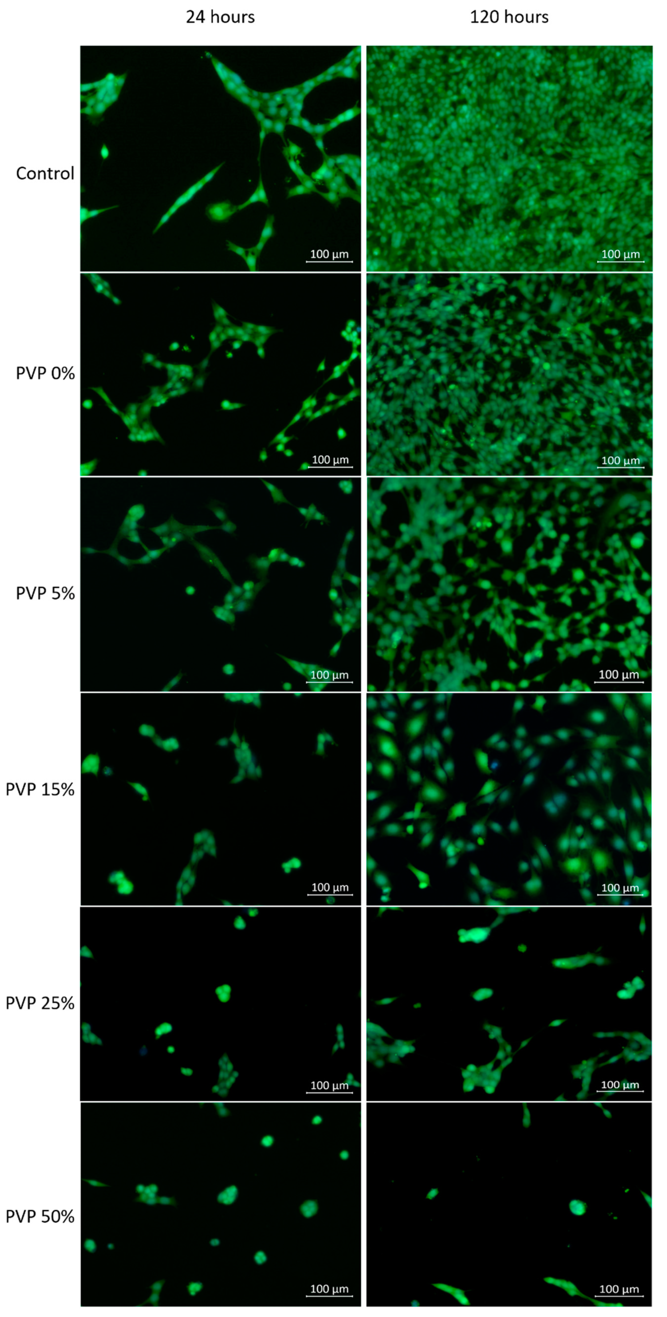
| Specimens Names | Number of Fibroblasts (pcs/mm2), Me (Q1;Q3) | |
|---|---|---|
| 24 h | 120 h | |
| Control | 240 (175;291) | 1214 (980;1312) |
| PVP content (%) | ||
| 0 | 220 (182; 232) * | 1112 (967;1290) |
| 5 | 146 (83; 187) * | 790 (693;843) |
| 15 | 92 (69; 102) *,** | 376 (198;452) |
| 25 | 35 (9.0;37.0) *,** | 158 (40;172) |
| 50 | 27 (5.5;33.0) *,** | 67 (34;76) |
Publisher’s Note: MDPI stays neutral with regard to jurisdictional claims in published maps and institutional affiliations. |
© 2020 by the authors. Licensee MDPI, Basel, Switzerland. This article is an open access article distributed under the terms and conditions of the Creative Commons Attribution (CC BY) license (http://creativecommons.org/licenses/by/4.0/).
Share and Cite
Tverdokhlebova, T.S.; Antipina, L.S.; Kudryavtseva, V.L.; Stankevich, K.S.; Kolesnik, I.M.; Senokosova, E.A.; Velikanova, E.A.; Antonova, L.V.; Vasilchenko, D.V.; Dambaev, G.T.; et al. Composite Ferroelectric Membranes Based on Vinylidene Fluoride-Tetrafluoroethylene Copolymer and Polyvinylpyrrolidone for Wound Healing. Membranes 2021, 11, 21. https://doi.org/10.3390/membranes11010021
Tverdokhlebova TS, Antipina LS, Kudryavtseva VL, Stankevich KS, Kolesnik IM, Senokosova EA, Velikanova EA, Antonova LV, Vasilchenko DV, Dambaev GT, et al. Composite Ferroelectric Membranes Based on Vinylidene Fluoride-Tetrafluoroethylene Copolymer and Polyvinylpyrrolidone for Wound Healing. Membranes. 2021; 11(1):21. https://doi.org/10.3390/membranes11010021
Chicago/Turabian StyleTverdokhlebova, Tamara S., Ludmila S. Antipina, Valeriya L. Kudryavtseva, Ksenia S. Stankevich, Ilya M. Kolesnik, Evgenia A. Senokosova, Elena A. Velikanova, Larisa V. Antonova, Dmitry V. Vasilchenko, Georgiy T. Dambaev, and et al. 2021. "Composite Ferroelectric Membranes Based on Vinylidene Fluoride-Tetrafluoroethylene Copolymer and Polyvinylpyrrolidone for Wound Healing" Membranes 11, no. 1: 21. https://doi.org/10.3390/membranes11010021
APA StyleTverdokhlebova, T. S., Antipina, L. S., Kudryavtseva, V. L., Stankevich, K. S., Kolesnik, I. M., Senokosova, E. A., Velikanova, E. A., Antonova, L. V., Vasilchenko, D. V., Dambaev, G. T., Plotnikov, E. V., Bouznik, V. M., & Bolbasov, E. N. (2021). Composite Ferroelectric Membranes Based on Vinylidene Fluoride-Tetrafluoroethylene Copolymer and Polyvinylpyrrolidone for Wound Healing. Membranes, 11(1), 21. https://doi.org/10.3390/membranes11010021