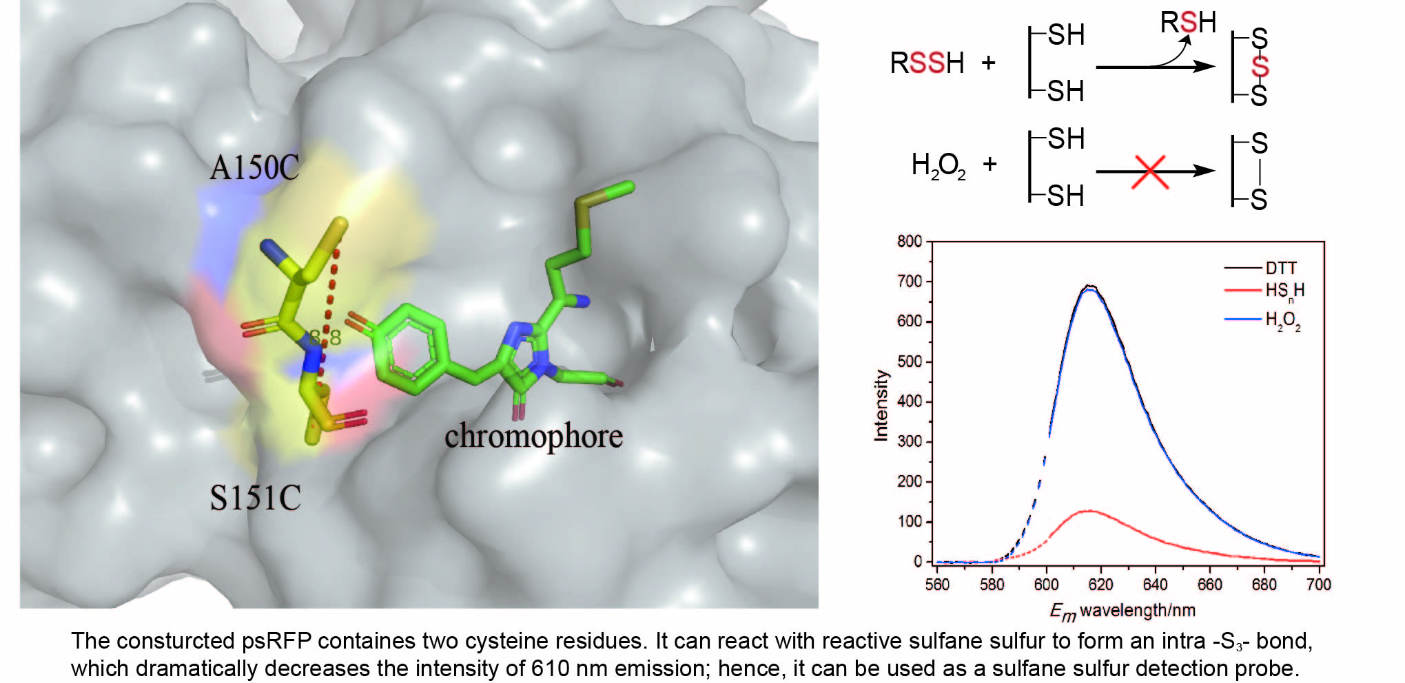
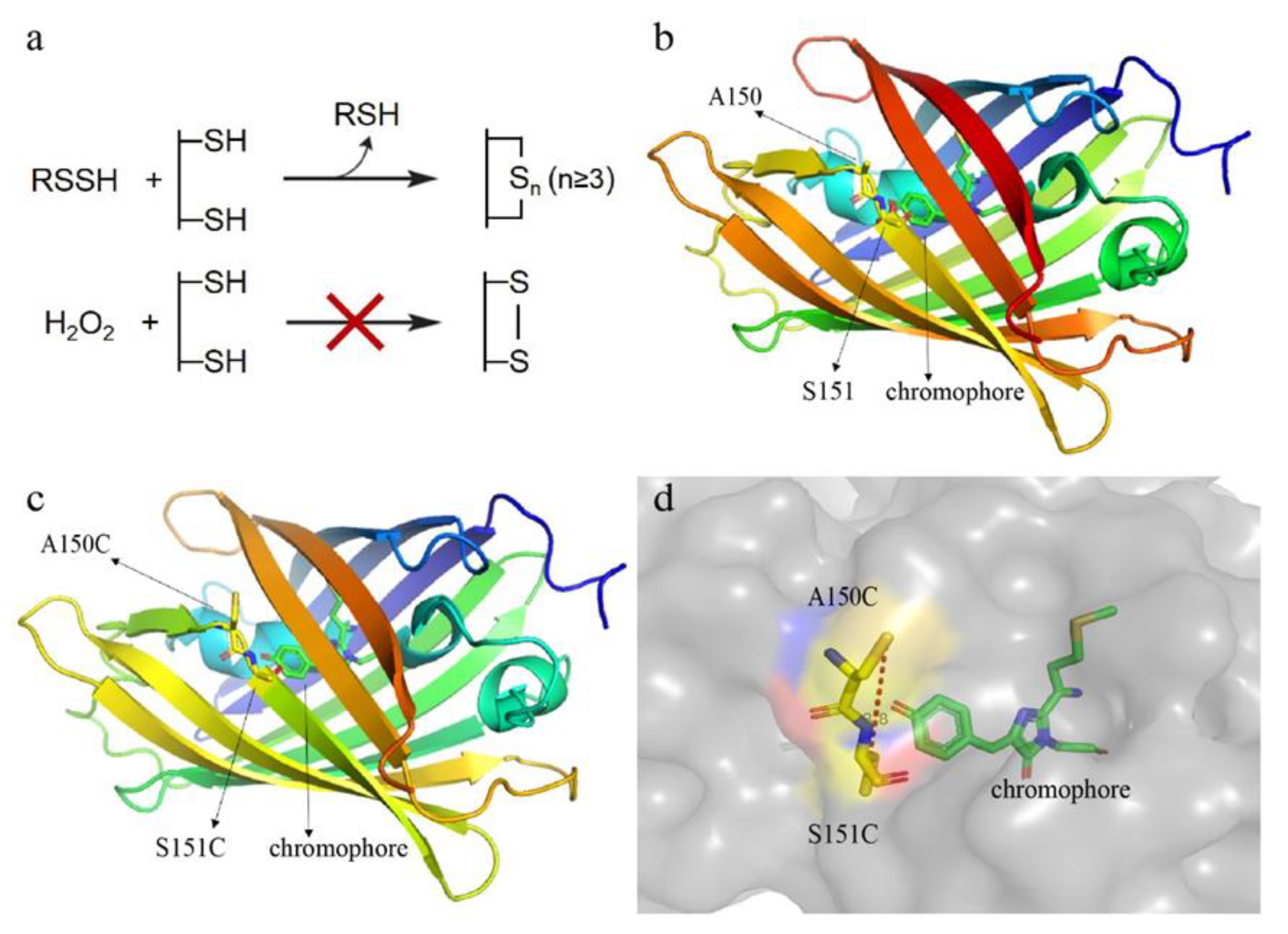
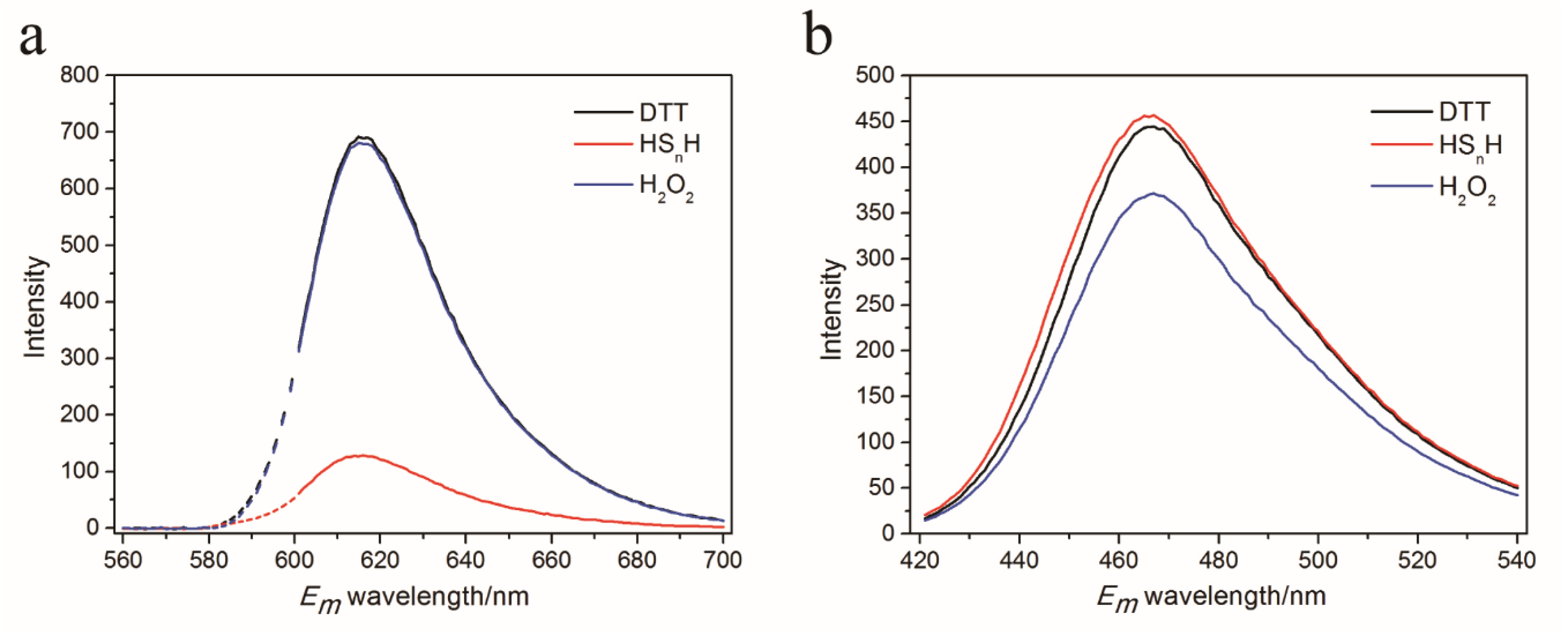
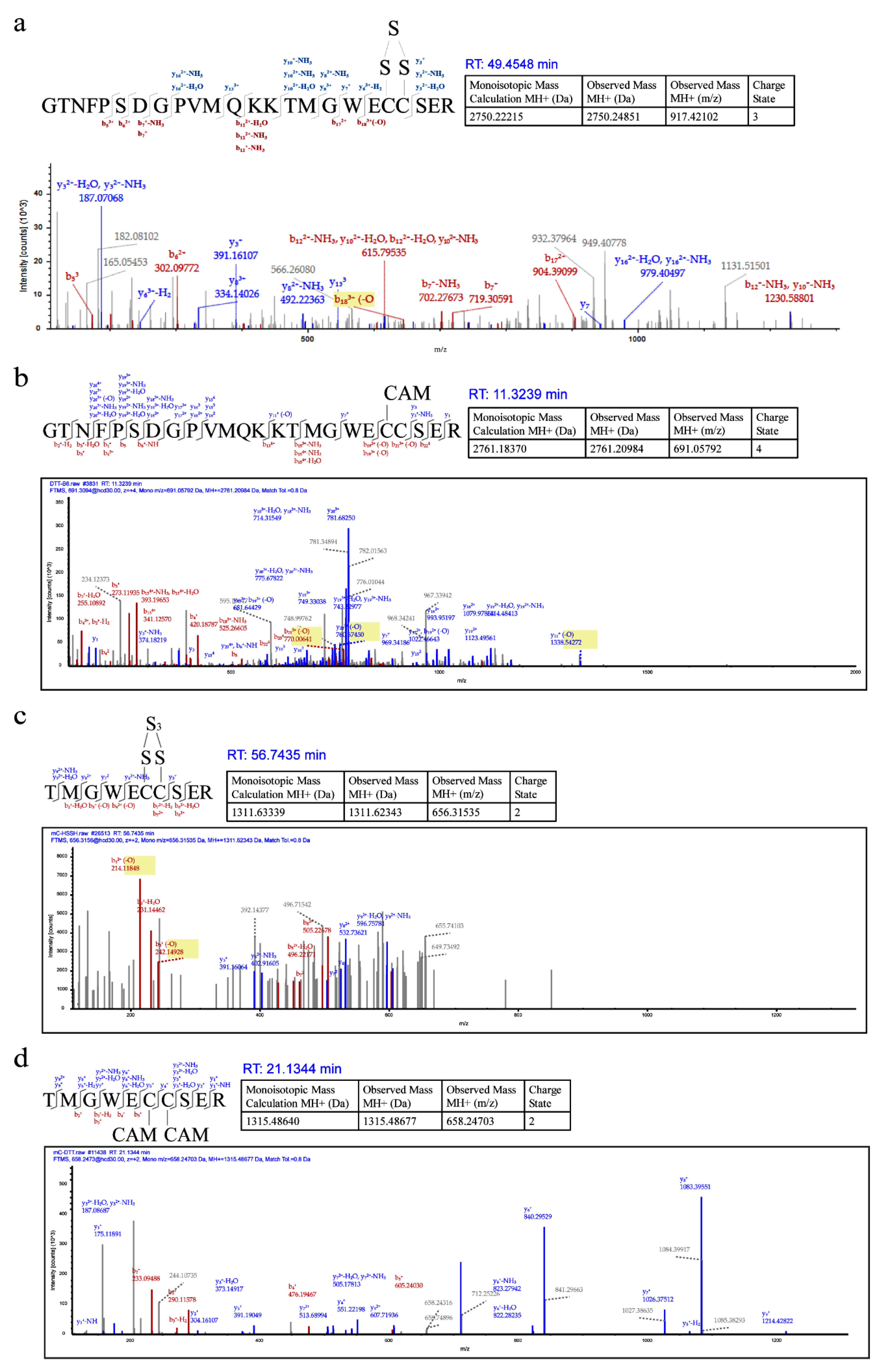
A Red Fluorescent Protein-Based Probe for Detection of Intracellular Reactive Sulfane Sulfur


Abstract
Share and Cite
Li, Z.; Wang, Q.; Xia, Y.; Xun, L.; Liu, H. A Red Fluorescent Protein-Based Probe for Detection of Intracellular Reactive Sulfane Sulfur. Antioxidants 2020, 9, 985. https://doi.org/10.3390/antiox9100985
Li Z, Wang Q, Xia Y, Xun L, Liu H. A Red Fluorescent Protein-Based Probe for Detection of Intracellular Reactive Sulfane Sulfur. Antioxidants. 2020; 9(10):985. https://doi.org/10.3390/antiox9100985
Chicago/Turabian StyleLi, Zimai, Qingda Wang, Yongzhen Xia, Luying Xun, and Huaiwei Liu. 2020. "A Red Fluorescent Protein-Based Probe for Detection of Intracellular Reactive Sulfane Sulfur" Antioxidants 9, no. 10: 985. https://doi.org/10.3390/antiox9100985
APA StyleLi, Z., Wang, Q., Xia, Y., Xun, L., & Liu, H. (2020). A Red Fluorescent Protein-Based Probe for Detection of Intracellular Reactive Sulfane Sulfur. Antioxidants, 9(10), 985. https://doi.org/10.3390/antiox9100985