Protective Effect of Multifloral Honey on Stem Cell Aging in a Dynamic Cell Culture Model


 , ,
, ,
Abstract
1. Introduction
2. Materials and Methods
2.1. Stem Cell Isolation, Culturing and Preparation of Other Cell Types
2.2. Preparation for Honey
2.3. Analysis of Phytochemical Compounds UHPLC-Orbitrap®-HRMS
2.4. Cytotoxic Effect of Honey on HFF1 Cells and SSCs
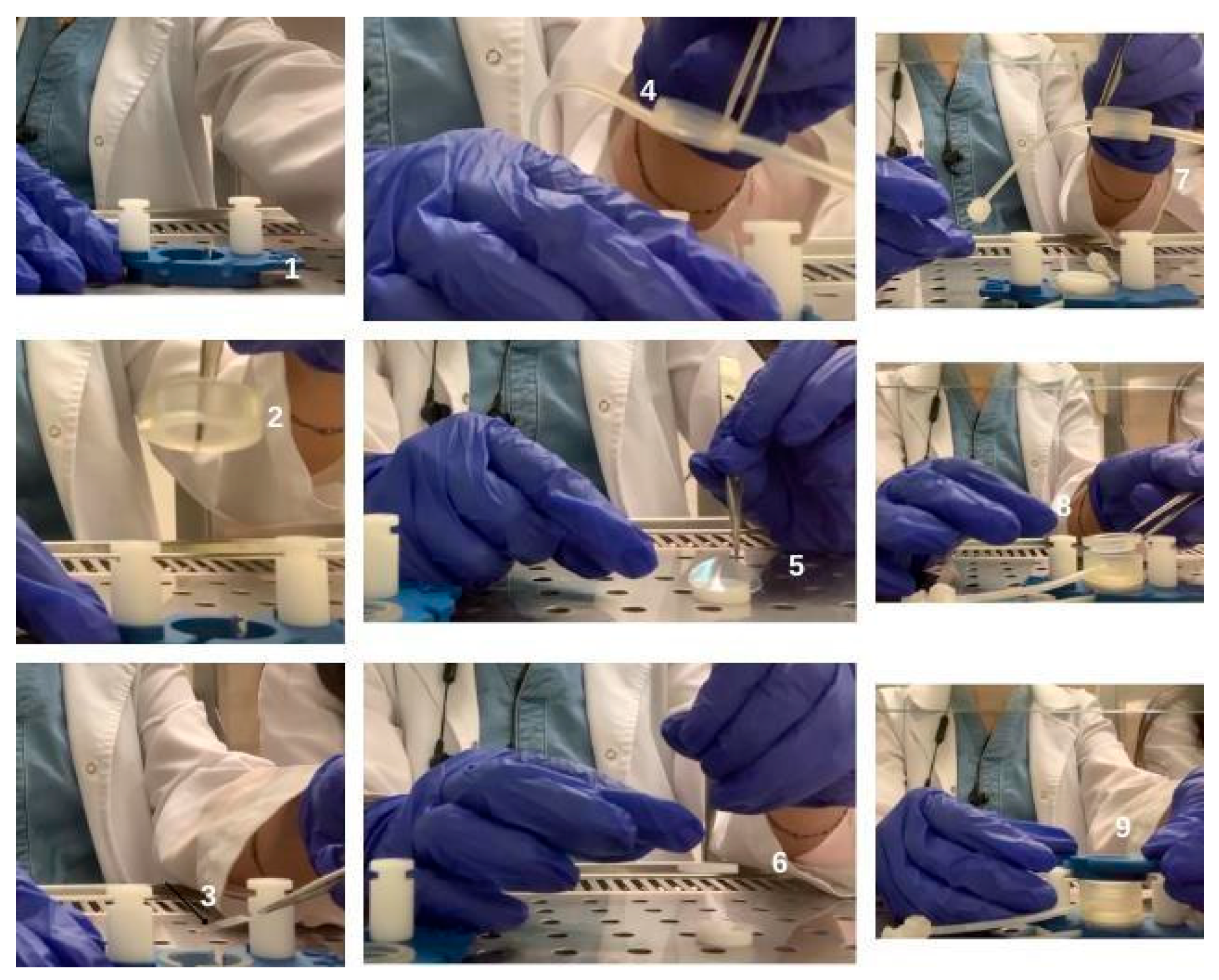
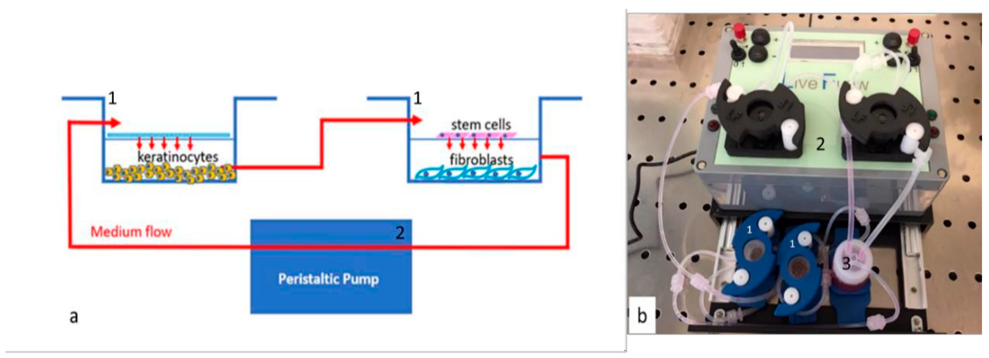
2.5. Setting up of Bioreactor and Cell Culture Conditions
2.6. Gene Expression Analysis
2.7. Nitrite Quantification
2.8. Total Antioxidant Capacity
2.9. Statistical Analysis
3. Results
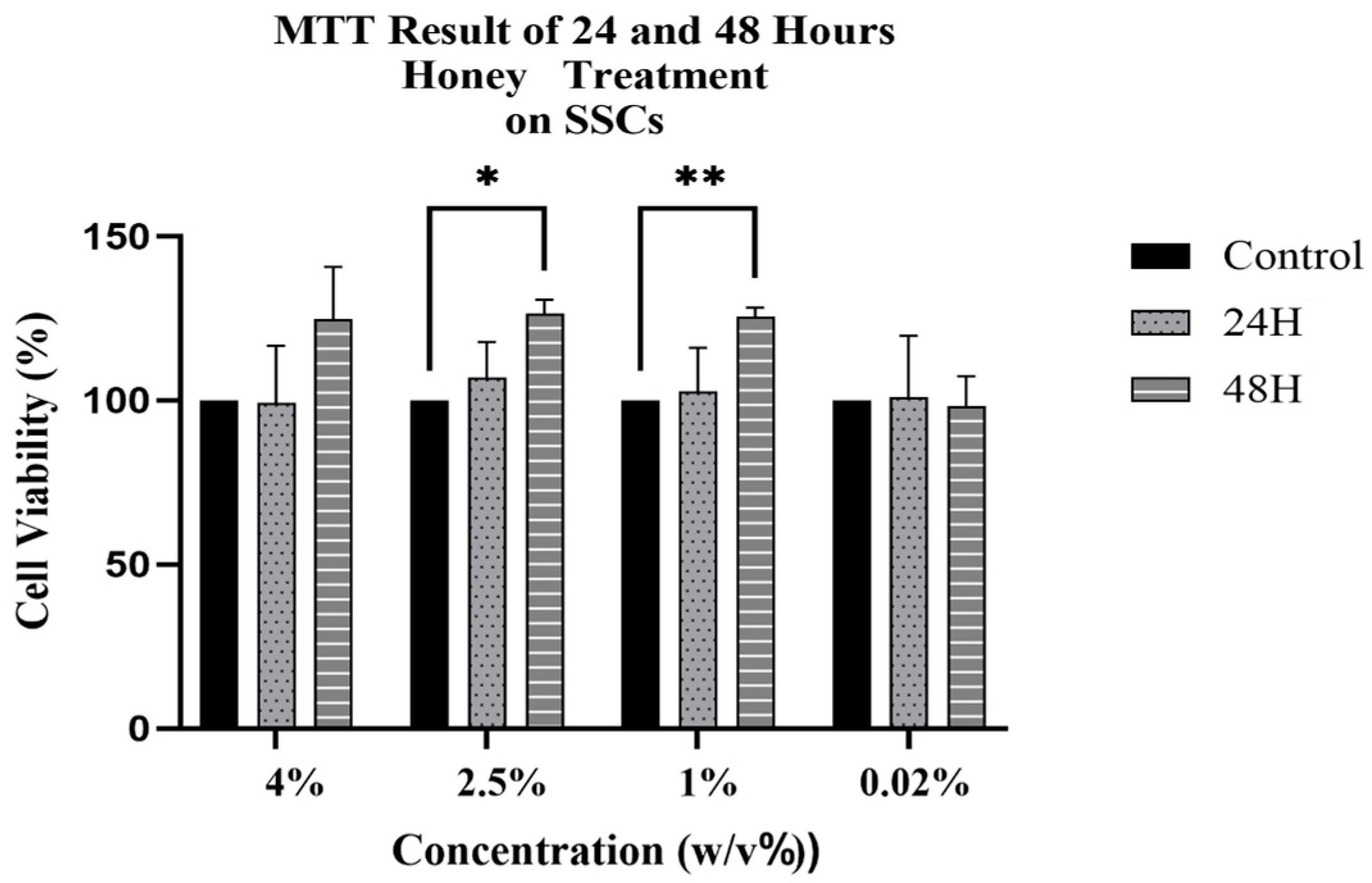
3.1. Cytotoxic Effect of Honey on SSCs
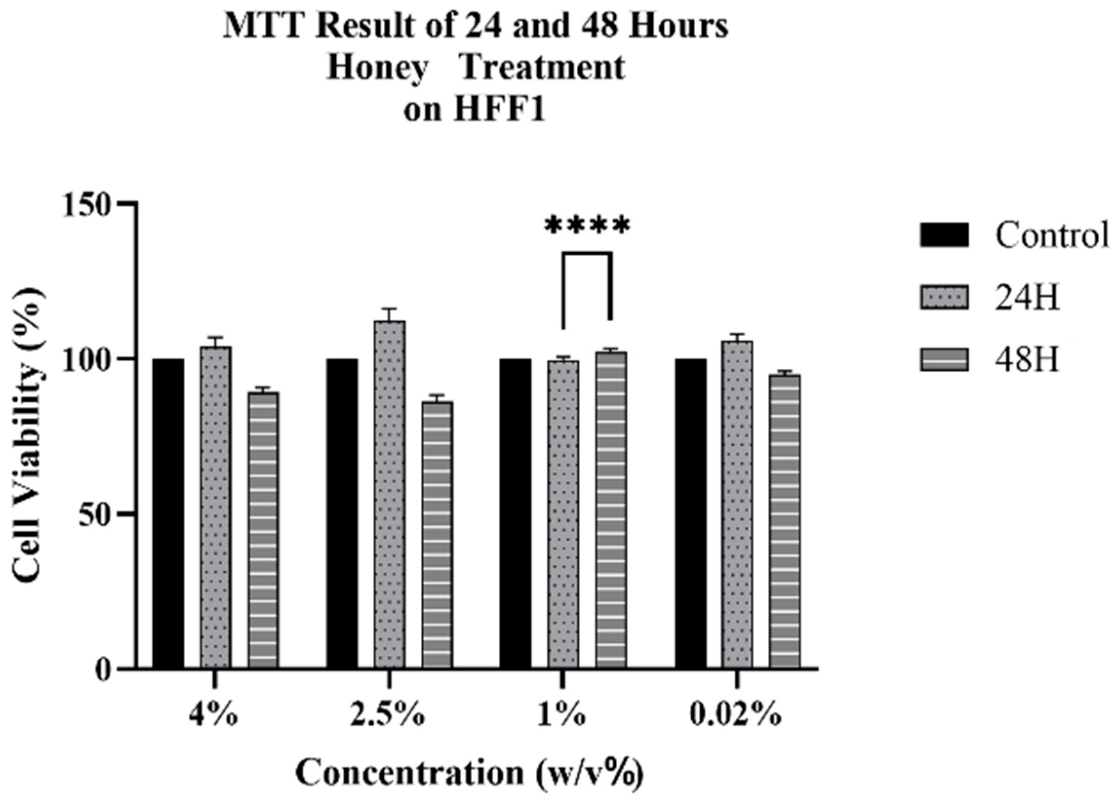
3.2. Cytotoxic Effect of Honey on HFF1
3.3. Gene Expression Analysis in SSCs and HFF1 Cells
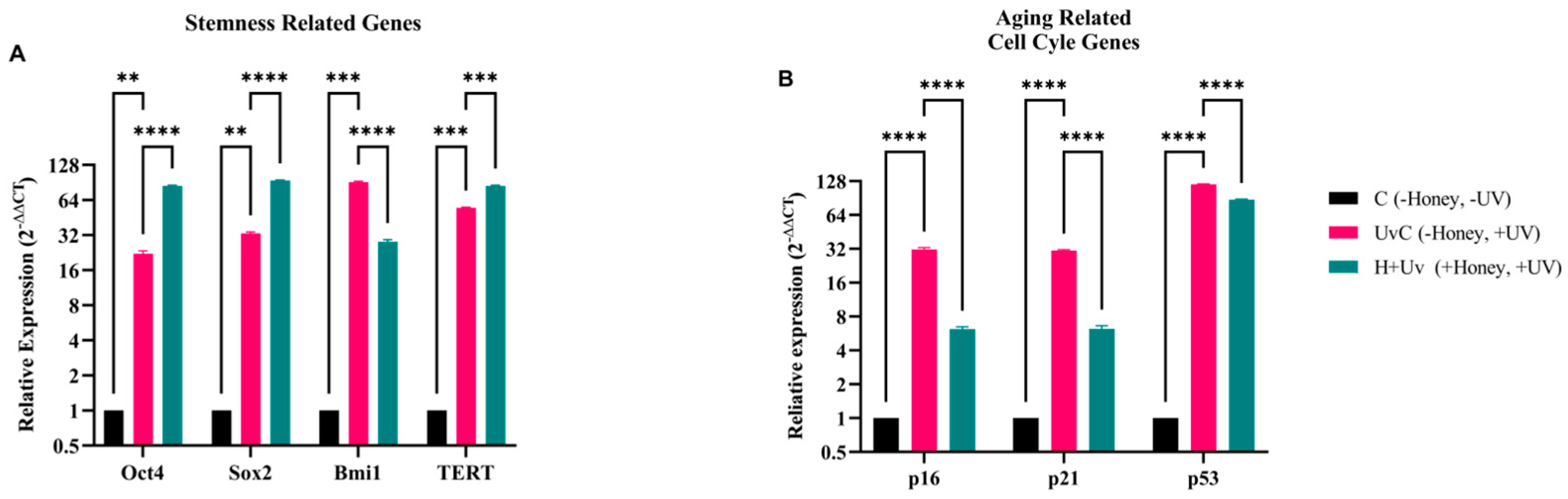
3.3.1. Gene Expression Analysis of Stem Cell and Cell Cycle Related Genes
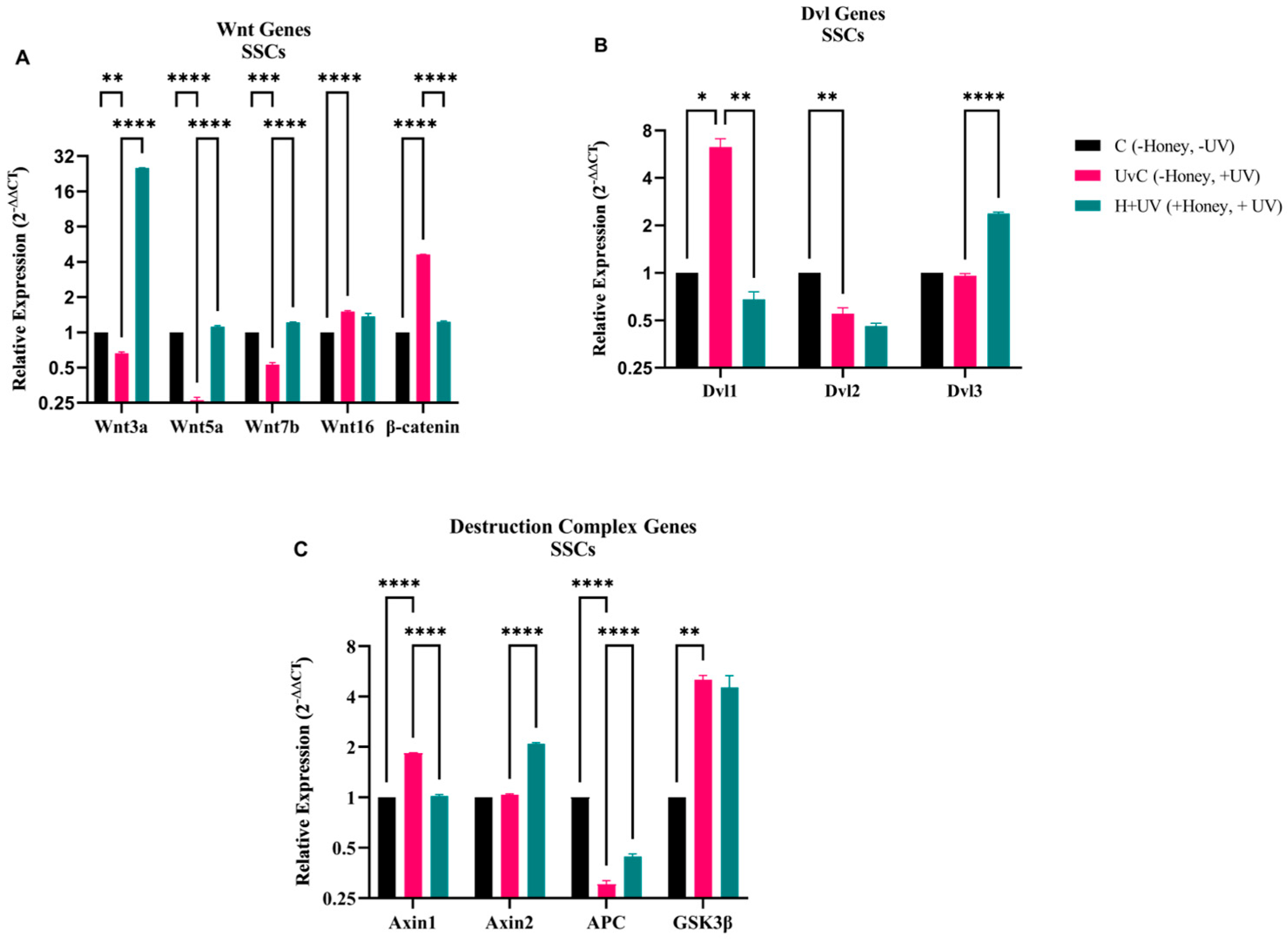
3.3.2. Gene Expression Analysis of Wnt Signaling Pathway in Stem Cells
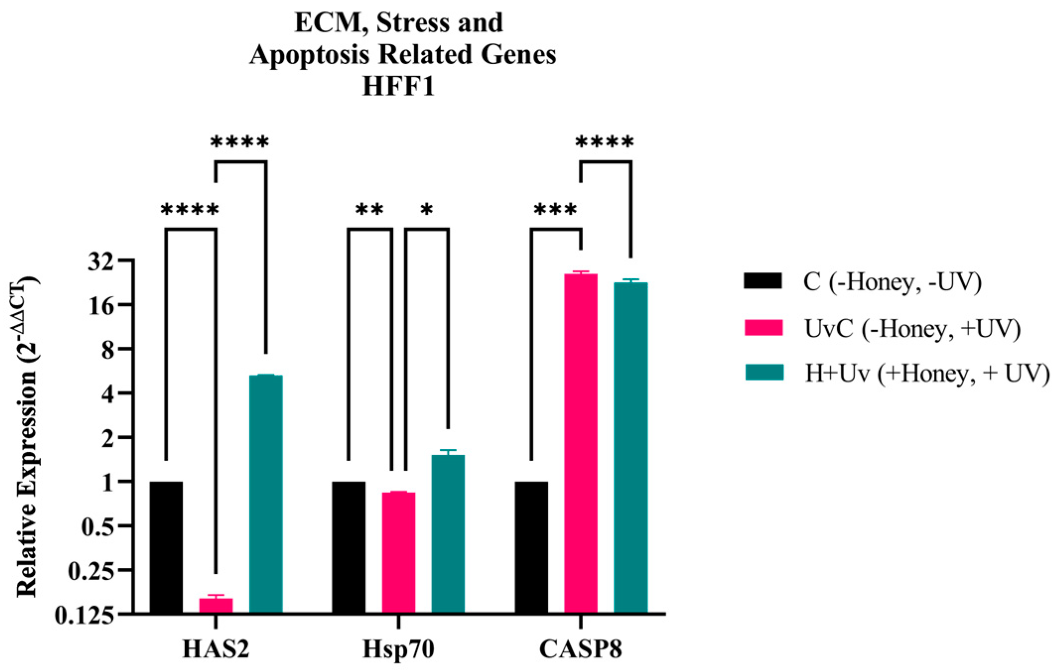
3.3.3. Gene Expression Analysis of Extracellular Matrix and Stress-Related Genes in HFF1
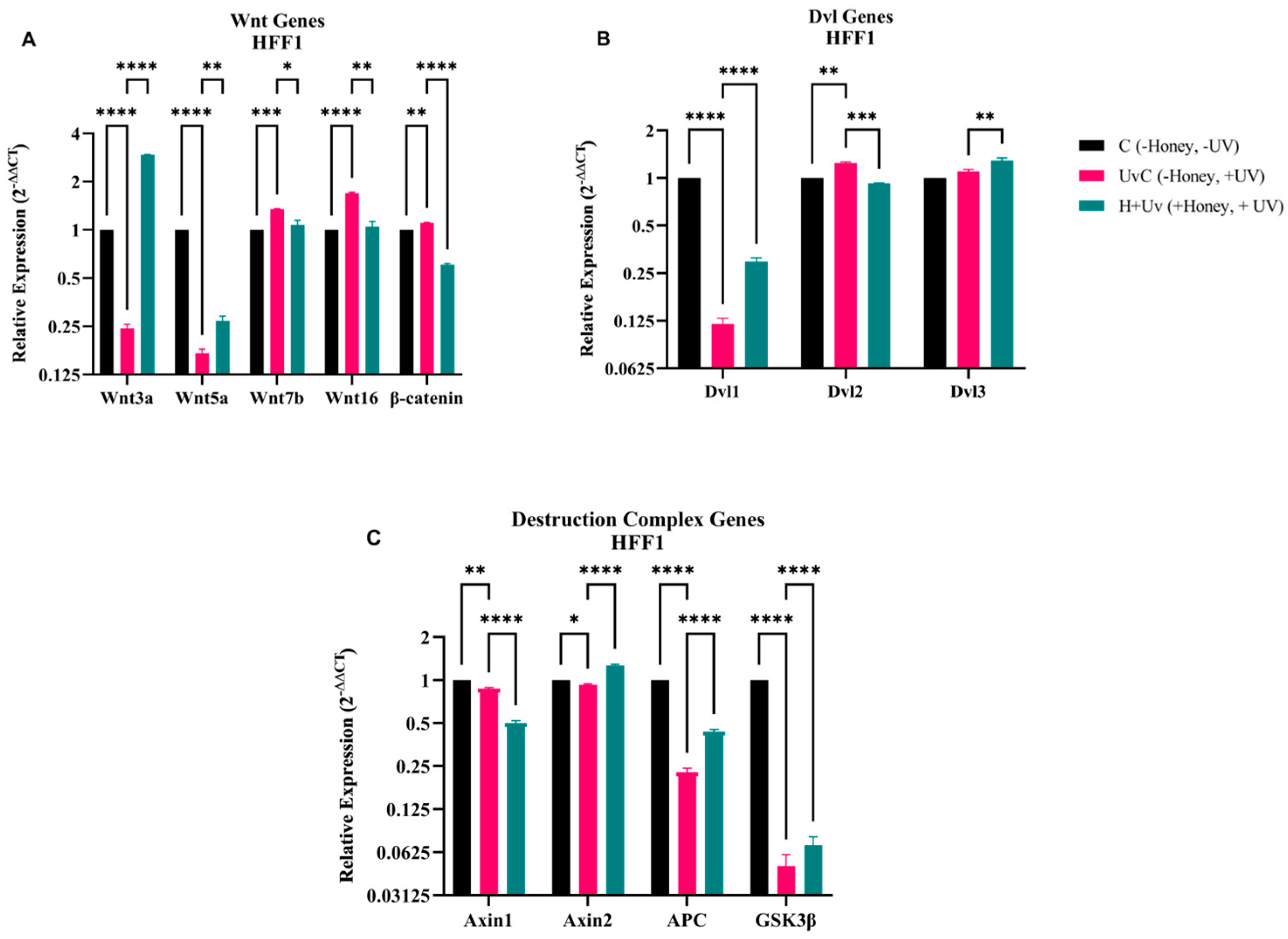
3.3.4. Gene Expression Analysis of Wnt Signaling Pathway in HFF1
3.4. Honey Phytochemical Characterization by UHPLC-Orbitrap®-HRMS
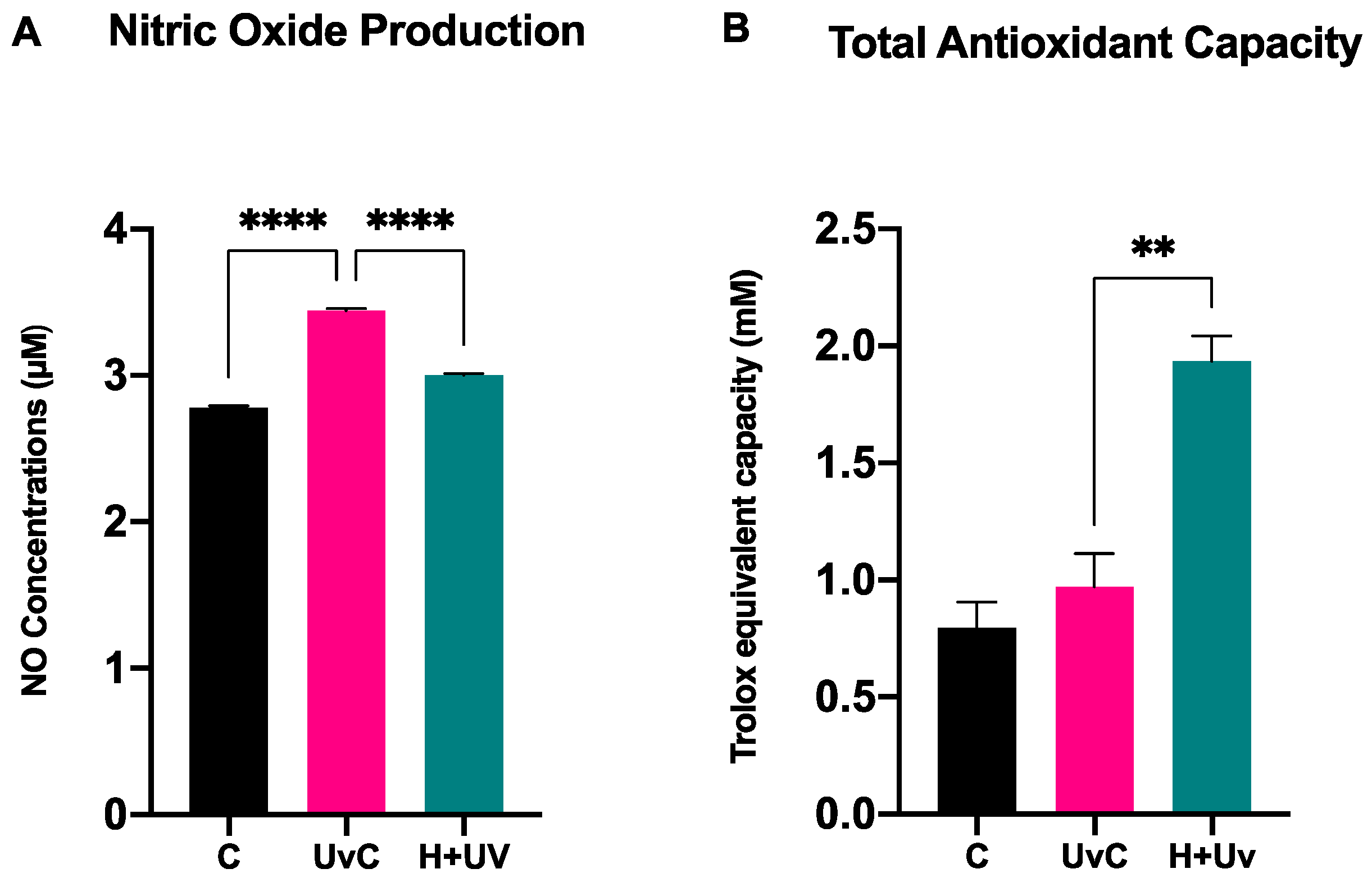
3.5. Antioxidant Activity in SSCs
4. Discussion

| Compound | Main Effects | Target Pathways/Genes |
|---|---|---|
| Acacetin | Anti-senescence, telomere protection, UVA protection | ↓ p53, ↓ p21, ↑ Sirt1/Sirt6, ↑ SIRT3, TGF-β/Smad3, mitochondrial function [10,11] |
| Pinocembrin | Antioxidant, wound healing, stemness support | ↑ Wnt/β-catenin (via Fzd1), ↓ NF-κB [7,8] |
| Chrysin | Context-dependent: protects normal stem cells, induces senescence/apoptosis in cancer | ↑ p53, ↑ p21, ↑ p16; ↓ ROS; maintains HSC self-renewal [52,53] |
| 4-Hydroxybenzoic acid (4-HBA) | Epigenetic modulator | HDAC6 inhibition → ↑ p53 activity, ↑ HIPK2/p53 pathway [54] |
| Caffeic acid | Anti-fibrotic, antioxidant; Anti-ionizing radiation | ↓ Nox4/ROS, ↓ α-SMA, ↓ MMP-1; ↑ Nrf2 [55,56] |
| Compound | Model/Cell Type | Dose/Treatment | Mechanism of Action/Main Findings |
|---|---|---|---|
| Acacetin | UVA-irradiated mice and human dermal fibroblasts (HDF), HaCaT keratinocytes, | 40–80 mg/kg topical (mice), 5–20 μg/mL (cells) | Prevented photoaging, preserving collagen and decreasing wrinkles and inflammation [10,57]; ↓ MAPK/AP-1 (JNK/c-Jun) and MMP-1/3, preserves SIRT3, ↓ ROS, ↑ TGF-β/Smad3 |
| Pinocembrin | Human keloid fibroblasts, mouse dermal fibroblasts, bleomycin-induced skin fibrosis (mice). HaCaT cell line | 20–80 µM (cells); intradermal in mice. 0.5–500 µM | Suppressed keratinocytes and fibroblast proliferation/migration, reducing fibrosis and scar formation [9,58]; ↓ TGF-β1/Smad, ↓ α-SMA, collagen I and fibronectin. ↑ MAPK and PI3K/Akt signaling pathways. |
| Chrysin | Human dermal fibroblasts, B16 murine melanoma cells, amiodarone-induced skin injury (rats) | 6.25–25 μM (cells); 10–40 mg/mL topical (rats) | Improved collagen production, reduced oxidative stress, accelerated wound closure and re-epithelialization [59,60]; ↓ degradation of collagen I synthesis, SA-β-Gal and MMP1 activity, ↓ IL-6/TNF-α, ↑ bFGF |
| 4-Hydroxybenzoic acid | UV-damaged skin cells (mainly from plant extracts) | Not specified | Reduced inflammation, supported repair and antioxidant activity, protecting against photoaging [61]; ↓ UV-induced pathways |
| Syringic acid | Diabetic wound model (rats), HaCaT keratinocytes (C. acnes-induced inflammation); L-929 fibroblasts | 2.5–5% ointment (rats); 0.4–32 µM (cells); 1–200 μM (cells) | Accelerated wound healing improving collagen and angiogenesis; reduced oxidative/inflammatory markers and ROS, preventing lipid peroxidation [62,63,64]; ↓ NF-κB/NLRP3, ↑ PPARγ/Nrf2, ↓ MMPs, raises TIMPs, boosts TGF-β1 and VEGF; |
| Caffeic acid | Mouse skin incision model, NIH-3T3 fibroblasts, RAW 264.7 macrophages, MSCs; HDFa, albino mouse | 10 mg/kg oral (mice), 30–100 µM (cells); 10–100 μM; 40 μM (cells), 15 mg kg.b.wt (mouse) | Counteract photoaging, faster wound healing, reducing oxidative damages and inflammation, enhancing collagen deposition, and improving MSC survival under hypoxia [41,65,66]; ↓ MPO/ROS, ↓ PLA2 and PGE2, boosts collagen; ↓ PI3K and AKT and ↑ PTEN |
5. Conclusions
Limitations
Author Contributions
Funding
Institutional Review Board Statement
Informed Consent Statement
Data Availability Statement
Acknowledgments
Conflicts of Interest
Abbreviations
| α-SMA | Alpha Smooth Muscle Actin |
| APC | Adenomatous Polyposis Coli |
| AKT | Protein Kinase B |
| ATCC | American Type Culture Collection |
| ATP | Adenosine Triphosphate |
| Axin1 | Axis Inhibition Protein 1 |
| Axin2 | Axis Inhibition Protein 2 |
| bFGF | Basic Fibroblast Growth Factor |
| Bmi1 | B lymphoma Mo-MLV insertion region 1 |
| β-catenin | Beta catenin |
| CASP8 | Caspase 8 |
| CDKN1A | Cyclin Dependent Kinase Inhibitor 1A |
| CDKN2A | Cyclin Dependent Kinase Inhibitor 2A |
| CNR | Consiglio Nazionale delle Ricerche |
| Ct | Cycle Threshold |
| DMSO | Dimethyl Sulfoxide |
| DMEM | Dulbecco’s Modified Eagle Medium |
| DNA | Deoxyribonucleic Acid |
| DVL | Dishevelled Protein Family (Dvl1, Dvl2, Dvl3) |
| ECM | Extracellular Matrix |
| FBS | Fetal Bovine Serum |
| GAPDH | Glyceraldehyde 3 phosphate dehydrogenase |
| GSK3β | Glycogen Synthase Kinase 3 Beta |
| HaCaT | Human Keratinocyte Cell Line |
| HAS2 | Hyaluronan Synthase 2 |
| HDF | Human Dermal Fibroblast |
| HFF1 | Human Foreskin Fibroblast 1 |
| HIPK2 | Homeodomain Interacting Protein Kinase 2 |
| HRMS | High Resolution Mass Spectrometry |
| Hsp70 | Heat Shock Protein 70 |
| HSC | Hematopoietic Stem Cell |
| IL-1β | Interleukin 1 Beta |
| MAPK | Mitogen Activated Protein Kinase |
| MMP | Matrix Metalloproteinases (MMP-1, MMP-3) |
| MSCs | Mesenchymal Stem Cells |
| MTT | MTT Cell Viability Assay |
| NLRP3 | NLR Family Pyrin Domain Containing 3 |
| NF-κB | Nuclear Factor Kappa B |
| Nrf2 | Nuclear Factor Erythroid 2 related Factor 2 |
| OD | Optical Density |
| PBS | Phosphate-Buffered Saline |
| PCR | Polymerase Chain Reaction |
| PI3K | Phosphoinositide 3 Kinase |
| PPARγ | Peroxisome Proliferator Activated Receptor Gamma |
| p16 | Cyclin Dependent Kinase Inhibitor 2A |
| p21 | Cyclin Dependent Kinase Inhibitor 1A |
| p53 | Tumor Protein 53 |
| PTEN | Phosphatase and Tensin Homolog |
| RNA | Ribonucleic Acid |
| ROS | Reactive Oxygen Species |
| RT-qPCR | Real Time Quantitative Polymerase Chain Reaction |
| SIRT | Sirtuin Protein Family (SIRT1, SIRT3, SIRT6) |
| SSCs | Skin Stem Cells |
| TGF-β1 | Transforming Growth Factor Beta 1 |
| TERT | Telomerase Reverse Transcriptase |
| UV | Ultraviolet |
| UVB | Ultraviolet B |
| UVC | Ultraviolet C |
| VEGF | Vascular Endothelial Growth Factor |
| WNT | Wingless Type Signaling Proteins (Wnt3a, Wnt5a, Wnt7b, Wnt16) |
References
- Díaz-García, D.; Filipová, A.; Garza-Veloz, I.; Martinez-Fierro, M.L. A Beginner’s Introduction to Skin Stem Cells and Wound Healing. Int. J. Mol. Sci. 2021, 22, 11030. [Google Scholar] [CrossRef]
- Domaszewska-Szostek, A.; Puzianowska-Kuźnicka, M.; Kuryłowicz, A. Flavonoids in Skin Senescence Prevention and Treatment. Int. J. Mol. Sci. 2021, 22, 6814. [Google Scholar] [CrossRef]
- Chen, X.; Yang, C.; Jiang, G. Research progress on skin photoaging and oxidative stress. Adv. Dermatol. Allergol. 2021, 38, 931–936. [Google Scholar] [CrossRef]
- Panich, U.; Sittithumcharee, G.; Rathviboon, N.; Jirawatnotai, S. Ultraviolet Radiation-Induced Skin Aging: The Role of DNA Damage and Oxidative Stress in Epidermal Stem Cell Damage Mediated Skin Aging. Stem Cells Int. 2016, 2016, 7370642. [Google Scholar] [CrossRef]
- Jaganathan, S.K. Can flavonoids from honey alter multidrug resistance? Med. Hypotheses 2011, 76, 535–537. [Google Scholar] [CrossRef] [PubMed]
- Talebi, M.M.; Talebi, M.M.; Farkhondeh, T.; Samarghandian, S. Molecular mechanism-based therapeutic properties of honey. Biomed. Pharmacother. 2020, 130, 110590. [Google Scholar] [CrossRef] [PubMed]
- Li, J.; Peng, X.L.; Cheng, X.Y.; Yang, J.J.; Cui, C.; Liu, J.H. Pinocembrin Alleviates Postoperative Cognitive Dysfunction in Aged Mice by Modulating miR-384-5p/FZD1 Axis to Activate the Wnt/β-Catenin Pathway. Mol. Neurobiol. 2025, 62, 13975–13988. [Google Scholar] [CrossRef]
- Borriello, M.; Iannuzzi, C.; Sirangelo, I. Pinocembrin Protects from AGE-Induced Cytotoxicity and Inhibits Non-Enzymatic Glycation in Human Insulin. Cells 2019, 8, 385. [Google Scholar] [CrossRef]
- Ruttanapattanakul, J.; Wikan, N.; Potikanond, S.; Nimlamool, W. Molecular Targets of Pinocembrin Underlying Its Regenerative Activities in Human Keratinocytes. Pharmaceuticals 2022, 15, 954. [Google Scholar] [CrossRef] [PubMed]
- Mu, J.; Chen, H.; Ye, M.; Zhang, X.; Ma, H. Acacetin resists UVA photoaging by mediating the SIRT3/ROS/MAPKs pathway. J. Cell. Mol. Med. 2022, 26, 4624–4628. [Google Scholar] [CrossRef]
- Hong, Y.X.; Wu, W.Y.; Song, F.; Wu, C.; Li, G.R.; Wang, Y. Cardiac senescence is alleviated by the natural flavone acacetin via enhancing mitophagy. Aging 2021, 13, 16381–16403. [Google Scholar] [CrossRef]
- Oryan, A.; Alemzadeh, E.; Mohammadi, A.A. Application of honey as a protective material in maintaining the viability of adipose stem cells in burn wound healing: A histological, molecular and biochemical study. Tissue Cell 2019, 61, 89–97. [Google Scholar] [CrossRef]
- Das, A.; Datta, P.; Chowdhury, A.R.; Barui, A. Honey-incorporated nanofibre reduces replicative senescence of umbilical cord-derived mesenchymal stem cells. IET Nanobiotechnol. 2020, 14, 870–880. [Google Scholar] [CrossRef]
- de Jaime-Soguero, A.; Abreu de Oliveira, W.; Lluis, F. The Pleiotropic Effects of the Canonical Wnt Pathway in Early Development and Pluripotency. Genes 2018, 9, 93. [Google Scholar] [CrossRef] [PubMed]
- Sato, N.; Meijer, L.; Skaltsounis, L.; Greengard, P.; Brivanlou, A.H. Maintenance of pluripotency in human and mouse embryonic stem cells through activation of Wnt signaling by a pharmacological GSK-3-specific inhibitor. Nat. Med. 2004, 10, 55–63. [Google Scholar] [CrossRef] [PubMed]
- Govarthanan, K.; Vidyasekar, P.; Gupta, P.K.; Lenka, N.; Verma, R.S. Glycogen synthase kinase 3β inhibitor- CHIR 99021 augments the differentiation potential of mesenchymal stem cells. Cytotherapy 2020, 22, 91–105. [Google Scholar] [CrossRef] [PubMed]
- Zhang, D.Y.; Pan, Y.; Zhang, C.; Yan, B.X.; Yu, S.S.; Wu, D.L.; Shi, M.; Shi, K.; Cai, X.; Zhou, S.; et al. Wnt/β-catenin signaling induces the aging of mesenchymal stem cells through promoting the ROS production. Mol. Cell. Biochem. 2013, 374, 13–20. [Google Scholar] [CrossRef]
- Jeoung, J.Y.; Nam, H.Y.; Kwak, J.; Jin, H.J.; Lee, H.J.; Lee, B.W.; Baek, J.H.; Eom, J.S.; Chang, E.J.; Shin, D.M.; et al. A Decline in Wnt3a Signaling is Necessary for Mesenchymal Stem Cells to Proceed to Replicative Senescence. Stem Cells Dev. 2015, 24, 973–982. [Google Scholar] [CrossRef]
- Lehmann, J.; Narcisi, R.; Franceschini, N.; Chatzivasileiou, D.; Boer, C.G.; Koevoet, W.J.L.M.; Putavet, D.; Drabek, D.; van Haperen, R.; de Keizer, P.L.J.; et al. WNT/beta-catenin signalling interrupts a senescence-induction cascade in human mesenchymal stem cells that restricts their expansion. Cell. Mol. Life Sci. 2022, 79, 82. [Google Scholar] [CrossRef]
- Çalişkan, C.; Sercan, H.O. Kronik Miyeloid Lösemide Dishevelled Proteinlerinin Onkojenik Etkisinin Araştirilmasi. Ph.D. Thesis, Dokuz Eylül Üniversitesi Sağlik Bilimleri Enstitüsü, Izmir, Turkey, 2016. Available online: https://www.proquest.com/openview/0358657e0703d7e57e57ee308ce14891/1?pq-origsite=gscholar&cbl=2026366&diss=y (accessed on 11 April 2025).
- Park, I.-K.; Morrison, S.J.; Clarke, M.F. Bmi1, stem cells, and senescence regulation. J. Clin. Investig. 2004, 113, 175–179. [Google Scholar] [CrossRef]
- Lin, X.; Ojo, D.; Wei, F.; Wong, N.; Gu, Y.; Tang, D. A Novel Aspect of Tumorigenesis—BMI1 Functions in Regulating DNA Damage Response. Biomolecules 2015, 5, 3396–3415. [Google Scholar] [CrossRef]
- Matsuda, M.; Hoshino, T.; Yamakawa, N.; Tahara, K.; Adachi, H.; Sobue, G.; Maji, D.; Ihn, H.; Mizushima, T. Suppression of UV-Induced Wrinkle Formation by Induction of HSP70 Expression in Mice. J. Investig. Dermatol. 2013, 133, 919–928. [Google Scholar] [CrossRef] [PubMed]
- Bellu, E.; Garroni, G.; Balzano, F.; Satta, R.; Montesu, M.A.; Kralovic, M.; Fedacko, J.; Cruciani, S.; Maioli, M. Isolating stem cells from skin: Designing a novel highly efficient non-enzymatic approach. Physiol. Res. 2019, 68, S385–S388. [Google Scholar] [CrossRef]
- Bellu, E.; Garroni, G.; Cruciani, S.; Balzano, F.; Serra, D.; Satta, R.; Montesu, M.A.; Fadda, A.; Mulas, M.; Sarais, G.; et al. Smart Nanofibers with Natural Extracts Prevent Senescence Patterning in a Dynamic Cell Culture Model of Human Skin. Cells 2020, 9, 2530. [Google Scholar] [CrossRef] [PubMed]
- Rasulova, K.; Kavak, D.E.; Kavak, F.F.; Irtegun Kandemir, S.; Kizildag, S. The effect of multifloral honey on cytotoxicity and the mitochondrial apoptotic pathway in ovarian cancer cell. Med. J. Nutr. Metab. 2025, 18, 69–80. [Google Scholar] [CrossRef]
- Dursun, İ.; Sağlamtaş, R.; Fettahoğlu, K.; Zor, M.; Sinan, A.; Demirci, A.; Demir, Y.; Gülçin, İ. Antioxidant and antimicrobial activities of different extracts of Tragopogon dubius and Tragopogon porrifolium L. subsp. longirostris: Determination of their phytochemical contents by UHPLC-Orbitrap®-HRMS analysis. Food Biosci. 2025, 63, 105604. [Google Scholar] [CrossRef]
- Belay, A.; Haki, G.D.; Birringer, M.; Borck, H.; Lee, Y.-C.; Kim, K.-T.; Baye, K.; Melaku, S. Enzyme activity, amino acid profiles and hydroxymethylfurfural content in Ethiopian monofloral honey. J. Food Sci. Technol. 2017, 54, 2769–2778. [Google Scholar] [CrossRef]
- Bellu, E.; Cruciani, S.; Garroni, G.; Balzano, F.; Satta, R.; Montesu, M.A.; Fadda, A.; Mulas, M.; Sarais, G.; Bandiera, P.; et al. Natural Compounds and PCL Nanofibers: A Novel Tool to Counteract Stem Cell Senescence. Cells 2021, 10, 1415. [Google Scholar] [CrossRef]
- Origene. Available online: http://www.origene.com (accessed on 9 December 2025).
- Cruciani, S.; Garroni, G.; Pala, R.; Barcessat, A.R.P.; Facchin, F.; Ventura, C.; Fozza, C.; Maioli, M. Melatonin finely tunes proliferation and senescence in hematopoietic stem cells. Eur. J. Cell Biol. 2022, 101, 151251. [Google Scholar] [CrossRef]
- Li, X.; Ouyang, X.; Cai, R.; Chen, D. 3,8”-Dimerization Enhances the Antioxidant Capacity of Flavonoids: Evidence from Acacetin and Isoginkgetin. Molecules 2019, 24, 2039. [Google Scholar]
- Habtemariam, S. The Nrf2/HO-1 Axis as Targets for Flavanones: Neuroprotection by Pinocembrin, Naringenin, and Eriodictyol. Oxid. Med. Cell. Longev. 2019, 2019, 4724920. [Google Scholar] [CrossRef]
- Elbatreek, M.H.; Mahdi, I.; Ouchari, W.; Mahmoud, M.F.; Sobeh, M. Current advances on the therapeutic potential of pinocembrin: An updated review. Biomed. Pharmacother. 2023, 157, 114032. [Google Scholar] [CrossRef] [PubMed]
- Chen, J.H.; Ho, C.-T. Antioxidant Activities of Caffeic Acid and Its Related Hydroxycinnamic Acid Compounds. J. Agric. Food Chem. 1997, 45, 2374–2378. [Google Scholar] [CrossRef]
- Gülçin, İ. Antioxidant activity of caffeic acid (3,4-dihydroxycinnamic acid). Toxicology 2006, 217, 213–220. [Google Scholar] [CrossRef] [PubMed]
- López-Herrador, S.; Corral-Sarasa, J.; González-García, P.; Morillas-Morota, Y.; Olivieri, E.; Jiménez-Sánchez, L.; Díaz-Casado, M.E. Natural Hydroxybenzoic and Hydroxycinnamic Acids Derivatives: Mechanisms of Action and Therapeutic Applications. Antioxidants 2025, 14, 711. [Google Scholar] [CrossRef]
- Srinivasulu, C.; Ramgopal, M.; Ramanjaneyulu, G.; Anuradha, C.M.; Suresh Kumar, C. Syringic acid (SA)—A Review of Its Occurrence, Biosynthesis, Pharmacological and Industrial Importance. Biomed. Pharmacother. 2018, 108, 547–557. [Google Scholar] [CrossRef]
- Karapetsas, A.; Voulgaridou, G.-P.; Iliadi, D.; Tsochantaridis, I.; Michail, P.; Kynigopoulos, S.; Lambropoulou, M.; Stavropoulou, M.-I.; Stathopoulou, K.; Karabournioti, S.; et al. Honey Extracts Exhibit Cytoprotective Properties against UVB-Induced Photodamage in Human Experimental Skin Models. Antioxidants 2020, 9, 566. [Google Scholar] [CrossRef]
- Nichols, J.A.; Katiyar, S.K. Skin photoprotection by natural polyphenols: Anti-inflammatory, antioxidant and DNA repair mechanisms. Arch. Dermatol. Res. 2010, 302, 71–83. [Google Scholar] [CrossRef]
- Balupillai, A.; Nagarajan, R.P.; Ramasamy, K.; Govindasamy, K.; Muthusamy, G. Caffeic acid prevents UVB radiation induced photocarcinogenesis through regulation of PTEN signaling in human dermal fibroblasts and mouse skin. Toxicol. Appl. Pharmacol. 2018, 352, 87–96. [Google Scholar] [CrossRef]
- Janbandhu, V.C.; Singh, A.K.; Mukherji, A.; Kumar, V. p65 Negatively Regulates Transcription of the Cyclin E Gene. J. Biol. Chem. 2010, 285, 17453–17464. [Google Scholar] [CrossRef]
- Ahmad, I.; Jimenez, H.; Yaacob, N.S.; Yusuf, N. Tualang Honey Protects Keratinocytes from Ultraviolet Radiation-Induced Inflammation and DNA Damage. Photochem. Photobiol. 2012, 88, 1198–1204. [Google Scholar] [CrossRef]
- Ray, L.B. Wnt, the Fountain of Youth? Sci. STKE 2007, 2007, tw295. [Google Scholar] [CrossRef]
- Saraswati, S.; Bastakoty, D.; Young, P.P. Molecular and Signaling Pathways That Modulate Mesenchymal Stem Cell Self-renewal. In Stem Cells and Cancer Stem Cells; Springer: Dordrecht, The Netherlands, 2012; Volume 6, pp. 131–141. [Google Scholar] [CrossRef]
- Davidson, K.C.; Adams, A.M.; Goodson, J.M.; McDonald, C.E.; Potter, J.C.; Berndt, J.D.; Biechele, T.L.; Taylor, R.J.; Moon, R.T. Wnt/β-catenin signaling promotes differentiation, not self-renewal, of human embryonic stem cells and is repressed by Oct4. Proc. Natl. Acad. Sci. USA 2012, 109, 4485–4490. [Google Scholar] [CrossRef] [PubMed]
- Rognoni, E.; Goss, G.; Hiratsuka, T.; Sipilä, K.H.; Kirk, T.; Kober, K.I.; Lui, P.P.; Tsang, V.S.; Hawkshaw, N.J.; Pilkington, S.M.; et al. Role of distinct fibroblast lineages and immune cells in dermal repair following UV radiation-induced tissue damage. eLife 2021, 10, e71052. [Google Scholar] [CrossRef]
- Van Camp, J.K.; Beckers, S.; Zegers, D.; Van Hul, W. Wnt Signaling and the Control of Human Stem Cell Fate. Stem Cell Rev. Rep. 2014, 10, 207–229. [Google Scholar] [CrossRef] [PubMed]
- Wang, L.; Fu, Z.; Su, Y.; Yin, W.; Wang, X.; Zhao, W.; Wang, J.; Li, Y.; Liu, N.; Su, W.; et al. Cyclic Heptapeptide FZ1 Acts as an Integrin αvβ3 Agonist to Facilitate Diabetic Skin Wound Healing by Enhancing Angiogenesis. J. Med. Chem. 2025, 68, 19503–19520. [Google Scholar] [CrossRef] [PubMed]
- Li, Y.; Jia, Q.; Liu, N.; Yin, S.; Wang, J.; Ding, Y.; Yang, Y.; Peng, Y.; Ru, Z.; Zhang, S.; et al. Peptide RL-QN15 Regulates Functions of Epidermal Stem Cells to Accelerate Skin Wound Regeneration via the FZD8/β-Catenin Axis. Exploration 2025, 5, 20240090. [Google Scholar] [CrossRef]
- Yamada, T.; Hasegawa, S.; Iwata, Y.; Arima, M.; Kobayashi, T.; Numata, S.; Nakata, S.; Sugiura, K.; Akamatsu, H. UV irradiation-induced DNA hypomethylation around WNT1 gene: Implications for solar lentigines. Exp. Dermatol. 2019, 28, 723–729. [Google Scholar] [CrossRef]
- Pawar, J.S.; Mustafa, S.; Ghosh, I. Chrysin and Capsaicin induces premature senescence and apoptosis via mitochondrial dysfunction and p53 elevation in Cervical cancer cells. Saudi J. Biol. Sci. 2022, 29, 3838–3847. [Google Scholar] [CrossRef]
- Li, Y.; He, M.; Zhang, W.; Yang, M.; Ding, Y.; Xu, S.; Gu, J.; Li, Y.; Yin, J.; Gao, Y. Antioxidant Small Molecule Compound Chrysin Promotes the Self-Renewal of Hematopoietic Stem Cells. Front. Pharmacol. 2020, 11, 399. [Google Scholar] [CrossRef]
- Wang, X.N.; Wang, K.Y.; Zhang, X.S.; Yang, C.; Li, X.Y. 4-Hydroxybenzoic acid (4-HBA) enhances the sensitivity of human breast cancer cells to adriamycin as a specific HDAC6 inhibitor by promoting HIPK2/p53 pathway. Biochem. Biophys. Res. Commun. 2018, 504, 812–819. [Google Scholar] [CrossRef]
- Chung, S.W.; Park, I.H.; Hong, S.M.; Cho, J.S.; Moon, J.H.; Kim, T.H.; Lee, H.M. Role of Caffeic Acid on Collagen Production in Nasal Polyp-Derived Fibroblasts. Clin. Exp. Otorhinolaryngol. 2014, 7, 295. [Google Scholar] [CrossRef]
- Yao, J.; Liu, Y.; Lin, H.; Shao, C.; Jin, X.; Peng, T.; Liu, Y. Caffeic acid activates Nrf2 enzymes, providing protection against oxidative damage induced by ionizing radiation. Brain Res. Bull. 2025, 224, 111325. [Google Scholar] [CrossRef]
- Jeong, E.H.; Yang, H.; Kim, J.E.; Lee, K.W. Safflower Seed Oil and Its Active Compound Acacetin Inhibit UVB-Induced Skin Photoaging. J. Microbiol. Biotechnol. 2020, 30, 1567–1573. [Google Scholar] [CrossRef]
- Li, X.; Zhai, Y.; Xi, B.; Ma, W.; Zhang, J.; Ma, X.; Miao, Y.; Zhao, Y.; Ning, W.; Zhou, H.; et al. Pinocembrin Ameliorates Skin Fibrosis via Inhibiting TGF-β1 Signaling Pathway. Biomolecules 2021, 11, 1240. [Google Scholar] [CrossRef]
- Zhu, L.; Lu, Y.; Yu, W.G.; Zhao, X.; Lu, Y.H. Anti-photoageing and anti-melanogenesis activities of chrysin. Pharm. Biol. 2016, 54, 2692–2700. [Google Scholar] [CrossRef]
- Liu, L.; Mai, Y.; Liang, Y.; Zhou, X.; Chen, K. Experimental study on the effect of chrysin on skin injury induced by amiodarone extravasation in rats. Microvasc. Res. 2022, 139, 104257. [Google Scholar] [CrossRef] [PubMed]
- Kim, S.K.; Kim, S.B.; Lee, S.B.; Kim, K.S.; Son, S.R.; Choi, E.J.; Park, B.C.; Hong, E.; Kim, Y.A.; Moon, B.S.; et al. Investigating the antioxidant and anti-inflammatory potential of Nypa fruticans: A multifaceted approach to skin protection and aging. Appl. Biol. Chem. 2025, 68, 6. [Google Scholar] [CrossRef]
- Ren, J.; Yang, M.; Xu, F.; Chen, J.; Ma, S. Acceleration of wound healing activity with syringic acid in streptozotocin induced diabetic rats. Life Sci. 2019, 233, 116728. [Google Scholar] [CrossRef]
- Xiong, H.; Li, X.; Mou, X.; Huang, C.; Yi, S.; Xiong, X.; Zhou, Y.; Chen, Y. Syringic acid suppresses Cutibacterium acnes-induced inflammation in human keratinocytes via regulating the NLRP3/caspase-1/IL-1β signaling axis by activating PPARγ/Nrf2-antioxidant pathway. Int. Immunopharmacol. 2024, 139, 112708. [Google Scholar] [CrossRef] [PubMed]
- Nunes, K.C.; Lazarin-Bidoia, D.; Ueda-Nakamura, T.; de Oliveira Silva Lautenschlager, S.; Michel, R.; Auzély-Velty, R.; Nakamura, C.V. Syringic acid protective role: Combatting oxidative stress induced by UVB radiation in L-929 fibroblasts. J. Photochem. Photobiol. B Biol. 2025, 264, 113104. [Google Scholar] [CrossRef] [PubMed]
- Song, H.S.; Park, T.W.; Sohn, U.D.; Shin, Y.K.; Choi, B.C.; Kim, C.J.; Sim, S.S. The Effect of Caffeic Acid on Wound Healing in Skin-incised Mice. Korean J. Physiol. Pharmacol. 2008, 12, 343. [Google Scholar] [CrossRef] [PubMed]
- Shifa ul Haq, H.M.; Ashfaq, R.; Mehmood, A.; Shahid, W.; Azam, H.G.; Azam, M.; Tasneem, S.; Akram, S.J.; Malik, K.; Riazuddin, S. Priming with caffeic acid enhances the potential and survival ability of human adipose-derived stem cells to counteract hypoxia. Regen. Ther. 2023, 22, 115–127. [Google Scholar] [CrossRef] [PubMed]


| Experimental Grups | Honey Treatment | UV Damage |
|---|---|---|
| Control (C) | No | No |
| UV Control (UvC) | No | Yes |
| UV Honey (H + Uv) | Yes | Yes |
| Forward Primer | Reverse Primer | |
|---|---|---|
| β-Catenin | 5′-CTTACACCCACCATCCCACT-3′ | 5′-CCTCCACAAATTGCTGCTGT-3′ |
| Dvl1 | 5′-CCACCCTGAACCTCAACAGT-3′ | 5′-CCTTCACTCTGCTGACTCCC-3′ |
| Dvl2 | 5′-GCCTATCCAGGTTCCTCCTC-3′ | 5′-AGAGCCAGTCAACCACATCC-3′ |
| Dvl3 | 5′-TTCCATCCCTGACACAGAGC-3′ | 5′-TCCGTGAAGCCTTCCACATT-3′ |
| GAPDH | 5′-GCACCACCAACTGCTTA-3′ | 5′-AGTAGAGGCAGGGATGAT-3′ |
| Wnt3a | 5′-ACCTGAAGGCAGGGCTCCTC- 3′ | 5′-GCGTACGTGAAGGCCGTCTC-3′ |
| Wnt5a | 5′-TTTCTCCTTCGCCCAGGTTG-3′ | 5′-GCGTACGTGAAGGCCGTCTC-3′ |
| Wnt7b | 5′-AGAAGACCGTCTTCGGGCAAGA-3′ | 5′-AGTTGCTCAGGTTCCCTTGGCT-3′ |
| Wnt16 | 5′-CGCTGAACAGCCGCCAGAAG-3′ | 5′-ACAGCACAGGAGCCGGAAAC-3′ |
| GSK3β | 5-AATGAACCCAAACTACACAGAATTTAAA-3 | 5′-CAATTGCCTCCGGTGGAGT-3′ |
| APC | 5′-GCCCCTGACCAAAAAGGAAC-3′ | 5′-TGGCAGCAACAGTCCCACTA-3′ |
| Axin1 | 5′-AGCCGTGTCGGACATGGA-3′ | 5′-AAGTAGTACGCCACAACGATGCT-3′ |
| Axin2 | 5′-TGTGAGGTCCACGGAAACTG-3′ | 5′-CGTCAGCGCATCACTGGATA-3′ |
| Oct4 | 5′-CTCACCCTGGGGGTTCTATT-3′ | 5′-CTCCAGGTTGCCTCTCACTC-3′ |
| Sox2 | 5′-GCACATGAACGGCTGGAGCAACG-3′ | 5′-TGCTGCGAGTAGGACATGCTGTAGG-3′ |
| p16 | 5′-CATGAGTGTGGATCCAGCTTG-3′ | 5′-CCTGAATAAGCAGATCCATGG-3′ |
| p19 | 5′-CAACGCACCGAATAGTTACGG-3′ | 5′-AACTTCGTCCTCCAGAGTCGC-3′ |
| p21 | 5′-GCCTTCGGCTGACTGGCTGG-3′ | 5′-TCGTCCTCCAGAGTCGCCCG-3′ |
| p53 | 5′-CAAAGGCCCGCTCTACATCTT-3′ | 5′-AGGAACCTCTCATTCACCCGA-3′ |
| Bmi1 | 5′-TGGCCTTGAAACCACCTTTT-3′ | 5′-AACTACCAACCCACCAGCCAA-3′ |
| TERT | 5′-GCCAACAGCCCAGCAGGAGG-3′ | 5′-TTGGTGGTTACCGCTGGGGC-3′ |
| HAS2 | 5′-GACGTGGAAGATGAGCGTG-3′ | 5′-GACGACGTACACACTCATC-3′ |
| CASP8 | 5′-TCCACGAAAAGGGTCCCGGTGA-3′ | 5′-TCGTCCCAGTGCTCTGAAGGCT-3′ |
| Hsp70 | 5′-AGGAGGAGATGGAAAGGGAACTT-3′ | 5′-ACCTCAATTCTGATCTGCTCACTTCT-3′ |
| Target Compounds | Calculated Concentration (1) | Calculated Concentration (2) |
|---|---|---|
| Acacetin (5,7-Dihydroxy-2-(4-methoxyphenyl)-4H-chromen-4-one) | 19,292.94 | NS |
| Naringenin | 1169.75 | 1702.24 |
| 4-Hydroxybenzoic acid | 1075.68 | 879.35 |
| Quinic acid | 414.53 | NF |
| Salicylic acid | 354.04 | 274.83 |
| Caffeic acid | 312.2 | 290.87 |
| Epigallocatechin gallate | NF | 307.49 |
| Quercetin | 266.28 | 314.64 |
| Isorhamnetin (Quercetin 3′-methyl ether) | 266.12 | 334.74 |
| 2,4-dihydroxybenzoic acid (beta-Resorcylic acid) | 240.68 | NF |
| Nicotiflorin (Kaempferol 3-rutinoside, Kaempferol 3-O-β –rutinoside) | 235.95 | 188.84 |
| Syringic acid | 216.9 | 536.78 |
| Coumaric acid (trans-3-Hydroxycinnamic acid) | 216.83 | 219.25 |
| Galangin (3,5,7-Trihydroxy-2-phenyl-4H-chromen-4-one) | NF | 202.15 |
| Ferulic acid | NF | 191.4 |
| Rutin hydrate M-OH2 | 138.06 | NF |
| 3-(4-Hydroxyphenyl) propionic acid | 133.83 | NF |
| Vanillic acid | 120.88 | NF |
| Protocatechuic acid (3,4-Dihydroxybenzoic acid) | 113.03 | NF |
| Benzoic acid | NS | 71.81 |
| Narcissin (Narcissoside, Isorhamnetin 3-rutinoside) | 66.57 | 79.48 |
| Apigenin (5,7-Dihydroxy-2-(4-hydroxyphenyl)-4H-chromen-4-one) | 52.18 | 92.87 |
| Eriodictyol (3,4,5,7-Tetrahydroxyflavanone) | 42.34 | NF |
| Ellagic acid | 41.34 | NF |
Disclaimer/Publisher’s Note: The statements, opinions and data contained in all publications are solely those of the individual author(s) and contributor(s) and not of MDPI and/or the editor(s). MDPI and/or the editor(s) disclaim responsibility for any injury to people or property resulting from any ideas, methods, instructions or products referred to in the content. |
© 2026 by the authors. Licensee MDPI, Basel, Switzerland. This article is an open access article distributed under the terms and conditions of the Creative Commons Attribution (CC BY) license.
Share and Cite
Kavak, F.F.; Cruciani, S.; Garroni, G.; Serra, D.; Satta, R.; Pirim, I.; Pehlivan, M.; Maioli, M. Protective Effect of Multifloral Honey on Stem Cell Aging in a Dynamic Cell Culture Model. Antioxidants 2026, 15, 115. https://doi.org/10.3390/antiox15010115
Kavak FF, Cruciani S, Garroni G, Serra D, Satta R, Pirim I, Pehlivan M, Maioli M. Protective Effect of Multifloral Honey on Stem Cell Aging in a Dynamic Cell Culture Model. Antioxidants. 2026; 15(1):115. https://doi.org/10.3390/antiox15010115
Chicago/Turabian StyleKavak, Fikriye Fulya, Sara Cruciani, Giuseppe Garroni, Diletta Serra, Rosanna Satta, Ibrahim Pirim, Melek Pehlivan, and Margherita Maioli. 2026. "Protective Effect of Multifloral Honey on Stem Cell Aging in a Dynamic Cell Culture Model" Antioxidants 15, no. 1: 115. https://doi.org/10.3390/antiox15010115
APA StyleKavak, F. F., Cruciani, S., Garroni, G., Serra, D., Satta, R., Pirim, I., Pehlivan, M., & Maioli, M. (2026). Protective Effect of Multifloral Honey on Stem Cell Aging in a Dynamic Cell Culture Model. Antioxidants, 15(1), 115. https://doi.org/10.3390/antiox15010115