Communication

Analysis of Lipophilic Antioxidants in the Leaves of Kaempferia parviflora Wall. Ex Baker Using LC–MRM–MS and GC–FID/MS

1
Department of Bioresource Engineering, Sejong University, 209 Neungdong-ro, Gwangjin-gu, Seoul 05006, Korea
2
Department of Crop Science, Konkuk University, Seoul 05029, Korea
3
Department of Bioresources and Food Science, Institute of Natural Science and Agriculture, Konkuk University, Seoul 05029, Korea
*
Author to whom correspondence should be addressed.
Antioxidants 2021, 10(10), 1573; https://doi.org/10.3390/antiox10101573
Submission received: 31 August 2021 / Revised: 24 September 2021 / Accepted: 1 October 2021 / Published: 5 October 2021

Abstract

:
Lipophilic metabolites such as carotenoids, fatty acids, vitamin K1, phytosterols, and tocopherols are important antioxidants that are used in the cosmetics, foods, and nutraceutical industries. Recently, there has been a growing demand for the use of byproducts (wastes) as a potential source of industrially important compounds. The leaves of Kaempferia parviflora (black ginger) (KP-BG) are major byproducts of KP-BG cultivation and have been reported to contain several bioactive metabolites; however, the composition of lipophilic metabolites in KP-BG leaves has not been examined. In this study, the lipophilic antioxidant profile was analyzed in the leaves of KP-BG plants grown in vitro and ex vitro. Lipophilic compounds, namely carotenoids (80.40–93.84 µg/g fresh weight (FW)), tocopherols (42.23–46.22 µg/g FW), phytosterols (37.69–44.40 µg/g FW), and vitamin K1 (7.25–7.31 µg/g FW), were quantified using LC–MRM–MS. The fatty acid profile of the KP-BG leaves was identified using GC–FID/MS. The content of individual lipophilic compounds varied among the KP-BG leaves. Ex vitro KP-BG leaves had high levels of lutein (44.38 µg/g FW), α-carotene (14.79 µg/g FW), neoxanthin (12.30 µg/g FW), β-carotene (11.33 µg/g FW), violaxanthin (11.03 µg/g FW), α-tocopherol (39.70 µg/g FW), α-linolenic acid (43.12%), palmitic acid (23.78%), oleic acid (12.28%), palmitoleic acid (3.64%), total carotenoids (93.84 µg/g FW), and tocopherols (46.22 µg/g FW) compared with in vitro KP-BG leaves. These results indicate that ex-vitro-grown KP-BG leaves could be used as a valuable natural source for extracting important lipophilic antioxidants.

1. Introduction

The genus Kaempferia Linn. (Zingiberaceae) includes approximately 60 medicinal herbs native to Southeast Asia, China, and India [1]. Kaempferia species have been used in indigenous medicine for centuries [2]. Kaempferia extracts possess antimicrobial [3], antinociceptive [4], antitumor [5], antiallergenic [6], antiobesity [7], antiviral [8], antioxidant [9], anticholinesterase [10], anti-inflammatory [11], neuroprotective [12], and wound healing [13] properties. Several bioactive metabolites, namely diterpenoids, flavonoids, phenolics, steroids, triterpenes, and volatile oils, have been identified in Kaempferia species (reviewed in [2]).
Kaempferia parviflora, known as black ginger (KP-BG), is distributed in India, Laos, Myanmar, and Thailand. KP-BG is commercially produced in Thailand and other Southeast Asian countries using rhizomes. The rhizome of KP-BG is used to treat infantile colic, weakness, diabetes, male impotence, gout, and ulcers in folk medicine. Methoxyflavones (5,7-dimethoxyflavone, 3,4,5,7-tetramethoxyflavone, 5-hydroxy-3,7,3,4-tetramethoxyflavone, 5,3′-dihydroxy-3,7,4′-trimethoxyflavone, 5,7,4′-trimethoxyflavone, and 3,5,7,4,5-pentamethoxyflavone), kaempferiaosides, and terpenoids obtained from KP-BG rhizome extracts have been reported to possess anti-allergic [6], adaptogenic [14], antimutagenic [15], hepatoprotective [16], anti-osteoporotic [17], and antioxidant [17] properties. The foliage of Kaempferia spp., a major byproduct of Kaempferia cultivation, is also considered a good source of bioactive metabolites [18,19,20,21,22,23], and has been reported to have antinociceptive, anti-inflammatory [20], sedative [21], antioxidant, and tyrosinase inhibition [22] properties. The leaves of Kaempferia galanga and Kaempferia rotunda are also used as food flavoring agents and for preparing beverages [22,24,25]. The total carotenoid, flavonoid, phenolic, essential oil, and alpha-tocopherol contents in leaves of several Kaempferia spp. have been reported [22,23,26,27]. Chan et al. [22] reported that the total phenolic content and antioxidant capacity were higher in the leaf extract than in the rhizome extract of K. galanga. To date, there is only one study [23] on the bioactive metabolite profile and biological activity of KP-BG foliage extract. Park et al. [23] screened bioactive metabolites in both in-vitro-cultured and ex-vitro-grown KP-BG plant leaves. Their results showed the presence of many bioactive metabolites (phenolics and flavonoids) in KP-BG leaves that were previously reported in rhizomes. However, no literature is available about the lipophilic compounds present in the leaves of KP-BG. Carotenoids, fatty acids, phylloquinone, phytosterols, and tocopherols are lipophilic compounds that play important roles in plant development and defense against stress [28,29,30,31]. These lipophilic nutrients are also crucial for human health [32,33] because of their antioxidant, antiaging, anti-inflammatory, anticancer, and cardioprotective properties [34,35]. Thus, there is a growing demand for the evaluation of lipophilic substances in plants. The content of bioactive metabolites in field-grown plants is often influenced by climatic and soil conditions. In the present study, the profile and content of lipophilic compounds in KP-BG leaves of in-vitro-raised plantlets and ex-vitro (polyhouse)-grown plants were investigated. The analysis and quantification of these antioxidant components using LC–MRM–MS and GC–FID/MS is documented for the first time. The results show the presence of five classes of metabolites, namely carotenoids, fatty acids, phylloquinone, phytosterols, and tocopherols, in KP-BG byproducts that have beneficial effects on human health. The levels of several lipophilic compounds were higher in ex vitro KP-BG leaves than the in vitro KP-BG leaves. The high content of lipophilic metabolites, mainly α-carotene in KP-BG leaves, encourages scientists to explore this plant further.

2. Materials and Methods

2.1. Standards and Chemicals

Authentic standards of phylloquinone (Vitamin K1), γ-tocopherol (γ-Toc), β-tocopherol (β-toc), α-tocopherol (α-Toc), 24α-ethyl cholesterol, 24α-methyl cholesterol, trans-β-apo-8′-carotenal (internal standard), 5-α-Cholestan-3β-ol (internal standard), 37-component fatty acid methyl esters (FAME) mix, CRM47885 (standard fatty acid), ammonium formate (NH4HCO2), boron trifluoride (BF3)–methanol (MeOH) solution (14% in methanol), butylated hydroxytoluene (Bht), cyclohexane (Cyhex), dichloromethane (Dcm), isopropyl alcohol (Ipa), magnesium carbonate (MgCO3), sodium sulfate anhydrous (Na2SO4), sodium chloride (NaCl), and magnesium oxide (MgO) were purchased from Merck Ltd., Seoul, Korea. Acetone (Ace), water (H2O), hexane (Hex), methyl tert-butyl ether (MTBE), methanol (MeOH), chloroform (CHCl3), and ethanol (EtOH) were acquired from Daejung, Siheung-si, Korea. The major carotenoids, namely (all-E)-violaxanthin (Vio), (all-Z)-neoxanthin (Neo), (all-E)-lutein (Lut), and (all-E)-β-carotene (β-Car), used in this study were purified from lettuce (Romaine green: Lactuca sativa var. longifolia ‘Esse’) using preparative thin-layer chromatography (TLC), employing a kieselguhr and magnesium oxide (1:1, w/w)-coated TLC plate, and a mobile phase of Ace: Hex (1:1, v/v) [36]. Similarly, another carotenoid, (all-E)-α-carotene (α-Car), was purified from carrots.

2.2. Plant Material

Leaves (3 months old) of KP-BG were collected from in-vitro-raised plantlets and ex-vitro (polyhouse)-grown plants. KP-BG plantlets were raised according to Park et al. [23]. Briefly, surface-sterilized rhizome buds of KP-BG were cultivated on Murashige and Skoog (MS) basal medium with agar (0.8%), sucrose (3%), 6-benzyladenine (8.0 µM), and thidiazuron (0.5 µM) under 24 ± 1 °C and a 16 h photoperiod (40 µmol s−1 m−2) for shoot induction. After four weeks, the regenerated KP-BG shoots were grown on medium MS with agar (0.8%), sucrose (3%), and indole-3-butyric acid (2.0 µM) under 24 ± 1°C and a 16 h photoperiod (45 µmol s−1 m−2) for root induction. The cultures were subcultured on the same fresh rooting medium at 6-week intervals. After 6 weeks, rooted KP-BG plantlets were planted into plastic trays containing a mixture of peat moss, perlite, and vermiculite (40:30:30) and maintained in a growth room under a 16 h photoperiod (90 µmol s−1 m−2) at 24 ± 1 °C. They were fertigated with MS basal nutrient solutions at three-day intervals. After 5 weeks, well-developed KP-BG plantlets were transplanted into plastic pots containing peat moss, perlite, and vermiculite (40:30:30) and maintained in a polyhouse under a 12 h photoperiod (230 µmol s−1 m−2) at 26 ± 1 °C. They were fertigated with Hyponex nutrient solution (2 g L−1; N:P:K; 20:20:20) every four days. Lettuce and carrot were procured from E-mart (Seoul, Korea).

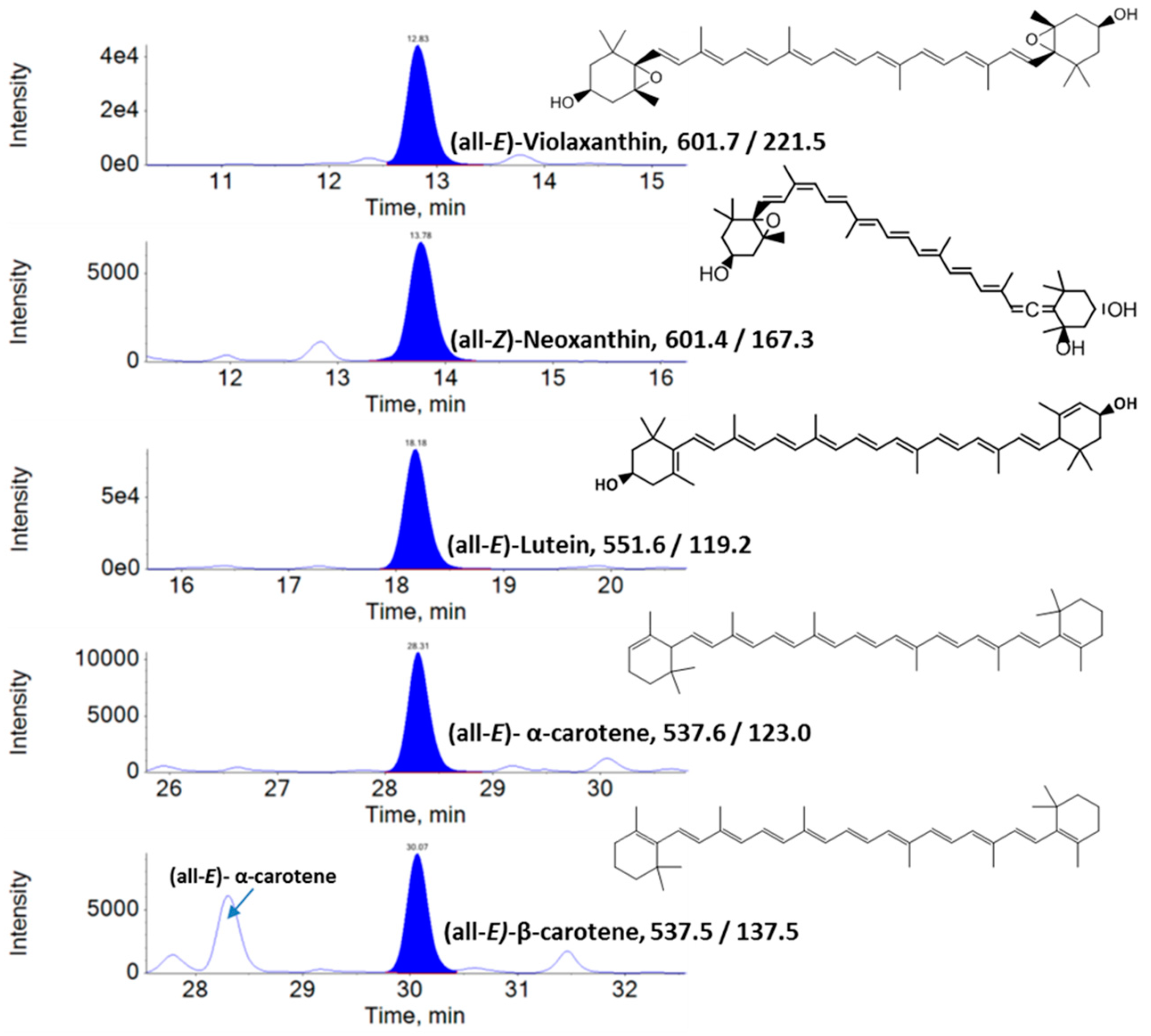
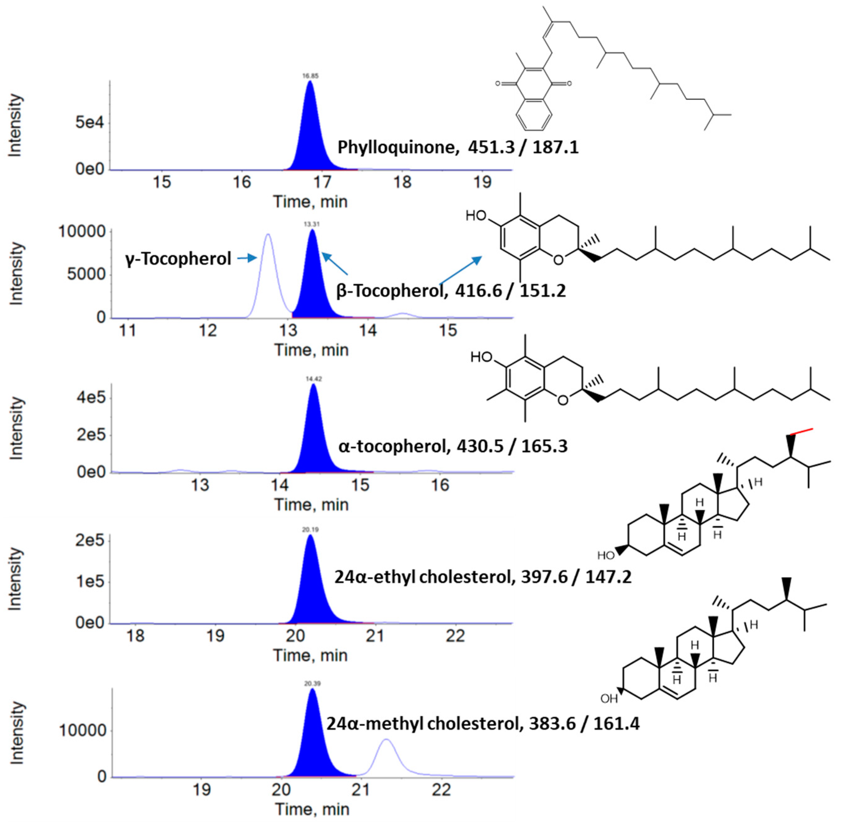
2.3. Analysis of Lipophilic Metabolites Using Liquid Chromatography–Multiple Reaction Monitoring–Mass Spectrometry (LC–MRM–MS)

The lipophilic metabolites were extracted from KP-BG leaves using Ace:Hex:EtOH (1:2:1) containing 0.1% (w/v) Bht (lipophilic extraction (LE) solution) as previously described [37]. Briefly, 4 g (exact to 0.001 g) of KP-BG leaves was placed in an amber glass vial containing 40 µL of trans-β-apo-8′-carotenal and 5-α-Cholestan-3β-ol (internal standards, dissolved in Ace, 1 mg/mL), a pinch of MgCO3, and 10 mL of LE solution. Samples were homogenized, sonicated (300 W, 60 Hz) for 15 min (JAC-2010, Sonics & Materials Inc., Newtown, CT, USA), centrifuged (5000× g for 5 min at 4 °C), and the supernatant was collected. The sample was re-extracted with 10 mL of fresh LE solution until the extract became colorless. Combined supernatants were evaporated at <35 °C using a rotary vacuum evaporator (RE 111, Büchi, Flawil, Switzerland), reconstituted with 4 mL of Ace comprising 0.1% BHT, and filtered (0.45 µm, Nylon syringe filter, Whatman) into an amber vial for LC–MRM–MS analysis.
Conditions for quantitative lipophilic antioxidant determination were adopted as described by Kim et al. [38]. Analysis was performed on a triple quadrupole mass spectrometer (SCIEX API 3200™, Applied Biosystems/MDS SCIEX, CA, USA) equipped with liquid chromatography (Exion LC™ system, Applied Biosystems/MDS SCIEX, Foster City, CA, USA). The LC separation was accomplished using a C30 carotenoid column YMC, 250 × 4.6 mm × 5 µm (Wilmington, NC) using mobile phases consisting of solvent A (MeOH/H2O (95:5, v/v) containing NH4HCO2 (5 mM)) and solvent B (MTBE/MeOH/H2O (90:7:3, v/v/v) containing NH4HCO2 (5 mM)) following the gradient elution starting from 0% B to 100% B in 45 min with a constant flow rate (1 mL/min) at 20 °C. Then, 20 mL of KP-BG leaf extract was injected into the YMC column using an autosampler. The optimized values of multiple reaction monitoring are listed in Table S1. Three independent extracts obtained from in vitro or ex vitro KP-BG leaves were analyzed in triplicate.

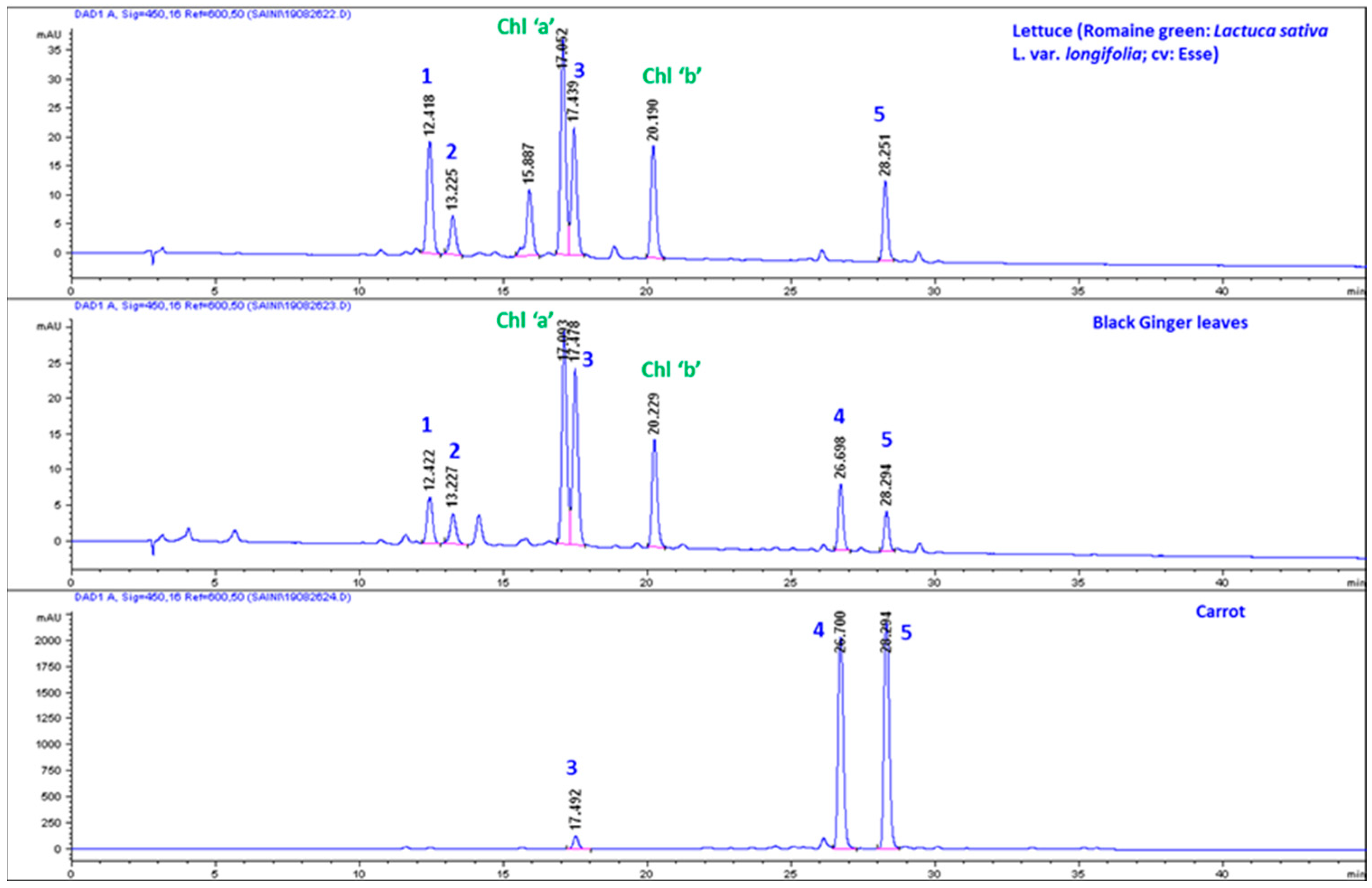
Comparative High-Performance Liquid Chromatography (HPLC) Analysis of Carotenoids in Carrot, KP-BG, and Lettuce

Extraction and analysis of carotenoids were performed following the protocols of Kim et al. [37] and Park et al. [39], respectively. The identification of (all-E)-α-carotene was performed by comparison of the RT with standard (all-E)-α-carotene) and absorption spectra were recorded by a diode array detector (DAD).

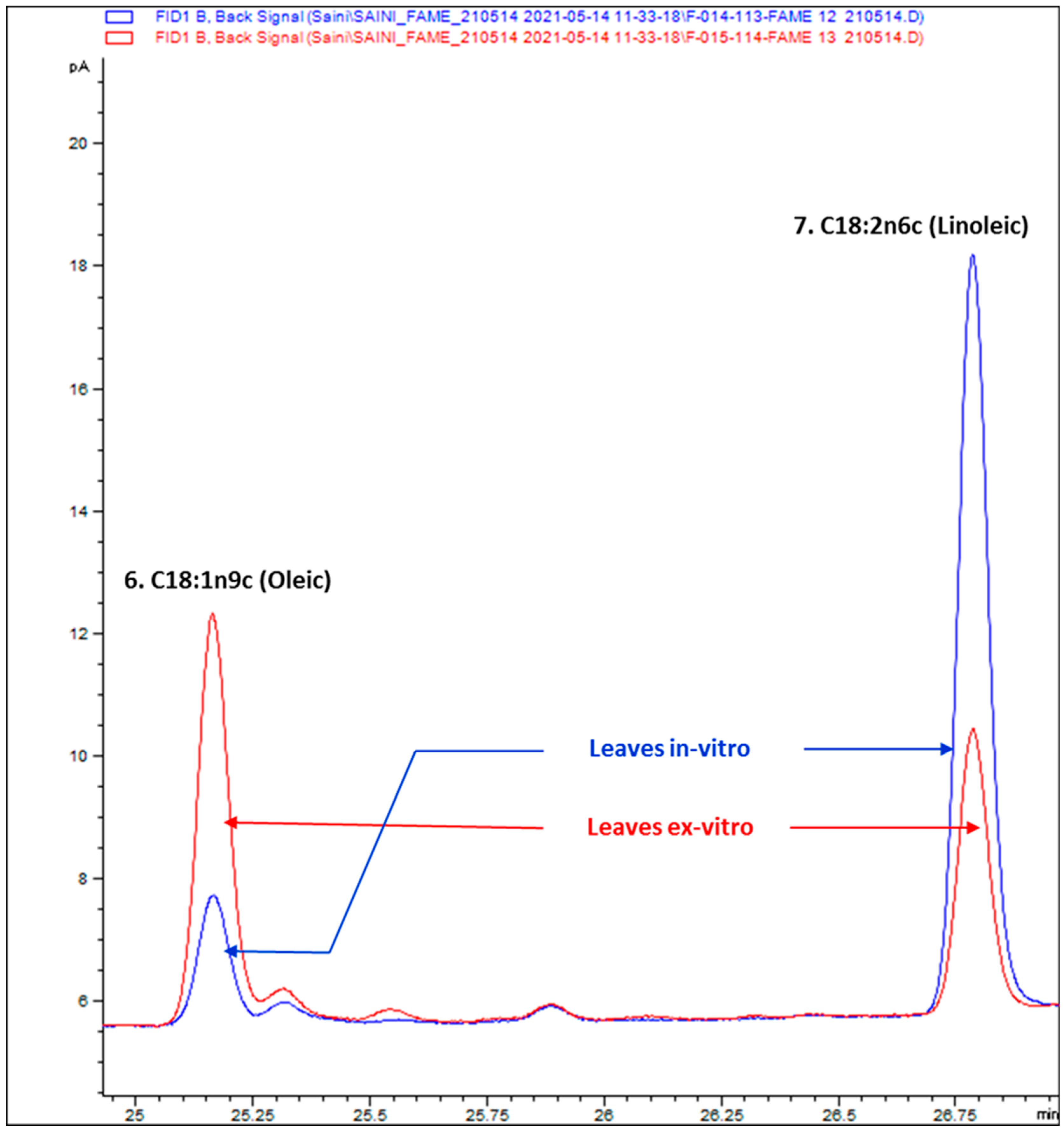
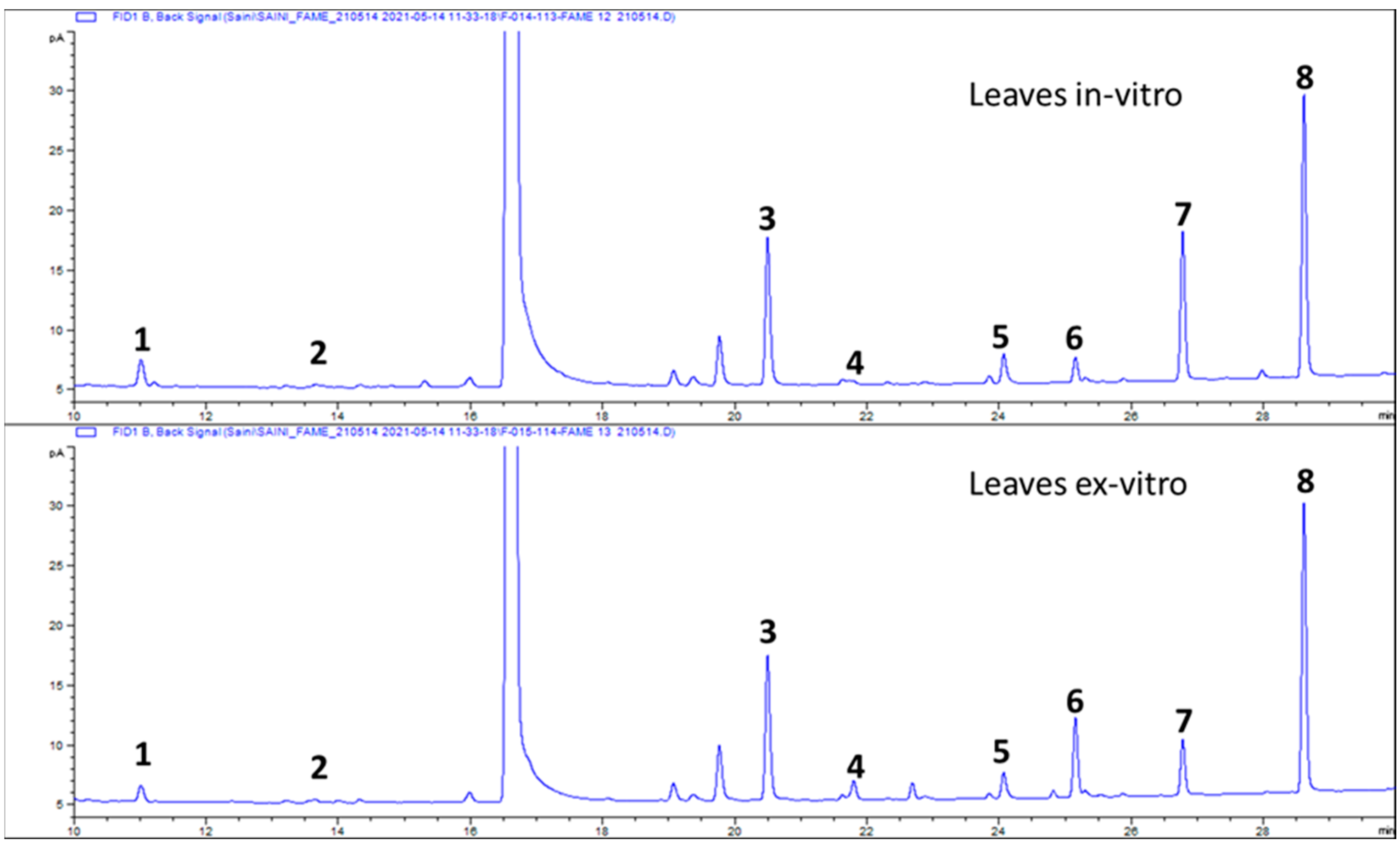
2.4. Composition of Fatty Acids

Lipids from KP-BG leaves were extracted following our optimized protocols [40,41], with minor modifications, originally based on a previous report [42]. First, 1 g of fresh leaves of KP-BG was mined with 20 mL of Ipa/Cyhex (10:12, v/v) and 0.075% Bht (w/v), sonicated for 10 min (JAC-2010; 300 W, 60 Hz), centrifuged (8000× g for 10 min at 4 °C), and the supernatant was collected. The sample was re-extracted with 20 mL of fresh solvent. Combined supernatants (approximately 40 mL) were partitioned with an equal volume of 1 M NaCl. The Ipa layer was collected, filtered through Na2SO4, and evaporated at 35 °C using a Büchi RE 111 rotary vacuum evaporator; the residue was dissolved in 3 mL of Cyhex/Dcm (1:3, v/v) containing 0.1% Bht, and stored at −20 °C.
Fatty acid methyl esters (FAMEs) were prepared using a BF3–MeOH solution (14% in methanol), according to the manufacturer’s guidelines, with minor modifications. First, 1 mL of KP-BG lipid sample was placed in a 5 mL glass vial and evaporated at 35 °C using a Büchi RE 111 rotary vacuum evaporator; 1 mL of BF3-MeOH solution was added, and then it was heated at 60 °C for 10 min. After cooling, the KP-BGFAME solution was washed with 1 M NaCl, recovered in 2 mL of Hex containing a small amount of Na2SO4, and filtered through a 0.45 µm nylon syringe filter into a vial.
FAMEs were quantitatively analyzed according to Saini et al. [41] using an Agilent (Agilent Technologies Canada, Inc.) 7890 B GC equipped with a flame ionization detector (FID), an autoinjector, and an SP-2560 100 m × 0.25 mm I.D. × 0.20 µm film thickness capillary GC column (Merck KGaA, Darmstadt, Germany). The column oven temperature was set to hold at 140 °C for 5 min, increased to 240 °C at a rate of 4 °C/min, and finally held at 240 °C for 15 min. Nitrogen was used as the carrier gas (2 mL/min). For GC–MS analysis, a QP2010 SE system (Shimadzu, Japan) was used. Three independent extracts (FAMEs) obtained from in vitro or ex vitro KP-BG leaves were analyzed in triplicate.

3. Results

3.1. Lipophilic Metabolite Content

The amounts of lipophilic metabolites in the KP-BG leaves are listed in Table 1. The lipophilic metabolites identified and measured in KP-BG leaves by LC–MRM–MS can be categorized as carotenoids, vitamin K1, tocopherols, and phytosterols. Higher amounts of total carotenoids (93.84 µg/g FW) and total tocopherols (46.22 µg/g FW) were found in the leaves of ex-vitro-raised KP-BG, whereas the levels of vitamin K1 (7.31 µg/g FW) and total phytosterols (44.40 µg/g FW) were higher in the leaves of in-vitro-grown KP-BG than ex-vitro-raised KP-BG.
LC–MRM–MS chromatograms of carotenoids found in KP-BG leaf extracts are shown in Figure 1. Five carotenoids were identified in the KP-BG leaves. Among them, Lut (39.42–44.38 µg/g FW) was the major carotenoid, followed by α-Car (10.85–14.79 µg/g FW), Neo (10.10–12.30 µg/g FW), β-Car (10.61–11.33 µg/g FW), and Vio (9.42–11.03 µg/g FW) (Table 1). LC–MRM–MS chromatograms of vitamin K1, tocopherols, and phytosterols in KP-BG leaf extracts are shown in Figure 2. α-Toc (31.08–39.07 µg/g FW) was the predominant tocopherol in KP-BG leaves, followed by β-Toc (3.44–5.66 µg/g FW) and γ-Toc (3.08–5.49 µg/g FW). Two phytosterols were identified in the leaves of KP-BG. Specifically, 24α-ethyl cholesterol (30.55–36.62 µg/g FW) was the dominant phytosterol, followed by 24α-methyl cholesterol (7.14–7.78 µg/g FW).

3.2. Composition of Fatty Acids

The GC chromatograms of FAMEs of KP-BG leaves are shown in Figure 3. Eight fatty acids were identified in the KP-BG leaves, as shown in Table 2. α-Linolenic (43.12%), palmitic (23.78%), oleic (12.28%), palmitoleic (3.64%), and lauric (1.14%) acids were the major fatty acids in the ex-vitro-raised KP-BG leaves, whereas linoleic (21.35%) and capric (5.47%) were the dominant fatty acids in the leaves of in-vitro-raised KP-BG. However, the stearic acid content (4.85–4.86%) in both leaf tissues was almost similar (Table 2). Higher levels of total saturated fatty acids (SFAs, 33.89%) and total polyunsaturated fatty acids (PUFAs, 61.03%) were found in KP-BG leaves grown in vitro. Ex-vitro-raised KP-BG leaves had a higher content of total monounsaturated fatty acids (MUFAs, 15.91%) than the KP-BG leaves grown in vitro (5.08%). The ratios of PUFAs:SFAs (1.80) and PUFAs:MUFAs (12.00) in the KP-BG leaves grown in vitro were higher than those in the ex-vitro-raised KP-BG leaves.

4. Discussion

In recent decades, there has been a growing interest in using byproducts (wastes) as a potential source for obtaining industrially important compounds. Ginger leaves have been reported to contain several bioactive metabolites [22,23,24,25,26,27]; however, the profile and content of lipophilic metabolites in KP-BG leaves have not been examined. Lipophilic compounds such as carotenoids, tocopherols, plant sterols, and fatty acids are important antioxidants that are used in the cosmetics, foods, and nutraceutical industries [32,43]. Plants and several microorganisms produce carotenoids as fat-soluble colored pigments, which are used as natural colorants in various industries [36,44]. These colored pigments are important components required for light harvesting and photoprotection in plants [28]. The profile and content of carotenoids in reproductive (flower, fruit, and seed) and vegetative (leaf and stem) organs vary with plant species, cultivar, growth stage, and environment. In this study, the ex-vitro-raised KP-BG leaves had higher total carotenoid content (93.84 µg/g FW) than the in-vitro-grown KP-BG leaves (80.40 µg/g FW). A similar result was observed in Aronia melanocarpa [45] and Sedum dasyphyllum [46]. Of the identified carotenoids, Lut was found to be a major component (44.38 µg/g FW) in KP-BG leaves (Table 1). Lut has been identified as the predominant carotenoid in leafy green vegetables and the mature leaves of many medicinal plants [37,39,45,46,47].
α-Car and β-Car are important provitamin A carotenoids found in Cucurbita pepo, Daucus carota, Ipomoea batatas, and some cultivars of apricot, beans, and squash at high concentrations [48,49]. However, these provitamin A carotenoids were found in green leaves at low concentrations. In addition, the β-Car content in leaf tissues is often higher than that α-Car [50]. In this study, the presence of a high amount of α-Car (similar to β-Car) in ginger foliage was surprising. Generally, in green foliage, complete conversion of α-Car to Lut causes the dominance of lutein and the absence of α-Car [51]. α-Car was absent in the leafy vegetables studied by Lakshminarayana et al. [51], including Basella rubra, Peucedanum sowa, Moringa oleifera, Trigonella foenum-graecum, Spinacia oleracea, Sesbania grandiflora, and Raphanus sativus. Similarly, α-Car was not recorded in the fresh-cut foliage of Romaine (red) (Lactuca sativa var. romana), Komatsuna (Brassica rapavar. perviridis), salad rocket (Garden rocket, Arugula) (Eruca sativa, syn. E. vesicaria subsp. sativa), wild rocket (perennial wall rocket) (Diplotaxis tenuifolia), and Batavian lettuce (Lactuca sativa L. var. acephala) [52]. Thus, considering the previous findings of the absence of α-Car in several green foliages and confirming its significant presence in ginger leaves, we recorded the absorbance spectrum by online HPLC–DAD, which was in addition to the confirmation by retention time and molecular mass of qualifier (Ql) and quantifier (Qt) transition ions of α-Car and other metabolites quantified by LC–MRM–MS. However, the MRM transition of m/z 537.5/137.5 is common between α-Car and β-Car. The retention time (28.31 and 30.07 min for α- and β-Car, respectively; Figure 1), transition of m/z 537.6/123.0 (produced selectively from α-carotene; Figure 1), and the absorbance spectrum can easily distinguish between α-Car (λmax: 446, 472) and β-Car (λmax: 450, 476) (Figure 4). Similarly, the chromatograms of lettuce and carrot were compared. Orange carrots are the most significant source of α-Car [53]. The α-Car peak was absent in lettuce, whereas it was dominant in ginger leaves and carrots (Figure 5).
Phylloquinone, another important antioxidant, is a lipid-soluble vitamin found in plants [54]. The phylloquinone content in KP-BG leaves (7.31 µg/g FW) (Table 1) was higher than that found in artichokes (16.2 µg·100 g−1 FW), broccoli (102 µg·100 g−1 FW), broccoli raab (242 µg·100 g−1 FW), carrot (8.3 µg·100 g−1 FW), celery (29.0 µg·100 g−1 FW), cucumber (16.4 µg·100 g−1 FW), lettuce ‘Green leaf’ (127 µg·100 g−1 FW), pepper (green) (7.1 µg·100 g−1 FW), potato (red) (3.2 µg·100 g−1 FW), sweet potato (1.8 µg·100 g−1 FW), radish (1.4 µg·100 g−1 FW) [55], and broccoli byproducts (leaves) (24.3 µg/g FW) [56]. Tocopherols are important bioactive compounds mainly obtained from photosynthetic organisms [57]. The levels of individual tocopherols differed among KP-BG leaves. γ-Toc (5.49 µg/g FW) and β-Toc (5.66 µg/g FW) levels were higher in in vitro KP-BG leaves than in ex vitro KP-BG leaves (Table 1). However, the major tocopherol (α-Toc) content in ex vitro KP-BG leaves (39.70 µg/g FW) was higher than that in in vitro KP-BG leaves (31.08 µg/g FW). The variation in the tocopherol content of KP-BG leaves may be due to differences in the growing environment, plant physiological status, and nutrient availability. The production of metabolites through plant organ cultures also changes according to environmental and nutritional factors [39,45,46]. It is worth mentioning that the α-Toc content in ex vitro KP-BG leaves (39.70 µg/g FW) is higher than that of various medicinal plants, such as Tanacetum vulgare (2.2 µg/g dry weight (DW), Silene vulgaris (11.21 µg/g DW), Urtica dioica (16.52 µg/g DW), and Rosa canina L. cv. Plovdiv 1 (34.05 µg/g DW) [58]. Moreover, 24α-ethyl cholesterol is a valuable phytosterol found abundantly in plants and has been reported to have antimicrobial, antioxidant, anti-inflammatory, antidiabetic, anticancer, and immunomodulatory activities [59]. In this study, 24α-ethyl cholesterol was found to be the major phytosterol in KP-BG leaves.
To the best of our knowledge, there are no reports on the fatty acid profile of KP-BG. In this study, the fatty acid composition of KP-BG leaves was examined using GC–FID/MS for the first time. α-Linolenic acid was found (39.68–43.12%) to be the most abundant fatty acid in KP-BG leaves (Table 2). Similarly, α-Linolenic acid was identified as the predominant fatty acid in the leaves of Mertensia maritima [39], Aronia melanocarpa [45], Sedum dasyphyllum [46], and Ajuga multiflora [60]. In the in-vitro-raised KP-BG leaves, the proportion of oleic acid (OA) decreased, and that of linoleic acid (LA) increased. A reverse trend was observed in the ex-vitro-grown KP-BG leaves (Table 2). In plants, ∆12-fatty acid desaturase catalyzes the conversion of C18:1n9c (OA) to C18:2n6c (LA) [61]. Comparison of OA:LA peaks can be discussed, as the OA:LA ratio is 0.17 in in-vitro-raised KP-BG leaves and 1.5 in leaves obtained from ex-vitro-grown KP-BG plants (Figure 6). OA has been reported to have anticancer activity and prevent cardiovascular diseases [62]. LA is an important PUFA that is essential for human life [63]. In this study, the KP-BG leaves raised in vitro had higher total PUFA content (61.03%) than the ex-vitro-grown KP-BG leaves (51.59%). In addition, the percentage of individual identified fatty acids varied among the KP-BG leaves. Similar results were also observed in in vitro cultures of Sedum dasyphyllum [46], Ajuga multiflora [60], Argania spinosa [64], and conifers [65]. It has been disclosed that plant growth regulators (PGR) could alter the fatty acid profile of treated plants [46,60]. The addition of PGR to in vitro culture media is often required to stimulate callus and shoot formation. Such calli and shoots (in-vitro-raised) may accumulate higher levels of fatty acids than ex-vitro-grown plants [46].

5. Conclusions

The lipophilic metabolite profiles and contents of KP-BG leaves were successfully analyzed using LC–MRM–MS and GC–FID/MS. The analysis and quantification of lipophilic components is an engaging task and, using the sophistication offered by the dual analytical approaches, we have been able to analyze and quantify the antioxidant compounds. The present report elaborately consolidates the bioactive components present in these leaves. In total, five carotenoids, three tocopherols, two phytosterols, eight fatty acids, and vitamin K were identified in KP-BG leaves. Further, the quantification data confirmed that Lut, α-Toc, 24α-ethyl cholesterol, and α-linolenic acid were the major lipophilic metabolites in KP-BG leaves. In addition, the content of α-car was higher than that of β-car. The findings of this study confirmed that KP-BG byproducts are a rich source of lipophilic antioxidants. However, the beneficial effects of the individual lipophilic compounds identified in KP-BG byproducts should be studied. This study confirms that KP-BG leaves are a rich reservoir of the numerous listed antioxidant-rich bioactive metabolites and emphasizes the need to exploit these resources for human benefit.

Supplementary Materials

The following are available online at https://www.mdpi.com/article/10.3390/antiox10101573/s1, Table S1: Optimized values of declustering potential (DP), entrance potential (EP), collision energy (CE), cell exit potential (CXP) and retention time (Rt) of qualifier (Ql) and quantifier (Qt) transition used for the liquid chromatography (LC)-multiple reaction monitoring (MRM)-mass spectrometry (MS) quantification of major lipophilic metabolites from the Kaempferia parviflora (black ginger) (KP-BG) leaves.

Author Contributions

Conceptualization, K.S. and I.S.; methodology, R.K.S. and I.S.; investigation, R.K.S. and I.S.; data curation, Y.-S.K.; writing—original draft preparation, R.K.S. and I.S.; writing—review and editing, K.S., Y.-S.K. and I.S. All authors have read and agreed to the published version of the manuscript.

Funding

This research received no external funding.

Institutional Review Board Statement

Not applicable.

Informed Consent Statement

Not applicable.

Data Availability Statement

Data is contained within the article.

Conflicts of Interest

The authors declare no conflict of interest.

References

  1. Techaprasan, J.; Klinbunga, S.; Ngamriabsakul, C.; Jenjittikul, T. Genetic variation of Kaempferia (Zingiberaceae) in Thailand based on chloroplast DNA (psbA-trnH and petA-psbJ) sequences. Genet. Mol. Res. 2010, 9, 1957–1973. [Google Scholar] [CrossRef]
  2. Elshamy, A.I.; Mohamed, T.A.; Essa, A.F.; Abd-El Gawad, A.M.; Alqahtani, A.S.; Shahat, A.A.; Yoneyama, T.; Farrag, A.R.H.; Noji, M.; El-Seedi, H.R.; et al. Recent advances in Kaempferia phytochemistry and biological activity: A comprehensive review. Nutrients 2019, 11, 2396. [Google Scholar] [CrossRef] [Green Version]
  3. Kochuthressia, K.; Britto, S.J.; Jaseentha, M.; Raphael, R. In vitro antimicrobial evaluation of Kaempferia galanga L. rhizome extract. Am. J. Biotechnol. Mol. Sci. 2012, 2, 1–5. [Google Scholar] [CrossRef]
  4. Ridtitid, W.; Sae-Wong, C.; Reanmongkol, W.; Wongnawa, M. Antinociceptive activity of the methanolic extract of Kaempferia galanga Linn. in experimental animals. J. Ethnopharmacol. 2008, 118, 225–230. [Google Scholar] [CrossRef]
  5. Ahmed, F.R.S.; Amin, R.; Hasan, I.; Asaduzzaman, A.; Kabir, S.R. Antitumor properties of a methyl-β-d-galactopyranoside specific lectin from Kaempferia rotunda against Ehrlich ascites carcinoma cells. Int. J. Biol. Macromol. 2017, 102, 952–959. [Google Scholar] [CrossRef]
  6. Kobayashi, S.; Kato, T.; Azuma, T.; Kikuzaki, H.; Abe, K. Anti-allergenic activity of polymethoxyflavones from Kaempferia parviflora. J. Funct. Foods 2015, 13, 100–107. [Google Scholar] [CrossRef]
  7. Hidaka, M.; Horikawa, K.; Akase, T.; Makihara, H.; Ogami, T.; Tomozawa, H.; Tsubata, M.; Ibuki, A.; Matsumoto, Y. Efficacy of Kaempferia parviflora in a mouse model of obesity-induced dermatopathy. J. Nat. Med. 2017, 71, 59–67. [Google Scholar] [CrossRef]
  8. Win, N.N.; Ngwe, H.; Abe, I.; Morita, H. Naturally occurring Vpr inhibitors from medicinal plants of Myanmar. J. Nat. Med. 2017, 71, 579–589. [Google Scholar] [CrossRef] [PubMed] [Green Version]
  9. Yeap, Y.S.Y.; Kassim, N.K.; Ng, R.C.; Ee, G.C.L.; Saiful Yazan, L.; Musa, K.H. Antioxidant properties of ginger (Kaempferia angustifolia Rosc.) and its chemical markers. Int. J. Food Prop. 2017, 20, 1158–1172. [Google Scholar] [CrossRef] [Green Version]
  10. Sawasdee, P.; Sabphon, C.; Sitthiwongwanit, D.; Kokpol, U. Anticholinesterase activity of 7-methoxyflavones isolated from Kaempferia parviflora. Phytother. Res. 2009, 23, 1792–1794. [Google Scholar] [CrossRef] [PubMed]
  11. Kaewkroek, K.; Wattanapiromsakul, C.; Matsuda, H.; Nakamura, S.; Tewtrakul, S. Anti-inflammatory activity of compounds from Kaempferia marginata rhizomes. Songklanakarin J. Sci. Technol. 2017, 39, 91–99. [Google Scholar]
  12. Plaingam, W.; Sangsuthum, S.; Angkhasirisap, W.; Tencomnao, T. Kaempferia parviflora rhizome extract and Myristica fragrans volatile oil increase the levels of monoamine neurotransmitters and impact the proteomic profiles in the rat hippocampus: Mechanistic insights into their neuroprotective effects. J. Tradit. Complement. Med. 2017, 7, 538–552. [Google Scholar] [CrossRef] [PubMed]
  13. Shanbhag, T.V.; Sharma, C.; Adiga, S.; Bairy, K.; Shenoy, S.; Shenoy, G. Wound healing activity of alcoholic extract of Kaempferia galanga in Wistar rats. Indian J. Physiol. Pharmacol. 2006, 50, 384–390. [Google Scholar]
  14. Pripdeevech, P.; Pitija, K.; Rujjanawate, C.; Pojanagaroon, S.; Kittakoop, P.; Wongpornchai, S. Adaptogenic-active components from Kaempferia parviflora rhizomes. Food Chem. 2012, 132, 1150–1155. [Google Scholar] [CrossRef] [PubMed]
  15. Azuma, T.; Kayano, S.-I.; Matsumura, Y.; Konishi, Y.; Tanaka, Y.; Kikuzaki, H. Antimutagenic and glucosidase inhibitory effects of constituents from Kaempferia parviflora. Food Chem. 2011, 125, 471–475. [Google Scholar] [CrossRef]
  16. Chaipech, S.; Morikawa, T.; Ninomiya, K.; Yoshikawa, M.; Pongpiriyadacha, Y.; Hayakawa, T.; Muraoka, O. Structures of two new phenolic glycosides, kaempferiaosides A and B, and hepatoprotective constituents from the rhizomes of Kaempferia parviflora. Chem. Pharm. Bull. 2012, 60, 62–69. [Google Scholar] [CrossRef] [Green Version]
  17. Thao, N.P.; Luyen, B.T.T.; Lee, S.H.; Jang, H.D.; Kim, Y.H. Anti-osteoporotic and antioxidant activities by rhizomes of Kaempferia parviflora wall. Ex Baker. Nat. Prod. Sci. 2016, 22, 13–19. [Google Scholar] [CrossRef] [Green Version]
  18. Tewtrakul, S.; Subhadhirasakul, S.; Karalai, C.; Ponglimanont, C.; Cheenpracha, S. Anti-inflammatory effects of compounds from Kaempferia parviflora and Boesenbergia pandurata. Food Chem. 2009, 115, 534–538. [Google Scholar] [CrossRef]
  19. Nakao, K.; Murata, K.; Deguchi, T.; Itoh, K.; Fujita, T.; Higashino, M.; Yoshioka, Y.; Matsumura, S.-I.; Tanaka, R.; Shinada, T. Xanthine oxidase inhibitory activities and crystal structures of methoxyflavones from Kaempferia parviflora rhizome. Biol. Pharm. Bull. 2011, 34, 1143–1146. [Google Scholar] [CrossRef] [Green Version]
  20. Sulaiman, M.R.; Zakaria, Z.A.; Duad, I.A.; Hidayat, M.T. Antinociceptive and anti-inflammatory activities of the aqueous extract of Kaempferia galanga leaves in animal models. J. Nat. Med. 2008, 62, 221–227. [Google Scholar] [CrossRef] [PubMed]
  21. Ali, M.S.; Dash, P.R.; Nasrin, M. Study of sedative activity of different extracts of Kaempferia galanga in Swiss albino mice. BMC Complement. Altern. Med. 2015, 15, 158. [Google Scholar] [CrossRef] [Green Version]
  22. Chan, E.W.C.; Lim, Y.Y.; Wong, L.F.; Lianto, F.S.; Wong, S.K.; Lim, K.K.; Joe, C.E.; Lim, T.Y. Antioxidant and tyrosinase inhibition properties of leaves and rhizomes of ginger species. Food Chem. 2008, 109, 477–483. [Google Scholar] [CrossRef]
  23. Park, H.Y.; Kim, K.-S.; Ak, G.; Zengin, G.; Cziáky, Z.; Jekő, J.; Adaikalam, K.; Song, K.; Kim, D.H.; Sivanesan, I. Establishment of a rapid micropropagation system for Kaempferia parviflora wall. Ex Baker: Phytochemical analysis of leaf extracts and evaluation of biological activities. Plants 2021, 10, 698. [Google Scholar] [CrossRef] [PubMed]
  24. Preetha, T.S.; Hemanthakumar, A.S.; Krishnan, P.N. A comprehensive review of Kaempferia galanga L. (Zingiberaceae): A high sought medicinal plant in tropical Asia. J. Med. Plants Stud. 2016, 4, 270–276. [Google Scholar]
  25. Chan, E.W.C.; Lim, Y.Y.; Wong, S.K.; Lim, K.K.; Tan, S.P.; Lianto, F.S.; Yong, M.Y. Effects of different drying methods on the antioxidant properties of leaves and tea of ginger species. Food Chem. 2009, 113, 166–172. [Google Scholar] [CrossRef]
  26. Bhuiyan, M.N.I.; Begum, J.; Anwar, M.N. Essential oils of leaves and rhizomes of Kaempferia galanga Linn. Chittagong Univ. J. Biol. Sci. 2008, 3, 65–76. [Google Scholar] [CrossRef] [Green Version]
  27. Norhayati, Y.; Nurulhidayah, A.; Rini Zunnurni, M.J.; Norliana, A.R.; Norliana, W.; Mohd Ifwat, I. Antioxidative constituents of selected Malaysian ‘Ulam’. Sci. Int. 2018, 30, 273–278. [Google Scholar]
  28. Sandmann, G. Antioxidant protection from UV- and light-stress related to carotenoid structures. Antioxidants 2019, 8, 219. [Google Scholar] [CrossRef] [Green Version]
  29. Lim, G.H.; Singhal, R.; Kachroo, A.; Kachroo, P. Fatty acid- and lipid-mediated signaling in plant defense. Annu. Rev. Phytopathol. 2017, 55, 505–536. [Google Scholar] [CrossRef]
  30. Cabianca, A.; Müller, L.; Pawlowski, K.; Dahlin, P. Changes in the plant β-sitosterol/stigmasterol ratio caused by the plant parasitic nematode Meloidogyne incognita. Plants 2021, 10, 292. [Google Scholar] [CrossRef]
  31. Ma, J.; Qiu, D.; Pang, Y.; Gao, H.; Wang, X.; Qin, Y. Diverse roles of tocopherols in response to abiotic and biotic stresses and strategies for genetic biofortification in plants. Mol. Breed. 2020, 40, 18. [Google Scholar] [CrossRef]
  32. Chang, K.H.; Cheng, M.L.; Chiang, M.C.; Chen, C.M. Lipophilic antioxidants in neurodegenerative diseases. Clin. Chim. Acta Int. J. Clin. Chem. 2018, 485, 79–87. [Google Scholar] [CrossRef] [PubMed]
  33. Pogačnik, L.; Ota, A.; Ulrih, N.P. An overview of crucial dietary substances and their modes of action for prevention of neurodegenerative diseases. Cells 2020, 9, 576. [Google Scholar] [CrossRef] [Green Version]
  34. Shin, G.H.; Kim, J.T.; Park, H.J. Recent developments in nanoformulations of lipophilic functional foods. Trends Food Sci. Technol. 2015, 46, 144–157. [Google Scholar] [CrossRef]
  35. Oppedisano, F.; Macrì, R.; Gliozzi, M.; Musolino, V.; Carresi, C.; Maiuolo, J.; Bosco, F.; Nucera, S.; Zito, M.C.; Guarnieri, L.; et al. The anti-inflammatory and antioxidant properties of n-3 PUFAs: Their role in cardiovascular protection. Biomedicines 2020, 25, 306. [Google Scholar] [CrossRef] [PubMed]
  36. Saini, R.K.; Moon, S.H.; Gansukh, E.; Keum, Y.S. An efficient one-step scheme for the purification of major xanthophyll carotenoids from lettuce, and assessment of their comparative anticancer potential. Food Chem. 2018, 266, 56–65. [Google Scholar] [CrossRef] [PubMed]
  37. Kim, D.E.; Shang, X.; Assefa, A.D.; Keum, Y.S.; Saini, R.K. Metabolite profiling of green, green/red, and red lettuce cultivars: Variation in health beneficial compounds and antioxidant potential. Food Res. Int. 2018, 105, 361–370. [Google Scholar] [CrossRef]
  38. Kim, D.H.; Enkhtaivan, G.; Saini, R.K.; Keum, Y.S.; Kang, K.W.; Sivanesan, I. Production of bioactive compounds in cladode culture of Turbinicarpus valdezianus (H. Moeller) Glass & R. C. Foster. Ind. Crop. Prod. 2019, 138, 111491. [Google Scholar]
  39. Park, H.Y.; Kim, D.H.; Saini, R.K.; Gopal, J.; Keum, Y.S.; Sivanesan, I. Micropropagation and quantification of bioactive compounds in Mertensia maritima (L.) gray. Int. J. Mol. Sci. 2019, 20, 2141. [Google Scholar] [CrossRef] [PubMed] [Green Version]
  40. Saini, R.K.; Assefa, A.D.; Keum, Y.-S. Spices in the Apiaceae family represent the healthiest fatty acid profile: A aystematic comparison of 34 widely used spices and herbs. Foods 2021, 10, 854. [Google Scholar] [CrossRef]
  41. Saini, R.K.; Rauf, A.; Khalil, A.A.; Ko, E.-Y.; Keum, Y.-S.; Anwar, S.; Alamri, A.; Rengasamy, K.R.R. Edible mushrooms show significant differences in sterols and fatty acid compositions. S. Afr. J. Bot. 2021, 141, 344–356. [Google Scholar] [CrossRef]
  42. Cruz, R.; Casal, S.; Mendes, E.; Costa, A.; Santos, C.; Morais, S. Validation of a single-extraction procedure for sequential analysis of vitamin E, cholesterol, fatty acids, and total fat in seafood. Food Anal. Methods 2012, 6, 1196–1204. [Google Scholar] [CrossRef] [Green Version]
  43. Dima, Ş.; Dima, C.; Iordăchescu, G. Encapsulation of functional lipophilic food and drug biocomponents. Food Eng. Rev. 2015, 7, 417–438. [Google Scholar] [CrossRef]
  44. Langi, P.; Kiokias, S.; Varzakas, T.; Proestos, C. Carotenoids: From plants to food and feed industries. Methods Mol. Biol. Clifton NJ 2018, 1852, 57–71. [Google Scholar]
  45. Sivanesan, I.; Saini, R.K.; Kim, D.H. Bioactive compounds in hyperhydric and normal micropropagated shoots of Aronia melanocarpa (michx.) Elliott. Ind. Crops Prod. 2016, 83, 31–38. [Google Scholar] [CrossRef]
  46. Park, H.Y.; Saini, R.K.; Gopal, J.; Keum, Y.-S.; Kim, D.H.; Lee, O.; Sivanesan, I. Micropropagation and subsequent enrichment of carotenoids, fatty acids, and tocopherol contents in Sedum dasyphyllum L. Front. Chem. 2017, 5, 77. [Google Scholar] [CrossRef] [Green Version]
  47. Yoon, G.-A.; Yeum, K.-J.; Cho, Y.-S.; Chen, C.-Y.O.; Tang, G.; Blumberg, J.B.; Russell, R.M.; Yoon, S.; Lee-Kim, Y.C. Carotenoids and total phenolic contents in plant foods commonly consumed in Korea. Nutr. Res. Pr. 2012, 6, 481–490. [Google Scholar] [CrossRef]
  48. Saini, R.K.; Nile, S.H.; Park, S.W. Carotenoids from fruits and vegetables: Chemistry, analysis, occurrence, bioavailability and biological activities. Food Res. Int. 2015, 76, 735–750. [Google Scholar] [CrossRef]
  49. Pan, Z.; Sun, Y.; Zhang, F.; Guo, X.; Liao, Z. Effect of thermal processing on carotenoids and folate changes in six varieties of sweet potato (Ipomoes batata L.). Foods 2019, 8, 215. [Google Scholar] [CrossRef] [Green Version]
  50. Ponder, A.; Hallmann, E. Phenolics and carotenoid contents in the leaves of different organic and conventional raspberry (Rubus idaeus L.) cultivars and their in vitro activity. Antioxidants 2019, 8, 458. [Google Scholar] [CrossRef] [Green Version]
  51. Lakshminarayana, R.; Raju, M.; Krishnakantha, T.P.; Baskaran, V. Determination of major carotenoids in a few Indian leafy vegetables by high-performance liquid chromatography. J. Agric. Food Chem. 2005, 53, 2838–2842. [Google Scholar] [CrossRef] [PubMed]
  52. Saini, R.K.; Shang, X.M.; Ko, E.Y.; Choi, J.H.; Kim, D.; Keum, Y.-S. Characterization of nutritionally important phytoconstituents in minimally processed ready-to-eat baby-leaf vegetables using HPLC–DAD and GC–MS. J. Food Meas. Charact. 2016, 10, 341–349. [Google Scholar] [CrossRef]
  53. Purkiewicz, A.; Ciborska, J.; Tańska, M.; Narwojsz, A.; Starowicz, M.; Przybyłowicz, K.E.; Sawicki, T. The impact of the method extraction and different carrot variety on the carotenoid profile, total phenolic content and antioxidant properties of juices. Plants 2020, 9, 1759. [Google Scholar] [CrossRef]
  54. Basset, G.J.; Latimer, S.; Fatihi, A.; Soubeyrand, E.; Block, A. Phylloquinone (vitamin K1): Occurrence, biosynthesis and functions. Mini Rev. Med. Chem. 2017, 17, 1028–1038. [Google Scholar] [CrossRef]
  55. Damon, M.; Zhang, N.Z.; Haytowitz, D.B.; Booth, S.L. Phylloquinone (vitamin K1) content of vegetables. J. Food Compos. Anal. 2005, 18, 751–758. [Google Scholar] [CrossRef]
  56. Liu, M.; Zhang, L.; Ser, S.; Cumming, J.; Ku, K. Comparative phytonutrient analysis of broccoli byproducts: The potentials for broccoli byproduct utilization. Molecules 2018, 23, 900. [Google Scholar] [CrossRef] [Green Version]
  57. Szewczyk, K.; Chojnacka, A.; Górnicka, M. Tocopherols and tocotrienols—Bioactive dietary compounds; what is certain, what is doubt? Int. J. Mol. Sci. 2021, 22, 6222. [Google Scholar] [CrossRef]
  58. Mihaylova, D.; Vrancheva, R.; Petkova, N.; Ognyanov, M.; Desseva, I.; Ivanov, I.; Popova, M.; Popova, A. Carotenoids, tocopherols, organic acids, charbohydrate and mineral content in different medicinal plant extracts. Z. Nat. C. J. Biosci. 2018, 73, 439–448. [Google Scholar] [CrossRef]
  59. Babu, S.; Jayaraman, S. An update on β-sitosterol: A potential herbal nutraceutical for diabetic management. Biomed. Pharmacother. 2020, 131, 110702. [Google Scholar] [CrossRef]
  60. Sivanesan, I.; Saini, R.K.; Noorzai, R.; Zamany, A.J.; Kim, D.H. In vitro propagation, carotenoid, fatty acid and tocopherol content of Ajuga multiflora Bunge. 3 Biotech 2016, 6, 91. [Google Scholar] [CrossRef] [Green Version]
  61. Chen, P.-Y.; Hsieh, M.-J.; Tsai, Y.-T.; Chung, H.-H.; Shyur, L.-F.; Hsieh, C.-H.; To, K.-Y. Transformation and characterization of Δ12-fatty acid acetylenase and Δ12-oleate desaturase potentially involved in the polyacetylene biosynthetic pathway from Bidens pilosa. Plants 2020, 9, 1483. [Google Scholar] [CrossRef] [PubMed]
  62. Sales-Campos, H.; Reis de Souza, P.; Crema Peghini, B.; Santana da Silva, J.; Ribeiro Cardoso, C. An overview of the modulatory effects of oleic acid in health and disease. Mini Rev. Med. Chem. 2013, 13, 201–210. [Google Scholar]
  63. Marangoni, F.; Agostoni, C.; Borghi, C.; Catapano, A.L.; Cena, H.; Ghiselli, A.; La Vecchia, C.; Lercker, G.; Manzato, E.; Pirillo, A. Dietary linoleic acid and human health: Focus on cardiovascular and cardiometabolic effects. Atherosclerosis 2020, 292, 90–98. [Google Scholar] [CrossRef] [PubMed] [Green Version]
  64. Koufan, M.; Belkoura, I.; Mazri, M.A.; Amarraque, A.; Essatte, A.; Elhorri, H.; Zaddoug, F.; Alaoui, T. Determination of antioxidant activity, total phenolics and fatty acids in essential oils and other extracts from callus culture, seeds and leaves of Argania spinosa (L.) Skeels. Plant Cell Tissue Organ Cult. 2020, 141, 217–227. [Google Scholar] [CrossRef]
  65. Semenova, N.V.; Makarenko, S.P.; Shmakov, V.N.; Konstantinov, Y.M.; Dudareva, L.V. Fatty acid composition of total lipids from needles and cultured calluses of conifers Pinus sylvestris L., Picea pungens Engelm., Pinus koraiensis Siebold & Zucc., and Larix sibirica Ledeb. Biochem. Suppl. Ser. A Membr. Cell Biol. 2017, 11, 287–295. [Google Scholar]
Figure 1. LC–MRM–MS chromatograms of carotenoids of KP-BG leaf extracts.
Antioxidants 10 01573 g001
Figure 2. LC–MRM–MS chromatograms of phylloquinone (Vitamin K1), tocopherols, and phytosterols of KP-BG leaf extracts.
Antioxidants 10 01573 g002
Figure 3. GC–FID chromatograms of FAMEs of KP-BG leaves. (1) C10:0 (Capric), (2) C12:0 (Lauric), (3) C16:0 (Palmitic), (4) C16:1 (Palmitoleic), (5) C18:0 (Stearic), (6) C18:1n9c (Oleic), (7) C18:2n6c (Linoleic), (8) C18:3n3 (α-Linolenic).
Antioxidants 10 01573 g003
Figure 4. Absorption spectra recorded by diode array detector (DAD).
Antioxidants 10 01573 g004
Figure 5. HPLC analysis of pigments in black ginger, carrot, and lettuce. (1) (all-E)-Violaxanthin; (2) (all-Z)-Neoxanthin; (3) (all-E)-Lutein; (4) (all-E)- α-carotene; (5) (all-E)-β-carotene; Chl: chlorophyll.
Antioxidants 10 01573 g005
Figure 6. Comparison of oleic: linoleic acid peaks.
Antioxidants 10 01573 g006
Table 1. Contents of major lipophilic metabolites from the KP-BG leaves, quantified (µg/g FW) by the LC–MRM–MS method.
S/NoAnalyteIn Vitro Leaves Ex Vitro Leaves
1(all-E)-Violaxanthin9.4211.03
2(all-Z)-Neoxanthin10.1012.30
3(all-E)-Lutein39.4244.38
4(all-E)-α-carotene10.8514.79
5(all-E)-β-carotene10.6111.33
6Total carotenoids (S/No. 1+2+3+4+5)80.4093.84
7Phylloquinone (Vitamin K1)7.317.25
8γ-Tocopherol5.493.08
9β-Tocopherol5.663.44
10α-Tocopherol31.0839.70
11Total tocopherols (S/No. 8+9+10)42.2346.22
1224α-ethyl cholesterol36.6230.55
1324α-methyl cholesterol7.787.14
14Total phytosterols44.4037.69
Table 2. Composition of fatty acids in KP-BG leaves.
S/NoFAMERT (Minute)In Vitro LeavesEx Vitro Leaves
1C10:0 (Capric)11.0295.473.03
2C12:0 (Lauric)13.6661.031.14
3C16:0 (Palmitic)20.50922.5323.78
4C16:1 (Palmitoleic)21.7441.373.64
5C18:0 (Stearic)24.0824.864.85
6C18:1n9c (Oleic)25.1693.7112.28
7C18:2n6c (Linoleic)26.78921.358.17
8C18:3n3 (α-Linolenic)28.62539.6843.12
Oleic: linoleic acid0.171.50
Total SFAs 33.8932.80
Total MUFAs 5.0815.91
Total PUFAs 61.0351.29
PUFAs: SFAs 1.801.56
PUFAs: MUFAs 12.003.22
Values are % of total fatty acids. SFAs: total saturated fatty acids; MUFAs: total monounsaturated fatty acids; PUFAs: total polyunsaturated fatty acids; RT: retention time.
Publisher’s Note: MDPI stays neutral with regard to jurisdictional claims in published maps and institutional affiliations.

