4. Discussion
PSE represents one of the most frequent and clinically relevant long-term sequelae of pediatric AIS.
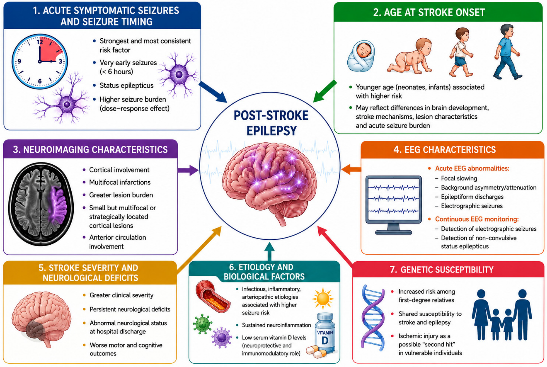
Among the various predictors investigated to date, only a limited number of clinical factors have been consistently confirmed across independent pediatric cohorts. In contrast, several proposed biological, neuroimaging, and electrophysiological predictors remain supported by relatively small, retrospective, or methodologically heterogeneous studies and should therefore be interpreted with caution.
Original pediatric cohort studies consistently demonstrate that the risk of epilepsy remains substantially higher than in adults and persists well beyond the acute post-stroke period, although reported cumulative incidences vary widely according to study design, duration of follow up, patient characteristics, and outcome definitions [
1,
2,
6]. Several original pediatric cohort studies have consistently shown that the temporal profile of seizures plays a critical role in determining long-term epileptogenic risk. In particular, acute symptomatic seizures, particularly those occurring within the first hours after stroke onset, represent the most consistently identified clinical predictor of PSE across pediatric cohort studies [
8,
9,
10,
15,
18]. This observation suggests that severe early cortical hyperexcitability may represent not only an acute consequence of ischemic injury, but also an early marker of maladaptive epileptogenic network dysfunction.
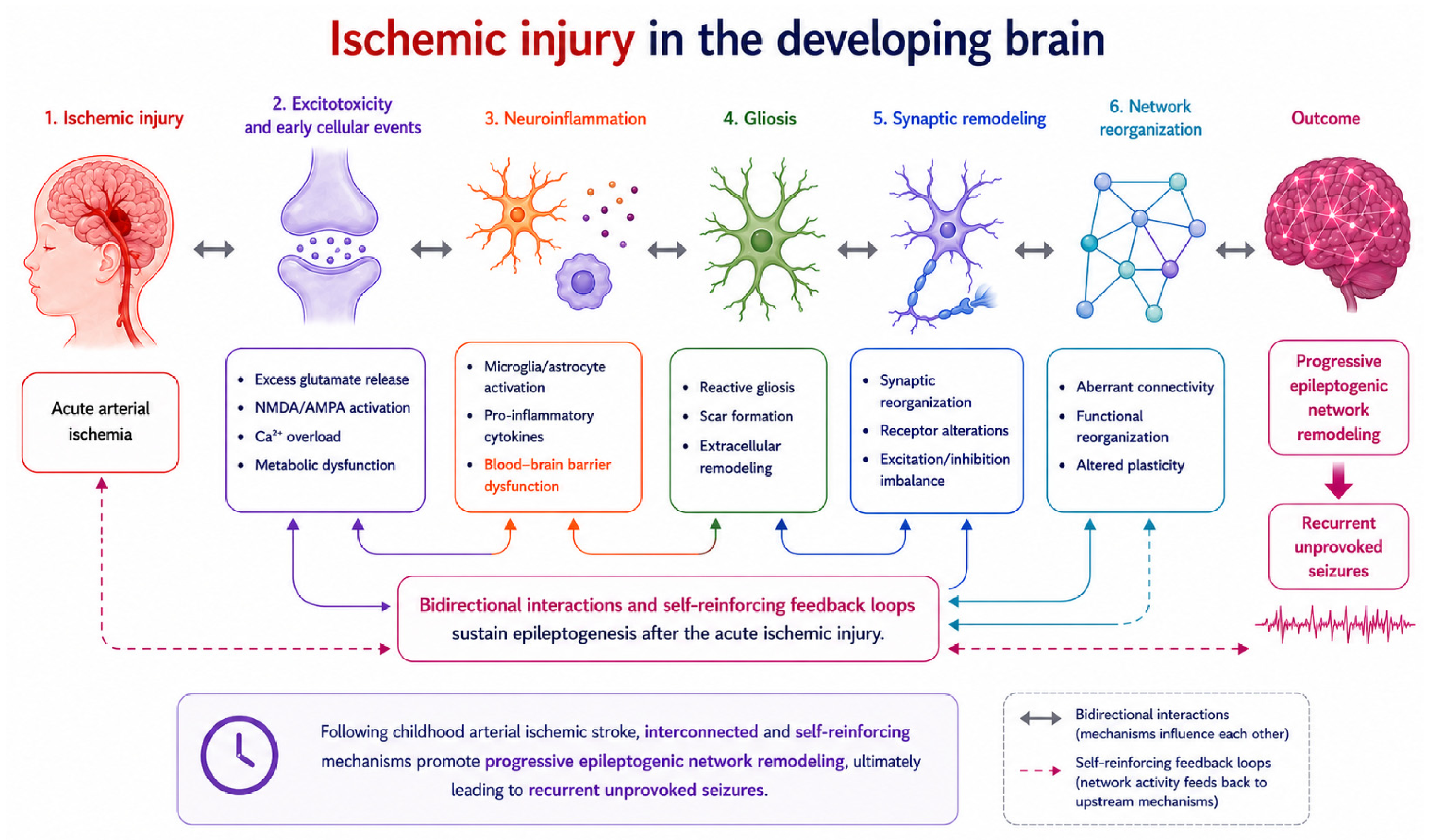
Very early seizures are thought to reflect the complex cascade triggered by cerebral ischemia, including excitotoxic neuronal injury, disruption of ionic homeostasis, metabolic dysfunction, mitochondrial impairment, and activation of inflammatory pathways. Experimental studies suggest that excessive glutamate release, ATP depletion, oxidative stress, and impaired astrocyte–neuron metabolic coupling contribute to persistent alterations in neuronal excitability and maladaptive synaptic remodeling, thereby facilitating epileptogenesis [
26]. In parallel, spreading and anoxic depolarizations increase metabolic demand in vulnerable peri-infarct tissue, potentially amplifying excitotoxic and inflammatory responses and promoting network remodeling [
27]. Ischemic injury also induces a sustained neuroinflammatory response characterized by activation of microglia and astrocytes, release of pro-inflammatory cytokines and recruitment of peripheral immune cells [
28]. Experimental and clinical evidence further suggests that BBB dysfunction may directly promote neuronal hyperexcitability through the extravasation of serum proteins and activation of astrocytic signaling pathways, providing an additional mechanism linking acute ischemic injury to epileptogenesis [
29]. Although these mechanisms provide a biologically plausible framework linking acute ischemic injury to post-stroke epileptogenesis, direct evidence supporting their specific contribution to pediatric post-stroke epilepsy remains limited.
In the developing brain, these processes may further interact with age-dependent neuronal plasticity and the ongoing maturation of inhibitory circuits, thereby facilitating maladaptive synaptic reorganization and long-term hyperexcitability. These developmental mechanisms are particularly relevant during the neonatal period, when reduced KCC2 expression, immature chloride homeostasis, and depolarizing GABAergic signaling contribute to increased seizure susceptibility [
30,
31]. In contrast, in post-neonatal childhood AIS, epileptogenesis is more likely to be driven by cortical injury, excitotoxicity, neuroinflammation, and maladaptive network remodeling, although developmental factors may continue to influence individual vulnerability. Accordingly, mechanistic findings derived from neonatal stroke models should not be directly extrapolated to childhood AIS without considering age-dependent differences in brain maturation, inhibitory neurotransmission, and epileptogenic pathways. Original observational studies have consistently shown a dose–response relationship between early seizure burden and subsequent epilepsy risk, with prolonged seizures and acute status epilepticus conferring the highest risk of later recurrent unprovoked seizures [
6,
15,
16]. These findings support the hypothesis that sustained early neuronal hyperexcitability and more severe cortical dysfunction may contribute to progressive epileptogenic network remodeling after pediatric stroke. Furthermore, prospective population-based studies suggest that the epileptogenic impact of acute seizures may evolve over time, with limited predictive value during early follow-up but significantly increased association with epilepsy at longer follow-up intervals, underscoring the progressive nature of post-stroke network remodeling in the developing brain [
17].
Younger age at stroke onset has been associated with an increased risk of PSE in several cohorts, although interpretation of this finding is complicated by the frequent inclusion of neonatal and childhood stroke populations within the same studies [
3,
14,
15].
A major limitation of the available literature is that several original cohort studies combine neonatal, presumed perinatal, and childhood stroke populations without performing age-stratified analyses [
3,
16]. This methodological heterogeneity complicates the interpretation of epilepsy risk because neonatal and childhood AIS differ substantially in developmental neurobiology, seizure susceptibility, underlying etiologies, and mechanisms of epileptogenesis [
19,
20,
21]. As a result, combining these populations within the same analyses may mask age-specific risk factors and partially explain the wide variability in reported epilepsy rates across studies.
Neonates exhibit distinct developmental neurobiology characterized by enhanced excitatory neurotransmission, immature inhibitory GABAergic systems, and unique patterns of cortical connectivity, all of which influence both seizure susceptibility and long-term epileptogenic potential. In addition, neonatal strokes are frequently related to perinatal hemodynamic disturbances, placental disorders, or perinatal thrombosis, whereas childhood strokes more commonly involve arteriopathies, cardiac disease, inflammatory disorders, or prothrombotic conditions [
19,
20,
21]. Consequently, combining these populations within single analyses may obscure age-specific predictors of epileptogenesis and contribute significantly to the wide variability in reported cumulative incidences of PSE across studies.
Despite these limitations, more recent cohort studies that separately analyzed neonatal and childhood stroke suggest that both shared and age-specific mechanisms contribute to epileptogenesis. Across pediatric age groups, acute symptomatic seizures, cortical involvement, multifocal infarctions, and abnormal neurological status at hospital discharge consistently emerge as the strongest independent predictors of PSE [
16]. However, the impact of these factors may differ across developmental stages, suggesting that epileptogenesis after pediatric stroke is not a uniform process but rather one that is influenced by brain maturation and age-related biological factors. These observations underscore the importance of age-stratified analyses and caution against considering younger age as an independent risk factor without accounting for the underlying clinical and neuroimaging characteristics of the stroke.
An important limitation of the available evidence is the substantial heterogeneity across pediatric stroke studies, which limits the direct comparability of reported incidence estimates and risk factors.
Studies differ in patient age and composition, with some cohorts combining neonatal, presumed perinatal, and childhood stroke populations, lesion characteristics, duration of follow-up, and definitions and ascertainment of post-stroke epilepsy.
In addition, much of the available evidence derives from retrospective, single-center studies with relatively small sample sizes, limiting the generalizability of their findings. Consequently, although several clinical and neuroimaging factors have been consistently associated with PSE, the strength and independence of these associations should be interpreted cautiously, particularly for factors supported by a limited number of heterogeneous studies.
Neuroimaging findings play a key role in assessing epilepsy risk after pediatric AIS. Among neuroimaging features, cortical involvement is the most consistently identified predictor of post-stroke epilepsy (PSE) following childhood AIS, as demonstrated by original pediatric cohort studies [
4,
6,
9,
15,
16]. Cortical injury is thought to increase epileptogenic risk by disrupting both local cortical circuits and large-scale functional brain networks, thereby promoting maladaptive synaptic remodeling and persistent neuronal hyperexcitability. Moreover, cortical lesions may directly impair the balance between excitatory and inhibitory neuronal activity while altering long-range structural and functional connectivity, ultimately facilitating the development of epileptogenic networks.
Likewise, infarctions affecting the anterior circulation, particularly within the middle cerebral artery territory, have been linked to an increased risk of epilepsy, probably owing to the involvement of highly interconnected cortical regions responsible for sensorimotor, language, and higher-order cognitive functions [
4,
6,
9]. These lesions may facilitate widespread cortical network dysfunction and maladaptive synaptic reorganization, thereby promoting chronic hyperexcitability. Beyond lesion location, the extent and spatial distribution of ischemic injury also appear to substantially influence epileptogenic risk. Several studies have shown that multifocal infarctions and greater lesion burden are associated with higher rates of PSE, particularly when cortical regions are involved, suggesting a cumulative effect of distributed cortical injury on epileptogenic network remodeling, although direct mechanistic evidence in pediatric populations remains limited [
2,
6,
7,
24]. Indeed, emerging evidence indicates that even relatively small infarcts may confer significant epilepsy risk when lesions are multifocal or strategically located within highly interconnected cortical regions, emphasizing the importance of lesion topology and network involvement rather than infarct volume alone [
15,
16]. These observations support the hypothesis of a network-based model of post-stroke epileptogenesis in childhood, in which distributed cortical injury, altered interhemispheric connectivity, and maladaptive large-scale network reorganization may contribute to the progressive development of chronic cortical hyperexcitability and recurrent unprovoked seizures. However, although this model provides a biologically plausible framework, direct evidence in childhood AIS remains limited and is largely inferred from experimental studies and adult stroke populations.
Stroke etiology may also influence epileptogenic risk after pediatric AIS, although the available evidence remains heterogeneous. Original pediatric cohort studies suggest that inflammatory, infectious, and arteriopathic stroke mechanisms—particularly focal cerebral arteriopathy (FCA)—may be associated with an increased risk of PSE, potentially reflecting the contribution of persistent neuroinflammatory activation, blood–brain barrier dysfunction, and ongoing vascular injury to epileptogenesis [
4,
8,
16]. In particular, FCA has emerged as one of the etiological subtypes most frequently associated with PSE, although this association is based primarily on retrospective studies and requires confirmation in larger prospective cohorts. Mechanistically, inflammatory arteriopathies may promote chronic cortical instability through recurrent ischemic injury, endothelial dysfunction, blood–brain barrier disruption, and sustained inflammatory signaling.
Interestingly, despite representing one of the most common etiological categories of childhood AIS, congenital and acquired cardiac disorders have not been consistently identified as independent predictors of PSE in pediatric cohorts. Any apparent association is likely mediated by stroke-related factors, including cortical involvement, lesion burden, multifocal infarctions, and recurrent ischemic events [
2,
4,
14].
Interpretation of the current evidence is further complicated by the inclusion of mixed cerebrovascular populations in several pediatric cohorts and meta-analyses, including both arterial ischemic and hemorrhagic stroke without separate subgroup analyses. Given the important differences in neurobiological mechanisms, acute seizure propensity, and long-term epileptogenic pathways between ischemic and hemorrhagic brain injury, this heterogeneity likely represents a significant source of variability across studies.
Although EEG abnormalities have been proposed as potential biomarkers of epileptogenic risk, the available pediatric evidence remains limited and heterogeneous, precluding definitive conclusions regarding their independent prognostic value [
4,
5,
8]. Original pediatric studies evaluating EEG after AIS remain relatively limited and heterogeneous, with important differences in acquisition protocols, timing of recording, and availability of continuous EEG monitoring. Consequently, EEG abnormalities have not been consistently identified as independent predictors of PSE across pediatric cohorts. This variability was also highlighted by the recent systematic review by Alqahtani and Makke, in which EEG findings could not be confirmed as independent predictors of epilepsy [
8].
Continuous EEG monitoring may improve the detection of electrographic-only seizures and non-convulsive status epilepticus, particularly in critically ill children with persistent altered mental status or unexplained encephalopathy, in whom clinical seizure recognition may be challenging [
14,
22,
24]. This issue is particularly relevant in pediatric stroke because unrecognized electrographic seizures and non-convulsive status epilepticus may increase overall seizure burden and potentially contribute to worse neurological outcomes [
23,
32]. Nevertheless, systematic cEEG monitoring has not been consistently implemented in most pediatric stroke cohorts, limiting accurate estimation of electrographic seizure burden and its relationship with long-term epileptogenesis. In children with acute AIS, cEEG should be considered in the presence of persistent impairment of consciousness, failure to return to neurological baseline after a clinical seizure, suspected non-convulsive status epilepticus, recurrent subtle paroxysmal events of uncertain nature, ongoing status epilepticus, or when sedation or pharmacological paralysis may obscure clinical seizure activity [
22]. Although at least 24 h of monitoring is generally recommended in critically ill patients, the optimal duration and independent prognostic value of electrographic seizure burden specifically in childhood AIS remain uncertain because cEEG has not been systematically implemented across pediatric stroke cohorts [
8,
14,
22]. Importantly, the prognostic value of electrographic seizure burden for the subsequent development of PSE remains uncertain because cEEG use, timing, and duration have varied considerably across pediatric stroke cohorts. Therefore, prospective studies using standardized monitoring protocols are needed to determine whether electrographic seizure burden independently predicts long-term epileptogenesis. Beyond seizure detection, EEG may also provide insight into mechanisms of network dysfunction after ischemic injury. Persistent focal slowing, abnormal background organization, and epileptiform discharges may reflect disrupted cortical connectivity, impaired maturation of neuronal networks, and maladaptive synaptic reorganization within structurally injured regions, processes that are thought to contribute to post-stroke epileptogenesis [
7,
12].
Greater stroke severity and persistent neurological deficits have been consistently identified as robust clinical predictors of PSE following childhood AIS [
14,
15,
16,
24]. These associations likely reflect more extensive and functionally disruptive brain injury, serving as indirect markers of widespread cortical damage, impaired network recovery, and increased epileptogenic potential.
Children who develop PSE consistently experience poorer long-term neurological outcomes than those who remain seizure-free, suggesting that epileptogenesis may represent both a consequence and a marker of more severe underlying brain injury and network disruption after pediatric AIS [
2,
3,
17,
24].
Although PSE has been consistently associated with poorer overall functional outcomes, its independent contribution to long-term cognitive, neuropsychological, behavioral, and academic impairment remains insufficiently characterized. Most pediatric stroke studies have focused primarily on global neurological disability and functional status, whereas detailed longitudinal assessments of cognition, executive functions, psychosocial well-being, and educational achievement remain scarce [
33]. Consequently, the extent to which PSE independently contributes to long-term neuropsychological sequelae, beyond the effects of the initial ischemic injury, remains an important area for future investigation.
In addition to established clinical and neuroimaging risk factors, emerging population-based data suggest that inherited biological susceptibility may contribute to the risk of PSE; however, direct evidence from pediatric genetic studies remains limited, and these findings require further validation. Large nationwide registry studies have reported an increased prevalence of epilepsy among first-degree relatives of affected children, supporting the hypothesis that inherited biological factors may modify individual susceptibility to epileptogenesis after stroke. These findings raise the prospect that genetic factors, or other inherited mechanisms influencing neuronal excitability and brain resilience, may contribute to the risk of developing epilepsy after pediatric stroke [
1]. In this context, ischemic injury may act as a precipitating factor capable of unmasking latent epileptogenic predisposition in susceptible individuals [
25]. These observations are consistent with emerging concepts of precision medicine, in which genetic susceptibility may interact with acquired brain injury to determine individual epileptogenic risk.
Emerging evidence suggests that modifiable biological factors may also contribute to epileptogenic risk after pediatric stroke. Low serum vitamin D levels have recently been proposed as a potential risk factor, but current evidence is based on a limited number of observational studies and should be considered preliminary pending confirmation in prospective multicenter cohorts [
15,
18]. Although the underlying mechanisms remain incompletely understood, vitamin D is known to exert neuroprotective, anti-inflammatory, antioxidant, and immunomodulatory effects, and may influence neuronal excitability and post-ischemic network remodeling. These observations raise the possibility that part of the biological vulnerability to post-stroke epileptogenesis could be potentially modifiable.
Despite these advances, important knowledge gaps remain regarding the complex interplay between genetic susceptibility, inflammatory pathways, metabolic factors, and ischemia-induced network reorganization in pediatric PSE. Future prospective multicenter studies will be essential to better define the mechanisms underlying epileptogenesis and improve risk stratification.
Overall, the current evidence supports acute symptomatic seizures, status epilepticus, cortical involvement, and persistent neurological deficits as the most robust clinical predictors of PSE. By contrast, the prognostic value of lesion burden, EEG abnormalities, inflammatory biomarkers, vitamin D deficiency, genetic susceptibility, and network-based imaging markers remains promising but requires confirmation in larger prospective pediatric studies.
Finally, as this manuscript is a narrative review, the selection and synthesis of the available literature are inherently subject to methodological limitations, and no quantitative pooling or formal comparison of effect estimates across studies was performed. Prospective multicenter studies using standardized definitions, follow-up protocols, and outcome ascertainment are needed to better establish the incidence of PSE and validate potential risk factors across pediatric populations.
4.1. Clinical Implications
The consistently high incidence and long-term persistence of epilepsy risk after pediatric AIS underscore the need for prolonged neurological follow-up [
1,
2,
6,
8]. Data from pediatric cohorts indicate that acute symptomatic seizures may predict PSE over longer time horizons, even when short-term prediction is less robust, supporting the need for sustained follow-up beyond the early months after stroke [
6,
8,
17].
Children presenting with early seizures, cortical involvement, severe stroke, multifocal infarctions, focal cerebral arteriopathy, or persistent neurological deficits should be considered at particularly high risk and may benefit from closer surveillance, including targeted EEG monitoring [
2,
6,
8,
15,
16,
17,
24]. Improved risk stratification may facilitate family counseling, individualized follow-up strategies, and the design of future interventional studies aimed at preventing epileptogenesis.
Importantly, pediatric cohorts demonstrate that PSE is associated with worse neurological, motor, and cognitive outcomes after childhood AIS, further emphasizing the clinical value of early identification and longitudinal care pathways [
2,
24]. The identification of potentially modifiable risk factors may offer opportunities to improve long-term outcomes after pediatric stroke. In particular, strategies such as optimizing the management of acute symptomatic seizures and addressing vitamin D deficiency have emerged as potential targets for intervention, although prospective evidence supporting their effectiveness remains limited [
8,
15,
18].
Finally, growing evidence suggests that multimodal approaches integrating clinical features, EEG findings, neuroimaging markers, and biological factors may improve individualized risk prediction and support the development of future anti-epileptogenic strategies in pediatric stroke survivors [
8,
12].
Future studies integrating clinical, neuroimaging, and genetic data may help clarify individual vulnerability to PSE and improve long-term risk stratification. In particular, multimodal approaches that integrate advanced neuroimaging, electrophysiological biomarkers, inflammatory markers, and predictive computational models may improve the early identification of children at greatest risk of developing PSE. Advanced neuroimaging techniques, including structural and functional network-based MRI analyses, may provide valuable insights into patterns of cortical disconnection and maladaptive network reorganization that contribute to epileptogenesis following pediatric AIS. Similarly, quantitative EEG features, electrographic seizures, and early epileptiform abnormalities may represent promising biomarkers of cortical hyperexcitability and long-term epilepsy risk, although prospective pediatric studies using standardized EEG protocols remain limited. Growing evidence also supports the potential contribution of inflammatory and metabolic pathways to post-stroke epileptogenesis. Future investigations should therefore explore the role of circulating inflammatory biomarkers, blood–brain barrier dysfunction, genetic susceptibility, and modifiable biological factors such as vitamin D deficiency in determining individual epileptogenic vulnerability. Integration of genomic and molecular profiling may further improve understanding of shared susceptibility mechanisms linking stroke and epilepsy in childhood.
Prospective multicenter pediatric stroke registries incorporating standardized EEG acquisition, advanced neuroimaging protocols, and biological sampling will be essential to validate candidate biomarkers of epileptogenesis and to improve the reproducibility and generalizability of risk prediction models across pediatric populations.
In parallel, emerging artificial intelligence and machine learning approaches may facilitate development of predictive models integrating clinical variables, lesion topology, EEG characteristics, and multimodal imaging data to improve early risk stratification and individualized follow-up strategies. Ultimately, better characterization of epileptogenic pathways after stroke in children may support the development of targeted anti-epileptogenic interventions aimed at preventing or reducing long-term epilepsy burden in high-risk children.
4.2. Limitations
The findings summarized in this review should be interpreted in light of several limitations. First, most of the available evidence originates from retrospective observational studies with relatively small sample sizes, limiting statistical power and reducing the generalizability of the reported findings. Second, substantial heterogeneity exists across studies regarding patient age, stroke etiology, neuroimaging characteristics, follow-up duration, and definitions of acute symptomatic seizures and post-stroke epilepsy, making direct comparisons challenging. Third, EEG acquisition protocols, including the use and duration of continuous EEG monitoring, were inconsistently applied, likely resulting in under-recognition of electrographic seizures and limiting evaluation of their prognostic significance. Furthermore, most proposed biological, electrophysiological, and neuroimaging biomarkers remain supported by limited observational evidence and require prospective validation. Finally, as this is a narrative review, a formal systematic methodology, risk-of-bias assessment, and quantitative meta-analysis were not performed; therefore, some degree of selection bias cannot be excluded.