Neuropsychological Sequelae and Neuroradiological Correlates of Arachnoid Cysts in Adults: A Systematic Review


 and
and
Abstract
1. Introduction
2. Materials and Methods
2.1. Search Strategy
2.2. Eligibility Criteria
2.3. Study Selection
2.4. Data Extraction and Synthesis
2.5. Risk of Bias and Methodological Quality Assessment
3. Results
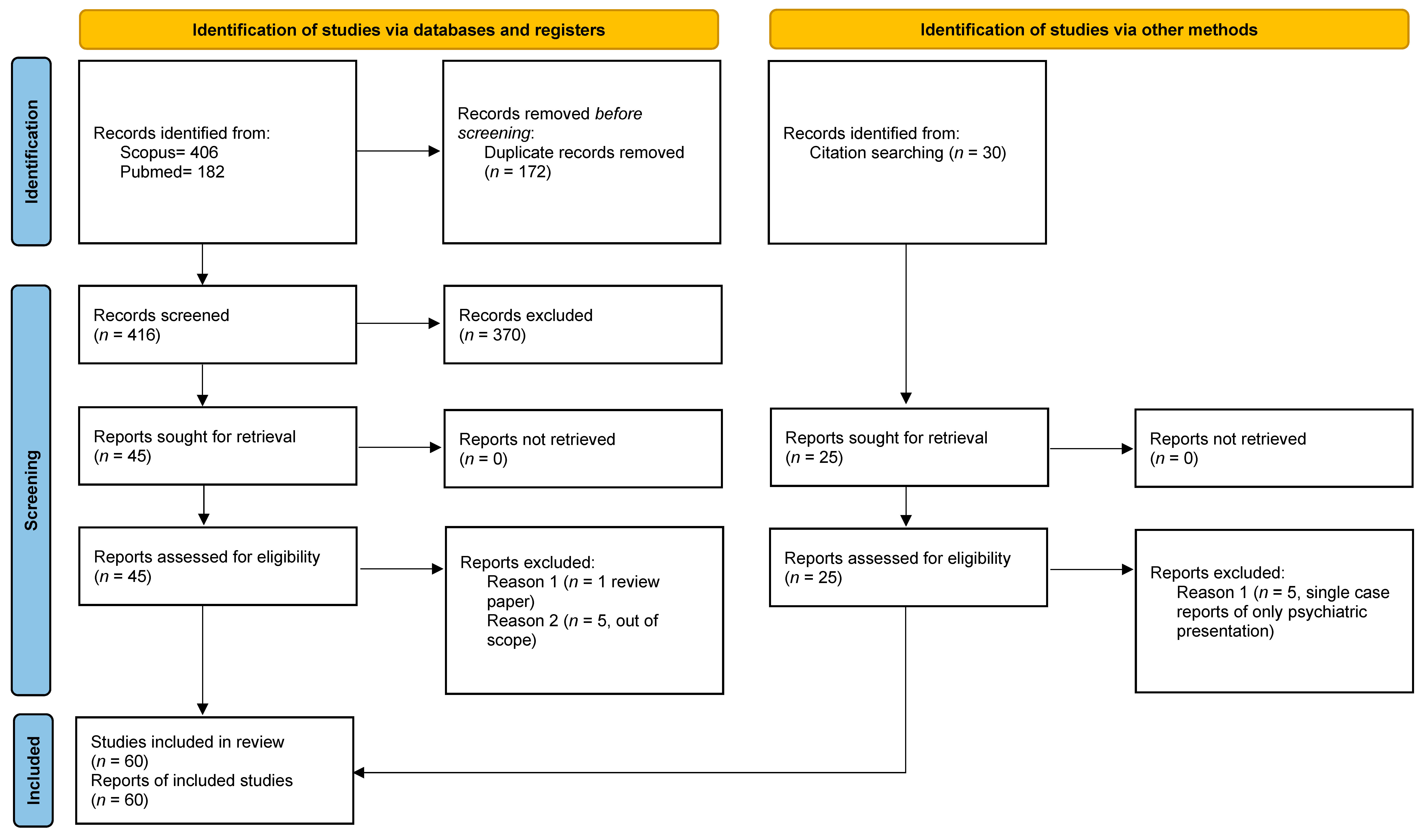
3.1. Study Selection and Characteristics
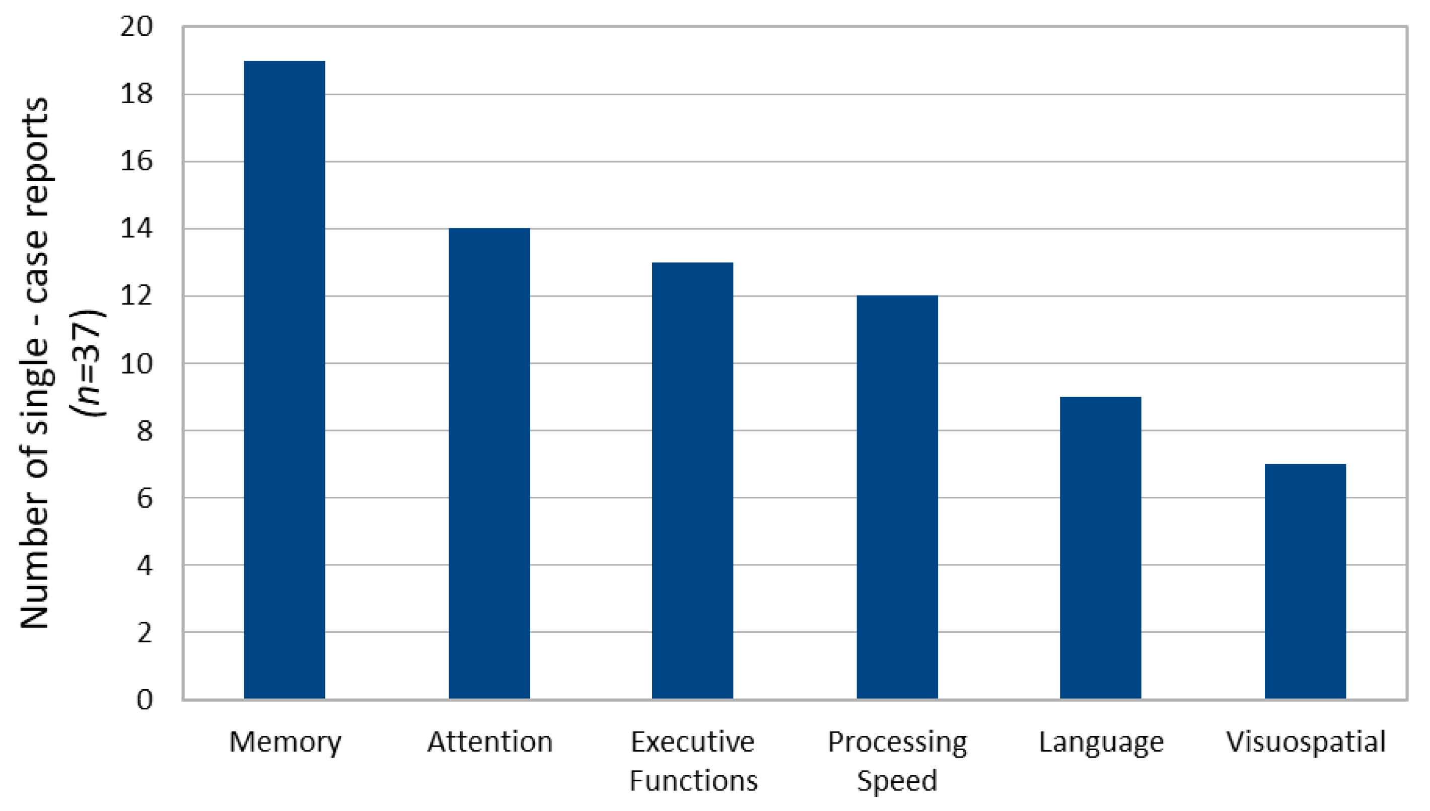
3.2. Single Case Reports
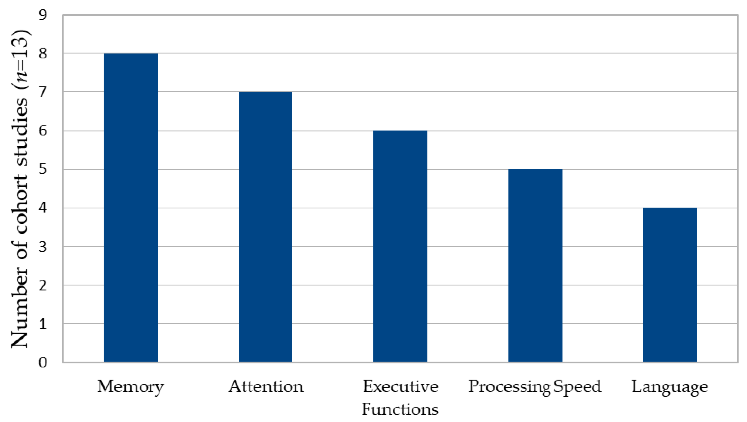
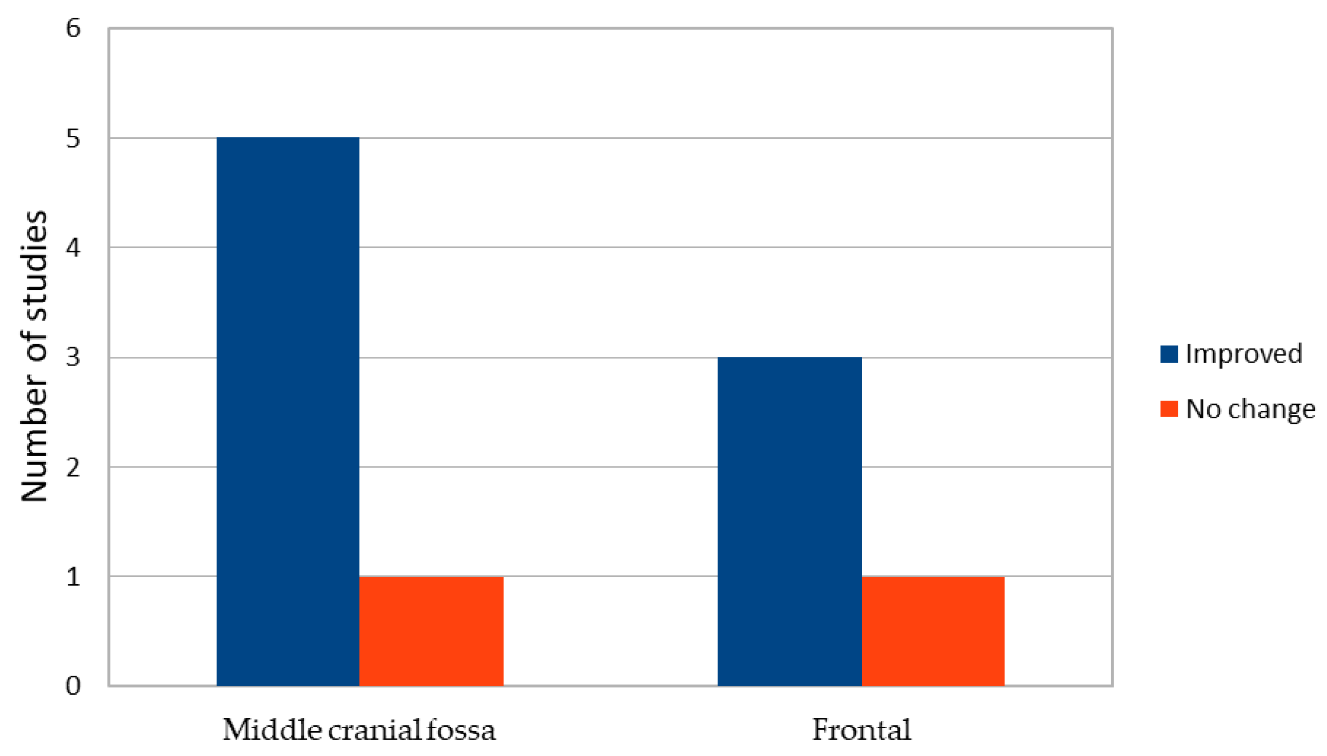
3.3. Case Series and Group Studies
3.4. Functional Neuroimaging Studies
4. Discussion
4.1. Cognitive Profiles and Anatomical Correlates
4.2. Evidence for Reversibility and Surgical Effects
4.3. Functional Neuroimaging and Pathophysiological Mechanisms
4.4. Compensation, Cognitive Reserve, and Threshold Effects
4.5. Clinical and Research Implications
4.6. Limitations
5. Conclusions
Supplementary Materials
Author Contributions
Funding
Data Availability Statement
Acknowledgments
Conflicts of Interest
References
- Cincu, R.; Agrawal, A.; Eiras, J. Intracranial arachnoid cysts: Current concepts and treatment alternatives. Clin. Neurol. Neurosurg. 2007, 109, 837–843. [Google Scholar] [CrossRef] [Scilit]
- Wester, K. Intracranial arachnoid cysts—Do they impair mental functions? J. Neurol. 2008, 255, 1113–1120. [Google Scholar] [CrossRef] [Scilit] [PubMed]
- Rabiei, K.; Jaraj, D.; Marlow, T.; Jensen, C.; Skoog, I.; Wikkelsø, C. Prevalence and symptoms of intracranial arachnoid cysts: A population-based study. J. Neurol. 2016, 263, 689–694. [Google Scholar] [CrossRef] [Scilit] [PubMed]
- Al-Holou, W.N.; Terman, S.; Kilburg, C.; Garton, H.J.L.; Muraszko, K.M.; Maher, C.O. Prevalence and natural history of arachnoid cysts in adults. J. Neurosurg. 2013, 118, 222–231. [Google Scholar] [CrossRef] [Scilit] [PubMed]
- Weber, F.; Knopf, H. Incidental findings in magnetic resonance imaging of the brains of healthy young men. J. Neurol. Sci. 2006, 240, 81–84. [Google Scholar] [CrossRef] [Scilit]
- Massimi, L.; Caldarelli, M.; Di Rocco, C. Intracranial congenital arachnoid cysts. In Textbook of Pediatric Neurosurgery; Springer: Cham, Switzerland, 2020. [Google Scholar]
- Wester, K.; Hugdahl, K. Intracranial arachnoid cysts and mental functions. In Arachnoid CystsClinical and Surgical Management; Wester, K., Ed.; Springer: Cham, Switzerland, 2018. [Google Scholar]
- Arai, H.; Sato, K.; Wachi, A.; Okuda, O.; Takeda, N. Arachnoid cysts of the middle cranial fossa: Experience with 77 patients. Neurosurgery 1996, 39, 1108–1113. [Google Scholar] [CrossRef] [Scilit]
- Lang, W.; Lang, M.; Kornhuber, A.; Gallwitz, A.; Kriebel, J. Neuropsychological and neuroendocrinological disturbances associated with extracerebral cysts of the anterior and middle cranial fossa. Eur. Arch. Psychiatry Neurol. Sci. 1985, 235, 38–41. [Google Scholar] [CrossRef] [Scilit]
- Raeder, M.B.; Helland, C.A.; Hugdahl, K.; Wester, K. Arachnoid cysts cause cognitive deficits that improve after surgery. Neurology 2005, 64, 160–162. [Google Scholar] [CrossRef] [Scilit]
- Gjerde, P.; Schmid, M.; Hammar, Å.; Wester, K. Intracranial arachnoid cysts: Impairment of higher cognitive functions and postoperative improvement. J. Neurodev. Disord. 2013, 5, 21. [Google Scholar] [CrossRef] [Scilit]
- Hodges, J.R. Pure apraxic agraphia with recovery after drainage of a left frontal cyst. Cortex 1991, 27, 469–473. [Google Scholar] [CrossRef] [Scilit]
- Soukup, V.M.; Patterson, J.; Trier, T.T.; Chen, J.W. Cognitive improvement despite minimal arachnoid cyst decompression. Brain Dev. 1998, 20, 589–593. [Google Scholar] [CrossRef] [Scilit]
- Lebowitz, B.K.; Schefft, B.K.; Testa, S.M.; Patton, J.A.; Yeh, H.S. Neurocognitive sequelae of a giant arachnoid cyst: Case study. Neurocase 2006, 12, 339–345. [Google Scholar] [CrossRef] [Scilit]
- Miskey, H.M.; Gross, P.L. Neuropsychological assessment of a veteran with a large arachnoid cyst. Appl. Neuropsychol. Adult 2016, 23, 464–470. [Google Scholar] [CrossRef] [Scilit] [PubMed]
- Dunn, C.B.; Price, C.C.; Schwab, N.A.; Wagle Shukla, A. Arachnoid cyst masquerading as corticobasal degeneration. Clin. Neuropsychol. 2012, 26, 1342–1357. [Google Scholar] [CrossRef] [Scilit] [PubMed]
- Wester, K.; Hugdahl, K. Arachnoid cysts of the left temporal fossa: Impaired preoperative cognition and postoperative improvement. J. Neurol. Neurosurg. Psychiatry 1995, 59, 293–298. [Google Scholar] [CrossRef] [Scilit] [PubMed]
- Gundersen, H.; Helland, C.; Raeder, M.; Hugdahl, K.; Wester, K. Visual attention in patients with intracranial arachnoid cysts. J. Neurol. 2007, 254, 60–66. [Google Scholar] [CrossRef] [Scilit]
- Torgersen, J.; Helland, C.; Flaatten, H.; Wester, K. Reversible dyscognition in patients with a unilateral, middle fossa arachnoid cyst revealed by using a laptop based neuropsychological test battery (CANTAB). J. Neurol. 2010, 257, 1909–1916. [Google Scholar] [CrossRef] [Scilit]
- Agopian-Dahlenmark, L.; Mathiesen, T.; Bergendal, Å. Cognitive dysfunction and subjective symptoms in patients with arachnoid cyst before and after surgery. Acta Neurochir. 2020, 162, 1041–1050. [Google Scholar] [CrossRef] [Scilit]
- Rabiei, K.; Hellström, P.; Högfeldt-Johansson, M.; Tisell, M. Does subjective improvement in adults with intracranical arachnoid cyst justify surgical treatment? J. Neurosurg. 2018, 128, 250–257. [Google Scholar] [CrossRef] [Scilit]
- Stowe, L.A.; Go, K.G.; Pruim, J.; Dunnen, W.D.; Meiners, L.; Paans, A.J. Language localization in cases of left temporal lobe arachnoid cysts: Evidence against interhemispheric reorganization. Brain Lang. 2000, 75, 347–358. [Google Scholar] [CrossRef] [Scilit]
- Hund-Georgiadis, M.; von Cramon, D.Y.; Kruggel, F.; Preul, C. Do quiescent arachnoid cysts alter CNS functional organization? A fMRI and morphometric study. Neurology 2002, 59, 1935–1939. [Google Scholar] [CrossRef] [Scilit] [PubMed]
- Nickel, J.; Jörgens, S.; Bussfeld, P.; Seitz, R.J. Asymptomatic motor cortex displacement due to a giant arachnoid cyst. Neurocase 2007, 13, 328–333. [Google Scholar] [CrossRef] [Scilit] [PubMed]
- Alkadhi, H.; Crelier, G.R.; Imhof, H.G.; Kollias, S.S. Somatomotor functional MRI in a large congenital arachnoid cyst. Neuroradiology 2003, 45, 153–156. [Google Scholar] [CrossRef] [Scilit] [PubMed]
- Martínez-Lage, J.F.; Valentí, J.A.; Piqueras, C.; Ruiz-Espejo, A.M.; Román, F.; de la Rosa, J.A.N. Functional assessment of intracranial arachnoid cysts with TC99 m-HMPAO SPECT: A preliminary report. Childs Nerv. Syst. 2006, 22, 1091–1097. [Google Scholar] [CrossRef] [Scilit]
- Bohnen, N.I.; Haugen, J.; Kluin, K.; Kotagal, V. Motor speech apraxia in a 70-year-old man with left dorsolateral frontal arachnoid cyst: A [18F] FDG-PET-CT study. Case Rep. Neurol. Med. 2016, 2016, 8941035. [Google Scholar] [CrossRef] [Scilit]
- Taskapilioglu, O.; Taskapilioglu, M.O.; Hakyemez, B.; Bekar, A. Brain function—Not size—Is important. Eur. Res. J. 2017, 3, 273–275. [Google Scholar] [CrossRef] [Scilit]
- Page, M.J.; McKenzie, J.E.; Bossuyt, P.M.; Boutron, I.; Hoffmann, T.C.; Mulrow, C.D.; Shamseer, L.; Tetzlaff, J.M.; Akl, E.A.; Brennan, S.E.; et al. The PRISMA 2020 statement: An updated guideline for reporting systematic reviews. BMJ 2021, 372, n71. [Google Scholar] [CrossRef] [Scilit]
- Moola, S.; Munn, Z.; Tufanaru, C.; Aromataris, E.; Sears, K.; Sfetic, R.; Currie, M.; Lisy, K.; Qureshi, R.; Mattis, P.; et al. Chapter 7Systematic reviews of etiology and risk. In JBI Manual for Evidence Synthesis; Aromataris, E., Munn, Z., Eds.; JBI: Adelaide, Australia, 2020. [Google Scholar]
- Munn, Z.; Barker, T.H.; Moola, S.; Tufanaru, C.; Stern, C.; McArthur, A.; Stephenson, M.; Aromataris, E. Methodological quality of case series studies: An introduction to the JBI critical appraisal tool. JBI Evid. Synth. 2020, 18, 2127–2133. [Google Scholar] [CrossRef] [Scilit]
- Wells, G.A.; Shea, B.; O’Connell, D.; Peterson, J.; Welch, V.; Losoa, M.; Tugwell, P. The Newcastle–Ottawa Scale (NOS) for Assessing the Quality of Non-Randomised Studies in Meta-Analyses; Ottawa Hospital Research Institute: Ottawa, ON, Canada, 2000. [Google Scholar]
- Babinsky, R.; Maier, B.; Calabrese, P.; Markowitsch, H.J.; Gehlen, W. Atraumatic temporal lobe pathology and autobiographical memory. Eur. Neurol. 1994, 34, 290–291. [Google Scholar] [CrossRef] [Scilit]
- Blackshaw, S.; Bowen, R.C. A case of atypical psychosis associated with alexithymia and a left fronto-temporal lesion: Possible correlations. Can. J. Psychiatry 1987, 32, 688–692. [Google Scholar] [CrossRef] [Scilit] [PubMed]
- Boomkens, H.R.; van der Cammen, T.J.; Mattace-Raso, F.U. Primary arachnoid cyst mimicking depressive symptoms in an elderly woman. Int. Psychogeriatr. 2010, 22, 832–833. [Google Scholar] [CrossRef] [Scilit] [PubMed]
- Cullum, C.M.; Heaton, R.K.; Harris, M.J.; Jeste, D.V. Neurobehavioral and neurodiagnostic aspects of late-onset psychosis. Arch. Clin. Neuropsychol. 1994, 9, 371–382. [Google Scholar] [CrossRef] [Scilit] [PubMed]
- Da Silva, J.A.; Alves, A.; Talina, M.; Carreiro, S.; Guimarães, J.; Xavier, M. Arachnoid cyst in a patient with psychosis: Case report. Ann. Gen. Psychiatry 2007, 6, 16. [Google Scholar] [CrossRef] [Scilit]
- Das, S.; Kartha, A.; Purushothaman, S.T.; Rajan, V. Arachnoid cyst and psychosis: The troublemaker or innocent bystander. Indian J. Psychol. Med. 2017, 39, 194–195. [Google Scholar] [CrossRef] [Scilit]
- Geniş, B.; Coşar, B. A case of arachnoid cyst presenting with cognitive impairment and hypomania symptoms. Turk. Psikiyatri Derg. 2020, 31, 131–136. [Google Scholar] [CrossRef] [Scilit]
- Heidrich, A.; Schmidtke, A.; Lesch, K.P.; Hofmann, E.; Becker, T. Cerebellar arachnoid cyst in a firesetter: The weight of organic lesions in arson. J. Psychiatry Neurosci. 1996, 21, 202–206. [Google Scholar]
- Kotil, K.; Balci, N.; Bilge, T. Intracranial symptomatic giant arachnoid cyst of the interhemispheric fissure presenting with frontal lobe syndrome. Turk. Neurosurg. 2007, 17, 147–151. [Google Scholar]
- Mormont, E.; Dorban, S.; Gustin, T. Parkinsonism and cognitive impairment due to a giant frontal arachnoid cyst improving after shunting. Acta Neurol. Belg. 2017, 117, 347–349. [Google Scholar] [CrossRef] [Scilit]
- Nuara, A.; Nicolini, Y.; D’Orio, P.; Cardinale, F.; Rizzolatti, G.; Avanzini, P.; Fabbri-Destro, M.; De Marco, D. Catching the imposter in the brain: The case of Capgras delusion. Cortex 2020, 131, 295–304. [Google Scholar] [CrossRef] [Scilit]
- Paradis, C.M.; Horn, L.; Lazar, R.M.; Schwartz, D.W. Brain dysfunction and violent behavior in a man with a congenital subarachnoid cyst. Hosp. Community Psychiatry 1994, 45, 714–716. [Google Scholar] [CrossRef] [Scilit] [PubMed]
- Patrikelis, P.; Korfias, S.; Messinis, L.; Kossivas, A.; Zournatzidi, C.; Alexoudi, A.; Lucci, G.; Gatzonis, S.; Nasios, G. Chronic hematoma superimposed on a large frontotemporal arachnoid cyst: Evidence against crowding of audioverbal memory. Neurocase 2022, 28, 337–343. [Google Scholar] [CrossRef] [Scilit] [PubMed]
- Raj, R.; Venkatanarayan, A.; Sharayah, A.; Ross, D. Giant left parietal lobe arachnoid cyst presenting as early-onset dementia. BMJ Case Rep. 2018, 2018, bcr2018224837. [Google Scholar] [CrossRef] [Scilit] [PubMed]
- Stracciari, A.; Ciucci, G.; Bissi, G. Transient global amnesia associated with a large arachnoid cyst of the middle cranial fossa. Ital. J. Neurol. Sci. 1987, 8, 609–611. [Google Scholar] [CrossRef] [Scilit]
- Tonna, M.; Ottoni, R.; Ossola, P.; De Panfilis, C.; Marchesi, C. Late-onset obsessive–compulsive disorder associated with left cerebellar lesion. Cerebellum 2014, 13, 531–535. [Google Scholar] [CrossRef] [Scilit]
- Tsurushima, H.; Harakuni, T.; Saito, A.; Tominaga, D.; Hyodo, A.; Yoshii, Y. Symptomatic arachnoid cyst of the left frontal convexity presenting with memory disturbance. Neurol. Med.-Chir. 2000, 40, 339–341. [Google Scholar] [CrossRef] [Scilit]
- Varshney, P.; Raghuraman, B.S.; Ganjekar, S.; Sharma, M.; Chaturvedi, S. Cerebellar arachnoid cyst masquerading as schizophrenia and later obsessive–compulsive disorder. Asian J. Psychiatry 2020, 47, 101838. [Google Scholar] [CrossRef] [Scilit]
- Wahl, A.S.; Löffler, M.; Hausner, L.; Ruttorf, M.; Nees, F.; Frölich, L. A giant arachnoid cyst masking Alzheimer’s disease. BMC Psychiatry 2019, 19, 274. [Google Scholar] [CrossRef] [Scilit]
- Zwagerman, N.T.; Pardini, J.; Mousavi, S.H.; Friedlander, R.M. A refractory arachnoid cyst presenting with tremor, expressive dysphasia, and cognitive decline. Surg. Neurol. Int. 2016, 7, S431–S433. [Google Scholar] [CrossRef] [Scilit]
- Bechter, K.; Wittek, R.; Seitz, K.; Antoniadis, G. Personality disorders improved after arachnoid cyst neurosurgery, then rediagnosed as ‘minor’ organic personality disorders. Psychiatry Res. 2010, 184, 196–200. [Google Scholar] [CrossRef] [Scilit]
- Guell, X.; Anteraper, S.A.; Ghosh, S.S.; Gabrieli, J.D.E.; Schmahmann, J.D. Neurodevelopmental and Psychiatric Symptoms in Patients with a Cyst Compressing the Cerebellum: An Ongoing Enigma. Cerebellum 2020, 19, 16–29. [Google Scholar] [CrossRef] [Scilit] [PubMed]
- Škarić, M.; Aukst Margetić, B.; Bogović Dijaković, A.; Karlović, D. Cognitive and psychotic symptoms in a patient with infratentorial arachnoid cyst: Case report. Acta Clin. Croat. 2021, 60, 304–308. [Google Scholar] [CrossRef] [Scilit] [PubMed]
- Yunes, C.P. A-84 Interdisciplinary Collaboration: Neuropsychology’s Role in Elucidating Neurocognitive Symptoms in a Patient with Arachnoid Cyst. Arch. Clin. Neuropsychol. 2021, 36, 1130–1131. [Google Scholar] [CrossRef] [Scilit]
- Bahk, W.M.; Pae, C.U.; Chae, J.H.; Jun, T.Y.; Kim, K.S. A case of brief psychosis associated with an arachnoid cyst. Psychiatry Clin. Neurosci. 2002, 56, 203–205. [Google Scholar] [CrossRef] [Scilit] [PubMed]
- Claussen, M.C.; Rohde, J.; Seifritz, E.; Schuepbach, D. Arachnoid cyst in a patient with bipolar disorder: Just an incidental finding? Schweiz. Arch. Für Neurol. Und Psychiatr. 2013, 164, 252–254. [Google Scholar]
- Cohen, A.J. Localized neuropsychiatric symptoms of an intracranial cyst. J Neuropsychiatr. Clin. Neurosci. 1989, 1, 445–446. [Google Scholar] [CrossRef] [Scilit]
- Desseilles, M.; Thiry, J.C.; Monville, J.F.; Ansseau, M.; Makhinson, M. Electroconvulsive therapy for depression in a patient with an intracranial arachnoid cyst. J. ECT 2009, 25, 64–66. [Google Scholar] [CrossRef] [Scilit]
- Lanczik, M.; Fritze, J.; Classen, W.; Ihl, R.; Maurer, K. Schizophrenia-like psychosis associated with an arachnoid cyst visualized by mapping of EEG and P300. Psychiatr. Res. 1989, 29, 421–423. [Google Scholar] [CrossRef] [Scilit]
- Parvizi, J.; Schiffer, R. Exaggerated crying and tremor with a cerebellar cyst. J Neuropsychiatr. Clin. Neurosci. 2007, 19, 187–190. [Google Scholar] [CrossRef]
- Stanishevskiy, A.; Gizatullin, S.; Davydov, D. Giant arachnoid cyst in adult presented with secondary epileptiform activity. Surg. Neurol. Int. 2021, 12, 418. [Google Scholar] [CrossRef] [Scilit]
- Isaksen, E.; Leet, T.; Helland, C.; Wester, K. Maze learning in patients with intracranial arachnoid cysts. Acta Neurochir. 2013, 155, 841–848. [Google Scholar] [CrossRef] [Scilit] [PubMed]
- Kohn, R.; Lilly, R.B.; Sokol, M.S.; Malloy, P.F. Psychiatric presentations of intracranial cysts. J. Neuropsychiatry Clin. Neurosci. 1989, 1, 60–66. [Google Scholar] [CrossRef] [Scilit] [PubMed]
- Kunz, U.; Rückert, N.; Tägert, J.; Dietz, H. Clinical and neuropsychological results after operative and conservative treatment of arachnoid cysts of the perisylvian region. Acta Neurochir. Suppl. 1988, 42, 216–220. [Google Scholar] [PubMed]
- Wester, K.; Hugdahl, K. Verbal laterality and handedness in patients with intracranial arachnoid cysts. J. Neurol. 2003, 250, 36–41. [Google Scholar] [CrossRef] [Scilit]
- Caruso, R.; Colonnese, C. Somatomotor fMRI in a hypertensive arachnoid cyst. Acta Neurochir. 2006, 148, 801–803. [Google Scholar] [CrossRef] [Scilit]
- de Melo Neto, V.L.; Lopes, F.L.; Magalhães, F.V.; Domingues, R.C.; Hallak, J.E.; Crippa, J.A.; Nardi, A.E. Anterior cingulate cortex activation in panic disorder associated with an arachnoid cyst: An fMRI study. Prog. Neuropsychopharmacol. Biol. Psychiatry 2009, 33, 1565–1567. [Google Scholar] [CrossRef] [Scilit]
- Hubele, F.; Imperiale, A.; Kremer, S.; Namer, I.J. Asymptomatic giant arachnoid cyst. Clin. Nucl. Med. 2012, 37, 982–983. [Google Scholar] [CrossRef] [Scilit]
- Neesgaard, V.E.; Poulsen, F.R.; Holst, A.V.; Mathiesen, T.I. Arachnoid cysts can be associated with cognitive and affective symptoms. Ugeskr. Laeger 2022, 184, V06210525. [Google Scholar]




| Neuropsychological Outcome | Intervention | Neuropsychological Findings | Neuropsychological Tests | Cyst Location | Cyst Size | Clinical Presentation | Sex | Age | Study |
|---|---|---|---|---|---|---|---|---|---|
| Writing restored | Cyst excision | BDAE, word reading, token test, ROCFT, oral-written spelling, apraxia testing | Comprehensive neuropsychological assessment | Left Frontal Lobe | “Large” | APRAXIC AGRAPHIA | F | 61 | Hodges, 1991 [12] |
| Significant improvement was demonstrated in visual–perceptual abilities, constructional skill, verbal learning/memory, conceptual shifting, and psychomotor speed. Performance IQ | Fenestration of the cyst and cysto-peritoneal shunt | Expressive-language functions involving confrontation naming and verbal fluency, coordination speed, verbal learning and delayed, spontaneous recall of words, Facial recognition memory | Comprehensive neuropsychological assessment | Left temporal fossa, extending to the frontal and parietal region | 15 × 3.5 × 2.5 cm | Incidental | M | 20 | Soukup et al., 1998 [13] |
| N/A | No | Visual information processing and memory dysfunction, word generation, inhibitory control, and motor function | Comprehensive neuropsychological assessment | Left Frontotemporoparietal | Galassi 3 | Frontal headaches | M | 33 | Lebowitz et al., 2006 [14] |
| N/A | None | Processing speed, naming, executive attention | Comprehensive neuropsychological assessment | Left Anterior and Middle Cranial Fossa | 16.4 × 7.7 cm | Complaints of memory impairment | M | 65 | Miskey & Gross, 2016 [15] |
| N/A | No | Processing speed Visuomotor processing speed, semantics and language, set shifting, abstract reasoning, problem solving, learning and memory for verbal information. | WASI, WTAR, TMT, SCWT, WCST, WMS-III, HVLT-R, BNT—Naming and Word Retrieval; COWAT Letter and Category Fluency Florida Token Test—Sentence Comprehension Benton Judgment of Line Orientation—Spatial Orientation Benton Facial Recognition Test—Meshulam’s Symbol Cancellation Test, Ideomotor and Ideational Praxis; Finger Tapping Test, Luria Motor Tests, BDI-II, GDS, GAS | Left Parietal, extending frontally occipitally, midline shift | Galassi 3 | Symptoms and neuropsychological profile alluding to Corticobasal Degeneration | F | 69 | Dunn et al., 2012 [16] |
| Improvements in verbal initiation, articulation, word. Retrieval, and comprehension skills. | Outpatient course speech and communication therapy | Impaired word finding and impaired verbal fluency | Not fully specified | Left Dorsolateral Frontal | N/A | Speech changes | M | 70 | Bohnen et al., 2016 [27] |
| N/A | No | Autobiographical memory | WAIS, Corsi block, TMT A, WMS-R, German aphasia test | Left Temporal pole | Galassi 1 | Diplopia, headache | M | 45 | Babinksy et al., 1994 [33] |
| N/A | Trifluoperazine | Visuo-motor perception and coordination, ability to appraise situations and comprehension of whole and part relationships, processing speed | MMPI, WAIS, SDMT | Left Frontotemporal | 3.5 × 5 cm | Emaciation and dehydration, alexithymia and psychotic symptoms | M | 40 | Blackshaw & Bowen, 1987 [34] |
| MMSE (28/30) | Fenestration | MMSE (23/30), executive functions not specified | Left Frontotemporal | 6.2 × 4.3 cm | Dizziness | F | 81 | Boomkens et al., 2010 [35] | |
| 3 years follow-up unchanged | No | Generalized cerebral dysfunction (verbal memory, visual attention, task-switching, and executive control, Reduced verbal fluency, Impairments in fine motor coordination, auditory discrimination and rhythm recognition) | MMSE (27/30), DRS, WAIS-R, Boston Naming Test, Verbal Fluency Tests—Category and Letter, CVLT, TMT A-B, Grooved Pegboard Test, Seashore Rhythm Test | Right Sylvian Fissure | N/A | Psychotic symptoms. | F | 63 | Cullum et al., 1994 [36] |
| N/A | No | MMSE (29/30) | Psychotic symptoms | M | 21 | Da Silva et al., 2007 [37] | |||
| N/A | No | No findings | MMSE | Right Middle Cranial Fossa | N/A | Alcohol dependence | M | 26 | Das et al., 2017 [38] |
| N/A | No | Cognitive inflexibility perseverations, processing speed, attention | SCWT, RAVLT, WCST, MMPI, Rorschach (mentioned, presumed comprehensive) | Left from the inferior to the vertex level. | 5.5 × 10.5 × 12.5 cm | Headache, nervousness and attention problems. | F | 44 | Genis & Cosar, 2020 [39] |
| No reduction in psychiatric symptom severity | Cyst fenestration 4 years prior to assessment | No findings | WAIS-R subtests Similarities, Picture Completion and Block Design, | Posterior Fossa | Galassi 1–2 | Pseudologia fantastica | F | 52 | Heidrich et al., 1996 [40] |
| Improvement in all WMS subscales | Cysto-peritoneal shunt | Verbal memory | WMS-R | Left Frontal Lobe | 6.1 × 6.5 × 6.9 cm | Headaches, acute vertigo and memory disturbances | F | 70 | Kotil et al., 2007 [41] |
| Verbal fluency improved, MMSE | Cyst-peritoneal shunt | Dysexecutive syndrome, selective Attention and deficit of free recall in episodic memory, | Comprehensive neuropsychological assessment | Right frontal | 9.5 × 6.5 × 5.8 cm | Gait and cognitive slowing | F | 75 | Mormont et al., 2016 [42] |
| N/A | No | No findings | Clock Drawing Test, MMSE, Three Objects Three Places Test, emotion recognition, face discrimination, famous faces identification, | Right-anterior Temporal | Galassi 2 | Capgras delusion | M | 87 | Nuara et al., 2020 [43] |
| N/A | No | Fine motor speed | Comprehensive neuropsychological assessment | Left-sided arachnoid cyst located in the middle cranial fossa | “Large” | Violent Behavior | M | 65 | Paradis et al., 1994 [44] |
| N/A | No | Auditory-verbal memory and learning (RALVT, BSRT), selective attention, divided attention | Comprehensive neuropsychological assessment | Left Frontotemporal | 11 × 4 cm | Trigeminal neuralgia, subdural/epidural hematoma superimposed to the cyst, subjective memory complaints | M | 50 | Patrikelis et al., 2022 [45] |
| Resolution of expressive aphasia and mild improvement in cognitive function (measurement not specified) | Microsurgical excision | MMSE (20/30) | Left Parietal Lobe | 6.7 × 5.3 cm | Difficulty with speaking for 1 month | F | 56 | Raj et al., 2018 [46] | |
| N/A | No | None | “Formal and extensive” | Middle cranial fossa | Large | Transient global amnesia | M | 60 | Stracciari et al., 1987 [47] |
| N/A | Sertraline, haloperidol | None | MMSE (29/30) | Left posterior fossa | 2.6 × 1.5 cm | Obsessive–compulsive disorder | F | 62 | Tonna et al., 2014 [48] |
| Improvement in all WMS-R subscales | Cyst excision | Verbal memory | WMS-R | Left frontal convexity | N/A | Memory disturbance | F | 48 | Tsurushima et al., 2000 [49] |
| N/A | No | Attention, information processing, verbal and visuospatial attention, working memory, planning, problem solving, encoding of information across all memory tasks, recognition and inhibitory control. | NIMHANS Neuropsychology Battery | Retro-cerebellar | 4.2 × 3.2 cm | Schizophrenia, later OCD | M | 45 | Varshney et al., 2020 [50] |
| N/A | No | Verbal memory | Comprehensive neuropsychological assessment | Left frontotemporal | Galassi 3 | Alzheimer’s disease | M | 66 | Wahl et al., 2019 [51] |
| Expressive speech, verbal fluency, psychomotor speed, bilateral fine motor speed, and mood. Memory verbal fluency unimproved. | Cyst fenestration | Expressive language, cognitive flexibility, bilateral manual dexterity, psychomotor slowing, attention | Not specified presumed comprehensive | Left orbital apex | Galassi 1 | Left-sided headache, double vision, and left-sided facial numbness, aphasia | F | 49 | Zwagerman et al., 2016 [52] |
| Improved RTs in vigilance | Fenestration (frontal only) | Vigilance, reaction time | Wiener Test System | Frontal and Temporopolar | 1.5 × 2.5 × 5 cm (frontal) and 3 × 1.5 × 2 cm (temporopolar) | Emotionally unstable personality disorder | M | 20 | Bechter et al., 2010 [53] |
| N/A | No | Phonemic and category switching fluency, digit span forward and backwards, cube draw and copy, and affect, with preservation of verbal learning and recall | Cerebellar Cognitive Affective/Schmahmann Syndrome Scale | Retro-cerebellar | Neurodevelopmental and psychiatric symptoms | M | 32 | Guell et al., 2020 [54] | |
| Improvement in mental control and concrete and abstract thinking; | No (clozapine 125 mg/day, lamotrigine 100 mg/day and diazepam) | Logical thinking, learning ability and mental control, as well as impairment in executive functions, His intellectual efficacy, psychomotor speed and memorizing ability were decreased to the level of mild intellectual disability, and his concrete and abstract thinking to the level of moderate intellectual disability. | BETA-II, WMS, Cornell index, WDCT, Mosaic Test, WCST | Posterior cranial fossa. | 6.3 × 5.9 × 4.4 cm | Imperative auditory hallucinations | M | 22 | Škarić et al., 2021 [55] |
| N/A | No | Deficits on measures of language, visuoperception, memory, and abstract reasoning in the context of relatively intact auditory attention and working memory | WAIS, RBANS | Left cerebral convexity | “Large” | Overdose admission | F | 68 | Yunes & Posada, 2021 [56] |
| Psychotic symptoms resolved | Risperidone | None reported | MMSE (27/30) | Anteromedial aspect of middle cranial fossa | 3 × 2.5 × 2 cm | Psychosis | M | 57 | Bahk et al., 2002 [57] |
| Improvement, outpatient treatment | No (valproic acid and atypical neuroleptic medication) | Attention deficits, including short-term and working memory. Visual perception/visual construction | Not specified | Left temporal pole, with extension to the insula cisterna | N/A | Bipolar disorder | F | 51 | Claussen et al., 2013 [58] |
| Not reported | Not reported | No findings | Not specified | Right Anterior Temporal Lobe (Uncus) | 1 cm | Depression With psychotic features. | Female | 58 | Cohen, 1989 [59] |
| N/A | N/A | None | MMSE (30/30) | Right Anterior Temporal and Right Lateral Prefrontal | 7.6 × 4 × 8.1 cm | Major depression | M | 58 | Deseilles et al., 2009 [60] |
| N/A | No | Stroop test | Presumed comprehensive | Left Temporal Fossa | N/A presumed large | Psychosis | M | 32 | Lanczik et al., 1989 [61] |
| N/A | No, amitriptyline | Sustained attention and working memory, and on learning, sequencing and switching, mild perseveration and “disinhibition”, visuospatial reconstruction | Comprehensive neuropsychological assessment | Midline Cerebellum | N/A | Pathological crying, ataxia | M | 70 | Parvizi & Schiffer, 2007 [62] |
| N/A | Microsurgical cystostomy | No findings | MMSE, FAB, MOCA, TMT A-B | Right temporal region | 7 × 8 × 7 cm | Everyday intensive headaches in the right temporal region | M | N/A | Stanishevskiy et al., 2021 [63] |
| Neuropsychological Outcome in Operated Group | Operated | Neuropsychological Findings | Neuropsychological Tests | Cyst Location | Cyst Size | Clinical Presentation | Sex | Age | N | Study |
|---|---|---|---|---|---|---|---|---|---|---|
| Improvement, ROCFT, MMSE, SRB, COWAT | Y | Confrontational naming, Figure copy | BNT, ROCFT, DSTB, MMSE, COWAT | 17 left temporal, 2 right temporal, 1 parietal left | 4 × 2 × 2 to 8 × 6 × 6 cm | Headache 12, Dizziness 1 | 10 F, 11 M | 40.1 | 21 | Agopian-Dahlenmark et al., 2020 [20] |
| Color-Word Interference inhibition/switching, color naming, Verbal Fluency test, letter fluency; condition 2, category fluency; condition 3, category switching; condition 4, total switching accuracy Tower test | Y | SCWT, Verbal Fluency, Tower test (compared to normal controls) | D-KEFS (Color-Word Interference test, Verbal Fluency test, and Tower test) | 14 left, 8 right, 19 temporal fossa, 3 frontal | Galassi type I; n = 9, Galassi type II to III; n = 10 | Headache and Dyscognition | 10 F, 12 M | 43 | 22 | Gjerde et al., 2013 [11] |
| Improvements in visual attention: reduced RTs and subjective reports; no change in response accuracy | Y | Prolonged RTs pre-surgery, right hemisphere cysts impaired attention shift, left hemisphere cysts impaired visual search. | Posner cue-target paradigm and visual search paradigm | 21 temporal 6 frontal | Gallasi I–III | N/A | 10 F, 17 M | 41 | 27 | Gundersen et al., 2006 [18] |
| Improvement in time navigating through the labyrinth and in errors | Y | Same number of errors, longer time navigating | labyrinth test | temporal fossa: 31 left 14 right | 17 cysts type Galassi I, 22 Galassi type II and 6 as larger than type Galassi II (type III or II–III) | Dyscognition, Epilepsy, Headache, Vertigo | 26 M, 19 F | 41.3 | 45 | Isaksen et al., 2013 [64] |
| Psychiatric symptom resolution post-surgery in operated cases; pharmacological treatment for others. | 2 patients | Three cases (n. 4, 5, 8) showed evidence of “frontal lobe” deficits in executive and self-regulatory behaviors. One patient (Case 3) had an I.Q. in the intellectually disabled range, and one (Case 8) was in the borderline range. | Pfeiffer Mental Status Exam, Wechsler Memory Scale, Benton Word fluency, WCST, Boston Naming Test, Hooper Visual Organization Test, Greek Cross and Drawings, WAIS | right parietal-occipital, right temporal, right frontal, right basal ganglia, left thalamus, left temporal, quadrigeminal plate | Psychiatric symptoms: delusions, hallucinations, psychosis, Tourette’s, conversion disorder | 6 F, 2 M | 8 | Kohn et al., 1989 [65] | ||
| Only some improved | 14 patients | Most patients performed within normal ranges; memory disturbances and initiative deficits in seizure cases; no direct correlation between cyst side and cognitive deficits; some patients had mild language or motor delays in childhood. | WAIS, ROCFT, BVRT | 18 left-sided, 10 right-sided | Seizures (13 cases), Headache, Hemiparesis, Dyslexia, Delayed speech development, Stuttering. | 6 F, 22 M | 5 to 59 (M = 28) | 28 | Kunz et al., 1988 [66] | |
| N/A | N/A | psychomotor reduction, reduced capacity for information processing and a difference between fluid and premorbid intelligence, memory | vocabulary-test, information processing. LGT-3 (Learning and Memory Test), verbal and non-verbal. Attention capacity and psychomotility were measured (Aufmerksamkeits-Belastungstest, d2,; Wiener Determinations-Test), MMPI | - | 9 cysts in medial cranial fossa (3 extending to anterior cranial fossa), 1 cyst in paramedian and basal left anterior cranial fossa | Headache, Temporal Lobe Epilepsy Abnormalities in behavior | 10 M | 21–26 | 10 | Lang et al., 1985 [9] |
| No significant post-operative differences | Y | None | MMSE, Bingley visual memory, Identical Forms, RAVLT, ROCF, Swedish Stroop, Grooved Pegboard, Target Reaction Time. | 16 temporal 9 frontal 3 parietal 5 occipital 13 posterior fossa 1 suprasellar 1 intraventricular 1mesencephalic 1ambient cistern | 49.5 (52.4) mL | Headache, Dizziness or Imbalance, Trauma, Visual disturbance, Seizures, Cognitive impairment, Focal neurological signs | 57 F, 68 M | 43 | 125 | Rabiei et al., 2018 [21] |
| N/A | N | No differences in MMSE or MADRS scores between subjects with cysts and controls; cysts were incidental findings with no cognitive or psychological impact. | MMSE | Galassi type I | No significant difference in headache, dizziness, cognitive impairment, depression, dementia, epilepsy, or previous head trauma between cases and controls. | 24 F, 5 M | 76.7 | 29 | Rabiei et al., 2016 [3] | |
| All tests | Y | All tests lower relative to control group | BVRT, Street Gestalt Test, SCWT, TMT A-B | temporal (43 patients; 28 left, 15 right), frontal (11 patients; 10 right, 1 left), and right parieto-occipital (1 patient; right) | Headache (48 patients), Dizziness/Nausea (9 patients), Epilepsy (8 patients) | 17 F, 38 M | Median 36/37 | 55 | Raeder et al., 2005 [10] | |
| Inhibition, switching | Y | CANTAB: PAL and DMS assessed temporal lobe functions, while SOC and IED | Middle fossa, unilateral (left: 14, right: 8), | Galassi types I–III | Headache (77.9%), dizziness (24.8%), Dyscognition, Seizures. | 9 F, 13 M | 42.9 | 22 | Torgersen et al., 2010 [19] | |
| dichotic perception and memory | Y | Better LE recall than RE recall, forced attention | DMT, DLT | Left Temporal fossa | Most Galassi II to III, 2 Galassi I | Headache, Epilepsy, Hemiparesis | 2 F, 11 M | 13 | Wester & Hugdahl, 1995 [17] | |
| postoperative improvement in verbal laterality and cognitive function; normalization of REA in patients with left temporal and frontal cysts. | Y | Preoperative DL test: 51% REA, 39% LEA, 10% NEA; Postoperative DLT: 73% REA, significant cognitive normalization after surgery | DLT with consonant-vowel syllables, Raczkowski Handedness questionnaire. | 34 left (6 frontal) 12 right (3 frontal) | Temporal cysts: 11 Type I, 15 Type II, 5 Type III (left); 5 Type I, 4 Type II, 2 Type III (right); | Headache, Cognitive suppression, Language and Memory impairment (preoperatively) No pathological left-handedness observed. | 24 F, 27 M | 51 | Wester & Hugdahl, 2003 [67] |
| Observed Deficits | Neuropsychological Tests Used | Key Findings | Clinical Presentation | Cyst Location | Age | Sample Size (N) | Study |
|---|---|---|---|---|---|---|---|
| No pre-surgery deficits; post-surgery showed cortical reorganization | fMRI and motor functional tests | Reorganized motor control pathways | Asymptomatic but cortical displacement | Left Frontal | 36 | 1 | Alkhadi et al., 2003 [25] |
| Apraxia and aphasia prior to treatment | Verbal and motor neuropsychological tests | Focal glucose hypometabolism near cyst | Motor speech apraxia and aphasia | Dorsolateral Left Frontal | 70 | 1 | Bohnen et al., 2016 [27] |
| Improved post-surgical cognitive deficits | fMRI and cognitive tests post-surgery | Improvement post-surgery with fMRI changes | Headache, vertigo, and limb weakness | Right Sylvian Fissure | 45 | 1 | Caruso & Colonese, 2006 [68] |
| ACC hyperactivation during panic stimuli | fMRI | ACC activation on fMRI during panic stimuli | Panic disorder with agoraphobia | Right Temporal | 33 | 1 | de Melo Neto et al., 2009 [69] |
| None observed | None | Normal glucose metabolism in adjacent cortex | Asymptomatic | Right Frontal | 51 | 1 | Hubele et al., 2013 [70] |
| No observed language lateralization deficits | Language dominance tested via fMRI | Language lateralization unaffected by cysts | Language dominance maintained despite displacement | Left Temporal Fossa | 19–51 | 5 | Hund–Georgiadis et al., 2002 [23] |
| Adult patients showed cognitive normalization post-surgery | SPECT with rCBF for adult subset | Significant rCBF improvement post-treatment; cognitive functions normalized in adults | Headache, cognitive decline | Sylvian Fissure | 18–42 | 6 | Martínez-Lage et al., 2006 [26] |
| Impaired rCBF prior to treatment; normalized post-surgery | SPECT with rCBF assessment | Impaired rCBF normalized post-treatment | Headache, developmental delay, seizures | Sylvian Fissure | 2–42 | 11 | Martínez-Lage et al., 2006 [26] |
| None observed | None | Functional motor cortex displacement with normal function | Motor cortex displacement without symptoms | Right Hemispheric | 66 | 1 | Nickel et al., 2007 [24] |
| No observed deficits in language processing | fMRI for language localization | Language localized without interhemispheric reorganization | No significant language reorganization | Left Temporal | - | 4 | Stowe et al., 2000 [22] |
| Mild cognitive decline (specifics not reported) | Neuropsychological tests (unspecified) | Cortical plasticity preserved despite large cyst | Headache and mild cognitive decline | Frontal Convexity | 38 | 1 | Taskapilioglu et al., 2017 [28] |
Disclaimer/Publisher’s Note: The statements, opinions and data contained in all publications are solely those of the individual author(s) and contributor(s) and not of MDPI and/or the editor(s). MDPI and/or the editor(s) disclaim responsibility for any injury to people or property resulting from any ideas, methods, instructions or products referred to in the content. |
© 2026 by the authors. Licensee MDPI, Basel, Switzerland. This article is an open access article distributed under the terms and conditions of the Creative Commons Attribution (CC BY) license.
Share and Cite
Lorentzos, O.; Patrikelis, P.; Lucci, G.; Messinis, L.; Korfias, S. Neuropsychological Sequelae and Neuroradiological Correlates of Arachnoid Cysts in Adults: A Systematic Review. Brain Sci. 2026, 16, 103. https://doi.org/10.3390/brainsci16010103
Lorentzos O, Patrikelis P, Lucci G, Messinis L, Korfias S. Neuropsychological Sequelae and Neuroradiological Correlates of Arachnoid Cysts in Adults: A Systematic Review. Brain Sciences. 2026; 16(1):103. https://doi.org/10.3390/brainsci16010103
Chicago/Turabian StyleLorentzos, Odysseas, Panayiotis Patrikelis, Giuliana Lucci, Lambros Messinis, and Stefanos Korfias. 2026. "Neuropsychological Sequelae and Neuroradiological Correlates of Arachnoid Cysts in Adults: A Systematic Review" Brain Sciences 16, no. 1: 103. https://doi.org/10.3390/brainsci16010103
APA StyleLorentzos, O., Patrikelis, P., Lucci, G., Messinis, L., & Korfias, S. (2026). Neuropsychological Sequelae and Neuroradiological Correlates of Arachnoid Cysts in Adults: A Systematic Review. Brain Sciences, 16(1), 103. https://doi.org/10.3390/brainsci16010103