Spontaneous Subarachnoid Hemorrhage in a Patient with a Co-Existent Posterior Communicating Artery Aneurysm and Cervical Spine Aneurysm Associated with Ventral Arterio-Venous Fistula


Abstract
1. Introduction
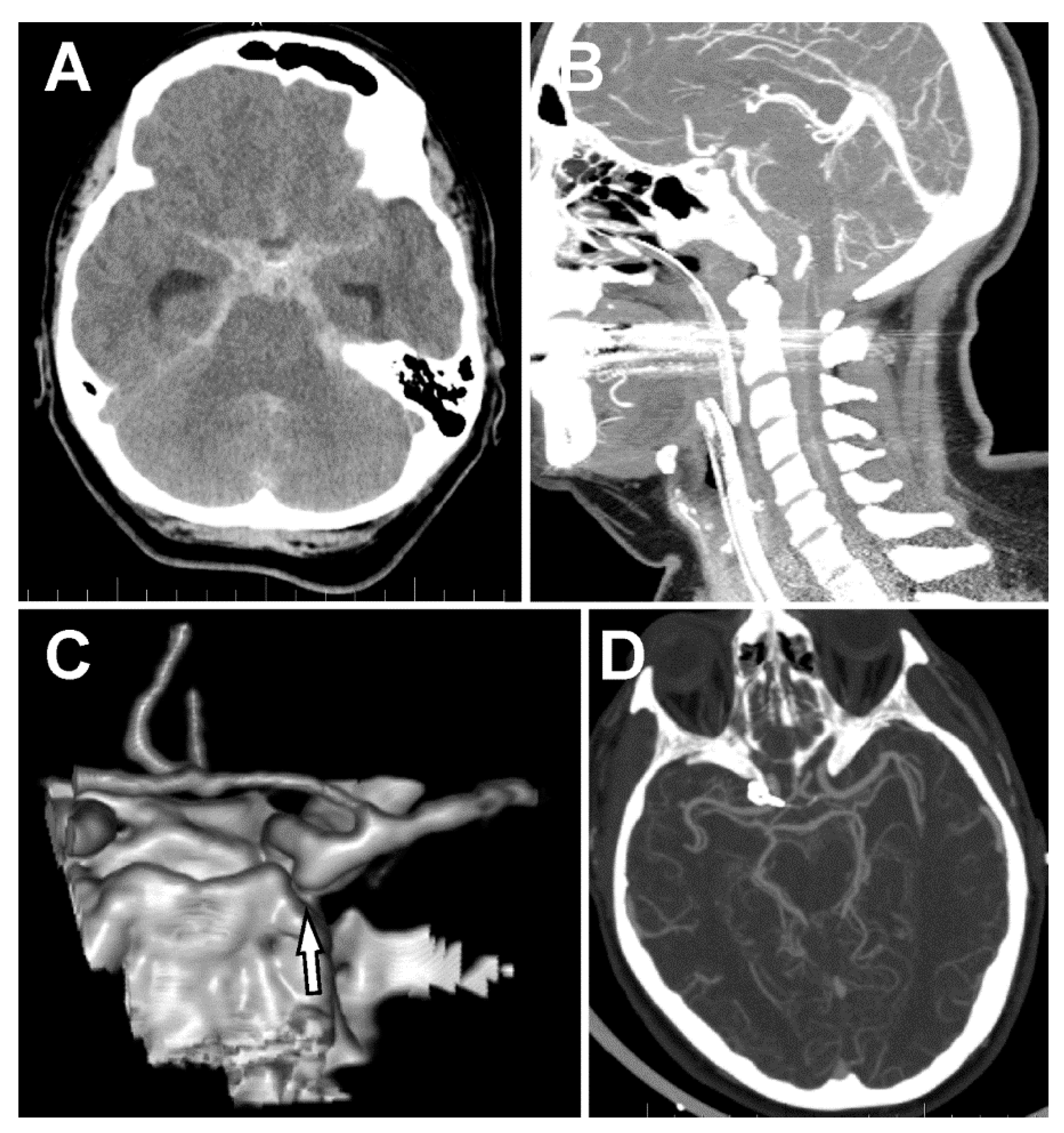
2. Case Report
3. Discussion
4. Conclusions
Author Contributions
Funding
Conflicts of Interest
References
- van Dijk, E.J.; Hupperts, R.M.; van der Jagt, M.; Bijvoet, H.W.; Hasan, D. Diagnosis of perimesencephalic nonaneurysmal subarachnoid hemorrhage with computed tomography. J. Stroke Cerebrovasc. Dis. 2001, 10, 247–251. [Google Scholar] [CrossRef] [PubMed]
- Konczalla, J.; Kashefiolasl, S.; Brawanski, N.; Senft, C.; Seifert, V.; Platz, J. Increasing numbers of nonaneurysmal subarachnoid hemorrhage in the last 15 years: Antithrombotic medication as reason and prognostic factor? J. Neurosurg. 2016, 124, 1731–1737. [Google Scholar] [CrossRef] [PubMed]
- Akcakaya, M.O.; Aydoseli, A.; Aras, Y.; Sabanci, P.A.; Barburoglu, M.; Alkir, G.; Sencer, A.; Sencer, S.; Aydin, K.; Kiris, T.; et al. Clinical Course of Non-Traumatic Non-Aneurysmal Subarachnoid Hemorrhage: A Single Institution Experience over 10 Years and Review of the Contemporary Literature. Turk. Neurosurg. 2017, 27, 732–742. [Google Scholar] [PubMed]
- Rangel-Castilla, L.; Russin, J.J.; Zaidi, H.A.; Martinez-Del-Campo, E.; Park, M.S.; Albuquerque, F.C.; McDougall, C.G.; Nakaji, P.; Spetzler, R.F. Contemporary management of spinal AVFs and AVMs: Lessons learned from 110 cases. Neurosurg. Focus 2014, 37, E14. [Google Scholar] [CrossRef]
- Hejčl, A.R.T.; Sameš, M. Our experience with lateral supraorbital approach in surgery of intracranial aneurysms. Ceska Slovenska Neurologie Neurochirurgie 2012, 75, 203–207. [Google Scholar]
- Hernesniemi, J.; Ishii, K.; Niemela, M.; Smrcka, M.; Kivipelto, L.; Fujiki, M.; Shen, H. Lateral supraorbital approach as an alternative to the classical pterional approach. Acta Neurochir. Suppl. 2005, 94, 17–21. [Google Scholar]
- Kim, L.J.; Spetzler, R.F. Classification and surgical management of spinal arteriovenous lesions: Arteriovenous fistulae and arteriovenous malformations. Neurosurgery 2006, 59, S195–S201. [Google Scholar] [CrossRef]
- Heros, R.C.; Debrun, G.M.; Ojemann, R.G.; Lasjaunias, P.L.; Naessens, P.J. Direct spinal arteriovenous fistula: A new type of spinal AVM. Case report. J. Neurosurg. 1986, 64, 134–139. [Google Scholar] [CrossRef]
- Lantigua, H.; Ortega-Gutierrez, S.; Schmidt, J.M.; Lee, K.; Badjatia, N.; Agarwal, S.; Claassen, J.; Connolly, E.S.; Mayer, S.A. Subarachnoid hemorrhage: Who dies, and why? Crit. Care 2015, 19, 309. [Google Scholar] [CrossRef]
- Kapadia, A.; Schweizer, T.A.; Spears, J.; Cusimano, M.; Macdonald, R.L. Nonaneurysmal perimesencephalic subarachnoid hemorrhage: Diagnosis, pathophysiology, clinical characteristics, and long-term outcome. World Neurosurg. 2014, 82, 1131–1143. [Google Scholar] [CrossRef]
- Vates, G.E.; Quiñones-Hinojosa, A.; Halbach, V.V.; Lawton, M.T. Conus perimedullary arteriovenous fistula with intracranial drainage: Case report. Neurosurgery 2001, 49, 457–461. [Google Scholar] [PubMed]
- Germans, M.R.; Coert, B.A.; Majoie, C.B.; van den Berg, R.; Verbaan, D.; Vandertop, W.P. Spinal axis imaging in non-aneurysmal subarachnoid hemorrhage: A prospective cohort study. J. Neurol. 2014, 261, 2199–2203. [Google Scholar] [CrossRef] [PubMed]
- Koch, C.; Gottschalk, S.; Giese, A. Dural arteriovenous fistula of the lumbar spine presenting with subarachnoid hemorrhage. Case report and review of the literature. J. Neurosurg. 2004, 100, 385–391. [Google Scholar] [PubMed]
- Matsui, T.; Taniguchi, T.; Saitoh, T.; Kamijoh, K.; Nakamura, T.; Yamashita, A.; Takayanagi, S.; Sakamoto, M.; Ishikawa, T. Hematomyelia caused by ruptured intramedullary spinal artery aneurysm associated with extramedullary spinal arteriovenous fistula—Case report. Neurol. Med. Chir. (Tokyo) 2007, 47, 233–236. [Google Scholar] [CrossRef] [PubMed]
- Spetzler, R.F.; Detwiler, P.W.; Riina, H.A.; Porter, R.W. Modified classification of spinal cord vascular lesions. J. Neurosurg. 2002, 96, 145–156. [Google Scholar] [CrossRef]
- Hida, K.; Iwasaki, Y.; Ushikoshi, S.; Fujimoto, S.; Seki, T.; Miyasaka, K. Corpectomy: A direct approach to perimedullary arteriovenous fistulas of the anterior cervical spinal cord. J. Neurosurg. 2002, 96, 157–161. [Google Scholar] [CrossRef]
- Mansour, A.; Endo, T.; Inoue, T.; Sato, K.; Endo, H.; Fujimura, M.; Tominaga, T. Clipping of an anterior spinal artery aneurysm using an endoscopic fluorescence imaging system for craniocervical junction epidural arteriovenous fistula: Technical note. J. Neurosurg. Spine 2019, 26, 1–6. [Google Scholar] [CrossRef]
- Markert, J.M.; Chandler, W.F.; Deveikis, J.P.; Ross, D.A. Use of the extreme lateral approach in the surgical treatment of an intradural ventral cervical spinal cord vascular malformation: Technical case report. Neurosurgery 1996, 38, 412–415. [Google Scholar] [CrossRef]
- Konczalla, J.; Platz, J.; Schuss, P.; Vatter, H.; Seifert, V.; Guresir, E. Non-aneurysmal non-traumatic subarachnoid hemorrhage: Patient characteristics, clinical outcome and prognostic factors based on a single-center experience in 125 patients. BMC Neurol. 2014, 14, 140. [Google Scholar] [CrossRef]
- Inoue, T.; Endo, T.; Sato, K.; Fesli, R.; Ogawa, Y.; Fujimura, M.; Matsumoto, Y.; Tominaga, T. Massive Intramedullary Hemorrhage After Subarachnoid Hemorrhage in Patient with Vertebrovertebral Arteriovenous Fistula. World Neurosurg. 2019, 129, 432–436. [Google Scholar] [CrossRef]
- Liu, C.L.; Su, Y.C.; Chen, C.C.; Chong, C.F.; Wang, T.L. Ruptured cervical arteriovenous fistulas presenting with subarachnoid hemorrhage and quadriplegia: An uncommon case. Am. J. Emerg. Med. 2008, 26, 249.e1-2. [Google Scholar] [CrossRef] [PubMed]
- Akter, M.; Hirai, T.; Kitajima, M.; Kai, Y.; Morioka, M.; Sasao, A.; Utsunomiya, D.; Uetani, H.; Korogi, Y.; Yamashita, Y. Type 1 perimedullary arteriovenous fistula with subarachnoid hemorrhage: Utility of contrast-enhanced 3D gradient-echo technique. Magn. Reson. Med. Sci. 2011, 10, 143–147. [Google Scholar] [CrossRef] [PubMed][Green Version]
- Hayashi, K.; Takahata, H.; Nakamura, M. Two cases of spinal arteriovenous malformation presenting with subarachnoid hemorrhage. No Shinkei Geka 2004, 32, 605–611. [Google Scholar] [PubMed]
- Kai, Y.; Hamada, J.; Morioka, M.; Yano, S.; Mizuno, T.; Kuratsu, J. Arteriovenous fistulas at the cervicomedullary junction presenting with subarachnoid hemorrhage: Six case reports with special reference to the angiographic pattern of venous drainage. AJNR Am. J. Neuroradiol. 2005, 26, 1949–1954. [Google Scholar] [PubMed]
- Alonso Fernández, L.; Nzau, M.; Ventureyra, E. Spinal intradural arteriovenous fistula with unusual presentation: Case report and literature review. Childs Nerv. Syst. 2008, 24, 1349–1353. [Google Scholar] [CrossRef] [PubMed]
- Poisson, A.; Vasdev, A.; Brunelle, F.; Plauchu, H.; Dupuis-Girod, S.; French Italian HHT network. Acute paraplegia due to spinal arteriovenous fistula in two patients with hereditary hemorrhagic telangiectasia. Eur. J. Pediatr. 2009, 168, 135–139. [Google Scholar] [CrossRef]
- Lv, X.; Li, Y.; Yang, X.; Jiang, C.; Wu, Z. Endovascular embolization for symptomatic perimedullary AVF and intramedullary AVM: A series and a literature review. Neuroradiology 2012, 54, 349–359. [Google Scholar] [CrossRef]
- Ohmori, Y.; Hamada, J.I.; Morioka, M.; Yoshida, A. Spinal aneurysm arising from the feeding pedicle of a thoracic perimedullary arteriovenous fistula: Case report. Surg. Neurol. 2005, 64, 468–470. [Google Scholar] [CrossRef]
- Bagherpour, A.N.; Rodriguez, G.J.; Moorthy, C.; Trier, T.T.; Maud, A. Combined surgical and endovascular treatment of complex high-flow conus medullaris arteriovenous fistula associated with Parkes Weber syndrome: Case report. J. Neurosurg. Spine 2016, 25, 234–238. [Google Scholar] [CrossRef]
- Ohba, S.; Onozuka, S.; Horiguchi, T.; Kawase, T.; Yoshida, K. Perimedullary arteriovenous fistula at the craniocervical junction—Case report. Neurol. Med. Chir. (Tokyo) 2011, 51, 299–301. [Google Scholar] [CrossRef][Green Version]
- Kominami, S.; Liu, Y.; Alvarez, H.; Rodesch, G.; Coubes, P.; Lasjaunias, P. A case of vertebrovertebral arteriovenous fistula presenting with subarachnoid haemorrhage. A case report. Interv. Neuroradiol. 1996, 2, 229–233. [Google Scholar] [CrossRef]
- Alshekhlee, A.; Edgell, R.C.; Kale, S.P.; Kitchener, J.; Vora, N. Endovascular therapy of a craniocervical pial AVF fed by the anterior spinal artery. J. Neuroimaging 2013, 23, 102–104. [Google Scholar] [CrossRef]
- Wakai, S.; Inoh, S.; Iwanaga, H.; Nagai, M.; Sato, T.; Izumi, J. Successful surgical obliteration of a huge intradural arteriovenous fistula of the spinal cord in a child. Childs Nerv. Syst. 1992, 8, 347–350. [Google Scholar] [CrossRef]


| Author | Year | Patient Age | Spine Level | Outcome | Treatment |
|---|---|---|---|---|---|
| Inoue et al. [20] | 2019 | 59 | High Cervical | mRS5 | refused treatment |
| Liu et al. [21] | 2008 | 26 | High Cervical | mRS0 | surgical resection |
| Akter et al. [22] | 2011 | 68 | High Cervical | N/A | surgical resection |
| Akter et al. | 2011 | 53 | High Cervical | N/A | surgical resection |
| Akter et al. | 2011 | 56 | Cervical | N/A | surgical resection |
| Akter et al. | 2011 | 60 | Thoracic | N/A | surgical resection |
| Hayashi et al. [23] | 2004 | 67 | Cervical | mRS0 | embolization |
| Kai et al. [24] | 2005 | 54 | High Cervical | mRS1 | surgical resection |
| Kai et al. | 2005 | 56 | High Cervical | mRS1 | surgical resection |
| Fernandéz et al. [25] | 2008 | 5 | High Cervical | mRS1 | surgical resection |
| Poisson et al. [26] | 2008 | 8 months | Low thoracic | mRS3 | embolization |
| Lv et al. [27] | 2012 | 17 | High Cervical | mRS2 | embolization |
| Ohmori et al. [28] | 2005 | 42 | Low thoracic | mRS0 | surgical resection |
| Bagherpour et al. [29] | 2016 | 14 | Conus medullaris | mRS0 | surgical resection |
| Ohba et al. [30] | 2011 | 55 | High Cervical | mRS1 | surgical resection |
| Vates et al. [11] | 2001 | 65 | Conus medullaris | mRS4 | surgical resection |
| Kominami et al. [31] | 1996 | 12 | High Cervical | N/A | embolization |
| Alshekhlee et al. [32] | 2011 | 57 | High Cervical | mRS4 | embolization |
| Hida et al. [16] | 2002 | 58 | High Cervical | N/A | surgical resection |
| Hida et al. | 2002 | 59 | High Cervical | N/A | surgical resection |
| Hida et al. | 2002 | 62 | High Cervical | N/A | surgical resection |
| Hida et al. | 2002 | 62 | Low Cervical | N/A | surgical resection |
| Hida et al. | 2002 | 34 | Low Cervical | N/A | combination |
| Wakai et al. [33] | 1992 | 8 | Low Cervical | mRS1 | surgical resection |
© 2020 by the authors. Licensee MDPI, Basel, Switzerland. This article is an open access article distributed under the terms and conditions of the Creative Commons Attribution (CC BY) license (http://creativecommons.org/licenses/by/4.0/).
Share and Cite
Hejčl, A.; Lodin, J.; Cihlář, F.; Sameš, M. Spontaneous Subarachnoid Hemorrhage in a Patient with a Co-Existent Posterior Communicating Artery Aneurysm and Cervical Spine Aneurysm Associated with Ventral Arterio-Venous Fistula. Brain Sci. 2020, 10, 70. https://doi.org/10.3390/brainsci10020070
Hejčl A, Lodin J, Cihlář F, Sameš M. Spontaneous Subarachnoid Hemorrhage in a Patient with a Co-Existent Posterior Communicating Artery Aneurysm and Cervical Spine Aneurysm Associated with Ventral Arterio-Venous Fistula. Brain Sciences. 2020; 10(2):70. https://doi.org/10.3390/brainsci10020070
Chicago/Turabian StyleHejčl, Aleš, Jan Lodin, Filip Cihlář, and Martin Sameš. 2020. "Spontaneous Subarachnoid Hemorrhage in a Patient with a Co-Existent Posterior Communicating Artery Aneurysm and Cervical Spine Aneurysm Associated with Ventral Arterio-Venous Fistula" Brain Sciences 10, no. 2: 70. https://doi.org/10.3390/brainsci10020070
APA StyleHejčl, A., Lodin, J., Cihlář, F., & Sameš, M. (2020). Spontaneous Subarachnoid Hemorrhage in a Patient with a Co-Existent Posterior Communicating Artery Aneurysm and Cervical Spine Aneurysm Associated with Ventral Arterio-Venous Fistula. Brain Sciences, 10(2), 70. https://doi.org/10.3390/brainsci10020070