Neurofilaments: The C-Reactive Protein of Neurology

 ,
, 
Abstract
:1. Introduction
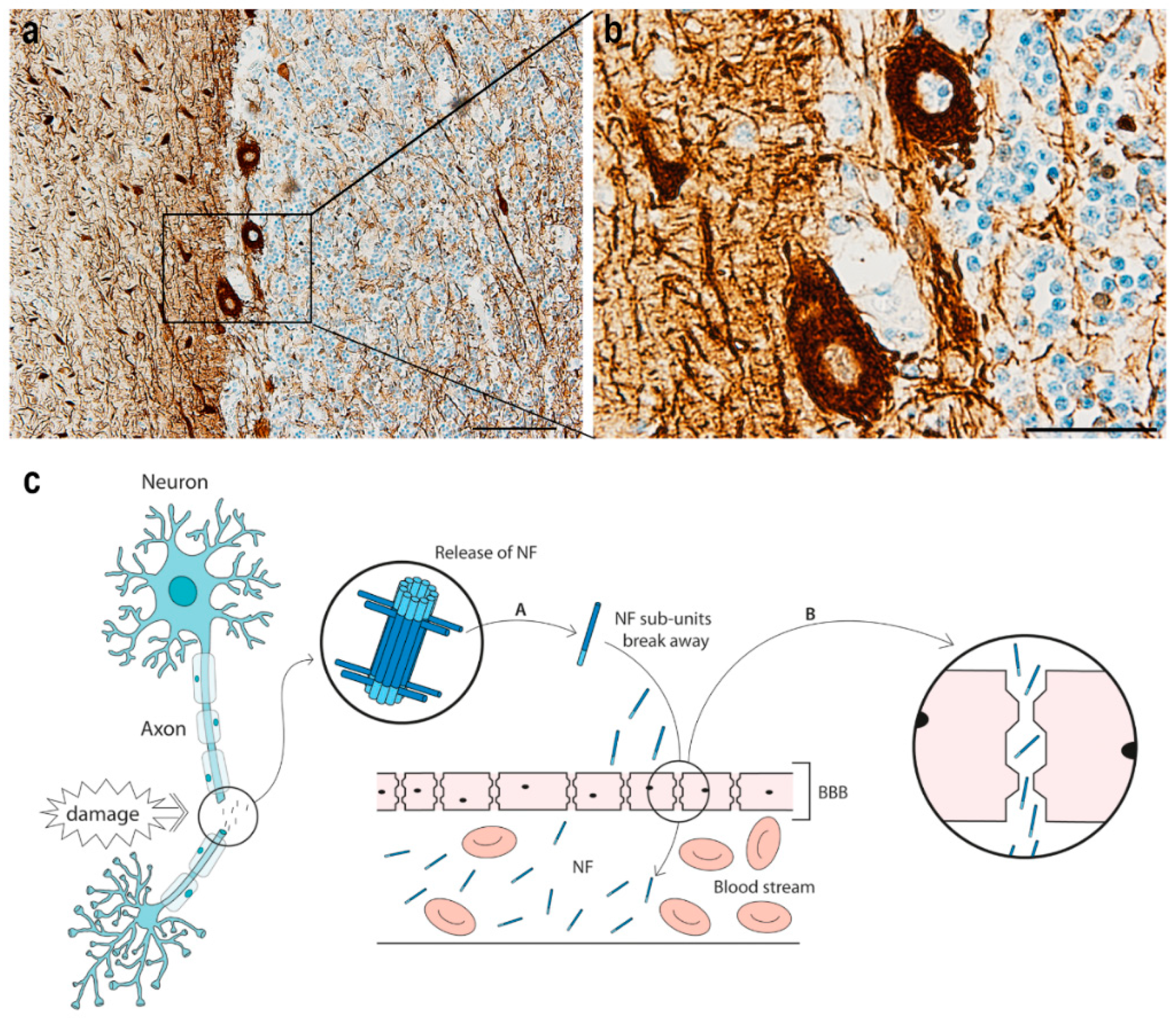
2. The Structure and Function of Neurofilament
3. Neurofilament in Neurological Disease
3.1. Peripheral Neuropathy
3.2. Motor Neuron Disease
3.3. Multiple Sclerosis
3.4. Alzheimer’s Disease
3.5. Huntington’s Disease
3.6. Parkinson’s Disease and Parkinsonian Disorders
3.7. Stroke
3.8. Traumatic Axonal Injury
3.9. Cardiac Arrest
3.10. Delirium
4. Discussion
5. Conclusions
Author Contributions
Funding
Acknowledgments
Conflicts of Interest
Abbreviations
| AD | Alzheimer’s disease |
| ALS | amyotrophic lateral sclerosis |
| APD | atypical parkinsonian disorder |
| APP | amyloid precursor protein |
| CBG | corticobasal degeneration |
| CSF | cerebrospinal fluid |
| CMT | Charcot–Marie–Tooth |
| CNS | central nervous system |
| CRP | C-reactive protein |
| DS | Down syndrome |
| ECL | electrochemilumninescence |
| EDSS | Extended Disability Status Scale |
| FTD | frontotemporal dementia |
| GBS | Guillain–Barré syndrome |
| GCS | Glasgow Coma Scale |
| HD | Huntington’s disease |
| HTT | huntingtin gene |
| mHTT | mutant huntingtin gene |
| MND | motor neuron disease |
| MRI | magnetic resonance imaging |
| MS | multiple sclerosis |
| MSA | multiple system atrophy |
| NF | neurofilament |
| NF-H | neurofilament heavy chain |
| NF-L | neurofilament light chain |
| NF-M | neurofilament medium chain |
| PD | Parkinson’s disease |
| PNS | peripheral nervous system |
| PSP | progressive supranuclear palsy |
| Simoa | single molecule array technology |
| TBI | traumatic brain injury |
References
- Rosengren, L.E.; Karlsson, J.E.; Karlsson, J.O.; Persson, L.I.; Wikkelso, C. Patients with amyotrophic lateral sclerosis and other neurodegenerative diseases have increased levels of neurofilament protein in CSF. J. Neurochem. 1996, 67, 2013–2018. [Google Scholar] [CrossRef]
- Gaiottino, J.; Norgren, N.; Dobson, R.; Topping, J.; Nissim, A.; Malaspina, A.; Bestwick, J.P.; Monsch, A.U.; Regeniter, A.; Lindberg, R.L.; et al. Increased neurofilament light chain blood levels in neurodegenerative neurological diseases. PLoS ONE 2013, 8, e75091. [Google Scholar] [CrossRef]
- Rissin, D.M.; Kan, C.W.; Campbell, T.G.; Howes, S.C.; Fournier, D.R.; Song, L.; Piech, T.; Patel, P.P.; Chang, L.; Rivnak, A.J.; et al. Single-molecule enzyme-linked immunosorbent assay detects serum proteins at subfemtomolar concentrations. Nat. Biotechnol. 2010, 28, 595–599. [Google Scholar] [CrossRef] [Green Version]
- Khalil, M.; Teunissen, C.E.; Otto, M.; Piehl, F.; Sormani, M.P.; Gattringer, T.; Barro, C.; Kappos, L.; Comabella, M.; Fazekas, F.; et al. Neurofilaments as biomarkers in neurological disorders. Nat. Rev. Neurol. 2018, 14, 577–589. [Google Scholar] [CrossRef]
- Gisslen, M.; Price, R.W.; Andreasson, U.; Norgren, N.; Nilsson, S.; Hagberg, L.; Fuchs, D.; Spudich, S.; Blennow, K.; Zetterberg, H. Plasma Concentration of the Neurofilament Light Protein (NFL) is a Biomarker of CNS Injury in HIV Infection: A Cross-Sectional Study. EBioMedicine 2016, 3, 135–140. [Google Scholar] [CrossRef] [Green Version]
- Lambertsen, K.L.; Finsen, B.; Clausen, B.H. Post-stroke inflammation-target or tool for therapy? Acta Neuropathol. 2018. [Google Scholar] [CrossRef] [Green Version]
- Clausen, B.H.; Lambertsen, K.L.; Dagnaes-Hansen, F.; Babcock, A.A.; von Linstow, C.U.; Meldgaard, M.; Kristensen, B.W.; Deierborg, T.; Finsen, B. Cell therapy centered on IL-1Ra is neuroprotective in experimental stroke. Acta Neuropathol. 2016. [Google Scholar] [CrossRef] [Green Version]
- Clausen, B.H.; Degn, M.; Sivasaravanaparan, M.; Fogtmann, T.; Andersen, M.G.; Trojanowsky, M.D.; Gao, H.; Hvidsten, S.; Baun, C.; Deierborg, T.; et al. Conditional ablation of myeloid TNF increases lesion volume after experimental stroke in mice, possibly via altered ERK1/2 signaling. Sci. Rep. 2016, 6, 29291. [Google Scholar] [CrossRef] [Green Version]
- Lambertsen, K.L.; Ostergaard, K.; Clausen, B.H.; Hansen, S.; Stenvang, J.; Thorsen, S.B.; Meldgaard, M.; Kristensen, B.W.; Hansen, P.B.; Sorensen, G.L.; et al. No effect of ablation of surfactant protein-D on acute cerebral infarction in mice. J. Neuroinflammation 2014, 11, 123. [Google Scholar] [CrossRef] [Green Version]
- Mariotto, S.; Farinazzo, A.; Magliozzi, R.; Alberti, D.; Monaco, S.; Ferrari, S. Serum and cerebrospinal neurofilament light chain levels in patients with acquired peripheral neuropathies. J. Peripher. Nerv. Syst. 2018, 23, 174–177. [Google Scholar] [CrossRef]
- Bischof, A.; Manigold, T.; Barro, C.; Heijnen, I.; Berger, C.T.; Derfuss, T.; Kuhle, J.; Daikeler, T. Serum neurofilament light chain: A biomarker of neuronal injury in vasculitic neuropathy. Ann. Rheum. Dis. 2018, 77, 1093–1094. [Google Scholar] [CrossRef]
- Axelsson, M.; Sjogren, M.; Andersen, O.; Blennow, K.; Zetterberg, H.; Lycke, J. Neurofilament light protein levels in cerebrospinal fluid predict long-term disability of Guillain-Barre syndrome: A pilot study. Acta Neurol. Scand. 2018, 138, 143–150. [Google Scholar] [CrossRef] [Green Version]
- Sandelius, A.; Zetterberg, H.; Blennow, K.; Adiutori, R.; Malaspina, A.; Laura, M.; Reilly, M.M.; Rossor, A.M. Plasma neurofilament light chain concentration in the inherited peripheral neuropathies. Neurology 2018, 90, e518–e524. [Google Scholar] [CrossRef] [Green Version]
- Lu, C.H.; Macdonald-Wallis, C.; Gray, E.; Pearce, N.; Petzold, A.; Norgren, N.; Giovannoni, G.; Fratta, P.; Sidle, K.; Fish, M.; et al. Neurofilament light chain: A prognostic biomarker in amyotrophic lateral sclerosis. Neurology 2015, 84, 2247–2257. [Google Scholar] [CrossRef] [Green Version]
- Verde, F.; Steinacker, P.; Weishaupt, J.H.; Kassubek, J.; Oeckl, P.; Halbgebauer, S.; Tumani, H.; von Arnim, C.A.F.; Dorst, J.; Feneberg, E.; et al. Neurofilament light chain in serum for the diagnosis of amyotrophic lateral sclerosis. J. Neurol. Neurosurg. Psychiatry 2019, 90, 157–164. [Google Scholar] [CrossRef]
- Weydt, P.; Oeckl, P.; Huss, A.; Muller, K.; Volk, A.E.; Kuhle, J.; Knehr, A.; Andersen, P.M.; Prudlo, J.; Steinacker, P.; et al. Neurofilament levels as biomarkers in asymptomatic and symptomatic familial amyotrophic lateral sclerosis. Ann. Neurol. 2016, 79, 152–158. [Google Scholar] [CrossRef]
- Skillback, T.; Mattsson, N.; Blennow, K.; Zetterberg, H. Cerebrospinal fluid neurofilament light concentration in motor neuron disease and frontotemporal dementia predicts survival. Amyotroph. Lateral Scler. Front. Degener. 2017, 18, 397–403. [Google Scholar] [CrossRef] [Green Version]
- Modvig, S.; Degn, M.; Horwitz, H.; Cramer, S.P.; Larsson, H.B.; Wanscher, B.; Sellebjerg, F.; Frederiksen, J.L. Relationship between cerebrospinal fluid biomarkers for inflammation, demyelination and neurodegeneration in acute optic neuritis. PLoS ONE 2013, 8, e77163. [Google Scholar] [CrossRef] [Green Version]
- Modvig, S.; Degn, M.; Sander, B.; Horwitz, H.; Wanscher, B.; Sellebjerg, F.; Frederiksen, J.L. Cerebrospinal fluid neurofilament light chain levels predict visual outcome after optic neuritis. Mult. Scler. 2016, 22, 590–598. [Google Scholar] [CrossRef]
- Modvig, S.; Degn, M.; Roed, H.; Sorensen, T.L.; Larsson, H.B.; Langkilde, A.R.; Frederiksen, J.L.; Sellebjerg, F. Cerebrospinal fluid levels of chitinase 3-like 1 and neurofilament light chain predict multiple sclerosis development and disability after optic neuritis. Mult. Scler. 2015, 21, 1761–1770. [Google Scholar] [CrossRef]
- Kuhle, J.; Kropshofer, H.; Haering, D.A.; Kundu, U.; Meinert, R.; Barro, C.; Dahlke, F.; Tomic, D.; Leppert, D.; Kappos, L. Blood neurofilament light chain as a biomarker of MS disease activity and treatment response. Neurology 2019, 92, e1007–e1015. [Google Scholar] [CrossRef]
- Siller, N.; Kuhle, J.; Muthuraman, M.; Barro, C.; Uphaus, T.; Groppa, S.; Kappos, L.; Zipp, F.; Bittner, S. Serum neurofilament light chain is a biomarker of acute and chronic neuronal damage in early multiple sclerosis. Mult. Scler. 2019, 25, 678–686. [Google Scholar] [CrossRef]
- Chitnis, T.; Gonzalez, C.; Healy, B.C.; Saxena, S.; Rosso, M.; Barro, C.; Michalak, Z.; Paul, A.; Kivisakk, P.; Diaz-Cruz, C.; et al. Neurofilament light chain serum levels correlate with 10-year MRI outcomes in multiple sclerosis. Ann. Clin. Transl. Neurol. 2018, 5, 1478–1491. [Google Scholar] [CrossRef] [Green Version]
- Barro, C.; Benkert, P.; Disanto, G.; Tsagkas, C.; Amann, M.; Naegelin, Y.; Leppert, D.; Gobbi, C.; Granziera, C.; Yaldizli, O.; et al. Serum neurofilament as a predictor of disease worsening and brain and spinal cord atrophy in multiple sclerosis. Brain 2018, 141, 2382–2391. [Google Scholar] [CrossRef]
- Sanchez-Valle, R.; Heslegrave, A.; Foiani, M.S.; Bosch, B.; Antonell, A.; Balasa, M.; Llado, A.; Zetterberg, H.; Fox, N.C. Serum neurofilament light levels correlate with severity measures and neurodegeneration markers in autosomal dominant Alzheimer’s disease. Alzheimers Res. Ther. 2018, 10, 113. [Google Scholar] [CrossRef]
- Preische, O.; Schultz, S.A.; Apel, A.; Kuhle, J.; Kaeser, S.A.; Barro, C.; Graber, S.; Kuder-Buletta, E.; LaFougere, C.; Laske, C.; et al. Serum neurofilament dynamics predicts neurodegeneration and clinical progression in presymptomatic Alzheimer’s disease. Nat. Med. 2019, 25, 277–283. [Google Scholar] [CrossRef]
- Zhou, W.; Zhang, J.; Ye, F.; Xu, G.; Su, H.; Su, Y.; Zhang, X.; Alzheimer’s Disease Neuroimaging, I. Plasma neurofilament light chain levels in Alzheimer’s disease. Neurosci. Lett. 2017, 650, 60–64. [Google Scholar] [CrossRef] [Green Version]
- Lewczuk, P.; Ermann, N.; Andreasson, U.; Schultheis, C.; Podhorna, J.; Spitzer, P.; Maler, J.M.; Kornhuber, J.; Blennow, K.; Zetterberg, H. Plasma neurofilament light as a potential biomarker of neurodegeneration in Alzheimer’s disease. Alzheimers Res. Ther. 2018, 10, 71. [Google Scholar] [CrossRef]
- Fortea, J.; Carmona-Iragui, M.; Benejam, B.; Fernandez, S.; Videla, L.; Barroeta, I.; Alcolea, D.; Pegueroles, J.; Munoz, L.; Belbin, O.; et al. Plasma and CSF biomarkers for the diagnosis of Alzheimer’s disease in adults with Down syndrome: A cross-sectional study. Lancet Neurol. 2018, 17, 860–869. [Google Scholar] [CrossRef]
- Mattsson, N.; Andreasson, U.; Zetterberg, H.; Blennow, K. Association of Plasma Neurofilament Light with Neurodegeneration in Patients with Alzheimer Disease. JAMA Neurol. 2017, 74, 557–566. [Google Scholar] [CrossRef]
- Lin, Y.S.; Lee, W.J.; Wang, S.J.; Fuh, J.L. Levels of plasma neurofilament light chain and cognitive function in patients with Alzheimer or Parkinson disease. Sci. Rep. 2018, 8, 17368. [Google Scholar] [CrossRef]
- Niemela, V.; Landtblom, A.M.; Blennow, K.; Sundblom, J. Tau or neurofilament light-Which is the more suitable biomarker for Huntington’s disease? PLoS ONE 2017, 12, e0172762. [Google Scholar] [CrossRef]
- Byrne, L.M.; Rodrigues, F.B.; Johnson, E.B.; Wijeratne, P.A.; De Vita, E.; Alexander, D.C.; Palermo, G.; Czech, C.; Schobel, S.; Scahill, R.I.; et al. Evaluation of mutant huntingtin and neurofilament proteins as potential markers in Huntington’s disease. Sci. Transl. Med. 2018, 10. [Google Scholar] [CrossRef] [Green Version]
- Wild, E.J.; Petzold, A.; Keir, G.; Tabrizi, S.J. Plasma neurofilament heavy chain levels in Huntington’s disease. Neurosci. Lett. 2007, 417, 231–233. [Google Scholar] [CrossRef]
- Byrne, L.M.; Rodrigues, F.B.; Blennow, K.; Durr, A.; Leavitt, B.R.; Roos, R.A.C.; Scahill, R.I.; Tabrizi, S.J.; Zetterberg, H.; Langbehn, D.; et al. Neurofilament light protein in blood as a potential biomarker of neurodegeneration in Huntington’s disease: A retrospective cohort analysis. Lancet Neurol. 2017, 16, 601–609. [Google Scholar] [CrossRef] [Green Version]
- Constantinescu, R.; Romer, M.; Oakes, D.; Rosengren, L.; Kieburtz, K. Levels of the light subunit of neurofilament triplet protein in cerebrospinal fluid in Huntington’s disease. Parkinsonism Relat. Disord. 2009, 15, 245–248. [Google Scholar] [CrossRef]
- Vinther-Jensen, T.; Bornsen, L.; Budtz-Jorgensen, E.; Ammitzboll, C.; Larsen, I.U.; Hjermind, L.E.; Sellebjerg, F.; Nielsen, J.E. Selected CSF biomarkers indicate no evidence of early neuroinflammation in Huntington disease. Neurol. Neuroimmunol. Neuroinflamm. 2016, 3, e287. [Google Scholar] [CrossRef] [Green Version]
- Hansson, O.; Janelidze, S.; Hall, S.; Magdalinou, N.; Lees, A.J.; Andreasson, U.; Norgren, N.; Linder, J.; Forsgren, L.; Constantinescu, R.; et al. Blood-based NfL: A biomarker for differential diagnosis of parkinsonian disorder. Neurology 2017, 88, 930–937. [Google Scholar] [CrossRef]
- Hall, S.; Surova, Y.; Ohrfelt, A.; Swedish Bio, F.S.; Blennow, K.; Zetterberg, H.; Hansson, O. Longitudinal Measurements of Cerebrospinal Fluid Biomarkers in Parkinson’s Disease. Mov. Disord. 2016, 31, 898–905. [Google Scholar] [CrossRef]
- Constantinescu, R.; Rosengren, L.; Eriksson, B.; Blennow, K.; Axelsson, M. Cerebrospinal fluid neurofilament light and tau protein as mortality biomarkers in parkinsonism. Acta Neurol. Scand. 2019, 140, 147–156. [Google Scholar] [CrossRef]
- Pedersen, A.; Stanne, T.M.; Nilsson, S.; Klasson, S.; Rosengren, L.; Holmegaard, L.; Jood, K.; Blennow, K.; Zetterberg, H.; Jern, C. Circulating neurofilament light in ischemic stroke: Temporal profile and outcome prediction. J. Neurol. 2019, 266, 2796–2806. [Google Scholar] [CrossRef] [Green Version]
- Onatsu, J.; Vanninen, R.; Jakala, P.; Mustonen, P.; Pulkki, K.; Korhonen, M.; Hedman, M.; Zetterberg, H.; Blennow, K.; Hoglund, K.; et al. Serum Neurofilament Light Chain Concentration Correlates with Infarct Volume but Not Prognosis in Acute Ischemic Stroke. J. Stroke Cerebrovasc. Dis. 2019, 28, 2242–2249. [Google Scholar] [CrossRef]
- Singh, P.; Yan, J.; Hull, R.; Read, S.; O’Sullivan, J.; Henderson, R.D.; Rose, S.; Greer, J.M.; McCombe, P.A. Levels of phosphorylated axonal neurofilament subunit H (pNfH) are increased in acute ischemic stroke. J. Neurol. Sci. 2011, 304, 117–121. [Google Scholar] [CrossRef]
- Tiedt, S.; Duering, M.; Barro, C.; Kaya, A.G.; Boeck, J.; Bode, F.J.; Klein, M.; Dorn, F.; Gesierich, B.; Kellert, L.; et al. Serum neurofilament light: A biomarker of neuroaxonal injury after ischemic stroke. Neurology 2018, 91, e1338–e1347. [Google Scholar] [CrossRef]
- De Marchis, G.M.; Katan, M.; Barro, C.; Fladt, J.; Traenka, C.; Seiffge, D.J.; Hert, L.; Gensicke, H.; Disanto, G.; Sutter, R.; et al. Serum neurofilament light chain in patients with acute cerebrovascular events. Eur. J. Neurol. 2018, 25, 562–568. [Google Scholar] [CrossRef] [Green Version]
- Hjalmarsson, C.; Bjerke, M.; Andersson, B.; Blennow, K.; Zetterberg, H.; Aberg, N.D.; Olsson, B.; Eckerstrom, C.; Bokemark, L.; Wallin, A. Neuronal and glia-related biomarkers in cerebrospinal fluid of patients with acute ischemic stroke. J. Cent. Nerv. Syst. Dis. 2014, 6, 51–58. [Google Scholar] [CrossRef] [Green Version]
- Traenka, C.; Disanto, G.; Seiffge, D.J.; Gensicke, H.; Hert, L.; Grond-Ginsbach, C.; Peters, N.; Regeniter, A.; Kloss, M.; De Marchis, G.M.; et al. Serum Neurofilament Light Chain Levels Are Associated with Clinical Characteristics and Outcome in Patients with Cervical Artery Dissection. Cerebrovasc. Dis. 2015, 40, 222–227. [Google Scholar] [CrossRef]
- Sellner, J.; Patel, A.; Dassan, P.; Brown, M.M.; Petzold, A. Hyperacute detection of neurofilament heavy chain in serum following stroke: A transient sign. Neurochem. Res. 2011, 36, 2287–2291. [Google Scholar] [CrossRef] [Green Version]
- Gattringer, T.; Pinter, D.; Enzinger, C.; Seifert-Held, T.; Kneihsl, M.; Fandler, S.; Pichler, A.; Barro, C.; Grobke, S.; Voortman, M.; et al. Serum neurofilament light is sensitive to active cerebral small vessel disease. Neurology 2017, 89, 2108–2114. [Google Scholar] [CrossRef]
- Martinez-Morillo, E.; Childs, C.; Garcia, B.P.; Alvarez Menendez, F.V.; Romaschin, A.D.; Cervellin, G.; Lippi, G.; Diamandis, E.P. Neurofilament medium polypeptide (NFM) protein concentration is increased in CSF and serum samples from patients with brain injury. Clin. Chem. Lab. Med. 2015, 53, 1575–1584. [Google Scholar] [CrossRef]
- Petzold, A.; Michel, P.; Stock, M.; Schluep, M. Glial and axonal body fluid biomarkers are related to infarct volume, severity, and outcome. J. Stroke Cerebrovasc. Dis. 2008, 17, 196–203. [Google Scholar] [CrossRef] [PubMed] [Green Version]
- Jonsson, M.; Zetterberg, H.; van Straaten, E.; Lind, K.; Syversen, S.; Edman, A.; Blennow, K.; Rosengren, L.; Pantoni, L.; Inzitari, D.; et al. Cerebrospinal fluid biomarkers of white matter lesions—Cross-sectional results from the LADIS study. Eur. J. Neurol. 2010, 17, 377–382. [Google Scholar] [CrossRef] [PubMed]
- Duering, M.; Konieczny, M.J.; Tiedt, S.; Baykara, E.; Tuladhar, A.M.; Leijsen, E.V.; Lyrer, P.; Engelter, S.T.; Gesierich, B.; Achmuller, M.; et al. Serum Neurofilament Light Chain Levels Are Related to Small Vessel Disease Burden. J. Stroke 2018, 20, 228–238. [Google Scholar] [CrossRef] [PubMed]
- Zanier, E.R.; Refai, D.; Zipfel, G.J.; Zoerle, T.; Longhi, L.; Esparza, T.J.; Spinner, M.L.; Bateman, R.J.; Brody, D.L.; Stocchetti, N. Neurofilament light chain levels in ventricular cerebrospinal fluid after acute aneurysmal subarachnoid haemorrhage. J. Neurol. Neurosurg. Psychiatry 2011, 82, 157–159. [Google Scholar] [CrossRef] [PubMed] [Green Version]
- Lewis, S.B.; Wolper, R.A.; Miralia, L.; Yang, C.; Shaw, G. Detection of phosphorylated NF-H in the cerebrospinal fluid and blood of aneurysmal subarachnoid hemorrhage patients. J. Cereb. Blood Flow Metab. 2008, 28, 1261–1271. [Google Scholar] [CrossRef]
- Thelin, E.; Al Nimer, F.; Frostell, A.; Zetterberg, H.; Blennow, K.; Nystrom, H.; Svensson, M.; Bellander, B.M.; Piehl, F.; Nelson, D.W. A Serum Protein Biomarker Panel Improves Outcome Prediction in Human Traumatic Brain Injury. J. Neurotrauma 2019, 36, 2850–2862. [Google Scholar] [CrossRef] [Green Version]
- Al Nimer, F.; Thelin, E.; Nystrom, H.; Dring, A.M.; Svenningsson, A.; Piehl, F.; Nelson, D.W.; Bellander, B.M. Comparative Assessment of the Prognostic Value of Biomarkers in Traumatic Brain Injury Reveals an Independent Role for Serum Levels of Neurofilament Light. PLoS ONE 2015, 10, e0132177. [Google Scholar] [CrossRef]
- Shahim, P.; Zetterberg, H.; Tegner, Y.; Blennow, K. Serum neurofilament light as a biomarker for mild traumatic brain injury in contact sports. Neurology 2017, 88, 1788–1794. [Google Scholar] [CrossRef] [Green Version]
- Hossain, I.; Mohammadian, M.; Takala, R.S.K.; Tenovuo, O.; Lagerstedt, L.; Ala-Seppala, H.; Frantzen, J.; van Gils, M.; Hutchinson, P.; Katila, A.J.; et al. Early Levels of Glial Fibrillary Acidic Protein and Neurofilament Light Protein in Predicting the Outcome of Mild Traumatic Brain Injury. J. Neurotrauma 2019, 36, 1551–1560. [Google Scholar] [CrossRef] [Green Version]
- McKee, A.C.; Cairns, N.J.; Dickson, D.W.; Folkerth, R.D.; Keene, C.D.; Litvan, I.; Perl, D.P.; Stein, T.D.; Vonsattel, J.P.; Stewart, W.; et al. The first NINDS/NIBIB consensus meeting to define neuropathological criteria for the diagnosis of chronic traumatic encephalopathy. Acta Neuropathol. 2016, 131, 75–86. [Google Scholar] [CrossRef] [Green Version]
- Wallace, C.; Smirl, J.D.; Zetterberg, H.; Blennow, K.; Bryk, K.; Burma, J.; Dierijck, J.; Wright, A.D.; van Donkelaar, P. Heading in soccer increases serum neurofilament light protein and SCAT3 symptom metrics. BMJ Open Sport Exerc. Med. 2018, 4, e000433. [Google Scholar] [CrossRef] [PubMed]
- Neselius, S.; Brisby, H.; Theodorsson, A.; Blennow, K.; Zetterberg, H.; Marcusson, J. CSF-biomarkers in Olympic boxing: Diagnosis and effects of repetitive head trauma. PLoS ONE 2012, 7, e33606. [Google Scholar] [CrossRef] [PubMed]
- Oliver, J.M.; Jones, M.T.; Kirk, K.M.; Gable, D.A.; Repshas, J.T.; Johnson, T.A.; Andreasson, U.; Norgren, N.; Blennow, K.; Zetterberg, H. Serum Neurofilament Light in American Football Athletes over the Course of a Season. J. Neurotrauma 2016, 33, 1784–1789. [Google Scholar] [CrossRef] [PubMed]
- Rubin, L.H.; Tierney, R.; Kawata, K.; Wesley, L.; Lee, J.H.; Blennow, K.; Zetterberg, H.; Langford, D. NFL blood levels are moderated by subconcussive impacts in a cohort of college football players. Brain Inj. 2019, 33, 456–462. [Google Scholar] [CrossRef] [PubMed]
- Guez, M.; Hildingsson, C.; Rosengren, L.; Karlsson, K.; Toolanen, G. Nervous tissue damage markers in cerebrospinal fluid after cervical spine injuries and whiplash trauma. J. Neurotrauma 2003, 20, 853–858. [Google Scholar] [CrossRef] [PubMed]
- Kuhle, J.; Gaiottino, J.; Leppert, D.; Petzold, A.; Bestwick, J.P.; Malaspina, A.; Lu, C.H.; Dobson, R.; Disanto, G.; Norgren, N.; et al. Serum neurofilament light chain is a biomarker of human spinal cord injury severity and outcome. J. Neurol. Neurosurg. Psychiatry 2015, 86, 273–279. [Google Scholar] [CrossRef] [PubMed]
- Rosen, H.; Karlsson, J.E.; Rosengren, L. CSF levels of neurofilament is a valuable predictor of long-term outcome after cardiac arrest. J. Neurol. Sci. 2004, 221, 19–24. [Google Scholar] [CrossRef]
- Rosen, C.; Rosen, H.; Andreasson, U.; Bremell, D.; Bremler, R.; Hagberg, L.; Rosengren, L.; Blennow, K.; Zetterberg, H. Cerebrospinal fluid biomarkers in cardiac arrest survivors. Resuscitation 2014, 85, 227–232. [Google Scholar] [CrossRef]
- Disanto, G.; Prosperetti, C.; Gobbi, C.; Barro, C.; Michalak, Z.; Cassina, T.; Kuhle, J.; Casso, G.; Agazzi, P. Serum neurofilament light chain as a prognostic marker in postanoxic encephalopathy. Epilepsy Behav. 2019, 101, 106432. [Google Scholar] [CrossRef]
- Yokobori, S.; Wang, K.K.K.; Yang, Z.; Zhu, T.; Tyndall, J.A.; Mondello, S.; Shibata, Y.; Tominaga, N.; Kanaya, T.; Takiguchi, T.; et al. Quantitative pupillometry and neuron-specific enolase independently predict return of spontaneous circulation following cardiogenic out-of-hospital cardiac arrest: A prospective pilot study. Sci. Rep. 2018, 8, 15964. [Google Scholar] [CrossRef] [Green Version]
- Rundgren, M.; Friberg, H.; Cronberg, T.; Romner, B.; Petzold, A. Serial soluble neurofilament heavy chain in plasma as a marker of brain injury after cardiac arrest. Crit. Care 2012, 16, R45. [Google Scholar] [CrossRef] [PubMed] [Green Version]
- Moseby-Knappe, M.; Mattsson, N.; Nielsen, N.; Zetterberg, H.; Blennow, K.; Dankiewicz, J.; Dragancea, I.; Friberg, H.; Lilja, G.; Insel, P.S.; et al. Serum Neurofilament Light Chain for Prognosis of Outcome After Cardiac Arrest. JAMA Neurol. 2019, 76, 64–71. [Google Scholar] [CrossRef] [PubMed] [Green Version]
- Gentil, B.J.; Tibshirani, M.; Durham, H.D. Neurofilament dynamics and involvement in neurological disorders. Cell Tissue Res. 2015, 360, 609–620. [Google Scholar] [CrossRef] [PubMed]
- Yuan, A.; Rao, M.V.; Veeranna; Nixon, R.A. Neurofilaments at a glance. J. Cell Sci. 2012, 125, 3257–3263. [Google Scholar] [CrossRef] [PubMed]
- Perrot, R.; Berges, R.; Bocquet, A.; Eyer, J. Review of the multiple aspects of neurofilament functions, and their possible contribution to neurodegeneration. Mol. Neurobiol. 2008, 38, 27–65. [Google Scholar] [CrossRef]
- Lee, M.K.; Cleveland, D.W. Neurofilament function and dysfunction: Involvement in axonal growth and neuronal disease. Curr. Opin. Cell Biol. 1994, 6, 34–40. [Google Scholar] [CrossRef]
- Perez-Olle, R.; Leung, C.L.; Liem, R.K. Effects of Charcot-Marie-Tooth-linked mutations of the neurofilament light subunit on intermediate filament formation. J. Cell Sci. 2002, 115, 4937–4946. [Google Scholar] [CrossRef] [Green Version]
- Perez-Olle, R.; Lopez-Toledano, M.A.; Liem, R.K. The G336S variant in the human neurofilament-M gene does not affect its assembly or distribution: Importance of the functional analysis of neurofilament variants. J. Neuropathol. Exp. Neurol. 2004, 63, 759–774. [Google Scholar] [CrossRef] [Green Version]
- Zhai, J.; Lin, H.; Julien, J.P.; Schlaepfer, W.W. Disruption of neurofilament network with aggregation of light neurofilament protein: A common pathway leading to motor neuron degeneration due to Charcot-Marie-Tooth disease-linked mutations in NFL and HSPB1. Hum. Mol. Genet. 2007, 16, 3103–3116. [Google Scholar] [CrossRef]
- Tradewell, M.L.; Durham, H.D.; Mushynski, W.E.; Gentil, B.J. Mitochondrial and axonal abnormalities precede disruption of the neurofilament network in a model of charcot-marie-tooth disease type 2E and are prevented by heat shock proteins in a mutant-specific fashion. J. Neuropathol. Exp. Neurol. 2009, 68, 642–652. [Google Scholar] [CrossRef] [Green Version]
- Yates, D.M.; Manser, C.; De Vos, K.J.; Shaw, C.E.; McLoughlin, D.M.; Miller, C.C. Neurofilament subunit (NFL) head domain phosphorylation regulates axonal transport of neurofilaments. Eur. J. Cell Biol. 2009, 88, 193–202. [Google Scholar] [CrossRef] [PubMed]
- Estrade, S.; Guiomard, C.; Fabry, V.; Baudou, E.; Cances, C.; Chaix, Y.; Cintas, P.; Meyer, P.; Cheuret, E. Prognostic factors for the sequelae and severity of Guillain-Barre syndrome in children. Muscle Nerve 2019, 60, 716–723. [Google Scholar] [CrossRef] [PubMed]
- Zou, Z.Y.; Zhou, Z.R.; Che, C.H.; Liu, C.Y.; He, R.L.; Huang, H.P. Genetic epidemiology of amyotrophic lateral sclerosis: A systematic review and meta-analysis. J. Neurol. Neurosurg. Psychiatry 2017, 88, 540–549. [Google Scholar] [CrossRef] [PubMed]
- McCombe, P.A.; Pfluger, C.; Singh, P.; Lim, C.Y.; Airey, C.; Henderson, R.D. Serial measurements of phosphorylated neurofilament-heavy in the serum of subjects with amyotrophic lateral sclerosis. J. Neurol. Sci. 2015, 353, 122–129. [Google Scholar] [CrossRef] [Green Version]
- Steinacker, P.; Huss, A.; Mayer, B.; Grehl, T.; Grosskreutz, J.; Borck, G.; Kuhle, J.; Lule, D.; Meyer, T.; Oeckl, P.; et al. Diagnostic and prognostic significance of neurofilament light chain NF-L, but not progranulin and S100B, in the course of amyotrophic lateral sclerosis: Data from the German MND-net. Amyotroph. Lateral Scler. Front. Degener. 2017, 18, 112–119. [Google Scholar] [CrossRef]
- Feneberg, E.; Oeckl, P.; Steinacker, P.; Verde, F.; Barro, C.; Van Damme, P.; Gray, E.; Grosskreutz, J.; Jardel, C.; Kuhle, J.; et al. Multicenter evaluation of neurofilaments in early symptom onset amyotrophic lateral sclerosis. Neurology 2018, 90, e22–e30. [Google Scholar] [CrossRef]
- Steinacker, P.; Feneberg, E.; Weishaupt, J.; Brettschneider, J.; Tumani, H.; Andersen, P.M.; von Arnim, C.A.; Bohm, S.; Kassubek, J.; Kubisch, C.; et al. Neurofilaments in the diagnosis of motoneuron diseases: A prospective study on 455 patients. J. Neurol. Neurosurg. Psychiatry 2016, 87, 12–20. [Google Scholar] [CrossRef]
- Gaiani, A.; Martinelli, I.; Bello, L.; Querin, G.; Puthenparampil, M.; Ruggero, S.; Toffanin, E.; Cagnin, A.; Briani, C.; Pegoraro, E.; et al. Diagnostic and Prognostic Biomarkers in Amyotrophic Lateral Sclerosis: Neurofilament Light Chain Levels in Definite Subtypes of Disease. JAMA Neurol. 2017, 74, 525–532. [Google Scholar] [CrossRef]
- Gille, B.; De Schaepdryver, M.; Goossens, J.; Dedeene, L.; De Vocht, J.; Oldoni, E.; Goris, A.; Van Den Bosch, L.; Depreitere, B.; Claeys, K.G.; et al. Serum neurofilament light chain levels as a marker of upper motor neuron degeneration in patients with Amyotrophic Lateral Sclerosis. Neuropathol. Appl. Neurobiol. 2019, 45, 291–304. [Google Scholar] [CrossRef]
- Comabella, M.; Montalban, X. Body fluid biomarkers in multiple sclerosis. Lancet Neurol. 2014, 13, 113–126. [Google Scholar] [CrossRef]
- Housley, W.J.; Pitt, D.; Hafler, D.A. Biomarkers in multiple sclerosis. Clin. Immunol. 2015, 161, 51–58. [Google Scholar] [CrossRef] [PubMed]
- Thouvenot, E. Multiple sclerosis biomarkers: Helping the diagnosis? Rev. Neurol. 2018, 174, 364–371. [Google Scholar] [CrossRef]
- Kuhle, J.; Leppert, D.; Petzold, A.; Regeniter, A.; Schindler, C.; Mehling, M.; Anthony, D.C.; Kappos, L.; Lindberg, R.L. Neurofilament heavy chain in CSF correlates with relapses and disability in multiple sclerosis. Neurology 2011, 76, 1206–1213. [Google Scholar] [CrossRef] [PubMed]
- Martinez, M.A.; Olsson, B.; Bau, L.; Matas, E.; Cobo Calvo, A.; Andreasson, U.; Blennow, K.; Romero-Pinel, L.; Martinez-Yelamos, S.; Zetterberg, H. Glial and neuronal markers in cerebrospinal fluid predict progression in multiple sclerosis. Mult. Scler. 2015, 21, 550–561. [Google Scholar] [CrossRef] [PubMed] [Green Version]
- Bergman, J.; Dring, A.; Zetterberg, H.; Blennow, K.; Norgren, N.; Gilthorpe, J.; Bergenheim, T.; Svenningsson, A. Neurofilament light in CSF and serum is a sensitive marker for axonal white matter injury in MS. Neurol. Neuroimmunol. Neuroinflamm. 2016, 3, e271. [Google Scholar] [CrossRef] [PubMed] [Green Version]
- Pawlitzki, M.; Sweeney-Reed, C.M.; Bittner, D.; Lux, A.; Vielhaber, S.; Schreiber, S.; Paul, F.; Neumann, J. CSF-Progranulin and Neurofilament Light Chain Levels in Patients with Radiologically Isolated Syndrome-Sign of Inflammation. Front. Neurol. 2018, 9, 1075. [Google Scholar] [CrossRef] [Green Version]
- Sejbaek, T.; Nielsen, H.H.; Penner, N.; Plavina, T.; Mendoza, J.P.; Martin, N.A.; Elkjaer, M.L.; Ravnborg, M.H.; Illes, Z. Dimethyl fumarate decreases neurofilament light chain in CSF and blood of treatment naive relapsing MS patients. J. Neurol. Neurosurg. Psychiatry 2019, 90, 1324–1330. [Google Scholar] [CrossRef]
- Disanto, G.; Barro, C.; Benkert, P.; Naegelin, Y.; Schadelin, S.; Giardiello, A.; Zecca, C.; Blennow, K.; Zetterberg, H.; Leppert, D.; et al. Serum neurofilament light: A biomarker of neuronal damage in multiple sclerosis. Ann. Neurol. 2017. [Google Scholar] [CrossRef]
- Lycke, J.N.; Karlsson, J.E.; Andersen, O.; Rosengren, L.E. Neurofilament protein in cerebrospinal fluid: A potential marker of activity in multiple sclerosis. J. Neurol. Neurosurg. Psychiatry 1998, 64, 402–404. [Google Scholar] [CrossRef] [Green Version]
- Damasceno, A.; Dias-Carneiro, R.P.C.; Moraes, A.S.; Boldrini, V.O.; Quintiliano, R.P.S.; da Silva, V.; Farias, A.S.; Brandao, C.O.; Damasceno, B.P.; Dos Santos, L.M.B.; et al. Clinical and MRI correlates of CSF neurofilament light chain levels in relapsing and progressive MS. Mult. Scler. Relat. Disord. 2019, 30, 149–153. [Google Scholar] [CrossRef]
- Novakova, L.; Axelsson, M.; Malmestrom, C.; Imberg, H.; Elias, O.; Zetterberg, H.; Nerman, O.; Lycke, J. Searching for neurodegeneration in multiple sclerosis at clinical onset: Diagnostic value of biomarkers. PLoS ONE 2018, 13, e0194828. [Google Scholar] [CrossRef] [PubMed] [Green Version]
- Peng, L.; Bi, C.; Xia, D.; Mao, L.; Qian, H. Increased cerebrospinal fluid neurofilament light chain in central nervous system inflammatory demyelinating disease. Mult. Scler. Relat. Disord. 2019, 30, 123–128. [Google Scholar] [CrossRef] [PubMed] [Green Version]
- Mariotto, S.; Ferrari, S.; Gastaldi, M.; Franciotta, D.; Sechi, E.; Capra, R.; Mancinelli, C.; Schanda, K.; Alberti, D.; Orlandi, R.; et al. Neurofilament light chain serum levels reflect disease severity in MOG-Ab associated disorders. J. Neurol. Neurosurg. Psychiatry 2019, 90, 1293–1296. [Google Scholar] [CrossRef] [PubMed]
- Yildiz, O.; Mao, Z.; Adams, A.; Dubuisson, N.; Allen-Philbey, K.; Giovannoni, G.; Malaspina, A.; Baker, D.; Gnanapavan, S.; Schmierer, K. Disease activity in progressive multiple sclerosis can be effectively reduced by cladribine. Mult. Scler. Relat. Disord. 2018, 24, 20–27. [Google Scholar] [CrossRef] [PubMed]
- Piehl, F.; Kockum, I.; Khademi, M.; Blennow, K.; Lycke, J.; Zetterberg, H.; Olsson, T. Plasma neurofilament light chain levels in patients with MS switching from injectable therapies to fingolimod. Mult. Scler. 2018, 24, 1046–1054. [Google Scholar] [CrossRef] [PubMed]
- Novakova, L.; Zetterberg, H.; Sundstrom, P.; Axelsson, M.; Khademi, M.; Gunnarsson, M.; Malmestrom, C.; Svenningsson, A.; Olsson, T.; Piehl, F.; et al. Monitoring disease activity in multiple sclerosis using serum neurofilament light protein. Neurology 2017, 89, 2230–2237. [Google Scholar] [CrossRef] [Green Version]
- Hyun, J.W.; Kim, Y.; Kim, G.; Kim, S.H.; Kim, H.J. Longitudinal analysis of serum neurofilament light chain: A potential therapeutic monitoring biomarker for multiple sclerosis. Mult. Scler. 2019. [Google Scholar] [CrossRef]
- Mellergard, J.; Tisell, A.; Blystad, I.; Gronqvist, A.; Blennow, K.; Olsson, B.; Dahle, C.; Vrethem, M.; Lundberg, P.; Ernerudh, J. Cerebrospinal fluid levels of neurofilament and tau correlate with brain atrophy in natalizumab-treated multiple sclerosis. Eur. J. Neurol. 2017, 24, 112–121. [Google Scholar] [CrossRef] [Green Version]
- Bhan, A.; Jacobsen, C.; Myhr, K.M.; Dalen, I.; Lode, K.; Farbu, E. Neurofilaments and 10-year follow-up in multiple sclerosis. Mult. Scler. 2018, 24, 1301–1307. [Google Scholar] [CrossRef]
- Pawlitzki, M.; Schreiber, S.; Bittner, D.; Kreipe, J.; Leypoldt, F.; Rupprecht, K.; Carare, R.O.; Meuth, S.G.; Vielhaber, S.; Kortvelyessy, P. CSF Neurofilament Light Chain Levels in Primary Progressive MS: Signs of Axonal Neurodegeneration. Front. Neurol. 2018, 9, 1037. [Google Scholar] [CrossRef] [Green Version]
- Kalatha, T.; Arnaoutoglou, M.; Koukoulidis, T.; Hatzifilippou, E.; Bouras, E.; Baloyannis, S.; Koutsouraki, E. Does cognitive dysfunction correlate with neurofilament light polypeptide levels in the CSF of patients with multiple sclerosis? J. Int. Med. Res. 2019, 47, 2187–2198. [Google Scholar] [CrossRef] [Green Version]
- Jakimovski, D.; Zivadinov, R.; Ramanthan, M.; Hagemeier, J.; Weinstock-Guttman, B.; Tomic, D.; Kropshofer, H.; Fuchs, T.A.; Barro, C.; Leppert, D.; et al. Serum neurofilament light chain level associations with clinical and cognitive performance in multiple sclerosis: A longitudinal retrospective 5-year study. Mult. Scler. 2019. [Google Scholar] [CrossRef] [PubMed]
- Hakansson, I.; Johansson, L.; Dahle, C.; Vrethem, M.; Ernerudh, J. Fatigue scores correlate with other self-assessment data, but not with clinical and biomarker parameters, in CIS and RRMS. Mult. Scler. Relat. Disord. 2019, 36, 101424. [Google Scholar] [CrossRef] [PubMed]
- Alzheimer’s Association. 2016 Alzheimer’s disease facts and figures. Alzheimers Dement. 2016, 12, 459–509. [Google Scholar] [CrossRef] [PubMed]
- Lane, C.A.; Hardy, J.; Schott, J.M. Alzheimer’s disease. Eur. J. Neurol. 2018, 25, 59–70. [Google Scholar] [CrossRef] [PubMed]
- Spangenberg, E.E.; Green, K.N. Inflammation in Alzheimer’s disease: Lessons learned from microglia-depletion models. Brain Behav. Immun. 2017, 61, 1–11. [Google Scholar] [CrossRef] [Green Version]
- Mantzavinos, V.; Alexiou, A. Biomarkers for Alzheimer’s Disease Diagnosis. Curr. Alzheimer Res. 2017, 14, 1149–1154. [Google Scholar] [CrossRef] [PubMed] [Green Version]
- Hampel, H.; Toschi, N.; Baldacci, F.; Zetterberg, H.; Blennow, K.; Kilimann, I.; Teipel, S.J.; Cavedo, E.; Melo Dos Santos, A.; Epelbaum, S.; et al. Alzheimer’s disease biomarker-guided diagnostic workflow using the added value of six combined cerebrospinal fluid candidates: Abeta1-42, total-tau, phosphorylated-tau, NFL, neurogranin, and YKL-40. Alzheimers Dement. 2018, 14, 492–501. [Google Scholar] [CrossRef]
- Lue, L.F.; Guerra, A.; Walker, D.G. Amyloid Beta and Tau as Alzheimer’s Disease Blood Biomarkers: Promise From New Technologies. Neurol. Ther. 2017, 6, 25–36. [Google Scholar] [CrossRef]
- Zetterberg, H. Applying fluid biomarkers to Alzheimer’s disease. Am. J. Physiol. Cell Physiol. 2017, 313, C3–C10. [Google Scholar] [CrossRef] [Green Version]
- Wiseman, F.K.; Al-Janabi, T.; Hardy, J.; Karmiloff-Smith, A.; Nizetic, D.; Tybulewicz, V.L.; Fisher, E.M.; Strydom, A. A genetic cause of Alzheimer disease: Mechanistic insights from Down syndrome. Nat. Rev. Neurosci. 2015, 16, 564–574. [Google Scholar] [CrossRef] [PubMed] [Green Version]
- Strydom, A.; Heslegrave, A.; Startin, C.M.; Mok, K.Y.; Hardy, J.; Groet, J.; Nizetic, D.; Zetterberg, H.; LonDown, S.C. Neurofilament light as a blood biomarker for neurodegeneration in Down syndrome. Alzheimers Res. Ther. 2018, 10, 39. [Google Scholar] [CrossRef] [PubMed]
- Ringholz, G.M.; Appel, S.H.; Bradshaw, M.; Cooke, N.A.; Mosnik, D.M.; Schulz, P.E. Prevalence and patterns of cognitive impairment in sporadic ALS. Neurology 2005, 65, 586–590. [Google Scholar] [CrossRef] [PubMed]
- Steinacker, P.; Anderl-Straub, S.; Diehl-Schmid, J.; Semler, E.; Uttner, I.; von Arnim, C.A.F.; Barthel, H.; Danek, A.; Fassbender, K.; Fliessbach, K.; et al. Serum neurofilament light chain in behavioral variant frontotemporal dementia. Neurology 2018, 91, e1390–e1401. [Google Scholar] [CrossRef] [PubMed]
- Ljubenkov, P.A.; Staffaroni, A.M.; Rojas, J.C.; Allen, I.E.; Wang, P.; Heuer, H.; Karydas, A.; Kornak, J.; Cobigo, Y.; Seeley, W.W.; et al. Cerebrospinal fluid biomarkers predict frontotemporal dementia trajectory. Ann. Clin. Transl. Neurol. 2018, 5, 1250–1263. [Google Scholar] [CrossRef] [PubMed]
- Meeter, L.H.H.; Vijverberg, E.G.; Del Campo, M.; Rozemuller, A.J.M.; Donker Kaat, L.; de Jong, F.J.; van der Flier, W.M.; Teunissen, C.E.; van Swieten, J.C.; Pijnenburg, Y.A.L. Clinical value of neurofilament and phospho-tau/tau ratio in the frontotemporal dementia spectrum. Neurology 2018, 90, e1231–e1239. [Google Scholar] [CrossRef] [Green Version]
- Paterson, R.W.; Slattery, C.F.; Poole, T.; Nicholas, J.M.; Magdalinou, N.K.; Toombs, J.; Chapman, M.D.; Lunn, M.P.; Heslegrave, A.J.; Foiani, M.S.; et al. Cerebrospinal fluid in the differential diagnosis of Alzheimer’s disease: Clinical utility of an extended panel of biomarkers in a specialist cognitive clinic. Alzheimers Res. Ther. 2018, 10, 32. [Google Scholar] [CrossRef]
- Abu-Rumeileh, S.; Mometto, N.; Bartoletti-Stella, A.; Polischi, B.; Oppi, F.; Poda, R.; Stanzani-Maserati, M.; Cortelli, P.; Liguori, R.; Capellari, S.; et al. Cerebrospinal Fluid Biomarkers in Patients with Frontotemporal Dementia Spectrum: A Single-Center Study. J. Alzheimers Dis. 2018, 66, 551–563. [Google Scholar] [CrossRef]
- Gangishetti, U.; Christina Howell, J.; Perrin, R.J.; Louneva, N.; Watts, K.D.; Kollhoff, A.; Grossman, M.; Wolk, D.A.; Shaw, L.M.; Morris, J.C.; et al. Non-beta-amyloid/tau cerebrospinal fluid markers inform staging and progression in Alzheimer’s disease. Alzheimers Res. Ther. 2018, 10, 98. [Google Scholar] [CrossRef]
- Wild, E.J.; Tabrizi, S.J. Huntington’s disease phenocopy syndromes. Curr. Opin. Neurol. 2007, 20, 681–687. [Google Scholar] [CrossRef]
- Byrne, L.M.; Wild, E.J. Cerebrospinal Fluid Biomarkers for Huntington’s Disease. J. Huntingt. Dis. 2016, 5, 1–13. [Google Scholar] [CrossRef] [PubMed] [Green Version]
- Constantinescu, R.; Rosengren, L.; Johnels, B.; Zetterberg, H.; Holmberg, B. Consecutive analyses of cerebrospinal fluid axonal and glial markers in Parkinson’s disease and atypical Parkinsonian disorders. Parkinsonism Relat. Disord. 2010, 16, 142–145. [Google Scholar] [CrossRef] [PubMed]
- Herbert, M.K.; Aerts, M.B.; Beenes, M.; Norgren, N.; Esselink, R.A.; Bloem, B.R.; Kuiperij, H.B.; Verbeek, M.M. CSF Neurofilament Light Chain but not FLT3 Ligand Discriminates Parkinsonian Disorders. Front. Neurol. 2015, 6, 91. [Google Scholar] [CrossRef] [PubMed]
- Abdo, W.F.; Bloem, B.R.; Van Geel, W.J.; Esselink, R.A.; Verbeek, M.M. CSF neurofilament light chain and tau differentiate multiple system atrophy from Parkinson’s disease. Neurobiol. Aging 2007, 28, 742–747. [Google Scholar] [CrossRef] [PubMed]
- Hall, S.; Ohrfelt, A.; Constantinescu, R.; Andreasson, U.; Surova, Y.; Bostrom, F.; Nilsson, C.; Hakan, W.; Decraemer, H.; Nagga, K.; et al. Accuracy of a panel of 5 cerebrospinal fluid biomarkers in the differential diagnosis of patients with dementia and/or parkinsonian disorders. Arch. Neurol. 2012, 69, 1445–1452. [Google Scholar] [CrossRef] [PubMed]
- Magdalinou, N.K.; Paterson, R.W.; Schott, J.M.; Fox, N.C.; Mummery, C.; Blennow, K.; Bhatia, K.; Morris, H.R.; Giunti, P.; Warner, T.T.; et al. A panel of nine cerebrospinal fluid biomarkers may identify patients with atypical parkinsonian syndromes. J. Neurol. Neurosurg. Psychiatry 2015, 86, 1240–1247. [Google Scholar] [CrossRef]
- Marques, T.M.; van Rumund, A.; Oeckl, P.; Kuiperij, H.B.; Esselink, R.A.J.; Bloem, B.R.; Otto, M.; Verbeek, M.M. Serum NFL discriminates Parkinson disease from atypical parkinsonisms. Neurology 2019, 92, e1479–e1486. [Google Scholar] [CrossRef]
- Sajjadi, S.A.; Acosta-Cabronero, J.; Patterson, K.; Diaz-de-Grenu, L.Z.; Williams, G.B.; Nestor, P.J. Diffusion tensor magnetic resonance imaging for single subject diagnosis in neurodegenerative diseases. Brain 2013, 136, 2253–2261. [Google Scholar] [CrossRef]
- Surova, Y.; Szczepankiewicz, F.; Latt, J.; Nilsson, M.; Eriksson, B.; Leemans, A.; Hansson, O.; van Westen, D.; Nilsson, C. Assessment of global and regional diffusion changes along white matter tracts in parkinsonian disorders by MR tractography. PLoS ONE 2013, 8, e66022. [Google Scholar] [CrossRef]
- Tsukamoto, K.; Matsusue, E.; Kanasaki, Y.; Kakite, S.; Fujii, S.; Kaminou, T.; Ogawa, T. Significance of apparent diffusion coefficient measurement for the differential diagnosis of multiple system atrophy, progressive supranuclear palsy, and Parkinson’s disease: Evaluation by 3.0-T MR imaging. Neuroradiology 2012, 54, 947–955. [Google Scholar] [CrossRef] [Green Version]
- Kuhle, J.; Barro, C.; Andreasson, U.; Derfuss, T.; Lindberg, R.; Sandelius, A.; Liman, V.; Norgren, N.; Blennow, K.; Zetterberg, H. Comparison of three analytical platforms for quantification of the neurofilament light chain in blood samples: ELISA, electrochemiluminescence immunoassay and Simoa. Clin. Chem. Lab. Med. 2016, 54, 1655–1661. [Google Scholar] [CrossRef]
- Johnson, W.; Onuma, O.; Owolabi, M.; Sachdev, S. Stroke: A global response is needed. Bull. World Health Organ. 2016, 94, 634. [Google Scholar] [CrossRef]
- Collaborators, G.B.D.S. Global, regional, and national burden of stroke, 1990–2016: A systematic analysis for the Global Burden of Disease Study 2016. Lancet Neurol. 2019, 18, 439–458. [Google Scholar] [CrossRef] [Green Version]
- Kaur, P.; Sharma, S. Recent Advances in Pathophysiology of Traumatic Brain Injury. Curr. Neuropharmacol. 2018, 16, 1224–1238. [Google Scholar] [CrossRef]
- Blennow, K.; Brody, D.L.; Kochanek, P.M.; Levin, H.; McKee, A.; Ribbers, G.M.; Yaffe, K.; Zetterberg, H. Traumatic brain injuries. Nat. Rev. Dis. Primers 2016, 2, 16084. [Google Scholar] [CrossRef]
- McCrory, P.; Meeuwisse, W.H.; Aubry, M.; Cantu, R.C.; Dvorak, J.; Echemendia, R.J.; Engebretsen, L.; Johnston, K.; Kutcher, J.S.; Raftery, M.; et al. Consensus statement on concussion in sport: The 4th International Conference on Concussion in Sport, Zurich, November 2012. J. Athl. Train. 2013, 48, 554–575. [Google Scholar] [CrossRef] [Green Version]
- Teasdale, G. Assessment of head injuries. Br. J. Anaesth. 1976, 48, 761–766. [Google Scholar] [CrossRef] [Green Version]
- Zetterberg, H.; Jonsson, M.; Rasulzada, A.; Popa, C.; Styrud, E.; Hietala, M.A.; Rosengren, L.; Wallin, A.; Blennow, K. No neurochemical evidence for brain injury caused by heading in soccer. Br. J. Sports Med. 2007, 41, 574–577. [Google Scholar] [CrossRef] [Green Version]
- Zetterberg, H.; Hietala, M.A.; Jonsson, M.; Andreasen, N.; Styrud, E.; Karlsson, I.; Edman, A.; Popa, C.; Rasulzada, A.; Wahlund, L.O.; et al. Neurochemical aftermath of amateur boxing. Arch. Neurol. 2006, 63, 1277–1280. [Google Scholar] [CrossRef]
- Neselius, S.; Brisby, H.; Granholm, F.; Zetterberg, H.; Blennow, K. Monitoring concussion in a knocked-out boxer by CSF biomarker analysis. Knee Surg. Sports Traumatol. Arthrosc. 2015, 23, 2536–2539. [Google Scholar] [CrossRef]
- Oliver, J.M.; Anzalone, A.J.; Stone, J.D.; Turner, S.M.; Blueitt, D.; Garrison, J.C.; Askow, A.T.; Luedke, J.A.; Jagim, A.R. Fluctuations in blood biomarkers of head trauma in NCAA football athletes over the course of a season. J. Neurosurg. 2018. [Google Scholar] [CrossRef] [Green Version]
- Bernick, C.; Zetterberg, H.; Shan, G.; Banks, S.; Blennow, K. Longitudinal Performance of Plasma Neurofilament Light and Tau in Professional Fighters: The Professional Fighters Brain Health Study. J. Neurotrauma 2018, 35, 2351–2356. [Google Scholar] [CrossRef] [Green Version]
- Iacono, D.; Shively, S.B.; Edlow, B.L.; Perl, D.P. Chronic Traumatic Encephalopathy: Known Causes, Unknown Effects. Phys. Med. Rehabil. Clin. N. Am. 2017, 28, 301–321. [Google Scholar] [CrossRef]
- Korley, F.K.; Yue, J.K.; Wilson, D.H.; Hrusovsky, K.; Diaz-Arrastia, R.; Ferguson, A.R.; Yuh, E.L.; Mukherjee, P.; Wang, K.K.W.; Valadka, A.B.; et al. Performance Evaluation of a Multiplex Assay for Simultaneous Detection of Four Clinically Relevant Traumatic Brain Injury Biomarkers. J. Neurotrauma 2018. [Google Scholar] [CrossRef]
- Young, G.B. Clinical practice. Neurologic prognosis after cardiac arrest. N. Engl. J. Med. 2009, 361, 605–611. [Google Scholar] [CrossRef]
- Halaas, N.B.; Blennow, K.; Idland, A.V.; Wyller, T.B.; Raeder, J.; Frihagen, F.; Staff, A.C.; Zetterberg, H.; Watne, L.O. Neurofilament Light in Serum and Cerebrospinal Fluid of Hip Fracture Patients with Delirium. Dement. Geriatr. Cogn. Disord. 2018, 46, 346–357. [Google Scholar] [CrossRef] [Green Version]
- Saller, T.; Petzold, A.; Zetterberg, H.; Kuhle, J.; Chappell, D.; von Dossow, V.; Klawitter, F.; Schurholz, T.; Hagl, C.; Reuter, D.A.; et al. A case series on the value of tau and neurofilament protein levels to predict and detect delirium in cardiac surgery patients. Biomed. Pap. Med. Fac. Univ. Palacky Olomouc Czech Repub. 2019, 163, 241–246. [Google Scholar] [CrossRef] [Green Version]
- Szwed, K.; Slomka, A.; Pawliszak, W.; Szwed, M.; Anisimowicz, L.; Zekanowska, E.; Borkowska, A. Novel markers for predicting type 2 neurological complications of coronary artery bypass grafting. Ann. Thorac. Surg. 2019. [Google Scholar] [CrossRef]
- Casey, C.P.; Lindroth, H.; Mohanty, R.; Farahbakhsh, Z.; Ballweg, T.; Twadell, S.; Miller, S.; Krause, B.; Prabhakaran, V.; Blennow, K.; et al. Postoperative delirium is associated with increased plasma neurofilament light. Brain 2020, 143, 47–54. [Google Scholar] [CrossRef]
- Mietani, K.; Sumitani, M.; Ogata, T.; Shimojo, N.; Inoue, R.; Abe, H.; Kawamura, G.; Yamada, Y. Dysfunction of the blood-brain barrier in postoperative delirium patients, referring to the axonal damage biomarker phosphorylated neurofilament heavy subunit. PLoS ONE 2019, 14, e0222721. [Google Scholar] [CrossRef] [Green Version]
- Ehler, J.; Petzold, A.; Wittstock, M.; Kolbaske, S.; Gloger, M.; Henschel, J.; Heslegrave, A.; Zetterberg, H.; Lunn, M.P.; Rommer, P.S.; et al. The prognostic value of neurofilament levels in patients with sepsis-associated encephalopathy—A prospective, pilot observational study. PLoS ONE 2019, 14, e0211184. [Google Scholar] [CrossRef] [Green Version]
- Steiner, I.; Gomori, J.M.; Melamed, E. Progressive brain atrophy during normal aging in man: A quantitative computerized tomography study. Isr. J. Med. Sci. 1985, 21, 279–282. [Google Scholar]
- Coffey, C.E.; Wilkinson, W.E.; Parashos, I.A.; Soady, S.A.; Sullivan, R.J.; Patterson, L.J.; Figiel, G.S.; Webb, M.C.; Spritzer, C.E.; Djang, W.T. Quantitative cerebral anatomy of the aging human brain: A cross-sectional study using magnetic resonance imaging. Neurology 1992, 42, 527–536. [Google Scholar] [CrossRef]
- Cai, L.; Huang, J. Neurofilament light chain as a biological marker for multiple sclerosis: A meta-analysis study. Neuropsychiatr. Dis. Treat. 2018, 14, 2241–2254. [Google Scholar] [CrossRef] [Green Version]
- Bridel, C.; van Wieringen, W.N.; Zetterberg, H.; Tijms, B.M.; Teunissen, C.E.; Alvarez-Cermeno, J.C.; Andreasson, U.; Axelsson, M.; Backstrom, D.C.; Bartos, A.; et al. Diagnostic Value of Cerebrospinal Fluid Neurofilament Light Protein in Neurology: A Systematic Review and Meta-analysis. JAMA Neurol. 2019. [Google Scholar] [CrossRef]
- Kuhle, J.; Barro, C.; Disanto, G.; Mathias, A.; Soneson, C.; Bonnier, G.; Yaldizli, O.; Regeniter, A.; Derfuss, T.; Canales, M.; et al. Serum neurofilament light chain in early relapsing remitting MS is increased and correlates with CSF levels and with MRI measures of disease severity. Mult. Scler. 2016, 22, 1550–1559. [Google Scholar] [CrossRef]
- Wilke, C.; Pujol-Calderon, F.; Barro, C.; Stransky, E.; Blennow, K.; Michalak, Z.; Deuschle, C.; Jeromin, A.; Zetterberg, H.; Schule, R.; et al. Correlations between serum and CSF pNfH levels in ALS, FTD and controls: A comparison of three analytical approaches. Clin. Chem. Lab. Med. 2019, 57, 1556–1564. [Google Scholar] [CrossRef] [Green Version]
- Hakansson, I.; Tisell, A.; Cassel, P.; Blennow, K.; Zetterberg, H.; Lundberg, P.; Dahle, C.; Vrethem, M.; Ernerudh, J. Neurofilament levels, disease activity and brain volume during follow-up in multiple sclerosis. J. Neuroinflamm. 2018, 15, 209. [Google Scholar] [CrossRef] [Green Version]
- Norgren, N.; Karlsson, J.E.; Rosengren, L.; Stigbrand, T. Monoclonal antibodies selective for low molecular weight neurofilaments. Hybrid. Hybridomics 2002, 21, 53–59. [Google Scholar] [CrossRef]

| Disease and Sample Size | Protein | Method | Time Profile, Association with Disease Activity, and Diagnostic and/or Prognostic Relevance | Reference |
|---|---|---|---|---|
| Neuropathies | ||||
| CSF NF-L and sNF-L | ECL | Elevated sNF-L and CSF NF-L in GBS patients compared to controls | [2] |
| CSF NF-L; pNF-L | Simoa | Elevated pNF-L and CSF NF-L in GBS, CIDP and antiMAG vs. controls | [10] |
| sNF-L | Simoa | Elevated sNF-L during active disease vs. controls | [11] |
| CSF NF-L | ELISA | Elevated CSF NF-L in GBS vs. controls, correlation with severity and outcome | [12] |
| pNF-L | Simoa | Elevated pNF-L vs. control correlated with severity | [13] |
| Amyotrophic Lateral Sclerosis | ||||
| CSF NF-L, sNF-L | ECL | Elevated sNF-L and CSF NF-L in ALS vs. controls | [2] |
| CSF NF-L, sNF-L, pNF-L | ECL | Blood-derived NF-L level is an easily accessible biomarker with prognostic value in ALS | [14] |
| sNF-L | Simoa | Serum NF-L is elevated in ALS and can distinguish between ALS mimics and correlate with prognosis | [15] |
| CSF NF-L, sNF-L | Simoa | Symptomatic carriers have higher levels of NFs in serum and CSF compared to controls, blood NF-L increases 12 months before symptom onset | [16] |
| CSF NF-L | Simoa | High levels of NF-L associated with shorter survival | [17] |
| Multiple Sclerosis | ||||
| CSF NF-L | ELISA | No correlation between NF-L and MS-risk parameters | [18] |
| CSF NF-L | ELISA | CSF NF-L predicted visual outcome after ON | [19] |
| CSF NF-L | ELISA | CSF NF-L predicted long-term physical and cognitive disability | [20] |
| pNF-L | Simoa | pNF-L levels associated with disease activity and have prognostic value | [21] |
| sNF-L | Simoa | sNF-L correlated with MRI activity | [22] |
| sNF-L | Simoa | sNF-L predicted 10 year lesion load and atrophy | [23] |
| sNF-L | Simoa | sNF-L correlated with concurrent and future clinical and MRI measures of disease activity and severity | [24] |
| Alzheimer’s Disease | ||||
| CSF NF-L, sNF-L | ECL | Elevated sNF-L and CSF NF-L in AD patients compared to controls | [2] |
| CSF NF-L, sNF-L | Simoa | sNF-L correlated with clinical and cognitive measures in ADAD and with CSF NF-L. Elevated sNF-L in symptomatic vs. asymptomatic carriers and controls | [25] |
| CSF NF-L, sNF-L | Simoa | Elevated sNF-L and CSF NF-L levels in ADAD compared to controls. sNF-L predicted disease progression and neurodegeneration at the early pre-symptomatic stages | [26] |
| pNF-L | Simoa | Elevated pNF-L in aMCI and AD patients compared to controls and elevated in AD compared to aMCI. pNF-L associated with cognition | [27] |
| pNF-L | Simoa | Elevated pNF-L in MCI and early AD patients compared to controls | [28] |
| CSF NF-L, pNF-L | ELISA | Elevated NF-L in prodromal and symptomatic AD compared to controls. pNF-L and CSF NF-L differentiated between asymptomatic, prodromal, and symptomatic AD. CSF NF-L and pNF-L correlated. | [29] |
| pNF-L | Simoa | Elevated pNF-L in MCI and AD patients compared to controls. CSF NF-L and pNF-L correlated. pNF-L associated with poor cognition, AD-related atrophy, and brain hypometabolism | [30] |
| pNF-L | Simoa | Elevated pNF-L in AD compared to controls. pNF-L associated with cognition | [31] |
| Huntington’s Disease | ||||
| CSF NF-L | ELISA | CSF NF-L correlated with 5 year probability of disease onset, functional capacity, and total motor score | [32] |
| CSF NF-L and pNF-L | ELISA | CSF and pNF-L were increased in premanifest and manifest HD patients vs. controls. Manifest HD displayed higher levels vs. premanifest HD patients | [33] |
| pNF-H | ELISA | No correlation between pNF-H and disease stage | [34] |
| pNF-L | Simoa | pNF-L correlated with clinical and MRI findings | [35] |
| CSF NF-L | ELISA | Elevated CSF NF-L in HD patients vs. controls. CSF NF-L correlated to functional capacity | [36] |
| CSF NF-L | ELISA | Elevated CSF NF-L in premanifest and manifest HD compared to controls, and increased levels in manifest compared to premanifest HD patients | [37] |
| Parkinson’s Disease and Parkinsonian Disorders | ||||
| sNF-L and pNF-L | Simoa | Elevated sNF-L and pNF-L in MSA, PSP, and CBS vs. PD and healthy controls. sNF-L and pNF-L discerned between PD and MSA, PSP, and CBS | [38] |
| pNF-L | Simoa | Elevated pNF-L in demented vs. non-demented and controls. pNF-L associated with cognition | [31] |
| CSF NF-L | ELISA | CSF NF-L levels increased in PD patients over 2 years, but not in controls | [39] |
| CSF NF-L | ELISA | CSF NF-L associated with increased mortality | [40] |
| Stroke—AIS, TIA and HS | ||||
| sNF-L | Simoa | sNF-L levels highest 3 months post-stroke. sNF-L associated with stroke severity and poor outcomes. sNF-L levels higher compared to controls. | [41] |
| sNF-L | Simoa | Elevated sNF-L in AIS compared to TIA. sNF-L correlated with final infarct volume. | [42] |
| s-pNF-H | ELISA | Elevated s-pNF-H in AIS vs. controls. s-pNF-H at week 3 correlated to stroke severity, size, and outcome | [43] |
| sNF-L | Simoa | Elevated sNF-L at admission until 6 months in AIS compared to controls. sNF-L correlated with infarct volume at day 7 | [44] |
| sNF-L | ECL | Elevated sNF-L in AIS vs. TIA. sNF-L associated with NIHSS and TIA diagnosis but not infarct size or functional outcome at 3 months | [45] |
| CSF NF-L | ELISA | Elevated NF-L in AIS compared to controls. NF-L correlated with the degree of WML | [46] |
| sNF-L | ECL | Elevated sNF-L in CeAD stroke vs. CeAD TIA. SNF-L associated with NIHSS. Elevated sNF-H levels within 24 h post-stroke | [47] |
| sNF-H | ELISA | Elevated sNF-L levels in RSSI at baseline and 3 months vs. controls. sNF-L associated with RSSI size and baseline WMH severity | [48] |
| sNF-L | Simoa | NF-M levels higher in HS vs. AIS and controls | [49] |
| CSF NF-M | ELISA | NF-H correlated with functional outcome at discharge | [50] |
| CSF NF-H and sNF-L | ELISA | No difference between AIS and controls | [51] |
| Stroke—Small Vessel Disease | ||||
| CSF NF-L | ELISA | CSF NF-L associated with volume of WMLs | [52] |
| sNF-L | Simoa | Elevated sNF-L in CADASIL and SVD patients compared to controls. sNF-L levels associated with imaging and clinical features of SVD | [53] |
| Stroke-SAH | ||||
| CSF NF-L | ELISA | Elevated CSF NF-L levels. No effect on secondary adverse events or long-term outcome | [54] |
| CSF pNF-H, s-pNF-H | ELISA | pNF-H levels differentiated between patients with poor and favorable outcomes | [55] |
| Traumatic Axonal Injury | ||||
| Severe Traumatic Brain Injury | ||||
| sNF-L | Simoa | sNF-L increased over 2 weeks, predicted 12 months outcome | [56] |
| CSF NF-L, sNF-L | ELISA | Higher NF-L CSF and serum levels correlated to GCS and predicted a poor clinical outcome | [57] |
| sNF-L | Simoa | High initial NF-L levels predicted poor clinical outcome at 1 year | [58] |
| Mild Traumatic Injury | ||||
| pNF-L | Simoa | Early levels of NF-L predicted outcome 6–12 months | [59] |
| sNF-L | Simoa | Older age and neurological diseases are associated with elevated serum NF-L levels in TBI and controls | [60] |
| Sport—Related Concussion | ||||
| sNF-L | Simoa | No difference in sNF-L with uncomplicated concussion | [61] |
| sNF-L, CSF NF-L | ELISA | Higher NF-L CSF and serum levels correlated to GCS and predicted a poor clinical outcome. Elevated CSF NF-L after bout and after 14 days | [62] |
| pNF-L | ELISA | Elevated p NF-L at 1 h and 1 month | [61] |
| sNF-L | Simoa | Increased sNF-L over time in football players | [63] |
| pNF-L | Simoa | The frequency and magnitude of head impacts associated with increased NF-L levels | [64] |
| sNF-L | Simoa | High NF-L levels after bout, returned to normal after 3 mths + higher NF-L levels predicted longer post-concussion symptoms | [58] |
| Spinal Cord Injury | ||||
| CSF NF-L | ELISA | NF-L increased in proportion to neurological deficits | [65] |
| sNF-L | Simoa | Serum NF-L concentrations in SCI patients closely correlated with acute severity and long-term outcome | [66] |
| Cardiac Arrest | ||||
| CSF NF-L | ELISA | CSF NF-L levels were a reliable measure of brain damage and predictive of poor outcome | [67] |
| CSF NF-L | ELISA | CSF NF-L levels increased in CA patients vs. controls. CSF NF-L levels highest in CA patients with poor outcome | [68] |
| sNF-L | Simoa | sNF-L levels associated with time to return of spontaneous circulation and brain damage. sNF-L levels were higher among CA patients who had died vs. CA patients alive at 1 months post-CA | [69] |
| s-pNF-H | ELISA | No difference in pNF-H levels between ROSC and non-ROSC CA patients | [70] |
| p-pNF-H | ELISA | p-pNF-H levels were higher at 2 and 36 h after CA in patients with poor outcome vs. those with good outcome. p-pNF-H levels correlated to neurological prognosis | [71] |
| sNF-L | Simoa | sNF-L levels were higher in patients with poor neurological outcome vs. those with good outcome | [72] |
© 2020 by the authors. Licensee MDPI, Basel, Switzerland. This article is an open access article distributed under the terms and conditions of the Creative Commons Attribution (CC BY) license (http://creativecommons.org/licenses/by/4.0/).
Share and Cite
Lambertsen, K.L.; Soares, C.B.; Gaist, D.; Nielsen, H.H. Neurofilaments: The C-Reactive Protein of Neurology. Brain Sci. 2020, 10, 56. https://doi.org/10.3390/brainsci10010056
Lambertsen KL, Soares CB, Gaist D, Nielsen HH. Neurofilaments: The C-Reactive Protein of Neurology. Brain Sciences. 2020; 10(1):56. https://doi.org/10.3390/brainsci10010056
Chicago/Turabian StyleLambertsen, Kate L., Catarina B. Soares, David Gaist, and Helle H. Nielsen. 2020. "Neurofilaments: The C-Reactive Protein of Neurology" Brain Sciences 10, no. 1: 56. https://doi.org/10.3390/brainsci10010056
APA StyleLambertsen, K. L., Soares, C. B., Gaist, D., & Nielsen, H. H. (2020). Neurofilaments: The C-Reactive Protein of Neurology. Brain Sciences, 10(1), 56. https://doi.org/10.3390/brainsci10010056