Article

Characterization of Biofilm Extracts from Two Marine Bacteria

by
Delphine Passerini
1,
Florian Fécamp
1,
Laetitia Marchand
1,
Laetitia Kolypczuk
1,
Sandrine Bonnetot
1,
Corinne Sinquin
1,
Véronique Verrez-Bagnis
1,
Dominique Hervio-Heath
2,
Sylvia Colliec-Jouault
1 and
Christine Delbarre-Ladrat
1,*
1
Ifremer, Atlantique Center, Microbial Ecosystems and Marine Molecules for the Biotechnologies, 44311 Rue de l’Ile d’Yeu, Nantes CEDEX 3, France
2
Ifremer, Bretagne Center, Health, Environment and Microbiology, 1625 Route de Sainte-Anne, 29280 Plouzané, France
*
Author to whom correspondence should be addressed.
Appl. Sci. 2019, 9(22), 4971; https://doi.org/10.3390/app9224971
Submission received: 17 September 2019 / Revised: 8 November 2019 / Accepted: 13 November 2019 / Published: 19 November 2019
(This article belongs to the Special Issue Polysaccharides from Marine Environments)

Abstract

:

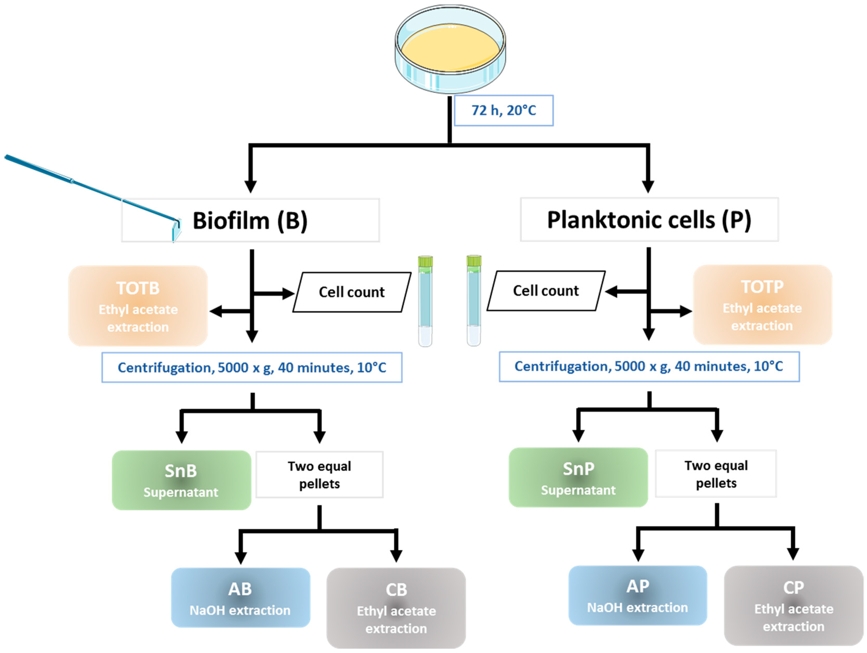
Featured Application

By analyzing extracts of biofilm formed by two marine bacteria and comparing them with planktonic extracts, we have shown that biofilm may induce the biosynthesis of potentially bioactive compounds and may open up new possibilities for compound discovery.

Abstract

In the marine environment, biofilm formation is an important lifestyle for microorganisms. A biofilm is comprised of cells embedded in an extracellular matrix that holds them close together and keeps the biofilm attached to the colonized surface. This predominant lifestyle and its main regulation pathway, namely quorum-sensing (QS), have been shown to induce specific bioactive metabolites. In this study, we investigated the biofilm formation by two marine bacteria belonging to the Vibrio species to discover potentially innovative bioactive compounds. We proposed a protocol to isolate biofilm extracts, to analyze their biochemical composition, and to compare them to planktonic cell extracts. Cells were grown attached to a plastic surface; extracts were prepared in water, NaOH, or in ethyl acetate and analyzed. Extracellular matrix components featured carbohydrates, proteins, lipids, and low amount of DNA. Carbohydrates appeared to be the main constituent of biofilm but also of the planktonic cell supernatant. Moreover, antimicrobial and QS-signaling activities were evidenced in extracts.

1. Introduction

Marine ecosystems host a wide microbial diversity with high biotechnological potential. Microorganisms have developed different strategies to adapt to the marine environment, which is characterized by a variety of dynamic conditions. In most cases, they grow into aggregates or biofilms attached on biotic or abiotic surfaces, such as artificial surfaces, rocks, particulate compounds, and organism surfaces [1]. This sessile microbial lifestyle is predominant and is characterized by cellular differentiation during adhesion followed by some physiological pathway modifications. Due to the importance of biofilms, a large number of reviews and book chapters have addressed this topic, as reviewed by Neu et al. [2].
A biofilm is a complex three-dimensional structured community featuring two main components: bacterial cells and the extracellular polymeric matrix (EPM) in which cells are embedded. The EPM is mainly produced by the cells themselves and can account for over 90% of the total dry mass of the biofilm [3]. A biofilm can be the cause of the corrosion and biofouling of marine equipment, resulting in its degradation and economic losses, and until now, most of the research efforts were focused on its inhibition [4]. In contrast, little is known about biofilm-specific metabolites. Indeed, a biofilm enables cell–cell interaction and communication by involving chemical signals, such as quorum-sensing (QS) autoinducers. Different metabolic pathways that remain inactive under the planktonic growth conditions are commonly used in the research laboratories where they can be activated [5,6]. Many biosynthetic pathways are regulated by QS, including the production of metabolites [7,8], of biosurfactants [9,10], and of antimicrobials [11].
Biofilm-specific EPM substances may be endowed with diverse physical/chemical properties and with biological activities and functionalities. Polysaccharides (PSs) have been evidenced as the most common components of a bacterial biofilm matrix [12,13,14]. Specific PSs can be produced during biofilm formation and development [14,15,16]. For example, cellulose production has been described at the air–liquid interface of static liquid cultures of bacteria [6,17]. Biotechnological applications of carbohydrate polymers are strongly influenced by their structural features and they have been investigated in various fields, including human health and the production of valuable metabolites [18,19,20,21].
Proteins in biofilms are the structural components of multimeric cellular appendages, such as flagella, pili, or fimbriae. They are involved in cell motility and in both the initial phase of biofilm formation and in its development [17]. Some of them are located on the cell surface to promote interactions with other extracellular matrix ligands, such as LecB in Pseudomonas aeruginosa [22] or the biofilm-associated proteins (Bap) [17,23]. Furthermore, many proteins are extracellular enzymes, such as proteases [12,24], which could be worth studying for their biotechnological potential. Among other less characterized biofilm components, extracellular DNA (eDNA) has also been found in EPMs and may play a structural role in biofilm development by promoting cell-to-cell interconnections [25,26,27]. Extracellular DNA release also takes part in both genetic material exchange and gene transfer [28]. Data on lipids as EPM components are still limited. In biofilms, two main groups have been identified, namely lipid vesicles, in particular in Vibrio fischeri biofilm [29], and lipoconjugates (glycolipid surfactants, lipopolysaccharides, lipoproteins) [30,31,32,33]. It appears then that, while EPM substances act to provide structure for the biofilm, their biotechnological potential is broad. New applications and markets for these molecules, including extracellular polymer-based bioproducts, are expected [34].
Biofilm recovery is the first step toward characterizing the specific metabolites. Subsequently, depending on whether the analysis targets a particular component or not, the samples can be homogenized, fractionated into separate cells and soluble components, usually via centrifugation, and/or submitted to extraction in aqueous or nonaqueous solvents [35,36,37,38,39]. Enzymatic digestion can also be applied to recover a targeted component [35,36].
In this study, we were interested in the biofilms formed by two Vibrio species known to produce exopolysaccharides in bioreactors. Their ability to form biofilms was evaluated. An extraction process was proposed to allow for the characterization of EPM components, as well as of both QS signaling and antimicrobial activities of the extracts. Furthermore, their features were compared with the metabolites synthesized by planktonic cells.

2. Materials and Methods

2.1. Bacterial Strains and Culture Conditions

Two bacterial strains isolated from deep-sea hydrothermal vents and stored in an Ifremer culture collection were studied. Vibrio diabolicus CNCM I-1629 was isolated from the dorsal integument of the hydrothermal vent polychaete annelid Alvinella pompejana near the active hydrothermal vent ELSA at a depth of 2600 m in a rift system of the East Pacific Rise (12°48.139 N, 103°56.309 W), accessed in 1991 during the French–American cruise HERO [40]. The MS969 strain was recovered from an artificial colonization module set at the Snake Pit site (23°22 N, 45°57 W—depth 3500 m) along the mid-Atlantic Ridge during the French oceanographic cruise “Microsmoke” in November 1995 [41]. Strains were stored in glycerol 20% (v/v) at −80 °C. They were grown in Zobell medium (4 g/L Tryptone, 1 g/L yeast extract, and 33.3 g/L aquarium salts).

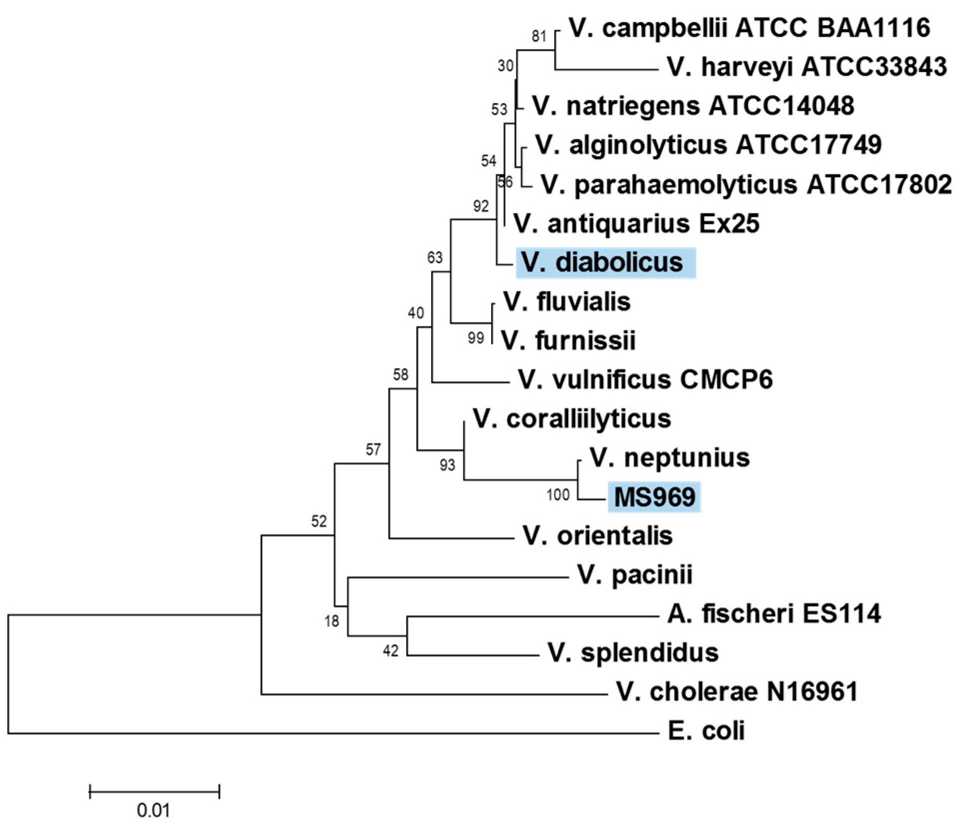
2.2. Identification of the MS969 Strain

The MS969 strain was cultured on Zobell medium at 28 °C over 24 h under 170 rpm agitation. A total of 500 µL of culture was centrifuged for 10 min at 7500 rpm. The pellet was washed with 300 µL phosphate buffer saline (PBS) solution and centrifuged again. Then, 200 µL of 10 mM Tris with 1mM EDTA (ethylenediaminetetraacetic acid) were added to the pellet; bacteria were lysed by heating at 95 °C for 10 min and cleared using centrifugation. The 16S rDNA gene was amplified directly from the supernatant (1 µL) using polymerase chain reaction (PCR) with 8F and 1489R primers as previously described [42]. PCR amplification was performed using a DreamTaq Green polymerase Master mix (Thermo Fisher Scientific, Illkirch, France). The sequencing of amplified products was carried out by Genoscreen (Lille, France). A consensus sequence was built using forward and reverse sequences with BioEdit software (Figure S1) [43]. Strain identification was performed using nucleotide Blast on the NCBI nr/nt Nucleotide Collection (National Center for Biotechnology Information). Phylogeny was inferred from the alignment with reference 16S rDNA sequences retrieved from Genbank using MEGA7 software (version 7.0) [44] (Table S1). The Escherichia coli type strain sequence (ATCC 11775T; X80725) was used as the outgroup. The optimal tree was built using the neighbor-joining method (NJ) [45]. The bootstrap support of nodes was assessed with 1000 replicates [46]. The evolutionary distances were computed using the Jukes–Cantor method [47].

2.3. Biofilm Assays

Bacterial strains were grown in Zobell medium at 30 °C under 180 rpm agitation for 24 h. After centrifugation, the pellet was rinsed twice with sterile phosphate buffer saline (PBS) solution and suspended in a PBS or Zobell medium to reach an average OD at 600 nm of 0.5. One volume of this suspension was used to inoculate various containers. After incubation under very gentle agitation, planktonic cells were removed, and the adherent biofilm was carefully rinsed with PBS and subsequently stained for 30 min with one volume of 0.5% (w/v) crystal violet (CV, Sigma-Aldrich, Lyon, France) prepared in water. The dye solution was removed and the biofilm was washed again thrice with PBS. The dye bound to the biofilm was solubilized via incubation for 15 min with one volume of 96% ethanol. The CV optical density was measured at 590 nm and directly reflected the amount of biofilm formed. It was normalized to the OD 600 nm of the broth.
Biofilm formation was evaluated in PBS and Zobell medium with or without 10 g/L glucose. Two incubation temperatures were tested. Three types of surfaces were also tested: glass, metal, and plastic containers; in that case, biofilm quantification using CV staining was normalized to the surface area. When possible, experiments were carried out in triplicates.

2.4. Biofilm Recovery

For each strain, the biofilm formation was performed in Zobell medium in 15 sterile plastic Petri dishes (90 mm diameter). A total of 10 mL of suspension was prepared in fresh Zobell as described in Section 2.3. and poured into each dish (OD 0.1 at 600 nm). Incubation was carried out at 20 °C for 72 h under very gentle agitation. After incubation, the upper broths containing the planktonic cells were collected and the plates were rinsed once with sterile PBS. The biofilm formed on the surface was collected with a sterile cell scraper with the addition of sterile PBS. The volume of the recovered adherent cells was measured and an aliquot was fixed in sterile seawater containing 2.5% formaldehyde; cells in the biofilm were counted in a Thoma chamber with an Optika microscope model B-192 (100×) (Dutscher, Brumath, France).

2.5. Preparation of Extracts

As illustrated in Figure 1, extracts TOTB and TOTP were obtained from the total biofilm sample and total planktonic cell broth, respectively, via ethyl acetate extraction carried out on 5 mL of each sample (see below). The remainder of each suspension was divided into two equal volumes and centrifuged (40 min, 8000× g, at 10 °C), giving two equal pellets and supernatants. Supernatants of the two same samples were mixed, filtered on a sterile 0.22 µm cellulose acetate membrane (VWR International, Fontenay-sous-Bois, France), dialyzed on a 3.5 kDa Spectra Por 3 membrane (Thermo Fisher Scientific, Illkirch, France), and freeze dried. Supernatants from the biofilm (B) and planktonic cells (P) were called SnB and SnP, respectively.
One of the pellets was extracted with NaOH via addition of 5 mL of 0.1% (w/v) NaOH. After 4 h of incubation, acetic acid was added to reach pH 7. The sample was centrifuged (30 min, 8000× g, at 10 °C) and the supernatant was filtered on a sterile 0.22 µm cellulose acetate membrane (VWR, France), dialyzed on a 3.5 kDa Spectra Por 3 membrane, and freeze dried. Extracts were called AB and AP for membrane “associated” compounds for biofilm and planktonic samples, respectively.
For the ethyl acetate extraction, 5 mL of ethyl acetate was added to 5 mL of the initial total biofilm cells and total planktonic cells, as well as to the second pellet, blended for 30 s, and agitated over 15 min to mix the aqueous and solvent phases. The solvent upper phase was collected and the aqueous phase was subjected to a second extraction using ethyl acetate. The two solvent phases were mixed and dried under N2. The resulting extracts were called CB and CP, respectively.
Sample volume and yield of each extract were registered.

2.6. Chemistry of the Biofilm Matrix

2.6.1. Composition Analysis

Composition analysis was carried out on SnB, SnP, AB, and AP extracts through colorimetric or fluorimetric methods. The protein content was analyzed using the bicinchoninic assay using a BCA kit (Sigma- Aldrich, Lyon, France) and bovine serum albumin as the standard [48]. Total carbohydrates were assayed with orcinol following the Tillmans and Philippi method modified by Rimington in 1931 [49,50] using glucose as the standard. DNA was measured with a Quant it Pico Green ds DNA assay (Invitrogen, Thermo Fisher Scientific, Illkirch, France); herring sperm DNA was used as the standard (Sigma- Aldrich, Lyon, France). To quantify the lipids, another extraction was necessary to remove carbohydrates that interfere with the sulfo-phospho-vanillin (SPV) assay in sulfuric acid. Lipids were extracted using the Bligh and Dyer method, as modified by Axelsson et al. [51,52]. Briefly, 250 µL of chloroform and 500 µL of methanol were blended with 200 µL of extract. Again, 250 µL of chloroform was added and mixed over 30 s, and finally, 250 µL of water was added. After centrifugation (5000 rpm, 10 min, 10 °C), the alcoholic upper phase was discarded and the organic phase was dried under N2 or overnight under an extraction hood. The SPV lipid assay was adapted from Frings et al.’s method [53]. The assay started by adding 200 µL of 98% sulfuric acid to the dried sample. After incubation for 15 min at 100 °C, and cooling under tap water, 500 µL of phospho-vanillin, prepared by mixing 120 mg isovanillin in 20 mL distilled water and adjusting the volume to 100 mL with 85% orthophosphoric acid, was added. The mixture was first incubated at 37 °C under agitation for 15 min, and subsequently at an ambient temperature in the dark for 45 min. The OD was read at 530 nm. Commercial olive oil at 0 to 2 mg/mL in sulfuric acid was used as the standard. Although the dosage assays were performed on a fraction of the suspension, each extract yield was calculated for the whole sample.

2.6.2. Electrophoretic Analysis of Proteins, DNA, and Glycopolymers

After the migration in 0.7% agarose gel was prepared as previously described [54], DNA was stained using SYBR SafeTM (Invitrogen, Thermo Fisher Scientific, France). The same gel was subsequently stained with the cationic carbocyanine dye Stains-All (SIGMA, France) to reveal polyanionic glycopolymers, as previously described [55]. Extracts were also analyzed using PAGE on 12% acrylamide gel and Stains-All staining or Coomassie Blue staining [56].

2.6.3. Carbohydrate Characterization Using Gas Chromatography

The monosaccharide composition was determined using gas chromatography according to the method of Kamerling et al. [57] and Montreuil et al. [58], as previously described [54].

2.6.4. Determination of Molecular Weight

Molecular weight profiles were determined using high-performance size-exclusion chromatography coupled with a multiangle light scattering detector (MALS, Dawn Heleos-II, Wyatt Technology, Santa Barbara, CA, USA), a differential refractive index (RI) detector (Optilab Wyatt technology, Santa Barbara, CA, USA), and a Prominence UV detector (Shimadzu Co., Kyoto, Japan). HPSEC system was composed of an HPLC Prominence system (Shimadzu Co., Kyoto, Japan), a PL aquagel-OH mixed, 8 μm (Agilent, Les Ulis, France) guard column (U 7.5 mm × L 50 mm), and a PL aquagel-OH mixed (Agilent, Les Ulis, France) separation column. The elution was carried out in 1 mL/min 0.1 M ammonium acetate.

2.7. Antimicrobial Activity Assay

Antimicrobial activities of the MS969 strain and of V. diabolicus extracts were assessed using thirteen indicator strains of aquacultural, food, or medical importance (Table S2) using a miniaturized method based on the spot-on-lawn assay [59]. Briefly, indicator strains, conserved in glycerol at −80 °C, were thawed at room temperature. One to four hundred microliters were inoculated in 2 mL of Trypto-casein soy broth (TSB, Biokar, Grosseron, Couéron, France) or brain heart infusion supplemented with 1.5% NaCl (BHIS, Biokar, Grosseron, Couéron, France) and incubated for 3 h in a 1.5 mL microtube at 30 °C. Then 500 µL of each suspension were seeded into 40 mL of 55 °C-incubated BHIS or TSB containing 1% agar and poured into a squared Petri dish 120 × 120 mm in size. After drying at ambient temperature, 5 µL of extracts were spotted onto the surface. Plates were incubated for 24–48 h at 30 °C. A clear halo around the spot provided evidence of the inhibition of the indicator strain growth.

2.8. Effect of Extracts on Quorum Sensing

The effect of samples on QS was evaluated by following the bioluminescence of Vibrio harveyi (LMG 4044). Each extract was prepared in water; thus, ethyl acetate extracts were dried under N2 and resolubilized in water. Aqueous extracts (20 µL) were added into the wells of flat- and clear-bottom 96-well white microplates containing 180 µL of Zobell medium seeded with a V. harveyi culture aliquot; this approximately corresponded to an initial concentration of 106 UFC/mL. A large volume of this suspension was prepared separately before distribution in order to ensure a good content homogeneity. Microplates were incubated at 30 °C under agitation. The optical density at 600 nm and bioluminescence were simultaneously monitored over 27 h using the multi-technology microplate reader VarioskanTMLUX (Thermo Fisher Scientific, Illkirch, France). The luminescence units were normalized by dividing them per OD 600 nm. Assays were performed four times and were statistically compared to 92 assays with V. harveyi alone using Student’s test.

2.9. Motility Assays

Motility was evaluated on Zobell or BHI (Biokar, Grosseron, Couéron, France) agar medium dried over 30 min under a security hood for microbiology. Agar was added at 0.4% (w/v) to detect swarming or 1.5% (w/v) to detect twitching. Ten microliter droplets of cultures were deposited on the gel and incubated at 25 °C. Petri dishes were photographed to follow the motility. When appropriate, glucose was added at a 30 g/L final concentration.

3. Results

3.1. Identification of the MS969 Strain

The analysis of the 16S rDNA sequence of the MS969 strain showed that this strain belongs to the Vibrio genus (Proteobacteria phylum, Vibrionaceae family) and its closest neighbors are Vibrio neptunius and Vibrio coralliilyticus, which are both representatives of the Coralliilyticus clade [60,61] (Figure 2).

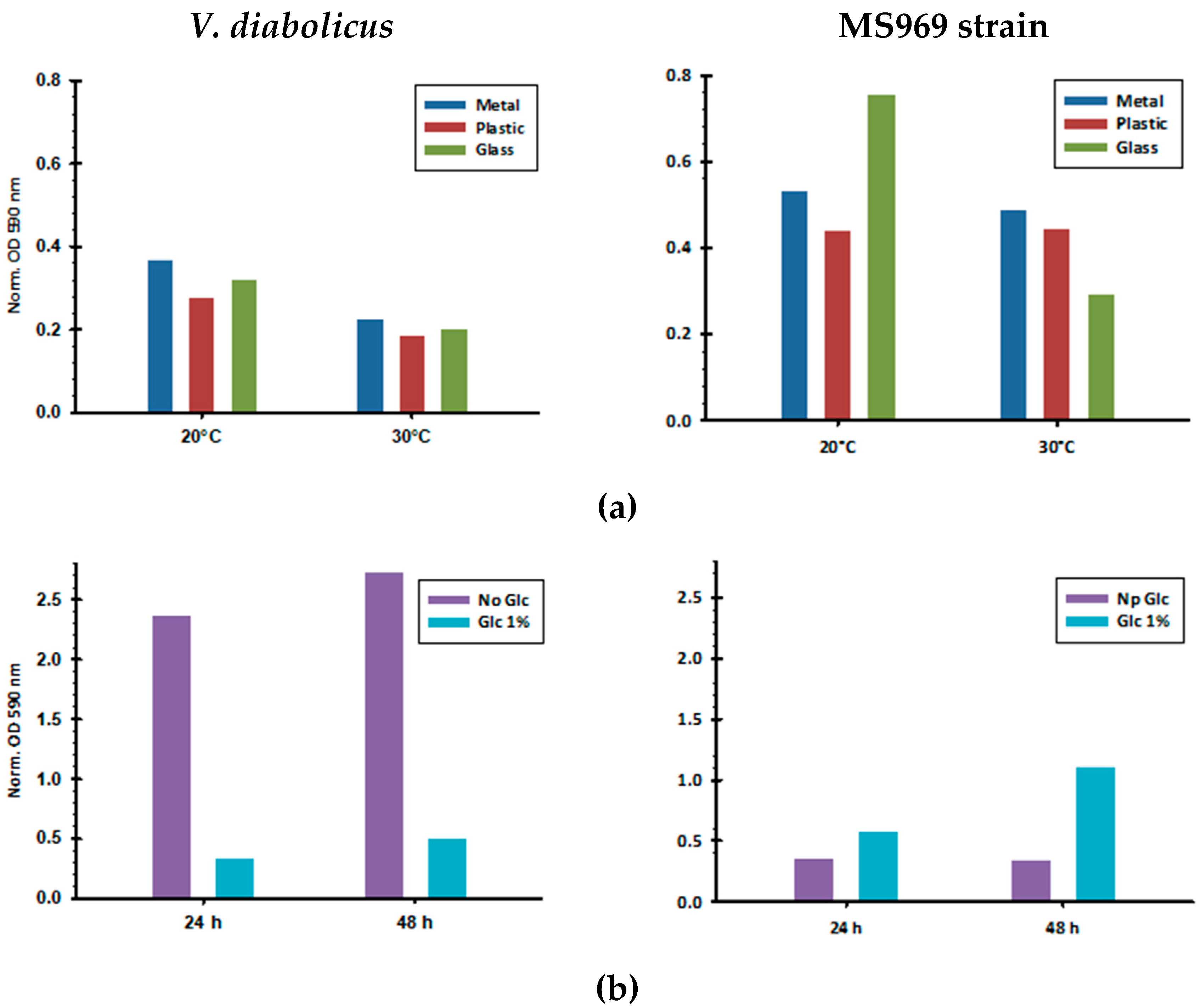
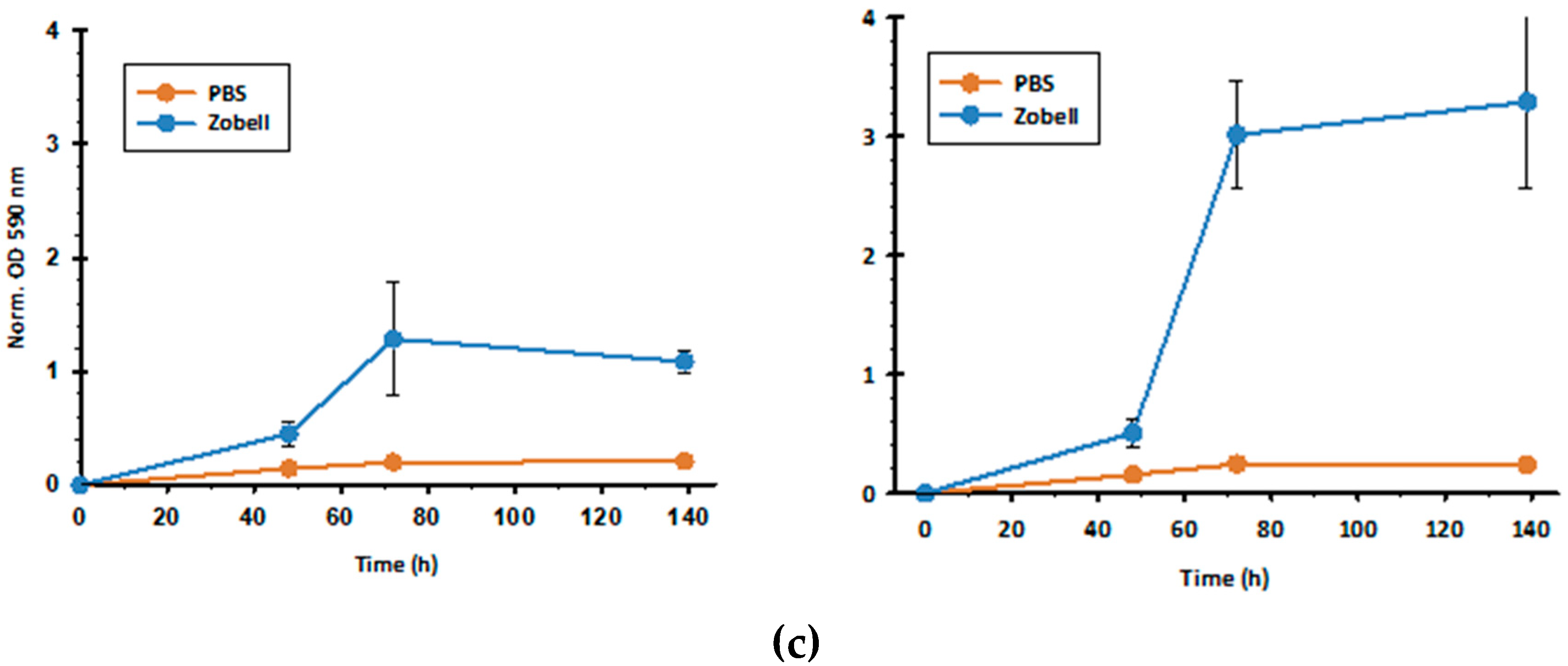
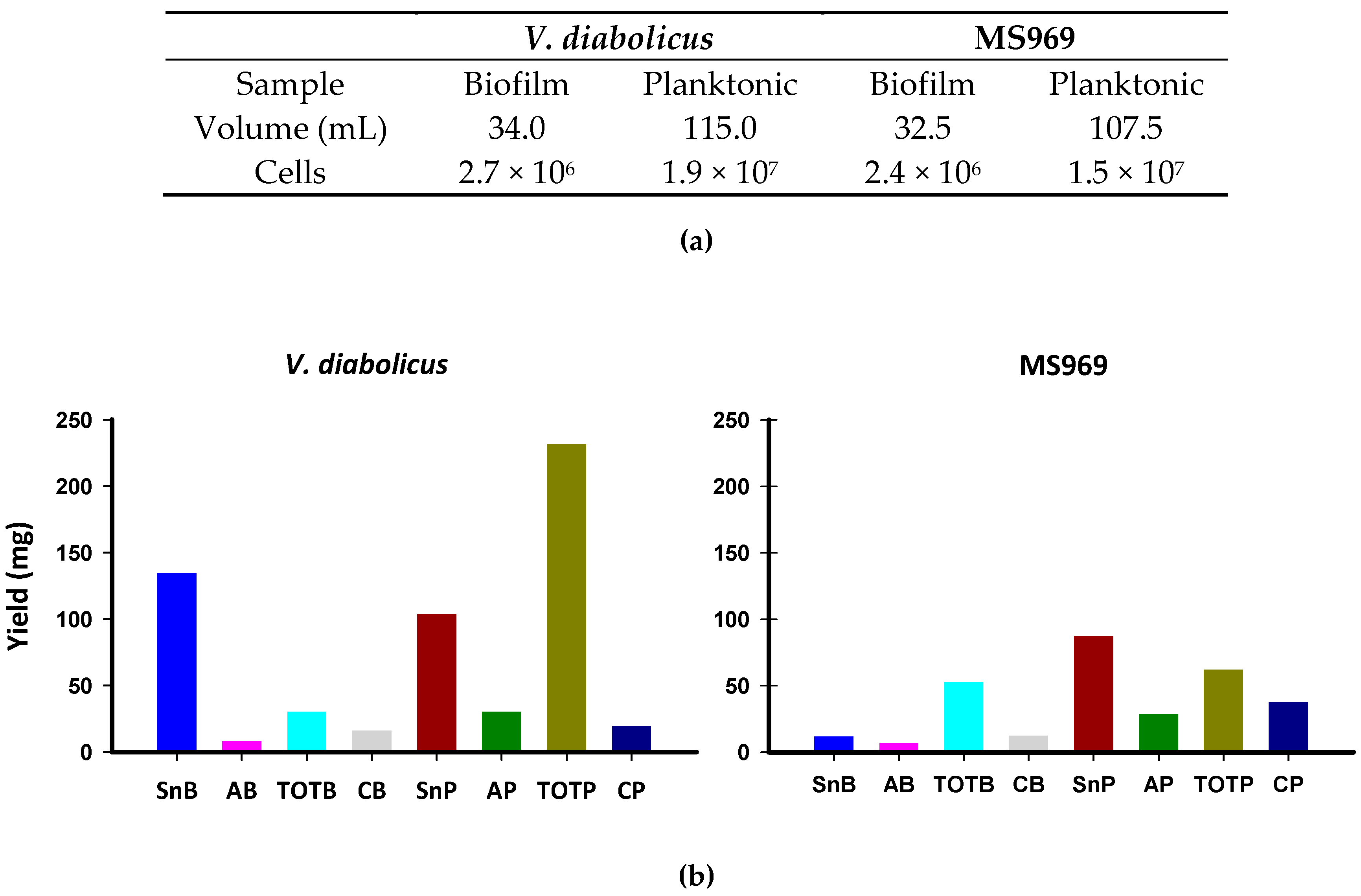
3.2. Biofilm Formation by V. diabolicus CNCM I-1629 and Vibrio sp. MS969

Two Vibrio species were chosen in this study; their origin allowed us to expect high biofilm formation. Both strains were isolated from deep-sea hydrothermal vents. Vibrio diabolicus CNCM I-1629 was an epibiont of a polychaete annelid and it produced a well-studied exopolysaccharide (EPS), namely HE800 [40,62,63], which resembles the well-known glycosaminoglycan hyaluronic acid. The Vibrio sp. MS969 strain was recovered on a colonization module and it was described for the first time in 2019 [54].
The biofilm formation by both strains was tested under various conditions, including temperature (20 °C and 30 °C), surface type (metal, plastic, glass), incubation time (24 h and 48 h), and suspension medium nature (PBS or Zobell medium, supplemented with glucose or not) (Figure 3). Whatever the bacterium, the biofilm amount was higher at 20 °C. V. diabolicus adhered better to metal, whereas the MS969 strain developed a more important biofilm on glass. Glucose inhibited V. diabolicus biofilm formation, while it promoted that of the MS969 strain. In the latter case, it was possible that glucose supported the bacterial growth, and thus, the observed effect on the biofilm was most probably indirect.
Subsequent experiments were performed at 20 °C without glucose and on a plastic surface.
The suspension of cells in the Zobell nutritive medium was tested and compared with PBS. Figure 3c shows that the suspension in the nutritive medium was better for the biofilm formation of both strains, suggesting a better cell development. The biofilm amount reached a plateau after 72 h of incubation.

3.3. Preparation of Biofilm Extracts

Under the optimal conditions determined in the above section, a strong biofilm was formed at the solid interface on the bottom of the plates and was recovered with a cell scraper. Different extracts were prepared, both from the biofilm sample and planktonic cells (Figure 1). The cell numbers in biofilm and planktonic samples were counted and the dry matter recovery yield was measured (Figure 4).
For both strains, cell numbers were in the same order of magnitude and planktonic cells were almost ten times higher than those recovered from the biofilm. Most of the extracted matter was recovered either from supernatants (SnB and SnP) or from ethyl acetate extraction of the total suspension (TOTB or TOTP). Nevertheless, for all the extracts, the recovery yield was higher from V. diabolicus, especially for the total ethyl acetate extract of planktonic suspension (TOTP). Soluble compounds collected in the supernatants and NaOH extracts (SnP or SnB and AP or AB, respectively) were largely equivalent between the biofilm and planktonic cells for V. diabolicus. However, for the MS969 strain, the extract amounts recovered from planktonic cells were always higher than the equivalent extract from biofilm cells.

3.4. Biofilm Water-Soluble Components

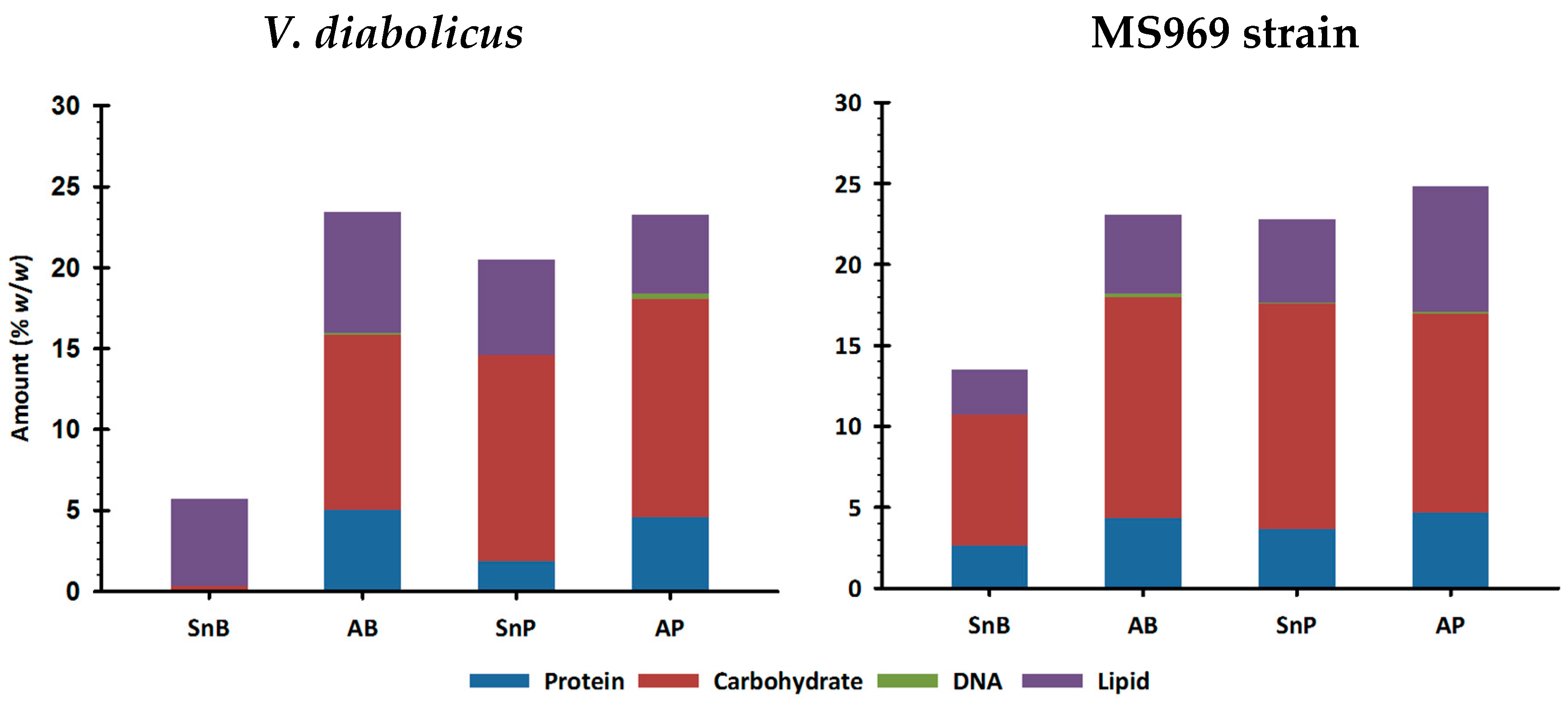
3.4.1. Biochemical Composition Determination Using Colorimetric or Fluorimetric Assays

Carbohydrate, protein, nucleic acid, and lipid amounts were quantified in water-soluble extracts using colorimetric and fluorimetric assays (Figure 5).
The total amount of analyzed molecules was limited to a maximum of 23% (w/w) of the dry mass. In several of the assays, the hydrolysis into monomers was necessary. Therefore, in this case, the hydrolysis might be incomplete, driving under-evaluated amounts. In addition, although the water-soluble extracts (Sn and A) were dialyzed, the presence of macromolecules, especially the polyanionic ones, might hinder the total elimination of salt. Nevertheless, carbohydrates represented the main component, except for the biofilm supernatant of V. diabolicus (SnB). We noticed that extracts were rich in lipids, a component that is rarely assayed. It represented up to 7.7% (w/w) of the extracts. In addition, the extracts contained proteins (except the SnB of V. diabolicus) and very low amounts of DNA.
Carbohydrates, proteins, and DNA have been the main compounds studied in biofilms. Conversely, lipids have rarely been evaluated. The compounds that constitute the EPM may probably favor the colonization of various surfaces. In addition, they play an important role in the stability of the EPS network through various interactions [12,36,64,65]. Our results are rather consistent with previous work since polysaccharides are usually described as the main component of Vibrio biofilms [66].

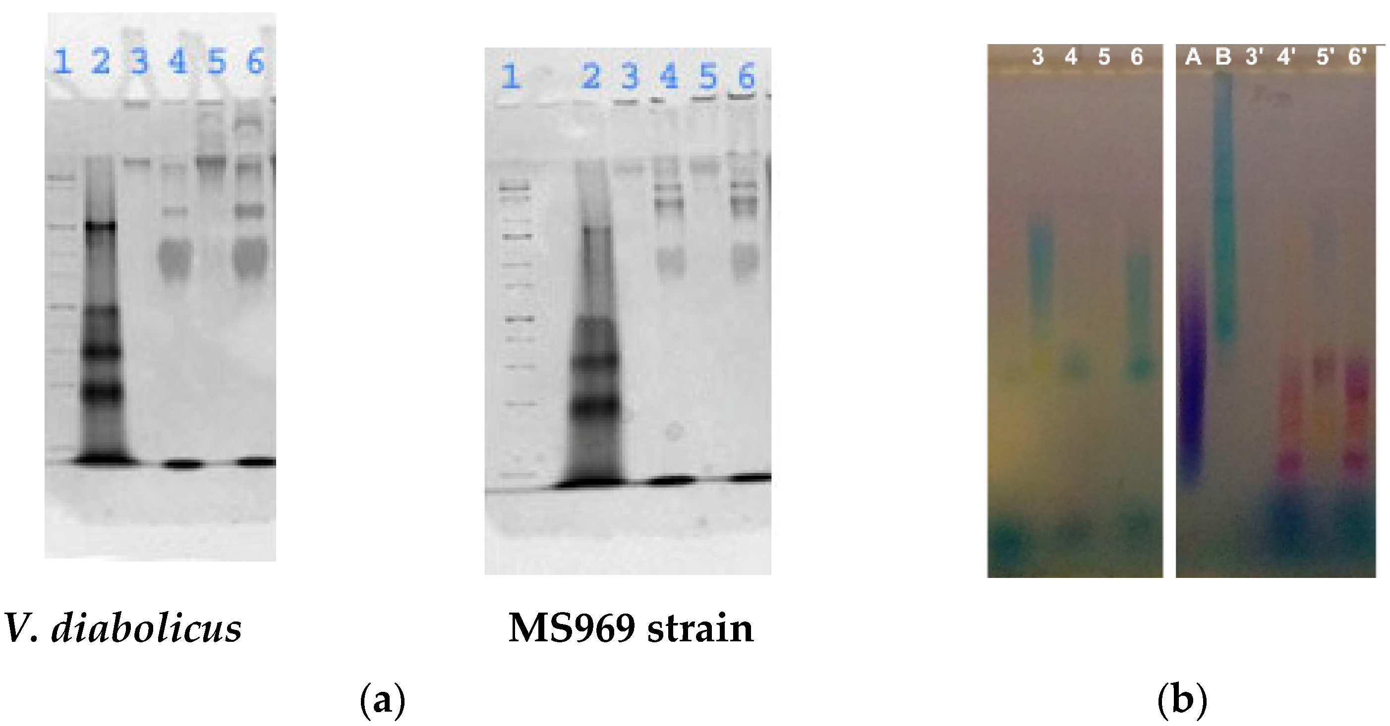
3.4.2. Electrophoretic Analysis

Each extract prepared either via centrifugation or by NaOH extraction was analyzed using electrophoresis (Figure 6).
Supernatants (Sn) of either surface-adhered (SnB) or planktonic cells (SnP) displayed the same migration pattern. NaOH extracts (AB and AP) also featured the same pattern on PAGE and displayed several bands. Agarose gel analysis showed that the V. diabolicus SnB extract contained a high-molecular-weight polydisperse compound that resembled the HE800 EPS produced by V. diabolicus on Zobell supplemented with 30 g/L glucose [54], although the colorimetric assay detected only traces of carbohydrates (Figure 5). This suggests that degradation of biofilm extracts into monomers for the colorimetric assay was not efficient. A similar band was detected in the planktonic NaOH extract (AP, lane 6). On the other hand, the extracts from the MS969 strain showed pink bands in the lower part of the gel, a color often observed for some sulfated glycosaminoglycans [69].
Neither DNA nor protein was detected in the electrophoresis gels. This result was correlated to the low amount of DNA and protein quantified using the colorimetric and fluorometric methods, as shown in Figure 5.

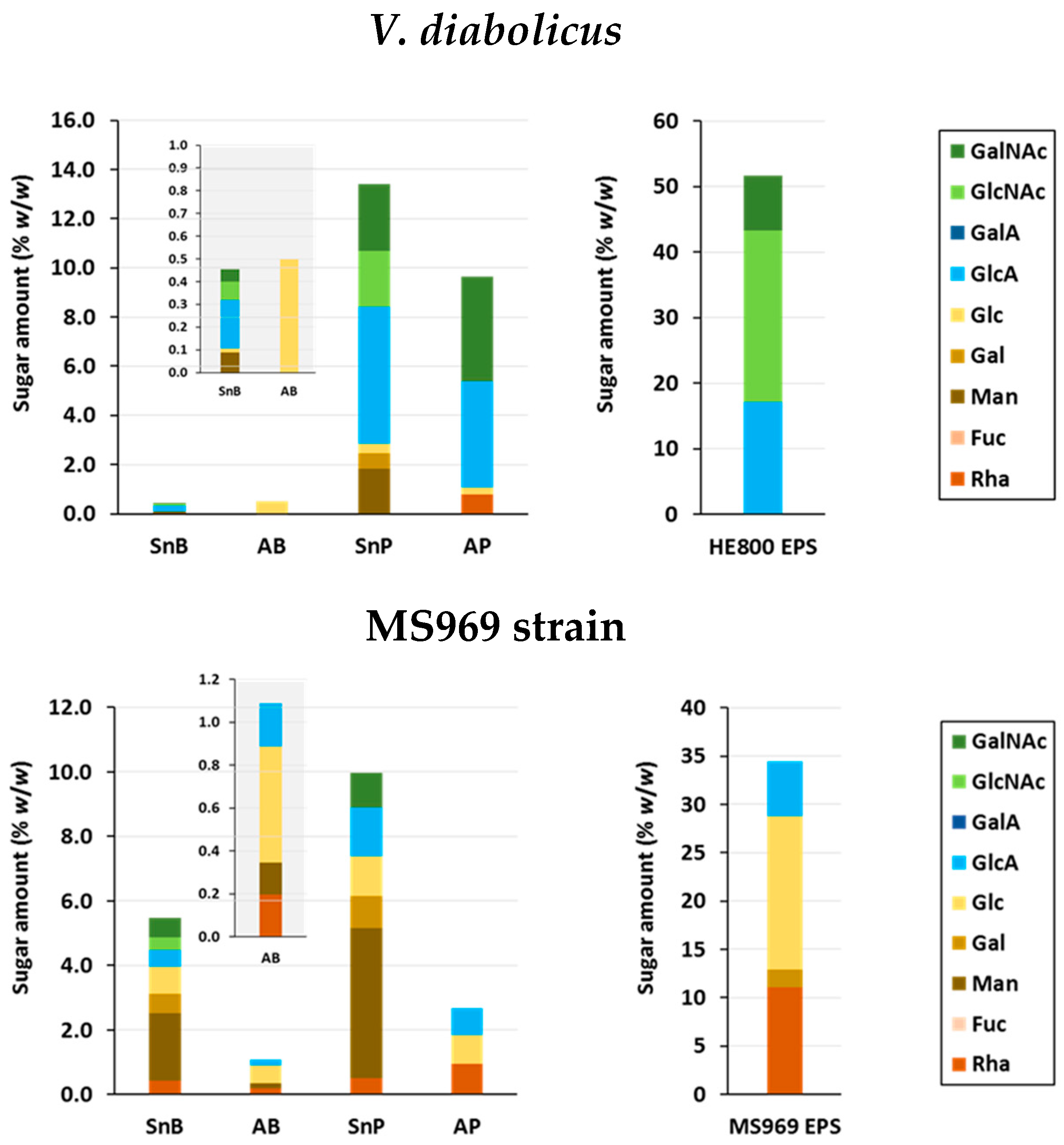
3.4.3. Osidic Composition

The osidic composition of samples were determined using gas chromatography (Figure 7).
The analyzed osidic amount of all extracts was low, especially in the biofilm cells. It reached a maximum of 10% (w/w) of dry mass. This might be explained by the uncomplete HCl/MeOH hydrolysis of these complex samples required by the gas chromatography analysis. Carbohydrate compounds seemed more difficult to recover from biofilm samples than from planktonic ones, especially the SnB and AB of V. diabolicus. Nevertheless, both V. diabolicus biofilm and planktonic cells soluble extracellular compounds (SnB and SnP) seemed to exhibit a similar osidic pattern. Glucuronic acid (GlcA) was the main component, followed by N-acetylglucosamine (GlcNAc) and N-acetylgalactosamine (GalNAc) in similar amounts. These two extracts also contained the neutral sugars mannose (Man), galactose (Gal), and glucose (Glc). The NaOH extract of biofilm cells (AB) consisted of glucose only, and that of planktonic cells (AP) featured GlcA and GalNAc as the main components. No GlcNAc was detected in AP.
GalNAc, GlcA, and neutral sugars (Glc, Gal, Man, and rhamnose (Rha)) were detected in soluble exopolymers of the MS969 strain biofilm, as well as in planktonic broth supernatant (SnB and SnP). SnB also contained GlcNAc. Mannose was the main component of SnB and SnP. On the contrary, NaOH extracts (AB and AP) featured Glc, Rha, and GlcA. In addition, Man was detected in AB.

3.4.4. Size Exclusion Chromatographic Profiles
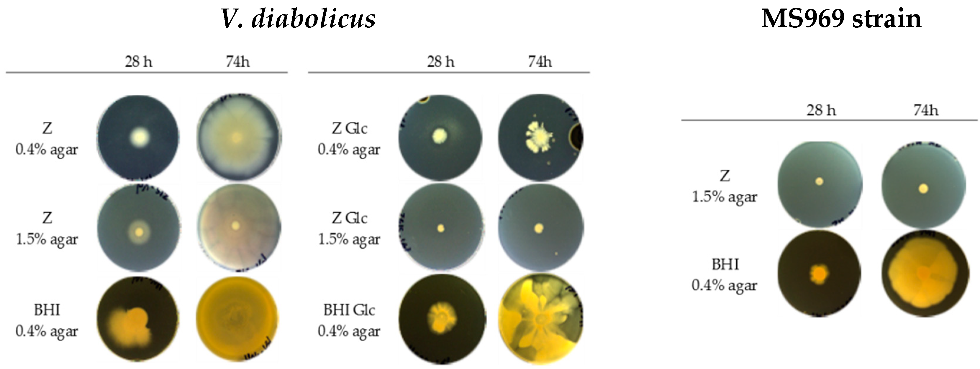
Each extract was analyzed using HPSEC-MALS to determine the molecular weight pattern. Elution profiles with refractive index (RI) and ultraviolet (UV) detection at 280 nm were obtained (Figure 8).
The refractometric index is related to the amount of carbohydrates and nucleic acids, and UV profiles are related to proteins, peptides, and other components that absorb wavelength of 280 nm. Both chromatographic profiles were in accordance with the previous observations. Indeed, a large salt peak was detected using RI at the column total volume (12-min elution). This might partly explain the low level of the total amount of analyzed molecules (Figure 5 and Figure 7).
All extracts of V. diabolicus exhibited three families of peaks (7–8 min, 8–10 min, and 10–11 min retention time) in the RI profile, corresponding to high-, medium-, and low-molecular-weight carbohydrates as determined by the MALS detector. Only SnP (planktonic cells supernatant) contained a high molecular weight carbohydrate with a small UV absorption. This could be attributed to an anionic polysaccharide. Medium-molecular-weight compounds were found in AB and AP extracts (NaOH extracts) with a low UV absorption. Both extracts presented quite similar peaks with a higher intensity for AP.
RI profiles of MS969 extracts were quite different in molecular weight repartition. There was no very high molecular weight compound in SnB extract, but all the extracts featured a peak at 8 min elution time, which absorbed UV. The AP extract presented two peaks, at 8 and 9.5 min, whereas AB extract peaks were detected at 8 min and 10.5 min. The UV profiles were very similar from one extract to another.
Since both Vibrio strains produced EPSs of biotechnological importance when glucose was added to planktonic growth medium, we were interested in the identification of those produced by the biofilm and planktonic cells. Sutherland et al. [15] suggested that EPSs present in biofilms certainly closely resemble the corresponding polymers synthesized by planktonic cells. The HE800 EPS is produced by V. diabolicus in shaken flasks or in aerated bioreactors containing the Zobell medium supplemented with glucose; its linear tetrasaccharidic repeating unit consists of two GlcA residues and two N-acetyl hexosamines: one GlcNAc and one GalNAc [68]. Although GC analysis of SnB gave only very low amounts of sugars, its overall osidic profile, together with SnP, seemed to be similar to the HE800 EPS. However, mannose was present in these two extracts, suggesting that another compound was co-extracted. Sugars recovered in the AB extract were also in very low amounts but only glucose was identified. This may suggest that the glycopolymer (as indicated by HPSEC-MALS) was similar to cellulose. Bacterial cellulose has already been described in biofilms of other Vibrionaceae, such as Aliivibrio fischeri [21]. Comparative sequence analyses also revealed the presence of a cellulose biosynthesis cluster in diverse bacteria, especially several Proteobacteria strains, of which the Vibrio species is a member [21].
The EPSs produced in excess of carbon source (glucose) by the MS969 strain cultured in shaken flasks was composed of neutral sugars including rhamnose, galactose, glucose, and glucuronic acid. Interestingly, it was the first time that a sulfate group, which is known to promote biological activities, was identified in an EPS produced by a Vibrio strain [54]. The pink color seen in the electrophoresis in this study might also suggest that extracts prepared from MS969 (SnB, SnP, AB, and AP) contained sulfated compounds. SnB and SnP osidic composition resembled that of the MS969 exopolysaccharide. Mannose was present in both extracts and seemed to be a common supplemental sugar in Sn extracts of both strains. This sugar could result from the extraction of cell membrane compounds. SnB also contained GlcNAc. Nevertheless, in V. cholerae EPSs (VPS), GlcNAc has already been described in 20% of the molecules, the rest of them contained Glc instead [66]. The AB and AP osidic profiles were similar. The RI profile from HPSEC analysis also suggested that carbohydrates may be slightly different in cell supernatants and cell surface (AB and AP extracts). It has already been found that exopolysaccharides have a great significance in biofilm formation by Vibrio spp. The symbiosis polysaccharide (Syp) is involved in the colonization of the squid Euprymna scolopes by V. fischeri [70]. Syp-like clusters have also been found in V. diabolicus and V. neptunius [71]. Other EPSs critical for biofilms have been described in Vibrio parahaemolyticus [72] or in Vibrio vulnificus [73].

3.5. Motility Assay

Colonization of a solid medium was tested at two agar concentrations on Zobell and BHI media that was eventually supplemented with 30 g/L glucose (Figure 9).
The motility of V. diabolicus on each medium (Zobell or BHI with 0.4 or 1.5% agar) was high since each Petri dish was totally colonized in 3 days. Movement in small groups on the nutritive surface was clearly visible. In media supplemented with glucose, motility on the BHI-based medium was conserved but reduced on Zobell 0.4% agar and inhibited on Zobell 1.5% agar.
The MS969 strain displayed a high motility on BHI 0.4% agar medium and colonized the whole Petri dish (90 mm diameter) in 3 days. This was also the case on Zobell with 0.4% agar but the mat appeared less dense (not shown). No motility was detected when agar was at a 1.5% concentration. Glucose, when added at 30 g/L, completely inhibited the biofilm and the strain was only able to form a colony with a maximum 14 mm diameter.
Three bacterial motility types have been described. First, swimming is an individual movement in liquid medium. Second, bacteria display swarming when they move in small groups and in an organized way on semi-solid surface. Depending on strain, cells undergo a morphology change by lengthening and one or two flagella forms, which allows for a rapid colonization of new surfaces. Swarming was detected on 0.4% (w/v) agar medium. Third, twitching mobility is due to type IV pili whose contraction allows the bacterial cells to move by slipping in small groups on a solid surface; it was evaluated on 1.5% agar medium. Flagella involved in swimming and swarming play a role during the adhesion initial phase of biofilm formation, whereas type IV pili participate in the biofilm maturation [74]. Flagella and pili are often present in Vibrio cells [75,76]. Raguenes et al. [40] have shown that V. diabolicus cells display one single flagellum in liquid medium and many lateral pili on a solid medium, which can contribute to swarming. In this study we saw that both Vibrio species displayed swarming motility and V. diabolicus also exhibited twitching motility.

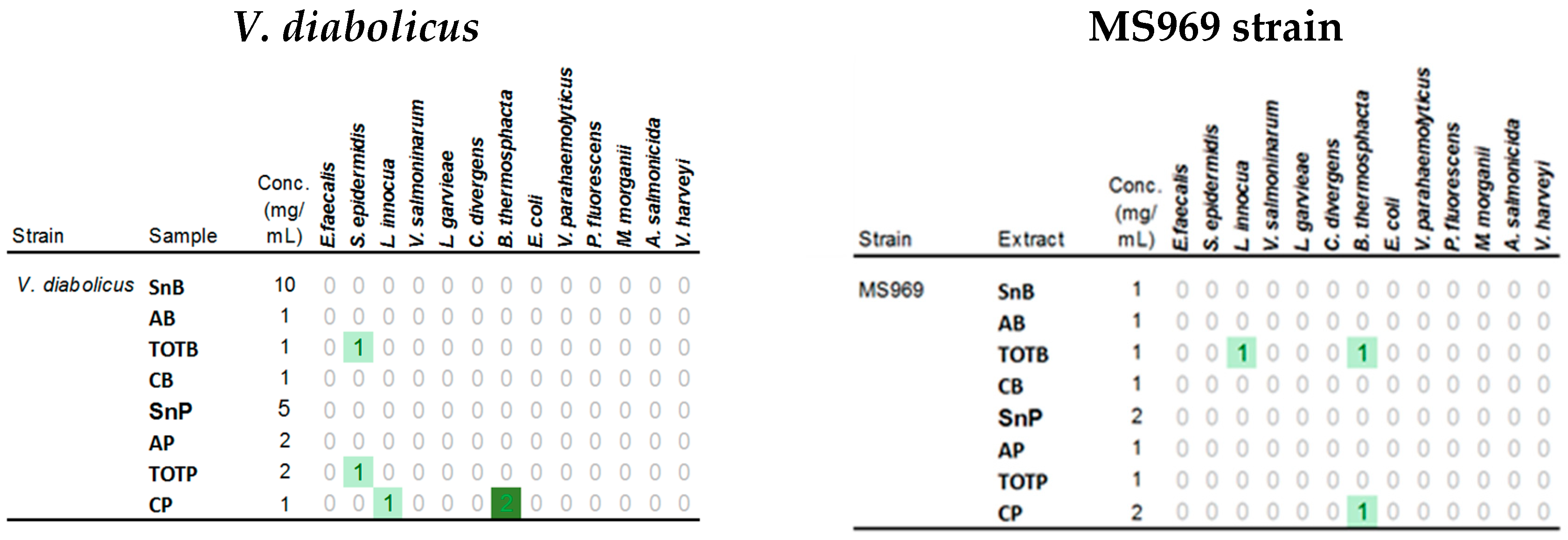
3.6. Antimicrobial Activities

Screening for antimicrobial activities of bacterial strains was carried out using the spot-on-lawn approach in which indicator cultures were grown within the solid medium [59]. Extracts of biofilm and planktonic cells produced in this study were tested against 13 indicator strains (Table S2, Figure 10).
For both strains, extracts obtained with ethyl acetate were the only ones to display some antimicrobial activities. Interestingly, inhibitory activities of both strains were detected against Listeria innocua and Brochothrix thermosphacta. In addition, V. diabolicus was able to inhibit the growth of Staphylococcus epidermidis (both TOTB and TOTP extracts). TOTB and CP extracts of the MS969 strain were able to limit the growth of B. thermosphacta.
Swarming can hinder the detection of antimicrobial activities using the classical spot-on-lawn method in which both the antimicrobial producer and indicator cultures are grown simultaneously on and in, respectively, the same solid media under the same conditions of incubation [59]. Preparation of extracts was therefore an efficient approach to studying the antimicrobial activities of swarming bacteria. In this study, we reported antimicrobial activities targeting S. epidermidis, L. innocua, and B. thermosphacta. Potential antibacterial compounds were previously identified in Vibrio spp. [77], especially in V. neptunius [78]. Moreover, we have shown that the extracts prepared from biofilm-grown cells may produce specific biologically active compounds. Although it needs to be confirmed, this result opens the way to the investigation of new antimicrobial compounds.

3.7. Quorum-Sensing Signaling

All extracts were added into microplate wells containing Zobell medium seeded with a V. harveyi culture aliquot. V. harveyi growth and quorum-sensing regulated bioluminescence were monitored over 24 h (Figure 11).
None of the extracts influenced V. harveyi growth but its luminescence was decreased by the NaOH extracts of V. diabolicus (AB and AP) and by the NaOH extract of the MS969 biofilm (AB). Interestingly, the amount of light produced was increased by the biofilm extracts of MS969 (SnB and TOTB) suggesting that the MS969 strain released quorum-sensing (QS) signal molecules in the extracellular matrix.
Quorum-sensing signaling is an important regulatory pathway in Vibrio species and has been well-studied in A. fischeri [79] and V. harveyi [80]. Bioluminescence in V. harveyi is regulated by the N-acyl homoserine lactone-based system, as well as two other systems, including an autoinducer AI-2 [81]. In the MS969 strain, biofilm-specifically released compounds have been shown to promote bioluminescence. This suggests that a quorum-sensing pathway similar to one of the three V. harveyi QS systems was involved in biofilm formation of the MS969 strain. The V. diabolicus biofilm may be regulated by signal molecules not recognized by V. harveyi to produce bioluminescence, or most probably, they were not enough to be effective.

4. Conclusions

In this study, we described a relevant protocol to characterize the biofilm components of two Vibrio species, both in total biofilm and in several aqueous or organic solvent extracts. The chemical composition, as well as QS signaling and antimicrobial activities of the extracts, were analyzed. In the future, the procedure should be further improved to better eliminate salts before the hydrolysis steps for colorimetric and GC analyses. The global composition (carbohydrate, protein, lipid, and nucleic acid) of the biofilm matrix showed that carbohydrate was the main component. The carbohydrates chemical composition and molecular weight may be slightly different in the biofilm cell supernatants and cell surface. Interestingly, in one case, a biofilm-specific carbohydrate, consisting only in glucose, was identified. The biofilm appeared thus as a specific bacterial phenotype. Therefore, it is potentially a source of biologically active compounds, such as polymers endowed with better biological and functional properties than those already known. Depending on their nature, chemical composition, and structure, the biotechnological potential of the biofilm polymer can be considered in a wide range of uses, such as cryoprotectants, emulsifiers, heavy metal chelators, bioactives for human health and well-being, and as a source of high-value sugar monomers or enzymes [18,19,20,82].
Antimicrobial resistance is a global public health issue affecting both human and animal health. At the same time, the presence of bacterial contaminants in foodstuffs is causing important economic losses. Therefore, it is urgent to market new antimicrobial molecules targeting, in particular, multi-resistant pathogenic strains, and thus presenting new mechanisms of action. Furthermore, while bacterial genome sequencing shows the existence of numerous genetic clusters that encode the biosynthesis of bioactive metabolites, a large majority of them remain inactive under the planktonic growth mode commonly used in lab experiments [83]. By investigating the underlying regulation pathways of biofilms, such as QS signaling, we will pave the way toward the future production of innovative metabolites.

Supplementary Materials

The following are available online at https://www.mdpi.com/2076-3417/9/22/4971/s1, Figure S1: 16S rDNA sequence of the MS969 strain, Table S1: 16S rDNA sequences of reference strains used in the phylogeny investigation, Table S2: Indicator strains used for antimicrobial activities screening.

Author Contributions

Conceptualization, D.P., D.H.-H., and C.D.-L.; methodology, D.P., F.F., and C.D.-L.; investigation and result analysis, D.P., F.F., L.M., L.K., S.B., C.S., V.V.-B., and C.D.-L.; writing—original draft preparation, D.P., V.V.-B., and C.D.L.; writing—review and editing, C.S., D.H.-H., S.C.-J., and C.D.-L.; supervision, C.D.-L.; project administration, C.D.-L.; funding acquisition, D.P., D.H.-H., and C.D.-L.

Funding

This research received Ifremer internal funding through the Vi-LAB project.

Acknowledgments

The authors are greatly indebted to other contributors to the Vi-LAB project from Marion Sorée and Solen Lozach.

Conflicts of Interest

The authors declare no conflict of interest. The funders had no role in the design of the study; in the collection, analyses, or interpretation of data; in the writing of the manuscript, or in the decision to publish the results.

References

  1. Flemming, H.-C.; Wuertz, S. Bacteria and archaea on Earth and their abundance in biofilms. Nat. Rev. Genet. 2019, 17, 247–260. [Google Scholar] [CrossRef]
  2. Neu, T.R.; Lawrence, J.R. Chapter 37—Extracellular polymeric substances in microbial biofilms. In Microbial Glycobiology; Academic Press: San Diego, CA, USA, 2010; pp. 733–758. [Google Scholar]
  3. Flemming, H.-C.; Wingender, J. The biofilm matrix. Nat. Rev. Genet. 2010, 8, 623–633. [Google Scholar] [CrossRef]
  4. de Carvalho, C.C.C.R. Marine Biofilms: A successful microbial strategy with economic implications. Front. Mar. Sci. 2018, 5, 126. [Google Scholar] [CrossRef]
  5. Nakhamchik, A.; Wilde, C.; Rowe-Magnus, D.A. Cyclic-di-GMP Regulates Extracellular Polysaccharide Production, Biofilm Formation, and Rugose Colony Development by Vibrio vulnificus. Appl. Environ. Microbiol. 2008, 74, 4199–4209. [Google Scholar] [CrossRef] [PubMed]
  6. Dickschat, J.S. Quorum sensing and bacterial biofilms. Nat. Prod. Rep. 2010, 27, 343–369. [Google Scholar] [CrossRef] [PubMed]
  7. Zeng, Z.; Cai, X.; Wang, P.; Guo, Y.; Liu, X.; Li, B.; Wang, X. Biofilm Formation and Heat Stress Induce Pyomelanin Production in Deep-Sea Pseudoalteromonas sp. SM9913. Front. Microbiol. 2017, 8, 1822. [Google Scholar] [CrossRef] [PubMed]
  8. Mitra, S.; Thawrani, D.; Banerjee, P.; Gachhui, R.; Mukherjee, J. Induced Biofilm Cultivation Enhances Riboflavin Production by an Intertidally Derived Candida famata. Appl. Biochem. Biotechnol. 2012, 166, 1991–2006. [Google Scholar] [CrossRef] [PubMed]
  9. Mnif, I.; Ghribi, D. Glycolipid biosurfactants: Potential related biomedical and biotechnological applications. Carbohydr. Res. 2015, 416, 59–69. [Google Scholar]
  10. Victor, I.U.; Kwiencien, M.; Tripathi, L.; Cobice, D.; McClean, S.; Marchant, R.; Banat, I.M. Quorum sensing as a potential target for increased production of rhamnolipid biosurfactant in Burkholderia thailandensis E264. Appl. Microbiol. Biotechnol. 2019, 103, 6505–6517. [Google Scholar] [CrossRef]
  11. Schmidt, S.; Blom, J.F.; Pernthaler, J.; Berg, G.; Baldwin, A.; Mahenthiralingam, E.; Eberl, L. Production of the antifungal compound pyrrolnitrin is quorum sensing-regulated in members of theBurkholderia cepaciacomplex. Environ. Microbiol. 2009, 11, 1422–1437. [Google Scholar] [CrossRef]
  12. Flemming, H.-C.; Wingender, J. Relevance of microbial extracellular polymeric substances (EPSs)—Part I: Structural and ecological aspects. Water Sci. Technol. 2001, 43, 1–8. [Google Scholar] [CrossRef] [PubMed]
  13. Moppert, X.; Fardeau, M.; Carrero, J.; Valadéz-González, A.; Guezennec, J.; Ortega-Morales, B.; Santiago-García, J.; Chan-Bacab, M.; Miranda-Tello, E.; Bartolo-Pérez, P.; et al. Characterization of extracellular polymers synthesized by tropical intertidal biofilm bacteria. J. Appl. Microbiol. 2007, 102, 254–264. [Google Scholar]
  14. Limoli, D.H.; Jones, C.J.; Wozniak, D.J. Bacterial Extracellular Polysaccharides in Biofilm Formation and Function. Microbiol. Spectr. 2015, 3, 223–247. [Google Scholar] [CrossRef] [PubMed]
  15. Sutherland, I.W. Biofilm exopolysaccharides: A strong and sticky framework. Microbiol. 2001, 147, 3–9. [Google Scholar] [CrossRef]
  16. Ostapska, H.; Howell, P.L.; Sheppard, D.C. Deacetylated microbial biofilm exopolysaccharides: It pays to be positive. PLoS Pathog. 2018, 14, e1007411. [Google Scholar] [CrossRef]
  17. Pamp, S.J.; Gjermansen, M.; Tolker-Nielsen, T. The biofilm matrix—A sticky framework. In The Biofilm Mode of Life; Kjelleberg, S., Givskov, M., Eds.; Horizon Press: Wymondham, UK, 2007; pp. 37–69. [Google Scholar]
  18. Senni, K.; Pereira, J.; Gueniche, F.; Delbarre-Ladrat, C.; Sinquin, C.; Ratiskol, J.; Godeau, G.; Fischer, A.-M.; Helley, D.; Colliec-Jouault, S. Marine Polysaccharides: A Source of Bioactive Molecules for Cell Therapy and Tissue Engineering. Mar. Drugs 2011, 9, 1664–1681. [Google Scholar] [CrossRef]
  19. Redcorn, R.M.; Hillman, E.T.; Solomon, K.V.; Engelberth, A.S. Xanthobacter-dominated biofilm as a novel source for high-value rhamnose. Appl. Microbiol. Biotechnol. 2019, 103, 4525–4538. [Google Scholar] [CrossRef]
  20. Caruso, C.; Rizzo, C.; Mangano, S.; Poli, A.; di Donato, P.; Finore, I.; Nicolaus, B.; di Marco, G.; Michaud, L.; Giudice, A.L. Production and biotechnological potential of extracellular polymeric substances from sponge-associated antarctic bacteria. Appl. Environ. Microbiol. 2018, 84, e01624-17. [Google Scholar] [CrossRef]
  21. Augimeri, R.V.; Varley, A.J.; Strap, J.L. Establishing a Role for Bacterial Cellulose in Environmental Interactions: Lessons Learned from Diverse Biofilm-Producing Proteobacteria. Front. Microbiol. 2015, 6, 402. [Google Scholar] [CrossRef]
  22. Tielker, D.; Hacker, S.; Loris, R.; Strathmann, M.; Wingender, J.; Wilhelm, S.; Rosenau, F.; Jaeger, K.-E. Pseudomonas aeruginosa lectin LecB is located in the outer membrane and is involved in biofilm formation. Microbiology 2005, 151, 1313–1323. [Google Scholar] [CrossRef]
  23. Lasa, Í.; Penadés, J.R. Bap: A family of surface proteins involved in biofilm formation. Res. Microbiol. 2006, 157, 99–107. [Google Scholar] [CrossRef] [PubMed]
  24. Sinsabaugh, R.L.; Repert, D.; Weiland, T.; Golladay, S.W.; Linkins, A.E. Exoenzyme accumulation in epilithic biofilms. Hydrobiologia 1991, 222, 29–37. [Google Scholar] [CrossRef]
  25. Seper, A.; Fengler, V.H.I.; Roier, S.; Wolinski, H.; Kohlwein, S.D.; Bishop, A.L.; Camilli, A.; Reidl, J.; Schild, S. Extracellular nucleases and extracellular DNA play important roles in Vibrio cholerae biofilm formation. Mol. Microbiol. 2011, 82, 1015–1037. [Google Scholar] [CrossRef] [PubMed]
  26. Pedraza, M.C.C.; Novais, T.F.; Faustoferri, R.C.; Quivey, R.G.; Terekhov, A.; Hamaker, B.R.; Klein, M.I. Extracellular DNA and lipoteichoic acids interact with exopolysaccharides in the extracellular matrix of Streptococcus mutansbiofilms. Biofouling 2017, 33, 722–740. [Google Scholar] [CrossRef]
  27. Kavanaugh, J.S.; Flack, C.E.; Lister, J.; Ricker, E.B.; Ibberson, C.B.; Jenul, C.; Moormeier, D.E.; Delmain, E.A.; Bayles, K.W.; Horswill, A.R. Identification of Extracellular DNA-Binding Proteins in the Biofilm Matrix. mBio 2019, 10, e01137-19. [Google Scholar] [CrossRef]
  28. Molin, S.; Tolker-Nielsen, T. Gene transfer occurs with enhanced efficiency in biofilms and induces enhanced stabilisation of the biofilm structure. Curr. Opin. Biotechnol. 2003, 14, 255–261. [Google Scholar] [CrossRef]
  29. Shibata, S.; Visick, K.L. Sensor kinase RscS induces the production of antigenically distinct outer membrane vesicles that depend on the symbiosis polysaccharide locus in Vibrio fischeri. J. Bacteriol. 2012, 194, 185–194. [Google Scholar] [CrossRef]
  30. Decho, A.W.; Gutierrez, T. Microbial Extracellular Polymeric Substances (EPSs) in Ocean Systems. Front. Microbiol. 2017, 8, 922. [Google Scholar] [CrossRef]
  31. Gehrke, T.; Telegdi, J.; Thierry, D.; Sand, W. Importance of Extracellular Polymeric Substances from Thiobacillus ferrooxidans for Bioleaching. Appl. Environ. Microbiol. 1998, 64, 2743–2747. [Google Scholar]
  32. Ojha, A.K.; Baughn, A.D.; Sambandan, D.; Hsu, T.; Trivelli, X.; Guerardel, Y.; Alahari, A.; Kremer, L.; Jacobs, W.R., Jr.; Hatfull, G.F. Growth of Mycobacterium tuberculosis biofilms containing free mycolic acids and harbouring drug-tolerant bacteria. Mol. Microbiol. 2008, 69, 164–174. [Google Scholar] [CrossRef]
  33. Davey, M.E.; Caiazza, N.C.; O’Toole, G.A. Rhamnolipid Surfactant Production Affects Biofilm Architecture in Pseudomonas aeruginosa PAO1. J. Bacteriol. 2003, 185, 1027–1036. [Google Scholar] [CrossRef] [PubMed]
  34. Seviour, T.; Derlon, N.; Dueholm, M.S.; Flemming, H.-C.; Girbal-Neuhauser, E.; Horn, H.; Kjelleberg, S.; Van Loosdrecht, M.C.; Lotti, T.; Malpei, M.F.; et al. Extracellular polymeric substances of biofilms: Suffering from an identity crisis. Water Res. 2019, 151, 1–7. [Google Scholar] [CrossRef] [PubMed]
  35. Mohammed, M.M.A.; Nerland, A.H.; Al-Haroni, M.; Bakken, V. Characterization of extracellular polymeric matrix, and treatment of Fusobacterium nucleatum and Porphyromonas gingivalis biofilms with DNase I and proteinase K. J. Oral Microbiol. 2013, 5, 623. [Google Scholar]
  36. Wai, S.N.; Mizunoe, Y.; Takade, A.; Kawabata, S.-I.; Yoshida, S.-I. Vibrio cholerae O1 Strain TSI-4 Produces the Exopolysaccharide Materials That Determine Colony Morphology, Stress Resistance, and Biofilm Formation. Appl. Environ. Microbiol. 1998, 64, 3648–3655. [Google Scholar] [PubMed]
  37. Ramirez-Mora, T.; Retana-Lobo, C.; Valle-Bourrouet, G. Biochemical characterization of extracellular polymeric substances from endodontic biofilms. PLoS ONE 2018, 13, e0204081. [Google Scholar] [CrossRef]
  38. Chang, S.; Lee, Y. Comparison of two chemical extraction methods for proteins and polysaccharides of Spirogyra fluviatilis in extracellular polymeric substances. In IOP Conference Series: Earth and Environmental Science; IOP Publishing: Bristol, UK, 2017; Volume 64, p. 012122. [Google Scholar]
  39. Brian-Jaisson, F.; Molmeret, M.; Fahs, A.; Guentas-Dombrowsky, L.; Culioli, G.; Blache, Y.; Cérantola, S.; Ortalo-Magné, A. Characterization and anti-biofilm activity of extracellular polymeric substances produced by the marine biofilm-forming bacteriumPseudoalteromonas ulvaestrain TC14. Biofouling 2016, 32, 547–560. [Google Scholar] [CrossRef]
  40. Raguenes, G.; Christen, R.; Guezennec, J.; Pignet, P.; Barbier, G. Vibrio diabolicus sp. nov., a New Polysaccharide-Secreting Organism Isolated from a Deep-Sea Hydrothermal Vent Polychaete Annelid, Alvinella pompejana. Int. J. Syst. Bacteriol. 1997, 47, 989–995. [Google Scholar] [CrossRef] [Green Version]
  41. Guezennec, J. Deep-sea hydrothermal vents: A new source of innovative bacterial exopolysaccharides of biotechnological interest? J. Ind. Microbiol. Biotechnol. 2002, 29, 204–208. [Google Scholar] [CrossRef]
  42. Delbarre-Ladrat, C.; Salas, M.L.; Sinquin, C.; Zykwinska, A.; Colliec-Jouault, S. Bioprospecting for Exopolysaccharides from Deep-Sea Hydrothermal Vent Bacteria: Relationship between Bacterial Diversity and Chemical Diversity. Microorganisms 2017, 5, 63. [Google Scholar] [CrossRef] [Green Version]
  43. Hall, T. BioEdit: A user-friendly biological sequence alignment editor and analysis program for Windows 95/98/NT. Nucl. Acids Symp. Ser. 1999, 41, 95–98. [Google Scholar]
  44. Kumar, S.; Stecher, G.; Tamura, K. MEGA7: Molecular Evolutionary Genetics Analysis version 7.0 for bigger datasets. Mol. Boil. Evol. 2016, 33, 1870–1874. [Google Scholar] [CrossRef] [PubMed] [Green Version]
  45. Saitou, N.; Nei, M. The neighbor-joining method: A new method for reconstructing phylogenetic trees. Mol. Biol. Evol. 1987, 4, 406–425. [Google Scholar] [PubMed]
  46. Felsenstein, J. Confidence Limits on Phylogenies: An Approach Using the Bootstrap. Evolution 1985, 39, 783–791. [Google Scholar] [CrossRef] [PubMed]
  47. Jukes, T.H.; Cantor, C.R. Evolution of Protein Molecules. In Mammalian Protein Metabolism; Academic Press: New York, NY, USA, 1969; pp. 21–132. [Google Scholar]
  48. Smith, P.; Krohn, R.; Hermanson, G.; Mallia, A.; Gartner, F.; Provenzano, M.; Fujimoto, E.; Goeke, N.; Olson, B.; Klenk, D. Measurement of protein using bicinchoninic acid. Anal. Biochem. 1985, 150, 76–85. [Google Scholar] [CrossRef]
  49. Tillmans, J.; Philippi, K. Über den Gehalt der wichtigsten Proteine der Nahrungsmittel an Kohlenhydrat und über ein kolorimetrisches Verfahren zur quantitativen Bestimmung von stickstoff-freiem Zucker im Eiweiß. Biochem. Z 1929, 215, 36–60. [Google Scholar]
  50. Rimington, C. The carbohydrate complex of the serum proteins improved method for isolation and re-determination of structure. Isolation of glucosaminodimannose from proteins of ox blood. Biochem. J. 1931, 25, 1062–1071. [Google Scholar]
  51. Bligh, E.G.; Dyer, W.J. A rapid method of total lipid extraction and purification. Can. J. Biochem. Physiol. 1959, 37, 911–917. [Google Scholar] [CrossRef]
  52. Axelsson, M.; Gentili, F. A Single-Step Method for Rapid Extraction of Total Lipids from Green Microalgae. PLoS ONE 2014, 9, e89643. [Google Scholar] [CrossRef] [Green Version]
  53. Frings, C.S.; Fendley, T.W.; Dunn, R.T.; A Queen, C. Improved determination of total serum lipids by the sulfo-phospho-vanillin reaction. Clin. Chem. 1972, 18, 673–674. [Google Scholar]
  54. Zykwinska, A.; Marchand, L.; Bonnetot, S.; Sinquin, C.; Colliec-Jouault, S.; Delbarre-Ladrat, C. Deep-sea Hydrothermal Vent Bacteria as a Source of Glycosaminoglycan-Mimetic Exopolysaccharides. Molecules 2019, 24, 1703. [Google Scholar] [CrossRef] [Green Version]
  55. Rigouin, C.; Ladrat, C.D.; Sinquin, C.; Colliec-Jouault, S.; Dion, M. Assessment of biochemical methods to detect enzymatic depolymerization of polysaccharides. Carbohydr. Polym. 2009, 76, 279–284. [Google Scholar] [CrossRef] [Green Version]
  56. Laemmli, U.K. Cleavage of Structural Proteins during the Assembly of the Head of Bacteriophage T4. Nature 1970, 227, 680–685. [Google Scholar] [CrossRef] [PubMed]
  57. Kamerling, J.P.; Gerwig, G.J.; Vliegenthart, J.F.G.; Clamp, J.R. Characterization by gas-liquid chromatography mass spectrometry of permethylsilylglycosides obtained in the methanolysis of glycoproteins and glycolipids. Biochem. J. 1975, 151, 491–495. [Google Scholar] [CrossRef] [PubMed] [Green Version]
  58. Montreuil, J.; Bouquelet, S.; Debray, H.; Fournet, B.; Spik, G.; Strecker, G. Glycoproteins. In Carbohydrate Analysis: A Practical Approach, 2nd ed.; Chaplin, M.F., Kennedy, J.K., Eds.; IRL Press: Oxford, UK, 1986; pp. 143–204. [Google Scholar]
  59. Gratia, A. Techniques sélectives pour la recherche systématique des germes antibiotiques. Comptes Rendus Seances Societe Biologie Ses Filiales 1946, 140, 1053–1055. [Google Scholar]
  60. Sawabe, T.; Ogura, Y.; Matsumura, Y.; Feng, G.; Amin, A.R.; Mino, S.; Nakagawa, S.; Sawabe, T.; Kumar, R.; Fukui, Y.; et al. Updating the Vibrio clades defined by multilocus sequence phylogeny: Proposal of eight new clades, and the description of Vibrio tritonius sp. nov. Front. Microbiol. 2013, 4, 414. [Google Scholar] [CrossRef] [PubMed] [Green Version]
  61. Romalde, J.L.; Dieguez, A.L.; Lasa, A.; Balboa, S. New Vibrio species associated to molluscan microbiota: A review. Front. Microbiol. 2013, 4, 413. [Google Scholar] [CrossRef]
  62. Senni, K.; Guéniche, F.; Changotade, S.; Septier, D.; Sinquin, C.; Ratiskol, J.; Lutomski, D.; Godeau, G.; Guezennec, J.; Colliec-Jouault, S. Unusual Glycosaminoglycans from a Deep Sea Hydrothermal Bacterium Improve Fibrillar Collagen Structuring and Fibroblast Activities in Engineered Connective Tissues. Mar. Drugs 2013, 11, 1351–1369. [Google Scholar] [CrossRef] [Green Version]
  63. Goudenège, D.; Boursicot, V.; Versigny, T.; Bonnetot, S.; Ratiskol, J.; Sinquin, C.; Lapointe, G.; Le Roux, F.; Delbarre-Ladrat, C. Genome sequence of Vibrio diabolicus and identification of the exopolysaccharide HE800 biosynthesis locus. Appl. Microbiol. Biotechnol. 2014, 98, 10165–10176. [Google Scholar] [CrossRef]
  64. Gupta, P.; Diwan, B. Bacterial exopolysaccharide mediated heavy metal removal: A review on biosynthesis, mechanism and remediation strategies. Biotechnol. Rep. 2017, 13, 58–71. [Google Scholar] [CrossRef]
  65. Hsieh, M.-L.; Hinton, D.M.; Waters, C.M. VpsR and cyclic di-GMP together drive transcription initiation to activate biofilm formation in Vibrio cholerae. Nucleic Acids Res. 2018, 46, 8876–8887. [Google Scholar] [CrossRef]
  66. Yildiz, F.; Fong, J.; Sadovskaya, I.; Grard, T.; Vinogradov, E. Structural Characterization of the Extracellular Polysaccharide from Vibrio cholerae O1 El-Tor. PLoS ONE 2014, 9, e86751. [Google Scholar] [CrossRef] [PubMed] [Green Version]
  67. Roger, O.; Kervarec, N.; Ratiskol, J.; Colliec-Jouault, S.; Chevolot, L. Structural studies of the main exopolysaccharide produced by the deep-sea bacterium Alteromonas infernus. Carbohydr. Res. 2004, 339, 2371–2380. [Google Scholar] [CrossRef] [PubMed] [Green Version]
  68. Rougeaux, H.; Kervarec, N.; Pichon, R.; Guezennec, J. Structure of the exopolysaccharide of Vibriodiabolicus isolated from a deep-sea hydrothermal vent. Carbohydr. Res. 1999, 322, 40–45. [Google Scholar] [CrossRef]
  69. Andrade, J.P.S.; Oliveira, C.P.; Tovar, A.M.F.; Mourão, P.A.D.S.; Vilanova, E. A color-code for glycosaminoglycans identification by means of polyacrylamide gel electrophoresis stained with the cationic carbocyanine dye Stains-all. Electrophoresis 2018, 39, 666–669. [Google Scholar] [CrossRef] [PubMed]
  70. Shibata, S.; Yip, E.S.; Quirke, K.P.; Ondrey, J.M.; Visick, K.L. Roles of the Structural Symbiosis Polysaccharide (syp) Genes in Host Colonization, Biofilm Formation, and Polysaccharide Biosynthesis in Vibrio fischeri. J. Bacteriol. 2012, 194, 6736–6747. [Google Scholar] [CrossRef] [PubMed] [Green Version]
  71. Lebellenger, L.; Verrez-Bagnis, V.; Passerini, D.; Delbarre-Ladrat, C. Comparative genomics reveals a widespread distribution of an exopolysaccharide biosynthesis gene cluster among Vibrionaceae. BMC Res. Notes 2018, 11, 102. [Google Scholar] [CrossRef] [Green Version]
  72. Kavita, K.; Mishra, A.; Jha, B. Isolation and physico-chemical characterisation of extracellular polymeric substances produced by the marine bacterium Vibrio parahaemolyticus. Biofouling 2011, 27, 309–317. [Google Scholar] [CrossRef]
  73. Guo, Y.; Rowe-Magnus, D.A. Identification of a c-di-GMP-Regulated Polysaccharide Locus Governing Stress Resistance and Biofilm and Rugose Colony Formation in Vibrio vulnificus. Infect. Immun. 2010, 78, 1390–1402. [Google Scholar] [CrossRef] [Green Version]
  74. Jarrell, K.F.; McBride, M.J. The surprisingly diverse ways that prokaryotes move. Nat. Rev. Genet. 2008, 6, 466–476. [Google Scholar] [CrossRef]
  75. Seper, A.; Pressler, K.; Kariisa, A.; Haid, A.G.; Roier, S.; Leitner, D.R.; Reidl, J.; Tamayo, R.; Schild, S. Identification of genes induced in Vibrio cholerae in a dynamic biofilm system. Int. J. Med Microbiol. 2014, 304, 749–763. [Google Scholar] [CrossRef] [Green Version]
  76. Tercero-Alburo, J.J.; González-Márquez, H.; Bonilla-González, E.; Quiñones-Ramírez, E.I.; Vázquez-Salinas, C. Identification of capsule, biofilm, lateral flagellum, and type IV pili in Vibrio mimicus strains. Microb. Pathog. 2014, 76, 77–83. [Google Scholar] [CrossRef] [PubMed]
  77. Mansson, M.; Gram, L.; Larsen, T.O. Production of Bioactive Secondary Metabolites by Marine Vibrionaceae. Mar. Drugs 2011, 9, 1440–1468. [Google Scholar] [CrossRef] [PubMed] [Green Version]
  78. Wietz, M.; Mansson, M.; Gotfredsen, C.H.; Larsen, T.O.; Gram, L. Antibacterial Compounds from Marine Vibrionaceae Isolated on a Global Expedition. Mar. Drugs 2010, 8, 2946–2960. [Google Scholar] [CrossRef] [PubMed] [Green Version]
  79. Verma, S.C.; Miyashiro, T. Quorum Sensing in the Squid-Vibrio Symbiosis. Int. J. Mol. Sci. 2013, 14, 16386–16401. [Google Scholar] [CrossRef] [PubMed] [Green Version]
  80. Lorenz, N.; Shin, J.Y.; Jung, K. Activity, Abundance, and Localization of Quorum Sensing Receptors in Vibrio harveyi. Front. Microbiol. 2017, 8, 267. [Google Scholar] [CrossRef] [PubMed]
  81. Henke, J.M.; Bassler, B.L. Three Parallel Quorum-Sensing Systems Regulate Gene Expression in Vibrio harveyi. J. Bacteriol. 2004, 186, 6902–6914. [Google Scholar] [CrossRef] [Green Version]
  82. Casillo, A.; Lanzetta, R.; Parrilli, M.; Corsaro, M.M. Exopolysaccharides from Marine and Marine Extremophilic Bacteria: Structures, Properties, Ecological Roles and Applications. Mar. Drugs 2018, 16, 69. [Google Scholar] [CrossRef] [Green Version]
  83. Zarins-Tutt, J.S.; Barberi, T.T.; Gao, H.; Mearns-Spragg, A.; Zhang, L.; Newman, D.J.; Goss, R.J.M. Prospecting for new bacterial metabolites: A glossary of approaches for inducing, activating and upregulating the biosynthesis of bacterial cryptic or silent natural products. Nat. Prod. Rep. 2016, 33, 54–72. [Google Scholar] [CrossRef] [Green Version]
Figure 1. Extraction scheme. TOT is the total extract obtained with ethyl acetate treatment; Sn refers to the broth supernatant; A contains the substances bound to the cell surface that were extracted using NaOH; C refers to the extraction using ethyl acetate on cell pellets. The extract names are followed by B for biofilm cells and P for planktonic cells.
Applsci 09 04971 g001
Figure 2. 16S rDNA sequence-based phylogenetic analysis of the MS969 strain.
Applsci 09 04971 g002
Figure 3. Evaluation of the biofilm formation by V. diabolicus and Vibrio sp. MS969. (a) Effect of the surface type at 20 and 30 °C in PBS; incubation was performed for 24 h. (b) Effect of the addition of glucose; biofilm was measured after 24 h and 48 h at 20 °C. (c) Kinetics of biofilm formation at 20 °C in plastic tubes; cells were suspended in PBS or in Zobell. The OD of fixed crystal violet was normalized by dividing it per OD 600 nm.
Applsci 09 04971 g003aApplsci 09 04971 g003b
Figure 4. Recovery yield of each extract from 15 Petri dishes. (a) Amount of recovered cells. (b) Yield (mg) recovered after freeze drying or evaporation calculated for the whole biofilm or planktonic sample.
Applsci 09 04971 g004
Figure 5. Composition analysis of supernatants (Sn) and NaOH extracts (A) from attached cells (B) or planktonic cells (P). Supernatants recovered directly using biofilm sample centrifugation were labeled “Sn” and those resulting from NaOH extraction were labeled “A”.
Applsci 09 04971 g005
Figure 6. Electrophoretic analysis of extracts. Gels were stained with Stains All. (a) PAGE of V. diabolicus extracts and MS969 strain extracts. 1: O’GeneRuler 1 kb Plus DNA Ladder, 2: Prestained Protein MW Marker (Thermo Scientific, 26612), 3: SnB, 4: AB, 5: SnP, and 6: AP. (b) Agarose gel of V. diabolicus (3: SnB, 4: AB, 5: SnP, 6: AP) and MS969 extracts (3′: SnB, 4′: AB, 5′: SnP, 6′: AP). A: GY785 EPS and B: HE800 EPS were used as standards [67,68].
Applsci 09 04971 g006
Figure 7. Osidic composition (% w/w) of V. diabolicus and MS969 extracts. GalNAc: N-Acetylgalactosamine, GlcNAc: N-Acetylglucosamine, GalA: Galacturonic acid, GlcA: Glucuronic acid, Glc: Glucose, Gal: Galactose, Man: Mannose, Fuc: Fucose, Rha: Rhamnose. Added inserts zoom on extracts containing low level of sugars. The osidic composition of EPS produced in the presence of 30 g/L glucose is indicated for both strains [54].
Applsci 09 04971 g007
Figure 8. RI (refractive index) and UV profiles on the HPSEC column of V. diabolicus and MS969 extracts.
Applsci 09 04971 g008
Figure 9. Mobility assays on media containing agar at two concentrations. Z: Zobell medium; Glc: 30 g/L glucose.
Applsci 09 04971 g009
Figure 10. Antimicrobial activities of extracts against 13 indicator strains. 0: no activity; 1: medium activity; 2: high activity.
Applsci 09 04971 g010
Figure 11. Effect of extracts on V. harveyi growth and luminescence. Results were registered at maximum luminescence (12 h growth). Vh: V. harveyi without the addition of the extract (reference). Sm: Streptomycin at 50 µg/mL. Blue stars: the significant effect on luminescence at p-value < 0.01. Red stars: the significant effect on growth (OD 600 nm) at p-value < 0.01. Assays were performed four times and were statistically compared to 92 assays with V. harveyi alone using Student’s test.
Applsci 09 04971 g011

