Article

Microbial Ecology Evaluation of an Iberian Pig Processing Plant through Implementing SCH Sensors and the Influence of the Resident Microbiota on Listeria monocytogenes

Hygiene and Food Inspection Unit, Department of Food and Animal Science, Universitat Autònoma de Barcelona, Barcelona 08193, Spain
*
Author to whom correspondence should be addressed.
Appl. Sci. 2019, 9(21), 4611; https://doi.org/10.3390/app9214611
Submission received: 29 September 2019 / Revised: 25 October 2019 / Accepted: 27 October 2019 / Published: 30 October 2019
(This article belongs to the Special Issue Biofilms in Focus: A Threat to Foods)

Abstract

:

Featured Application

An interesting control strategy for L. monocytogenes biofilm presence on surfaces of the food industry may be the growth of certain microbial communities that displace the pathogen.

Abstract

There is a whole community of microorganisms capable of surviving the cleaning and disinfection processes in the food industry. These persistent microorganisms can enhance or inhibit biofilm formation and the proliferation of foodborne pathogens. Cleaning and disinfection protocols will never reduce the contamination load to 0; however, it is crucial to know which resident species are present and the risk they represent to pathogens, such as Listeria monocytogenes, as they can be further used as a complementary control strategy. The aim of this study was to evaluate the resident surface microbiota in an Iberian pig processing plant after carrying out the cleaning and disinfection processes. To do so, surface sensors were implemented, sampled, and evaluated by culture plate count. Further, isolated microorganisms were identified through biochemical tests. The results show that the surfaces are dominated by Bacillus spp., Pseudomonas spp., different enterobacteria, Mannheimia haemolytica, Rhizobium radiobacter, Staphylococcus spp., Aeromonas spp., lactic acid bacteria, and yeasts and molds. Moreover, their probable relationship with the presence of L. monocytogenes in three areas of the plant is also explained. Further studies of the resident microbiota and their interaction with pathogens such as L. monocytogenes are required. New control strategies that promote the most advantageous profile of microorganisms in the resident microbiota could be a possible alternative for pathogen control in the food industry. To this end, the understanding of the resident microbiota on the surfaces of the food industry and its relation with pathogen presence is crucial.

1. Introduction

Despite all the efforts to eliminate the microorganisms present on the surfaces in the food industry, certain microbial communities can persist forming the resident microbiota. Important preventative measures against bacterial persistence are a judicious use of water, keeping the premises at a cold temperature, and frequent cleaning and disinfection [1]. In fact, multiple studies show the transfer capacity of microorganisms between food, surfaces, hands, and utensils, among others, highlighting the relevant role of cross-contamination in foodborne diseases [2,3,4,5,6,7]. Disinfection does not aim to sterilize surfaces but to reduce their microbial contamination to a safe, suitable level for their use [1]. There is a wide variety of bacterial families and variability in terms of the obtained results since many factors are at play such as the nature of the worked product. Nevertheless, Pseudomonas spp., Enterobacteriaceae, Acinetobacter spp., Bacillus spp., Staphylococcus spp., and lactic acid bacteria generally dominate on the surfaces of food facilities [8]. Persistent microorganisms can reach the final products through cross-contamination and consequently spoil them.
These resident microorganisms can either inhibit the proliferation of pathogens or, on the contrary, enhance their establishment in mixed biofilms [9]. Any microorganism, pathogen, or spoilage such as Pseudomonas spp. and Listeria monocytogenes can form biofilms [10]. The resident microbiota can have a significant effect on the probability of finding L. monocytogenes on food premises [11]. For example, in the presence of a natural microbiota on wooden shelves, inoculated L. monocytogenes remained stable or even decreased to 2 log (CFU/cm2) after twelve days of incubation at 15 °C under all conditions tested. However, L. monocytogenes increased to 4 log (CFU/cm2) when the resident biofilm was thermally inactivated [12], suggesting that the ecosystem residing in wooden shelves is able to control certain pathogens. L. monocytogenes can also frequently be isolated after sanitation and still remain the most challenging microbial threat to the food industry, including the meat processing industry [13]. L. monocytogenes persistence appears to be strongly linked to the manufacture of products and not to the sustained arrival of raw material. Ortiz et al. showed that some clones survived in a studied manufacturing area for three years [14]. On the other hand, resident bacteria may play a role in the persistence and spread of antimicrobial resistance genes [15]. For these reasons, progress in the identification of established bacteria in food processing environments is essential. Few studies have characterized the resident species and their interactions with foodborne pathogens such as L. monocytogenes. On a practical level, conventional methods for surface sampling are used, such as swabs or sponges, which in certain cases do not guarantee the complete recovery of cells within biofilms [16]. Additionally, the most common procedure is to culture samples in a non-specific media at a temperature of 30 °C, thus missing the opportunity to identify psychrotrophic microorganisms. Another relevant aspect is the time lapse and temperature between collecting and analyzing the samples because these can alter the results [8]. Last, the proposed approach to the microbiological control of food contact surfaces has previously been the maximum reduction of the microbial load. Products and strategies have been designed to maximize cleaning and disinfection operations. However, a potentially interesting approach that has not yet been considered is the use of microorganisms with the ability to compete with pathogens, thereby preventing their growth. A recent study proposed that the hygienic theory of the surfaces traditionally used in the food industry could be reconsidered using this type of microorganisms, provided they have no type of spoilage effect on the food products [16].
Overlapping with a macro quantitative study of the contamination load on the surfaces of an Iberian pig processing plant carried out by Ripolles-Avila et al., the objective of this study was to analyze the resident microbiota in the same thirteen areas of two meat processing plants [16]. The specific purpose was to identify the resident microorganisms (aerobic mesophilic bacteria, lactic acid bacteria, and yeasts and molds) after the cleaning and disinfection processes by means of implementing SCH surface sensors (SCH; Hygiene Control Sensor). Another objective was to compare the existing species in the different areas that could have a positive or negative effect on the presence of L. monocytogenes. As a long-term aim, this study was conducted to investigate the presence of possible inhibitors or enhancers of this persistent foodborne pathogen in the industry’s microbiota to reinforce the control strategies of L. monocytogenes and optimize cleaning and disinfection protocols.

2. Materials and Methods

2.1. Study Approach

The study was carried out in two Iberian pig processing plants (A and B) belonging to the same company. The activity in Plant A is mainly the slicing and packaging of raw meat provided by Plant B to produce cured meat products ready for consumption. The latter has a slaughterhouse which can slaughter 300 animals per day. The production process is generally based on the slaughter of the animals producing carcasses, which is followed by the meat cutting phase. After refrigeration, these products go for salting, chopping, or pickling to make sausages. The process finishes with curing or ripening and further dispatch [14]. This industry was one of the first Spanish slaughterhouses to export pork meat products to the United States. The company has difficulty controlling L. monocytogenes, which is repeatedly found in final products such as “chorizo”, a Spanish traditional sausage.
This ecological analysis overlapped temporally (16 non-consecutive weeks) with another long-term quantitative study of the same surfaces. Ripolles-Avila et al. monitored the microbial contamination of both plants for twenty-one months (May 2016–January 2018), taking a total of approximately 980 samples collected weekly from the thirteen locations on the surfaces (Table 1) where the SCH sensors were installed (Premiumlab, Barcelona, Spain) [16].

2.2. Surface Sensors

The SCH sensors were AISI 316 grade 2B stainless steel coupons (2 cm in diameter and 1 mm thick) coupled to a base through the action of neodymium magnets and coated with epoxy paint. These bases, which were also made of stainless steel, could hold three coupons simultaneously, thus facilitating their weekly analysis for three consecutive weeks. The supports were welded to the areas to be evaluated (Figure 1). This tool enabled the coupons to be in the same conditions as the rest of the surfaces in that area. The sensors were then subjected to the same contamination and cleaning/disinfection protocol as the surfaces to be sampled [17], allowing the natural biofilms that may have formed on the surfaces to be analyzed. To this effect, Moen et al. indicated that stainless steel coupons are suitable for analyzing the natural microbiota of the surrounding environment and they have been used in different subsequent studies [18]. More concretely, a variation of just 5.1% on species richness between the sensors and the sinks (the real study material of their research) was demonstrated.

2.3. Sampling Procedure and Recovery of Adhered Microorganisms

Stainless-steel coupons that had been present in the facilities for three weeks were collected every week for sampling. The sensor was extracted from its support using a sterile magnetized bar and deposited aseptically in a sterile flask. The flasks carrying the sensors were sent to the laboratory in an expanded polystyrene box to isolate the samples thermally.
The technique of recovery by agitation with pearls (UNE-EN 13697:2015) was chosen to detach the microorganisms from the sensors. The samples were transferred to sterile flasks containing 3.5 g of glass beads (2 mm diameter) and 9 mL of peptone water (BioMérieux, Marcy l’Etoile, France). The flasks were vortexed for 1.5 min at a frequency of 40 Hz. This stirring technique enables a high recovery of microorganisms, resulting in a real microbial load count and high reproducibility [17]. Decimal dilutions of the resulting suspension were done in peptone water and transferred to different culture media. The samples were sowed on Plate Count Agar plates (PCA; Oxoid, Madrid, Spain) and left for 48 h at 30 °C before proceeding with the identification of the resulting colonies. This process was also performed on Man, Rogosa and Sharpe agar (MRS, Oxoid, Madrid, Spain) for the isolation of lactic acid bacteria.

2.4. Identification Methodology

To identify a representative sample of the resident microorganisms on these surfaces, random isolations of 10% were made of each of the morphologically different colonies on the PCA plates. In the first phase, all the colonies to be isolated were picked with a flamed inoculation needle and transferred to a Tryptic Soy Agar medium (TSA; Oxoid, Madrid, Spain). This process was carried out for eight weeks. Three consecutive sows were then achieved in TSA agar for between 18–24 h at 37 °C to purify each isolated microorganism. Afterwards, different tests such as catalase, oxidase, KOH test, and optical microscopy observation with Gram stain were performed with the aim of using the information as the basis for the choice of subsequent biochemical tests [20]. The microorganisms were conserved in inclined TSA agar tubes under refrigeration conditions (±4 °C) until they were identified. In a third phase, a representative amount of the samples was identified using the BD BBL™ Crystal™ identification system for Gram positive and API® 20E and 20NE kits for Gram-negative Enterobacteriaceae and non-Enterobacteriaceae, respectively. The possible yeasts and molds found in the previous stage were cultured in Sabouraud Glucose Agar with chloramphenicol medium (SAB; Sigma-Aldrich, Madrid, Spain) (five to seven days at room temperature) and identified by API® 20C AUX. Later, the identification of lactic acid bacteria was undertaken. The same procedure was carried out with MRS agar (48 h at 30 °C). An API® 50 CHL test was carried out on all the catalase negative bacteria isolated from the MRS to identify the species present. The instructions of use provided by the manufacturing companies were followed for all the mentioned kits, inoculating multiple test strips that harbored a battery of specific biochemical tests, as performed by other researchers such as [21].

2.5. Statistical Analysis of the Data

The relationship between the studied areas was determined by means of the similarity of the mesophilic aerobes profile. Following other authors such as Feligini et al., these locations were classified into clusters through a hierarchical clusters analysis [22]. In this case, the analysis of hierarchical conglomerates was the most appropriate approach since the number of clusters was not known a priori and the number of areas to be classified was small (eleven areas). Thus, the statistical program measured the proximity between two conglomerates by calculating the average of the distances between objects in the two groups. A matrix of proximity between the objects was generated from the Euclidean distances between all the sites. Last, the distances between clusters of sampled areas were represented by a dendrogram.

3. Results and Discussion

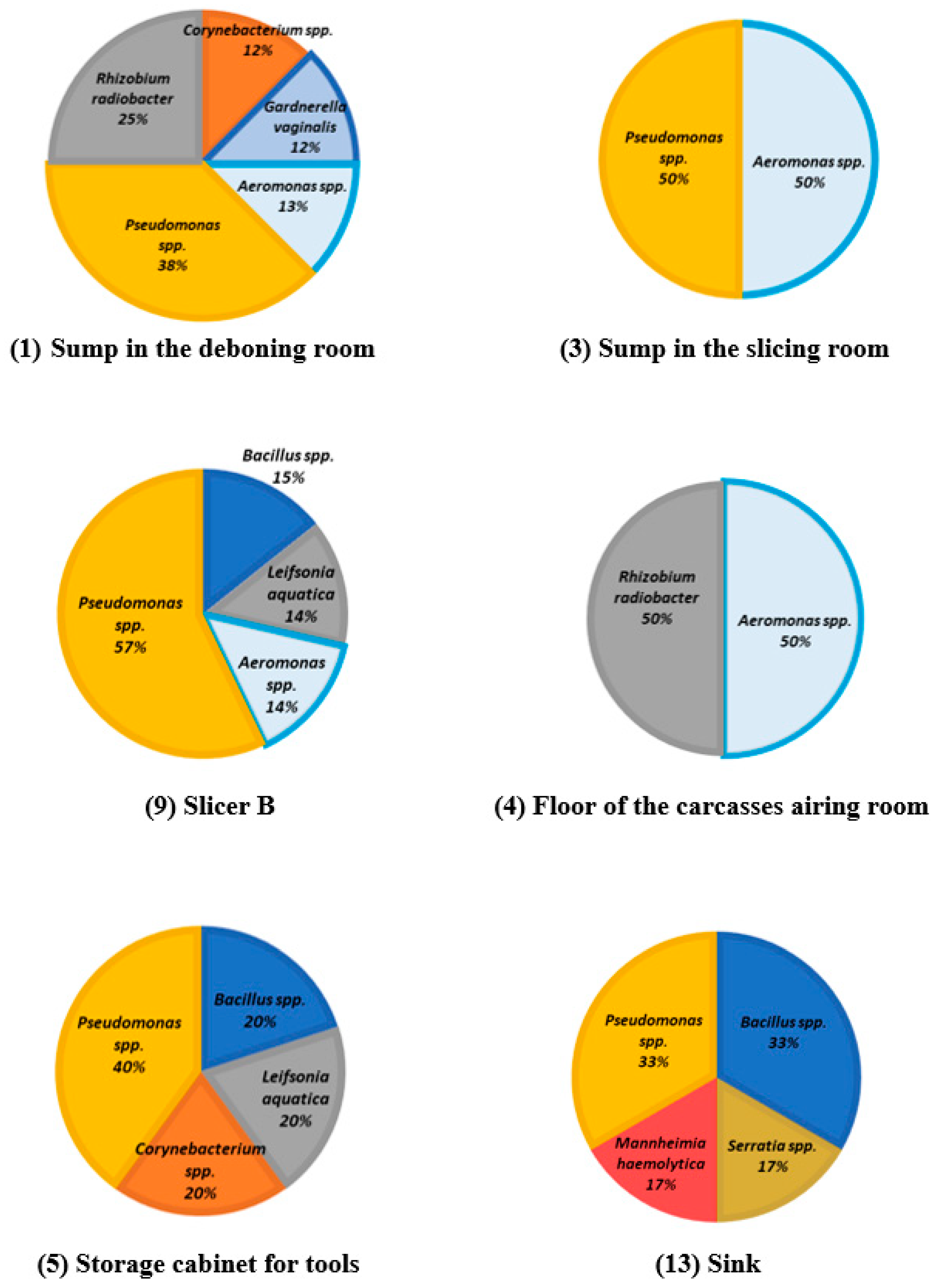
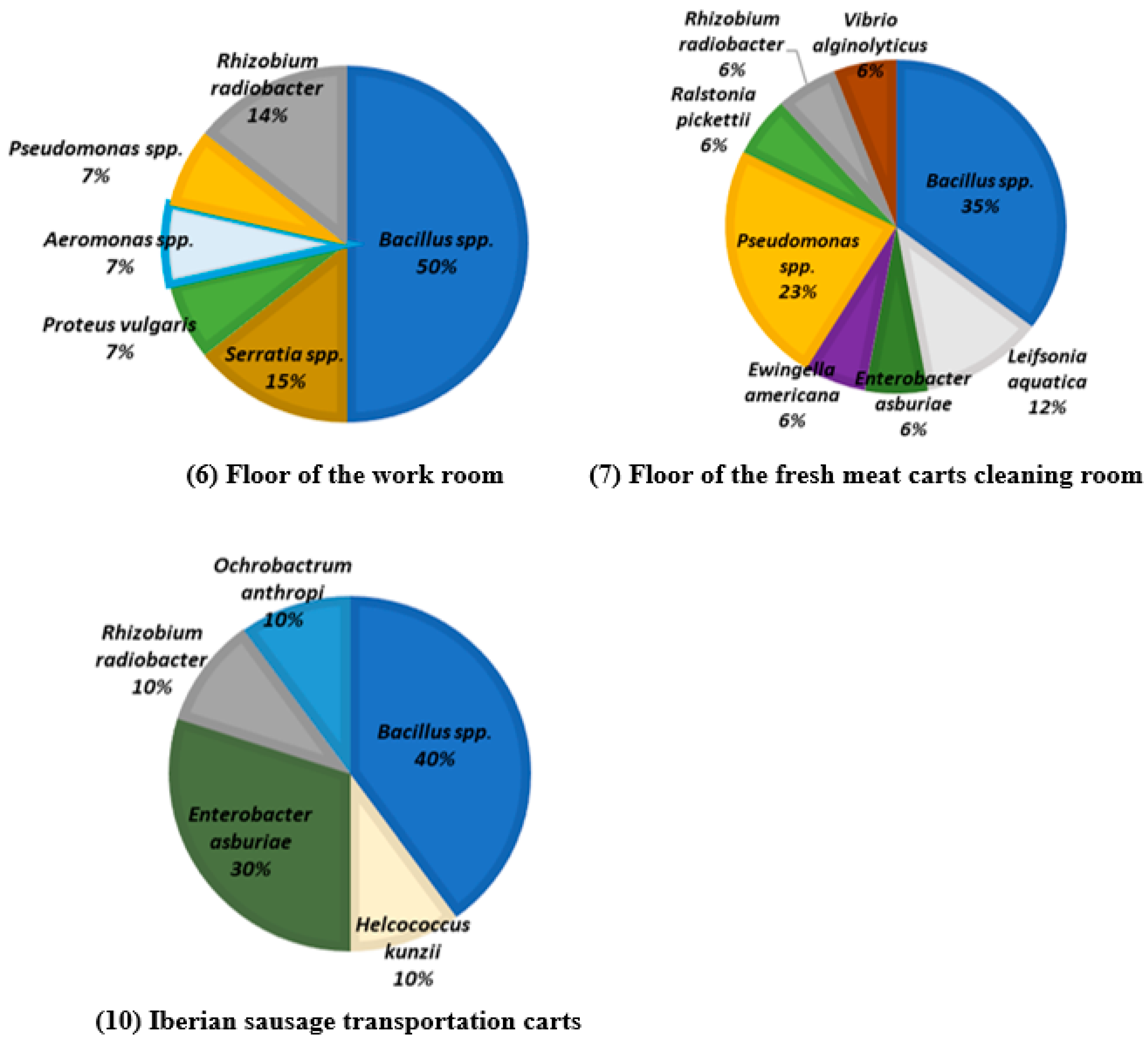
3.1. Identified Species

A total of 523 microorganisms were isolated from PCA and MRS agar culture media. Of these, 240 catalase positive isolates were discarded from the MRS medium since only the presence of lactic acid bacteria was investigated in this medium. Two-hundred microorganisms with different profiles were identified from these isolates, including mesophilic aerobic bacteria, lactic acid bacteria, and yeasts and molds. The results of the identifications are shown below in Table 2, Table 3 and Table 4, ordered from the greatest to the least presence on the surfaces.
Overall, in terms of mesophilic aerobes, there was a higher proportion of Gram-negative bacteria (57.27%) (Table 2). According to the study presented by Møretrø et al., Gram-negative bacteria such as Pseudomonas spp. have a greater capacity to form biofilms than Gram-positive bacteria [13].
The presence of the genera Bacillus spp., Pseudomonas spp., Staphylococcus spp., Aeromonas spp., Serratia spp., Enterobacter spp., Ralstonia spp., Proteus vulgaris, or Stenotrophomonas maltophilia has also been described in raw meat cold stores [23]. In the present study, the major bacterial genera found were Bacillus spp. (28.18% of the isolated bacteria) and Pseudomonas spp. (21.82%). The species identified within the genera Bacillus spp. were Bacillus subtilis (86.96%), Bacillus megaterium (6.52%), and Bacillus licheniformis (6.52%). The identified species of the genus Pseudomonas spp. were Pseudomonas fluorescens (40.00%), Pseudomonas luteola (40.00%), and Pseudomonas stutzeri (20.00%). These results are in concordance with other surfaces studies on which Pseudomonas spp. was predominant [24,25,26,27]. Stellato et al. also evaluated a beef and pork processing plant, identifying Pseudomonas spp. and several species of enterobacteria as major components of the surface microbiota [28]. In addition, according to Marouani-Gadri et al., the dominant genera in another meat industry (beef slaughterhouse and cutting room) were Staphylococcus spp. and Bacillus spp. [29]. Like in the present study, Lactobacillus spp., Staphylococcus spp., and Enterobacter spp. were also present.
Mannheimia haemolytica (9.17%), Rhizobium radiobacter (7.34%), Staphylococcus spp. (6.42%), and Aeromonas spp. (5.50%) (Table 2) were also found in the Iberian pig processing plant, in line with Hoodt and Zottola and Mertz et al. for Aeromonas spp. [27,30]. In another slaughterhouse, also analyzed after the cleaning and disinfection processes, the identified genera from a non-selective medium were Aerococcus spp., Acinetobacter spp., Pseudomonas spp., Staphylococcus spp., and Serratia spp. [13]. Furthermore, in another study conducted by Maes et al., the most abundant genera of the microbiota present on the food contact surfaces were Pseudomonas spp., Microbacterium spp., Stenotrophomonas spp., Staphylococcus spp., and Streptococcus spp. [26]. All these findings suggest that the type of microbiota found can be different depending on the surface area and the food industry, which may have a direct influence on the final hygiene of the food product.
It has been demonstrated that food industry surfaces, including those of the equipment, present an entire microbial ecosystem both during production and after cleaning and disinfection. The microbiota present is partly a reflection of the raw material used [31]. In this case, starter cultures are used to produce raw-cured meat. A typical starter for the preparation of raw-cured sausages is composed of Lactobacillus sakei, Staphylococcus xylosus, and Staphylococcus carnosus [32]. In this regard, different species of Lactobacillus spp. which can be used as starters were found in both Plant A and Plant B of the present study (Table 3), thus indicating that microbiota from food is transferred to the surfaces. Regarding lactic acid bacteria, the predominant species were Leuconostoc mesenteroides spp. cremoris, which commonly cause the spoilage of food packaged in a modified atmosphere and stored in cold conditions and were detected in abundance in a study carried out on sausages [25,33,34].
In this study, of the close to 30% of yeasts and molds, Candida spp. was the most abundant yeast followed by Cryptococcus spp. (Table 4). These results also coincide with other studies [24,35]. However, few studies that characterize the surface microbiota mention the isolated species of yeasts and molds, like Chevallier et al. and Talon et al. [36,37].

3.2. Ecological Profiles of the Different Areas: Cluster Analysis

According to the analysis by hierarchical clusters, the ecological profiles of the sampled areas can be grouped into four clusters, although only two of them are relevant. The first cluster was composed of areas 13 (sink), 5 (storage cabinet for tools), 9 (slicer B), 3 (sump in the slicing room), and 4 (carcasses airing room floor). The distance between these areas is short, so their ecological profiles can be considered as similar (Figure 2). The second main cluster is made up of areas 6 (work room floor), 7 (fresh meat carts cleaning room floor), and 10 (Iberian sausage transportation carts). The areas 8 (cured meat carts cleaning room floor) and 2 (slicer) form the other two clusters, each one on its own. These two profiles were remarkably different from the rest and could therefore be considered as outliers. As the technique of classification by hierarchical conglomerates is sensitive to the presence of outliers they can appear as two differentiated clusters.
Ecological profiles could not be established in areas 11 (vacuum machine slide) and 12 (heat treatment room floor) (Table 1) due to the lack of growth on the PCA plates. In fact, in the quantitative study carried out by Ripolles-Avila et al. these two areas were described as the least microbiologically contaminated [16].
Cluster 1 grouped the areas dominated by Pseudomonas spp. (Figure 3). These areas may have similar ecological profiles because they intervene in successive stages of the productive process. The carcasses are taken from the carcass airing room to the cutting room where the manipulated tools are stored in the storage cabinet. Therefore, how the different areas may be related to each other and how the movement of microbiota between them can occur are very relevant, especially in the transfer of pathogens such as L. monocytogenes. Area 13 (sink) also had the same proportion of Bacillus spp. and Pseudomonas spp., so it could be considered as belonging to both clusters. The work room receives products from the cutting room and processes such as chopping are carried out there. In this conglomerate, four of the six areas presented 13 to 50% of Aeromonas spp. and less abundantly Bacillus spp., Rhizobium radiobacter, Corynebacterium spp., and Leifsonia aquatica.
Like in the present study, Hoodt and Zottola identified the presence of Pseudomonas spp. and Aeromonas spp. in the sinks [30]. In other studies, Pseudomonas spp. [27,37], Enterobacteriaceae, and yeasts and molds have been found on knives, tables, and chopping machines [35]. Pseudomonas spp. is commonly found in soil and water where it plays an important role in the degradation of organic material. They are part of the normal microbiota of human skin and are found in the respiratory tract and intestines [38]. Aeromonas spp. is also ubiquitous, usually found in water sources, soil, arthropods, mollusks, mammals, birds, fish, and insects [39]. Corynebacterium spp., present in the sump in the deboning room and tools storage cabinet, also has a global distribution and is found in soils, water, animals, and plants, especially in temperate areas [40]. The less common Leifsonia aquatica, isolated from slicer B and the tool storage cabinet (Figure 3), could come from the water since it inhabits the aquatic environment. It is characterized by having a low growth rate and the capacity to form biofilms [41].
In this second cluster, areas 6 (work room floor), 7 (cured meat carts cleaning room floor), and 10 (Iberian sausage transportation carts) were mostly colonized by Bacillus spp. (Figure 4). This genus includes many of the most ubiquitous bacteria [42]. These areas are physically close and the same personnel and equipment, such as the transportation carts, raw material, and fresh products, circulate there. The fresh meat is processed in the work room and it is transported by the carts, so the same residues can be found in the work room as in the fresh meat carts cleaning room floor. Microbial communities are representative of each processing area and are influenced to a large extent by food debris, liquid effluents, and wash water [25]. R. radiobacter, isolated in the three areas that comprise this cluster, is also a ubiquitous bacterium and is usually found in soil, plants, etc. [43]. Serratia spp., another common microorganism in the area between the work room floor and the sink, has also been isolated from industries that manufacture sausages [44]. S. liquefaciens is one of the dominant enterobacteria in raw meat working plants. Another species of the genus Serratia spp. that is sometimes identified is S. marcescens [44]. The results obtained in the present study are in concordance with these findings, as both species were found on the surfaces in Plant B. Enterobacter spp., belonging to the family Enterobacteriaceae and widely distributed in nature, were found between the floor of the fresh meat cart cleaning room and the Iberian sausage transportation carts. They can be found in soil, water, and as part of the microbiota of animals, insects, and the human gastrointestinal tract [45].
Clusters 3 and 4 correspond to areas 8 (cured meat carts cleaning room floor) and 2 (slicer A), respectively. The analysis by hierarchical clusters determined that these two profiles were distant from the other two main clusters (1 and 2). This could be explained by the presence of non-isolated species such as Staphylococcus spp., Stenotrophomonas maltophilia in the other areas, and the strong presence of M. haemolytica in area 8 (cured meat carts cleaning room floor) (Figure 5). The atypical profile of location (8) could be explained by the fact that the carts transport a different raw material. They do not displace fresh products, but rather genera that have undergone a process of healing or maturation in which the microbiota is modified [35]. In addition, this room is on the upper floor of Plant B, unlike almost all the other areas where sensors were located (except for slicer B).

3.3. Unusual Identification of Mannheimia haemolytica

The identification of M. haemolytica in the sensors located in areas 2 (slicer A), 8 (cured meat carts cleaning room floor), and 13 (sink) was unexpected. This bacterium occasionally intervenes in the porcine respiratory complex, although it is much more frequent in bovines where it also causes respiratory disease. Ahmed and Sabiel also identified M. haemolytica in minced beef [21]. One hypothesis is that because this opportunistic pathogen can to be found in the respiratory tract of animals, it could contaminate the carcasses by means of incorrect handling in the respiratory tract removal stage. Another hypothesis could be a failure in identification when using the API® 20E test. In fact, Ahmed and Sabiel also used this commercial kit to identify their isolates [21]. According to the results provided by the API® 20E kit, in six of the ten isolates of this microorganism in this study, the probability percentage (ID %) was equal to or greater than 84.6%, reaching up to 98.5%. When API® kits had already been in use for twenty years, Hara et al. re-evaluated the method, concluding that it was reliable compared with traditional biochemical tests [46]. As a final aspect to be considered, M. haemolytica has undergone an extensive reclassification, previously known as Bacterium bipolare multocidum, Bacillus boviseptica, and Pasteurella haemolytica [47]. Despite the new classification, the correct identification of the isolates continues to be difficult and laborious [48].

3.4. Influence of the Microbiota on the Presence of L. Monocytogenes

In the quantitative study carried out by Ripolles-Avila et al., L. monocytogenes was isolated on five occasions from areas 5 (i.e., tools storage cabinet; serotype 4b), 8 (i.e., cured meat carts cleaning room floor; serotype 4b and 1/2a), and 10 (i.e., Iberian sausages transportation carts; serotype 1/2b) [16].
Analyzing the ecological profile of the areas where L. monocytogenes was found, the tools storage cabinet presented 40% of Pseudomonas spp., the majority genera of the community present (Figure 2). The effect of Pseudomonas spp. on the growth of L. monocytogenes has not been precisely described, since various authors have shown a positive [27], a negative, and no effect [8]. The cured meat carts cleaning room floor showed an atypical profile with the strong presence of M. haemolytica. It must be noted, however, that the genera with the most isolates was Pseudomonas spp., which has been shown to enhance the presence of L. monocytogenes [27,49] in some studies. It could also influence, in this sense, 7% of Serratia spp. since enterobacteria appear to favor L. monocytogenes growth [35,50]. Macroscopically, species such as Pseudomonas spp. can produce a viscous substance when colonizing a surface. Hoodt and Zottola suggested that these microorganisms produce a dense extracellular material that could allow the entrapment of other microorganisms [30]. These primary colonizers of meat processing plants could harbor pathogens such as L. monocytogenes. As for the Iberian sausage transportation carts, 30% of Enterobacter asburiae could be relevant since it has been observed that other enterobacteria have a positive effect on the growth of L. monocytogenes [35,50]. In this regard, the results obtained show that in the areas where L. monocytogenes was found, the surfaces contain a microbiota composed of microorganisms that could enhance L. monocytogenes growth. More in vitro studies are needed to demonstrate how ecological interrelations affect the formation of L. monocytogenes biofilms as a previous step to the application of this alternative at an industrial level. Some methodologies have been proposed for their formation and quantification that resemble real conditions [51,52]. The control of these microorganisms could also minimize the presence of this pathogen [16].

4. Conclusions

The ecological profiles of meat processing plants can vary depending on the raw material, temperature, humidity, and industrial processes carried out. The way to monitor these communities of microorganisms should be standardized both in the collection of the samples and the analysis. The microbiota of the studied Iberian pig processing plant was dominated by Bacillus spp. and Pseudomonas spp. The areas could be grouped into two main clusters and the profile of the areas and the ecological diversity was varied, enabling the correlation of the presence or absence of L. monocytogenes. This persistent pathogen seems to be associated with one or more specific bacterial groups such as Pseudomonas spp. and enterobacteria, which belong to the resident microbiota of the facilities. A high level of contamination does not always suggest the presence of these pathogens, but rather depends on the established species. Knowing the species that persist in the production plants and their interactions with L. monocytogenes can help to profile cleaning and disinfection programs. The control of resident microbiota is, therefore, a key element to guarantee food safety and the quality of the final products.

Author Contributions

A.-S.H.: Executed the experiments, interpreted the results, and wrote the manuscript; C.R.-A.: Designed experiments, interpreted the results, contributed to writing, and reviewing the manuscript; A.E.G.-N.: Contributed to the experiments, writing, and reviewing the manuscript; and J.J.R.-J.: Designed the experiments, interpreted the results, and contributed to writing the manuscript.

Funding

This study was supported by Research Project grants RTI2018-098267-R-C32 from the Spanish Ministerio de Ciencia, Innovación y Universidades.

Acknowledgments

The authors thank Dolors Busquets Soler for her technical assistance in the laboratory and Sarah Davies for the English grammar review.

Conflicts of Interest

The authors declare no conflict of interest.

References

  1. Fratamico, P.M.; Annous, B.A.; Gunther, N.W. Biofilms in the Food and Beverage Industries; Elsevier: Amsterdam, The Netherlands, 2009; ISBN 978-1-84-569362-6. [Google Scholar]
  2. Dantas, S.T.A.; Rossi, B.F.; Bonsaglia, E.C.R.; Castilho, I.G.; Hernandes, R.T.; Fernandes, A.; Rall, V.L.M. Cross-contamination and biofilm formation by Salmonella enterica serovar Enteritidis on various cutting boards. Foodborne Pathog. Dis. 2018, 15, 81–85. [Google Scholar] [CrossRef] [PubMed]
  3. Dourou, D.; Simpson, C.; Yoon, Y.; Belk, K.E.; Smith, G.C.; Nychas, G.E.; Sofos, J.N. Attachment and biofilm formation by E. coli O157:H7 at different temperatures, on various food-contact surfaces encountered in beef processing. Int. J. Food Microbiol. 2011, 149, 262–268. [Google Scholar] [CrossRef] [PubMed]
  4. Grove, S.F.; Suriyanarayanan, A.; Puli, B.; Zhao, H.; Li, M.; Li, D.; Schaffner, D.W.; Lee, A. Norovirus cross-contamination during preparation of fresh produce. Int. J. Food Microbiol. 2015, 198, 43–49. [Google Scholar] [CrossRef] [PubMed]
  5. Jensen, D.A.; Danyluk, M.D.; Harris, L.J.; Schaffner, D.W. Quantifying bacterial cross-contamination rates between fresh-cut produce and hands. J. Food Prot. 2017, 80, 213–219. [Google Scholar] [CrossRef]
  6. Kim, A.; Young Park, S.; Bae, S.-C.; Oh, M.-H.; Ha, S.-D. Survival of norovirus surrogate on various food-contact surfaces. Food Environ. Virol. 2014, 6, 182–188. [Google Scholar] [CrossRef]
  7. Sauders, B.D.; D’Amico, D.J. Listeria monocytogenes cross-contamination of cheese: Risk throughout the food supply chain. Epidemiol. Infect. 2016, 144, 2693–2697. [Google Scholar] [CrossRef]
  8. Møretrø, T.; Langsrud, S. Residential bacteria on surfaces in the food industry and their implications for food safety and quality. Compr. Rev. Food Sci. Food Saf. 2017, 16, 1022–1041. [Google Scholar] [CrossRef]
  9. Giaouris, E.; Heir, E.; Desvaux, M.; Hébraud, M.; Møretrø, T.; Langsrud, S.; Doulgeraki, A.; Nychas, G.J.; Kacániová, M.; Czaczyk, K.; et al. Intra- and inter-species interactions within biofilms of important foodborne bacterial pathogens. Front. Microbiol. 2015, 6, 1–26. [Google Scholar] [CrossRef]
  10. Parsek, M.R.; Singh, P.K. Bacterial biofilms: An emerging link to disease pathogenesis. Annu. Rev. Microbiol. 2003, 57, 677–701. [Google Scholar] [CrossRef]
  11. Carpentier, B.; Chassaing, D. Interactions in biofilms between Listeria monocytogenes and resident microorganisms from food industry premises. Int. J. Food Microbiol. 2004, 97, 111–122. [Google Scholar] [CrossRef]
  12. Mariani, C.; Oulahal, N.; Chamba, J.F.; Dubois-Brissonnet, F.; Notz, E.; Briandet, R. Inhibition of Listeria monocytogenes by resident biofilms present on wooden shelves used for cheese ripening. Food Control 2011, 22, 1357–1362. [Google Scholar] [CrossRef]
  13. Møretrø, T.; Langsrud, S.; Heir, E. Bacteria on meat abattoir process surfaces after sanitation: Characterisation of survival properties of Listeria monocytogenes and the commensal bacterial flora. Adv. Microbiol. 2013, 3, 255–264. [Google Scholar] [CrossRef]
  14. Ortiz, S.; López, V.; Villatoro, D.; López, P.; Dávila, J.C.; Martínez-Suárez, J.V. A 3-year surveillance of the genetic diversity and persistence of Listeria monocytogenes in an iberian pig slaughterhouse and processing plant. Foodborne Pathog. Dis. 2010, 7, 1177–1184. [Google Scholar] [CrossRef] [PubMed]
  15. Verraes, C.; Van Boxstael, S.; Van Meervenne, E.; Van Coillie, E.; Butaye, P.; Catry, B.; Huffel, M.-A.; Van Huffel, X.; Imberechts, H.; Dierick, K.; et al. Antimicrobial resistance in the food chain: A review. Int. J. Environ. Res. Public Health 2013, 10, 2643–2669. [Google Scholar] [CrossRef]
  16. Ripolles-Avila, C.; Hascoët, A.S.; Martínez-Suárez, J.V.; Capita, R.; Rodríguez-Jerez, J.J. Evaluation of the microbiological contamination of food processing environments through implementing surface sensors in an iberian pork processing plant: An approach towards the control of Listeria monocytogenes. Food Control 2019, 99, 40–47. [Google Scholar] [CrossRef]
  17. Montañez-Izquierdo, V.; Ríos-Castillo, A.G.; Fontecha-Umaña, F.; Rodríguez-Jerez, J.J. Uso de sensores de superficie y métodos rápidos para controlar la contaminación ambiental en las industrias alimentarias: Hacia el control on line. In XI Workshop de Métodos Rápidos y Automatización en Microbiología Alimentaria; Universidad Autónoma de Barcelona: Barcelona, Spain, 2012; pp. 1–6. [Google Scholar]
  18. Moen, B.; Røssvoll, E.; Måge, I.; Møretrø, T.; Langsrud, S. Microbiota formed on attached stainless steel coupons correlates with the natural biofilm of the sink surface in domestic kitchens. Can. J. Microbiol. 2016, 62, 148–160. [Google Scholar] [CrossRef]
  19. Ripolles-Avila, C. Supervivencia de Listeria monocytogenes Sobre Superficies de Contacto con Alimentos: Un Abordaje Multidisciplinar de un Problema Complejo. Ph.D. Thesis, Universitat Autònoma de Barcelona, Barcelona, Spain, 2018. [Google Scholar]
  20. Sharma, M.; Anand, S.K. Characterization of constitutive microflora of biofilms in dairy processing lines. Food Microbiol. 2002, 19, 627–636. [Google Scholar] [CrossRef]
  21. Ahmed, A.A.; Sabiel, Y.A. Detection of microbial contamination of processed beef meat by ssing API strips and automated Vitek 2 compact system. Microbiol. Res. J. Int. 2016, 13, 1–8. [Google Scholar] [CrossRef]
  22. Feligini, M.; Panelli, S.; Buffoni, J.N.; Bonacina, C.; Andrighetto, C.; Lombardi, A. Identification of microbiota present on the surface of Taleggio cheese using PCR-DGGE and RAPD-PCR. J. Food Sci. 2012, 77, 609–615. [Google Scholar] [CrossRef]
  23. Doulgeraki, A.I.; Ercolini, D.; Villani, F.; Nychas, G.E. Spoilage microbiota associated to the storage of raw meat in different conditions. Int. J. Food Microbiol. 2012, 157, 130–141. [Google Scholar] [CrossRef]
  24. Bagge-Ravn, D.; Ng, Y.; Hjelm, M.; Christiansen, J.N.; Johansen, C.; Gram, L. The microbial ecology of processing equipment in different fish industries—Analysis of the microflora during processing and following cleaning and disinfection. Int. J. Food Microbiol. 2003, 87, 239–250. [Google Scholar] [CrossRef]
  25. Dzieciol, M.; Schornsteiner, E.; Muhterem-Uyar, M.; Stessl, B.; Wagner, M.; Schmitz-Esser, S. Bacterial diversity of floor drain biofilms and drain waters in a Listeria monocytogenes contaminated food processing environment. Int. J. Food Microbiol. 2016, 223, 33–40. [Google Scholar] [CrossRef] [PubMed]
  26. Maes, S.; Heyndrickx, M.; Vackier, T.; Steenackers, H.; Verplaetse, A.; De Reu, K. Identification and spoilage potential of the remaining dominant microbiota on food contact surfaces after cleaning and disinfection in different food industries. J. Food Prot. 2019, 82, 262–275. [Google Scholar] [CrossRef] [PubMed]
  27. Mertz, A.W.; Kyung, O.; Bryan, C.A.O.; Morawicki, R.; Sirsat, S.A.; Neal, J.A.; Crandall, P.G.; Ricke, S.C. Microbial ecology of meat slicers as determined by denaturing gradient gel electrophoresis. Food Control 2014, 42, 242–247. [Google Scholar] [CrossRef]
  28. Stellato, G.; La Storia, A.; De Filippis, F.; Borriello, G.; Villani, F.; Ercolini, D. Overlap of spoilage-associated microbiota between meat and the meat processing environment in small-scale and large-scale retail distributions. Appl. Environ. Microbiol. 2016, 82, 4045–4054. [Google Scholar] [CrossRef] [PubMed]
  29. Marouani-Gadri, N.; Augier, G.; Carpentier, B. Characterization of bacterial strains isolated from a beef-processing plant following cleaning and disinfection-influence of isolated strains on biofilm formation by Sakaï and EDL 933 E. coli O157:H7. Int. J. Food Microbiol. 2009, 133, 62–67. [Google Scholar] [CrossRef] [PubMed]
  30. Hoodt, S.K.; Zottola, E.A. Isolation and identification of adherent gram-negative microorganisms from four meat-processing facilities. J. Food Prot. 1997, 60, 1135–1138. [Google Scholar] [CrossRef]
  31. Schön, K.; Schornsteiner, E.; Wagner, M.; Müller, M.; Schmitz-Esser, S.; Dzieciol, M. Microbial communities in dairy processing environment floor-drains are dominated by product-associated bacteria and yeasts. Food Control 2016, 70, 210–215. [Google Scholar] [CrossRef]
  32. Bañón, S.; Martínez, A.; López, A.M. Maduración de chorizo y salchichón de Chato Murciano con diferentes cultivos iniciadores (bacterias ácido-lácticas y estafilococos). An. Vet. Murcia 2011, 27, 101–118. [Google Scholar] [CrossRef]
  33. Hultman, J.; Rahkila, R.; Ali, J.; Rousu, J.; Johanna, K. Meat processing plant microbiome and contamination patterns of cold-tolerant bacteria causing food safety and spoilage risks in the manufacture of vacuum-packaged cooked sausages. Appl. Environ. Microbiol. 2015, 81, 7088–7097. [Google Scholar] [CrossRef]
  34. Padilla-Frausto, J.J.; Cepeda-Marquez, L.G.; Salgado, L.M.; Iturriaga, M.H.; Arvizu-Medrano, S.M. Detection and genotyping of Leuconostoc spp. in a sausage processing plant. J. Food Prot. 2015, 78, 2170–2176. [Google Scholar] [CrossRef] [PubMed]
  35. Gounadaki, A.S.; Skandamis, P.N.; Drosinos, E.H.; Nychas, G.E. Microbial ecology of food contact surfaces and products of small-scale facilities producing traditional sausages. Food Microbiol. 2008, 25, 313–323. [Google Scholar] [CrossRef] [PubMed]
  36. Chevallier, I.; Ammor, S.; Laguet, A.; Labayle, S.; Castanet, V.; Dufour, E.; Talon, R. Microbial ecology of a small-scale facility producing traditional dry sausage. Food Control 2006, 17, 446–453. [Google Scholar] [CrossRef]
  37. Talon, R.; Leroy, S.; Lebert, I. Microbial ecosystems of traditional fermented meat products: The importance of indigenous starters. Meat Sci. 2007, 77, 55–62. [Google Scholar] [CrossRef] [PubMed]
  38. Adler, R. Pseudomonas. Salem Press Encyclopedia of Health. Available online: http://mendeley.csuc.cat/fitxers/3b01d5716b505758e1d15ab62b7d7601 (accessed on 29 October 2019).
  39. Silva, O.F. Aeromonas spp. Rev. Chil. Infectol. 2011, 28, 157–158. [Google Scholar] [CrossRef]
  40. Cheney, R. Corynebacterium. Salem Press Encyclopedia of Health. Available online: http://mendeley.csuc.cat/fitxers/b50c6dd66f3a8698da8be0571a312e98 (accessed on 29 October 2019).
  41. Mühlhauser, M. Leifsonia aquatica. Rev. Chil. Infectol. 2016, 33, 313–314. [Google Scholar] [CrossRef]
  42. Kte’pi, B. Bacillus (bacteria). Salem Press Encyclopedia of Health. Available online: http://mendeley.csuc.cat/fitxers/315e4f3c33439a6298ababd24553641d (accessed on 29 October 2019).
  43. Stamou, A.; Pavlopoulos, C.; Roumeliotis, S.; Samoladas, E.; Xatzokos, I.; Kontopoulou, K. Nonunion humerous fracture infection caused by Rhizobium radiobacter in a 24-year-old healthy patient: A rare case report. Case Rep. Infect. Dis. 2018, 2018, 1–4. [Google Scholar] [CrossRef]
  44. Stiles, M.E.; Ng, L.K. Enterobacteriaceae associated with meats and meat handling. Appl. Environ. Microbiol. 1981, 41, 867–872. [Google Scholar] [Green Version]
  45. Silva, F.; Pabla Martínez, O.; Pabla, T.M. Complejo Enterobacter cloacae. Rev. Chil. Infectol. 2018, 35, 297–298. [Google Scholar] [CrossRef]
  46. Hara, C.M.O.; Rhoden, D.L.; Miller, J.M. Reevaluation of the API 20E identification system versus conventional biochemicals for identification of members of the family Enterobacteriaceae: A new look at an old product. J. Clin. Microbiol. 1992, 30, 123–125. [Google Scholar]
  47. Jaramillo-Arango, C.J.; Trigo Tavera, F.J.; Suárez-Güemes, F. Mannheimiosis bovina: Etiología, prevención y control. Vet. Mex. 2009, 40, 293–314. [Google Scholar]
  48. Angen, Ø.; Ahrens, P.; Bisgaard, M. Phenotypic and genotypic characterization of Mannheimia (Pasteurella) haemolytica-like strains isolated from diseased animals in Denmark. Vet. Microbiol. 2002, 84, 103–114. [Google Scholar] [CrossRef]
  49. Puga, C.H.; Dahdouh, E.; SanJose, C.; Orgaz, B. Listeria monocytogenes colonizes Pseudomonas fluorescens biofilms and induces matrix over-production. Front. Microbiol. 2018, 9, 1–12. [Google Scholar] [CrossRef] [PubMed]
  50. Jessen, B.; Lammert, L. Biofilm and disinfection in meat processing plants. Int. Biodeterior. Biodegrad. 2003, 51, 265–269. [Google Scholar] [CrossRef]
  51. Ripolles-Avila, C.; Cervantes-Huaman, B.H.; Hascoët, A.S.; Yuste, J.; Rodríguez-Jerez, J.J. Quantification of mature Listeria monocytogenes biofilm cells formed by an in vitro model: A comparison of different methods. Int. J. Food Microbiol. 2019, 289, 209–214. [Google Scholar] [CrossRef] [PubMed]
  52. Ripolles-Avila, C.; Hascoët, A.S.; Guerrero-Navarro, A.E.; Rodríguez-Jerez, J.J. Establishment of incubation conditions to optimize the in vitro formation of mature Listeria monocytogenes biofilms on food-contact surfaces. Food Control 2018, 92, 240–248. [Google Scholar] [CrossRef]
Figure 1. Examples of their placement in the collaborating industry: (a) Fresh meat carts cleaning room floor; (b) slicer B; and (c) vacuum machine slide. (d) Design of a SCH surface sensor [19].
Applsci 09 04611 g001
Figure 2. Analysis by hierarchical clusters showing the degree of similarity of the ecological profile of the sampled areas. There are two main clusters: One grouping with areas 3, 4, 5, 13, 9, and 1 and another with areas 6, 7, and 10.
Applsci 09 04611 g002
Figure 3. Individual profiles of the areas categorized as cluster 1. Pseudomonas spp. was predominant in most areas and there was also a notable presence of Aeromonas spp.
Applsci 09 04611 g003
Figure 4. Individual profiles of the areas categorized as cluster 2. Bacillus spp. was predominant in all the areas and there was a marked presence of Rhizobium radiobacter and Enterobacter asburiae.
Applsci 09 04611 g004
Figure 5. Individual profiles of the categorized areas, such as cluster 3, (8) cured meat carts cleaning room floor, and cluster 4, (2) slicer A.
Applsci 09 04611 g005
Table 1. Work surfaces from Plants A and B where the SCH sensors were installed (coded from 1 to 13) [16].
Processing PlantIDSurface
A1Sump in the deboning room
2Slicer A
3Sump in the slicing room
B4Floor of the carcasses airing room
5Storage cabinet for tools
6Floor of the work room
7Floor of the fresh meat carts cleaning room
8Floor of the cured meat carts cleaning room
9Slicer B
10Iberian sausage transportation carts
11Slide of vacuum machine
12Floor of the heat treatment room
13Sink
Table 2. Percentage of identified genera and species from Plate Count Agar (PCA) and its corresponding Gram stain (Gram positive or negative bacteria).
Identified Species% vs Isolated Bacteria% vs Isolated Microorganisms
Molds and yeasts-29.03
Bacillus spp.128.1820.00
Pseudomonas spp.221.8215.48
Mannheimia haemolytica9.096.45
Rhizobium radiobacter7.275.16
Staphylococcus spp.6.364.52
Aeromonas spp.5.453.87
Leifsonia aquatica3.642.58
Serratia spp.3.642,58
Enterobacter asburiae3.642.58
Proteus vulgaris1.821.29
Corynebacterium spp.1.821.29
Helcococcus kunzii0.910.65
Aerococcus urinae0.910.65
Gardnerella vaginalis0.910.65
Stenotrophomonas maltophilia0.910.65
Ewingella americana0.910.65
Ralstonia pickettii0.910.65
Vibrio spp.0.910.65
Ochrobactrum anthropi0.910.65
Total100.00100.00
Gram stain%
Gram positive42.73-
Gram negative57.27-
Total100.00-
1 Blue indicates that the bacteria are Gram-positive. 2 Orange indicates that the bacteria are Gram-negative.
Table 3. Identified lactic acid bacteria from Man, Rogosa and Sharpe agar (MRS), in number of isolates and respective percentage from the total of lactic acid bacteria.
Identified SpeciesTotal%
Leuconostoc mesenteroides ssp. cremoris23.051.1
Lactobacillus delbrueckii ssp. delbrueckii8.017.8
Lactococcus lactis ssp. lactis6.013.3
Leuconostoc mesenteroides ssp. mesenteroides4.08.9
Lactobacillus acidophilus1.02.2
Pediococcus damniosus1.02.2
Non-identified2.04.4
Total45.0100.0
Table 4. Identified molds and yeasts from cultures in PCA, in number of isolates and respective percentage from the total of lactic acid bacteria.
Identified SpeciesTotal%
Candida zeylanoides27.061.4
Cryptococcus uniguttulatus3.06.8
Candida krusei2.04.5
Candida ciferri2.04.5
Candida spp.2.04.5
Candida pelliculosa1.02.3
Candida magnoliae1.02.3
Cryptococcus terreus1.02.3
Non-identified5.011.4
Total44.0100.0

