Article

Lactobacillus acidophilus Derived Biosurfactant as a Biofilm Inhibitor: A Promising Investigation Using Microfluidic Approach

1
Department of Microbiology, Savitribai Phule Pune University, Pune 411007, Maharashtra, India
2
Department of Physics, Savitribai Phule Pune University, Pune 411007, Maharashtra, India
3
School of Biomedical Sciences, University of Ulster, Coleraine BT52 1SA, UK
*
Authors to whom correspondence should be addressed.
Appl. Sci. 2018, 8(9), 1555; https://doi.org/10.3390/app8091555
Submission received: 31 July 2018 / Revised: 18 August 2018 / Accepted: 29 August 2018 / Published: 4 September 2018
(This article belongs to the Special Issue Nano-systems for Antimicrobial Therapy)

Abstract

:
Background: Biomedical devices and implants are adversely affected by biofilm-associated infections that pose serious public health issues. Biosurfactants (BSs) can combat pathogenic biofilms through their antimicrobial, antibiofilm and antiadhesive capabilities. The objective of our research was to produce biosurfactant (BS) from Lactobacillus acidophilus NCIM 2903 and investigate its antibiofilm, antiadhesive potential using microfluidics strategies by mimicking the micro-environment of biofilm. Methods: Antibiofilm and antiadhesive potential was effectively evaluated using different methods like microfluidics assay, catheter assay, polydimethlysiloxane (PDMS) disc assay. Along with this chemical and physical characteristics of BS were also evaluated. Results: Cell free biosurfactant (CFBS) obtained was found to be effective against biofilm which was validated through the microfluidic (MF) or Lab on Chip (LOC) approach. The potency of CFBS was also evaluated on catheter tubing and PDMS surfaces (representative bioimplants). The efficacy of CFBS was also demonstrated through the reduction in surface tension, interfacial tension, contact angle and low critical micelle concentration. Conclusion: CFBS was found to be a potent antimicrobial and antibiofilm agent. We believe that perhaps this is the first report on demonstrating the inhibiting effect of Lactobacillus spp. derived CFBS against selected bacteria via LOC approach. These findings can be explored to design various BSs based formulations exhibiting antimicrobial, antibiofilm and antiadhesive potential for biomedical applications.

1. Introduction

Medical devices and equipment are routinely used by most healthcare professionals to support and treat patients. Bioimplants are amenable to develop microbial biofilms. Biofilms are microbial communities with abilities to attach to surfaces, exhibiting high resistance to many antimicrobial agents [1,2,3,4,5,6]. Pathogenic microbial biofilms exert harmful effects on human health. Increased resistance of bacteria to antibiotic therapy is a major concern for medical professionals worldwide. The inherent resistance of biofilms and their pervasive involvement in implant-related infections has prompted research towards the development of antibiofilm, antiadhesive agents. Biosurfactant (BS) producing microorganisms can disrupt biofilm on medical implants due to their antimicrobial, antibiofilm and antiadhesive potential. Lactic acid bacteria (LAB), such as Lactobacilli constitute an important part of the natural microbiota and are recognized as potentially useful bacteria through production of biosurfactant/s (BSs) [1,2,6]. Reports by Gomaa [7], Gudiña and co-workers [8,9,10] suggested that Lactobacilli originated BS is highly effective against several pathogenic microorganisms with distinct antimicrobial and anti-adhesive activities. Based on structural details, different types of BSs viz. Glycolipid [5,6], glycoprotein [8,11], glycolipoprotein/peptide [12,13] have been documented from Lactobacillus spp. [7,8,11]. Lactobacilli spp. are solely known to produce cell bound/cell associated BS (CABS) and cell free BS (CFBS) [14].
BS mediated synthesis of nanoparticles (NPs) is becoming an interesting approach for many researchers [15]. Currently, several researchers are synthesizing metal NPs from microbial origin and exploiting them for biomedical purposes [16,17]. Reddy et al. [18,19] reported synthesis of silver and gold NPs by using fractionated BS (as a stabilizer) with NaBH4 (as a reducing agent). BS plays a crucial role in the synthesis of gold and silver NPs from Bacillus subtilis ANR 88 by growing in non-hydrocarbon, agro industrial wastes. Synthesis of uniform size and shapes silver (spherical, 4–18 nm) and gold NPs (hexagonal, 40–60 nm) from BS producing Bacillus culture has been achieved [20].
Inspiring discoveries or novel formulations introduced by the engineering sector have improved our modern life. Currently incredible applications in the areas of protein crystallization, biochemical screening [21], DNA amplification, DNA sequencing and enzymatic kinetic assays [22] are driving forces to explore advanced developments of engineering. Microfluidics (MF) is one of the pioneering areas of engineering that deals with picolitre volumes of liquids [23]. MF terminology is popularly known as lab-on-a-chip (LOC) where experiments are carried out on small-scale in-vitro, mimicking in-vivo conditions [24]. MF devices have been reported to characterize cellular association in a flow system. Recently, Khor et al., 2018 [25] have cultured HUVECs on the surface of synthetic micro-vascular network in a MF device (SynVivo, INC). Such study provides an opportunity to explore possible interactions of molecules with live cells. MF devices having simple and straight micro-channels may not be sufficient to replicate the complex structures of desired systems. However, to some extent MF system are helpful in exploring interactions between NP with biological molecules. Implementation of fundamental experiments, applied, analytical and diagnostic are often very challenging due to the availability of compounds in negligible quantities. MF system offers better prospects to overcome these challenges and assist medicinal and therapeutic perspectives. It is also important to highlight that the monetary inputs along with labor cost are also reduced. Automation in the experimental set up facilitates the different combinations for experimental purposes [26].
The advantage of MF set up is that the experiments can be minimized so that extremely sensitive methods can be amalgamated. The design of this innovative technique promotes elucidating the unexplored biological and diagnostic challenges. Some BS related investigations are quite challenging due to the availability of limited amount of BS for in-vitro studies. In this study, we report the production, physico-chemical characterization of CFBS from Lactobacillus spp., some of the physical aspects of CFBS such as surface tension (SFT), critical micelle concentration (CMC), interfacial tension (IFT), contact angle (CA) and ionic character. In addition, the properties of BS like antimicrobial, antiadhesive and antibiofilm have been explored against pathogenic bacteria on catheter tubing and polydimethlysiloxane (PDMS) based bioimplant surfaces. To the best of our knowledge, perhaps this is the first report that describes the functional properties of Lactobacillus derived BS using MF approach.

2. Experimental

2.1. Microbial Cultures

L. acidophilus NCIM 2903 used for BS production was obtained from the National Collection of Industrial Microorganisms (NCIM), National Chemical Laboratory (NCL), Pune, Maharashtra, India. To visualize the effect of BS on microbial biofilms, bacterial cultures viz., Escherichia coli NCIM 2065, Staphylococcus aureus NCIM 2079, Proteus vulgaris NCIM 2027 were obtained from NCIM, NCL, Pune. Other cultures, B. subtilis MTCC 2423, Pseudomonas aeruginosa MTCC 2297 and Ps. putida MTCC 2467 were obtained from Microbial type culture collection (MTCC), Chandigarh, India. All cultures were grown and maintained as per supplier’s instructions.

2.2. Biosurfactant Production and Extraction

Fermentation medium (FM) used for production of CFBS was described in our recent communications [1]. To extract BS, cell free supernatant (CFS) was obtained by centrifuging the culture broth after 72 h at 15,000 rpm/20 min/4 °C and was acidified using 5 N HCl to bring down the pH (from 8.9 to 2.0). Further CFS was kept overnight at 4 °C to precipitate BS under acidic condition and further it was extracted with ethyl acetate and methanol (4:1) mixture. The organic layer was collected and anhydrous ammonium sulphate was added to remove the water content. Further, organic layer was evaporated to dryness using rotary evaporator [27] and was purified by column chromatographic technique [5,6].

2.3. Determination of Physical and Chemical Properties of Biosurfactant

Physical properties including SFT, IFT, CA, CMC, emulsification, stability at different pH, temperature and ionic character (IC) were explored for L. acidophilus NCIM 2903 derived CFBS. SFT, IFT, CMC measurements were carried out by pendant drop technique, while CA using sessile drop technique with the help of optical contact angle (OCA) Goniometer (DataPhysics, Stuttgart, Germany) [28]. In addition, IC and emulsification activity (%) was also determined. To find out the emulsification activity of CFBS; 2 mL of hydrocarbon (kerosene, n-decane, n-hexane, xylene, benzene and n-heptane-S. D. Fine Chemicals, Mumbai, Maharashtra, India) was vigorously mixed with an equal volume CFBS solution (CMC solution). For positive control the CMC solutions of four synthetic surfactants (Sodium dodecyl sulfate: SDS, Cetyl trimethyl ammonium bromide: CTAB (High Purity Laboratory Chemicals, Mumbai, India), Tween 80 (RFCL Ltd., New Delhi, India), Aerosol OT:AOT) (LR, Laboratory Rasayan, S.D. Fine Chemicals, Mumbai, India) were included. The mixture of each hydrocarbons and surfactants were mixed thoroughly by voxtexing 2 min at room temperature (RT) (30 °C)/1 h [15]. Immediate after mixing and settling down at RT, both the relative emulsion volume (REV, %) and the emulsion stability (ES, %) were measured at zero hour and after 24 h [29].
The stability of CFBS at different temperatures (4, 30, 60 to 121 °C) and pH (ranging between 2 and 14) were examined. Agar double diffusion technique was carried out to determine the ionic character of CFBS [30]. Two regularly-spaced rows of wells were punched in a soft agar (1% w/v). The upper row was filled with reference surfactants (with known ionic character) in around 20 mM concentration (SDS, AOT, CTAB, barium chloride) and lower row was filled with CFBS (10 mg/mL) and allowed to diffuse at RT. The set up was monitored on 4 h intervals for 48 h to detect the formation of a line of precipitation between cationic and anionic pair.
Chemical characterization of CFBS was carried out via thin layer chromatography (TLC, Merck, KGaA, Darmstadt, Germany) and Fourier-transform infrared (FTIR-Jasco FT/IR-6100, Hachioji, Tokyo, Japan) spectroscopy to demonstrate the presence of functional groups [1]. Rhamnolipid (RHL) BS (Agae, Technologies, Corvallis, OR, USA) was considered as a reference compound for both TLC and FTIR analysis. Different solvent systems and developers were used to detect presence of sugar and lipid moieties on pre-coated silica gel plates (Merck, KGaA, Darmstadt, Germany). The presence of UV active spots of CFBS was confirmed under UV light [5,6,11]. For FTIR analysis of CFBS, ~1 mg of CFBS paste was grounded with 100 mg of KBr. The translucent pellet was analysed by FTIR device (Jasco FT/IR-6100, Hachioji, Tokyo, Japan). The spectrum ranging between 400–4000 cm−1 at a resolution of 4 cm−1 was analyzed.

2.4. Determination of Antimicrobial Potential of Biosurfactant

Micro-dilution technique in 96-well flat-bottom plastic tissue culture plates (Greiner Bio-One GmbH, Frickenhausen, Germany) was carried out to determine the antimicrobial activity of CFBS compound (at a concentration 625 µg/mL) against six test bacterial cultures as described by Gudiña et al. [8,9,10] and Satpute et al. [1].

2.5. Preparation of Microfluidics (MF) Assembly

Polydimethylsiloxane (PDMS) based MF channels were designed by mixing elastomer and curing agent (Elastomer solution kit: 184 Sylgard, Dow Corning, Rheingaustrasse, Wiesbaden, Germany) in a ratio 10:1 (w:w). This solution was mixed thoroughly and then bubbles were removed by placing this solution in vacuum desiccators till a clear solution was achieved. This bubble free and clear solution was poured on Si wafer containing impression of rectangular micro-channels with width 200 µm and height 100 µm. While pouring the PDMS liquid, care was taken to prevent the formation of any cracks and minimize air entrapment. It was allowed to solidify at a temperature of 80–100 °C for 6 h in vacuum oven at a pressure of 100 Pa (10−3 to 1 Torr). Mixing process commences the curing reaction that is evident from the gradual increase in viscosity of the solution resulting in gelation and finally altering as a solid elastomer.
After completion of the solidification process, the MF chambers were cut in the shape of typical glass slide size (7.5 cm length and 2.5 cm wide). PDMS liquid was layered on a clean, grease free glass slide and MF chamber (cut previously) were placed on the glass slide. In this way the MF chamber channels were sealed from the bottom. The MF assembly preparation was allowed to solidify at 50–60 °C for overnight. After complete solidification, MF channels were ready to use for the experimental purpose. Loading of the reagents (medium, sample, reagents) was carried out using a Hamilton precision syringe (500 µL). In this case the flow rate of the reagents is not crucial as the method involved CFBS coating on the inner side of the MF channel. As shown in the Figure 1, the sample was loaded from a loading point (A)until the sample over flowed from the sample exit (B) point to ensure the complete filling/coating of MF channels. This ensured the complete filling/coating of MF channels representing the ‘LOC’ device ready for further experimental purpose. Around 2 µL sample is loaded in the MF channels and it takes ~3 s to coat the channels completely. The total length of the MF channel was 2.0 cm with a PDMS thickness of 0.2 cm and a diameter of hole (A: Sample loading and B: Sample exit points) is 1 mm. The channel width was 200 µm with a height of 100 µm. Figure 1 illustrates the actual construct of MF system used to conduct the experiment.

2.6. Preparation of Polydimethylsiloxane (PDMS) Surface

The procedure used for preparation of PDMS based surface as described in the Section 2.5. The clear solution of PDMS was poured in sterile disposable petri dish and after solidification at 50–60 °C for 1 h, circular PDMS disc (diameter 0.8 cm and thickness of 0.1 cm) were cut. Sterile discs were coated (4 °C/overnight) with sterile CFBS (625 µg/mL CMC solution) to evaluate its inhibiting effect on bacterial biofilm. Other set of disc coated only with PBS (without any CFBS) were considered as control. All discs were immersed in sterile culture medium inoculated with test cultures and incubated at 37 °C for 48 h. Further discs were removed and rinsed with sterile distilled water and scanning electron microscopic (SEM) images were taken.

2.7. Foley Catheters Assay

Biofilm formed in catheters were visualized as per assay described by Mireles et al. [31]. In brief about 10 μL of overnight bacterial culture (0.5 McFarland standard) was inoculated into 500 μL of medium and injected into 2 cm long clear sterile silicone based urethral catheters (Poly Medicure suction catheter, India). The catheter tubes were capped at both ends and incubated at 37 °C for 48 h. Media and growth conditions were followed as per instructions provided by suppliers. This set up were considered as control and other parallel set was first coated with glycolipid CFBS (625 µg/mL) and left at 4 °C for overnight and then the above procedure was continued. After 48 h, cultures were removed from the catheter (control and test) and OD630 nm was determined followed by rinsing the catheters with distilled water and further dried at RT for 15 to 20 min. After completing the air drying procedure, ~500 μL of crystal violet (CV) (1% w/v) were added in all catheter tubing and left at RT for 20 min. All biofilms developed on catheters appeared purple colour due to the CV staining. Further, excess CV stain was removed by washing with distilled water for several times and catheter tubes were examined for the presence of biofilms.

3. Results

The recurrence of infective diseases and the endless advancement in antibiotic resistance among disease-causing microbes has become one of the greatest threats to human health [32]. This kind of resistance has developed due to the ability of these microorganisms to form a biofilm on various surfaces where microorganism can shelter. BSs do possess some properties of significant therapeutic potential reflected through anti-microbial, anti-biofilm and anti-adhesive potentials. LAB has been recognized for BS production and beneficiary impact on human system [14]. Among LAB, Lactobacilli spp. have a highly competitive nature in gastrointestinal tract (GIT) by preventing the growth and adherence of pathogens. Consequently, in the current study we explored antibacterial anti-biofilm and anti-adhesion property of BSs derived from L. acidophilus.

3.1. Production and Extraction of Biosurfactant

In this paper, we used a comparatively simpler medium for synthesis of CFBS. Reduction in the SFT of FM with change in pH indicated the production of CFBS by strain NCIM 2903. In addition, gradual increase in the diameter of the drop of CFS on parafilm surface demonstrated the production of CFBS by the test culture. MRS medium (routinely use for growth and production of BS from Lactobacilli spp.) was found to be more appropriate for growth of NCIM 2903 culture but not for reducing the SFT of CFS. Therefore, FM was considered as a better medium for the production of BS throughout the experiment. CFS collected after 72 h of incubation was found to be suitable for extraction of CFBS. Further purification through column chromatography using chloroform: methanol (60:40) yielded 1.5 g/L of CFBS.

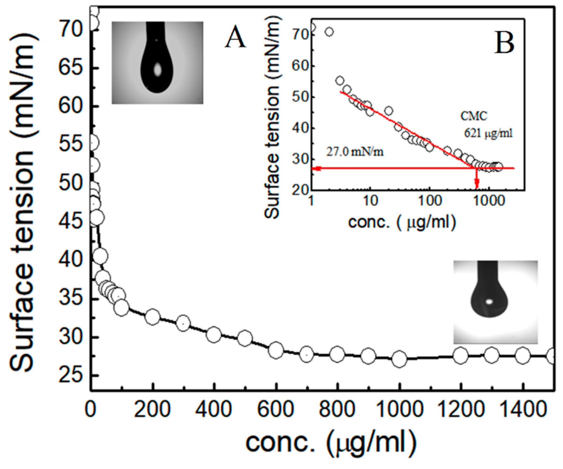
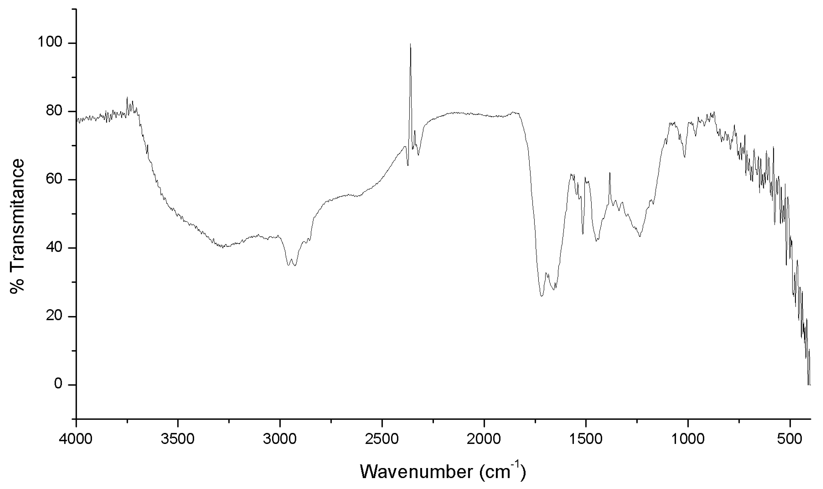
3.2. Analysis of Physico-Chemical Properties of Biosurfactant

Analysis of physical properties like SFT, IFT, CMC, CA, emulsification properties and ionic character proved the effectiveness of CFBS. The SFT of PBS was reduced from 72 to 27 mN/m with a CMC 625 µg/mL (Figure 2A). Figure 2B represents the semi-logarithmic reflection of the same CMC value from SFT versus CFBS concentration.
Other physical properties like IFT, CA, EA, ES also confirmed the efficacy of CFBS produced by NCIM 2903 with great certainty. The IFT measurements with immiscible liquids (hydrocarbons), showed lowest IFT against benzene (1.47 mN/m) followed by kerosene, o-xylene, n-hexane (Figure 3). There was good reduction in IFT values by CFBS against all hydrocarbons tested in comparison to other synthetic surfactants.
Effective spreading abilities for CFBS were tested through the measurement of CA. The highest reduction in CA was observed on polystyrene (From θ = 90° to 49°) and PDMS (from θ = 115° to 74°) followed by Teflon (from θ = 115° to 75°). Polystyrene and PDMS surfaces are used regularly in biomedical industries. CA on OHP transparent sheet was reduced from θ = 69° to 44°. However, on highly hydrophilic surface like glass; there was no significant reduction in CA values (from θ = 39° to 25°) (Figure 4).
The CA measurements for CFBS derived from Lactobacillus sp. have seldom been reported in literature. The emulsification capacity and stabilization activity of CFBS against different hydrocarbons-liquids (water insoluble) analysis showed that CFBS has relative EV between 25–65% and ES between 45–87% after 24 h. The highest EV (65%) was seen against n-decane with 87% of ES. CFBS demonstrated good EV (46%) and ES (87%) with xylene. Treatment of CFBS (CMC solution) at different pH (2.0 to 12.0) indicated higher stability between the pH of 6–10 and lowest SFT value at pH 7.0 indicating the potency of CFBS at neutral to alkaline conditions. However, treatment of BS at acidic pH; result its precipitation. Regarding the temperature effects, CFBS worked well at different temperatures (4, 30, 70 and 121 °C). Determination of ionic character by agar double diffusion technique indicated passive diffusion between CTAB (cationic compounds) and CFBS proving the presence of anionic charge on CFBS (Figure 5). A prominent line of precipitation formed on soft gel helped rapid determination of ionic character of CFBS.
Sugar and lipid moieties were detected using TLC confirming the presence of glycolipid typed CFBS. The chemical composition was confirmed by FTIR analysis (Figure 6).
The peak at 3320 cm−1 depicts the presence of OH stretching. The presence of hydrocarbons is confirmed by the peak at 2900 cm−1. The peaks at 1730 cm−1 significantly denote the presence C=O stretching in ester bond. The presence of ether moiety was confirmed by the presence of peak at 1230 while the presence of sugar moiety was clearly indicated by the peak at 1000 cm−1 (C-O stretching in sugars). The results of our study strongly suggest a glycolipid nature of CFBS.

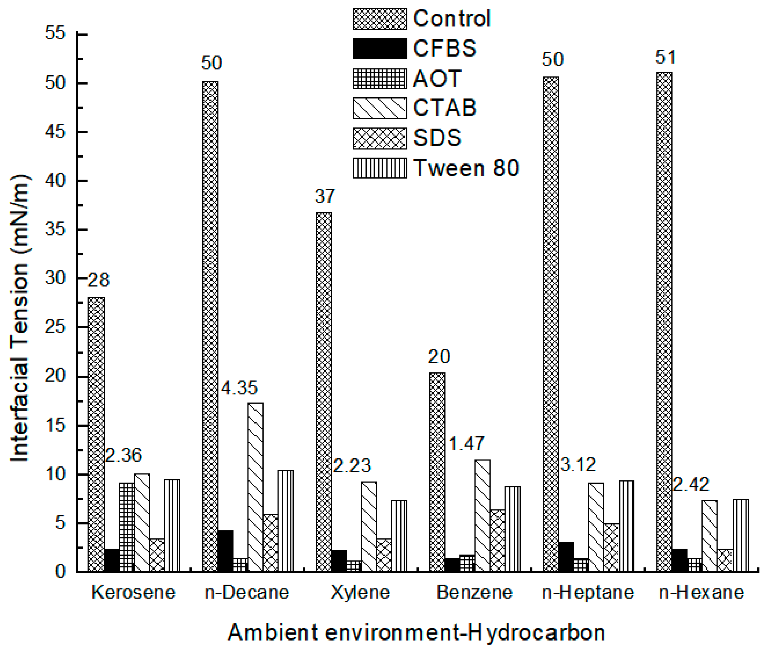
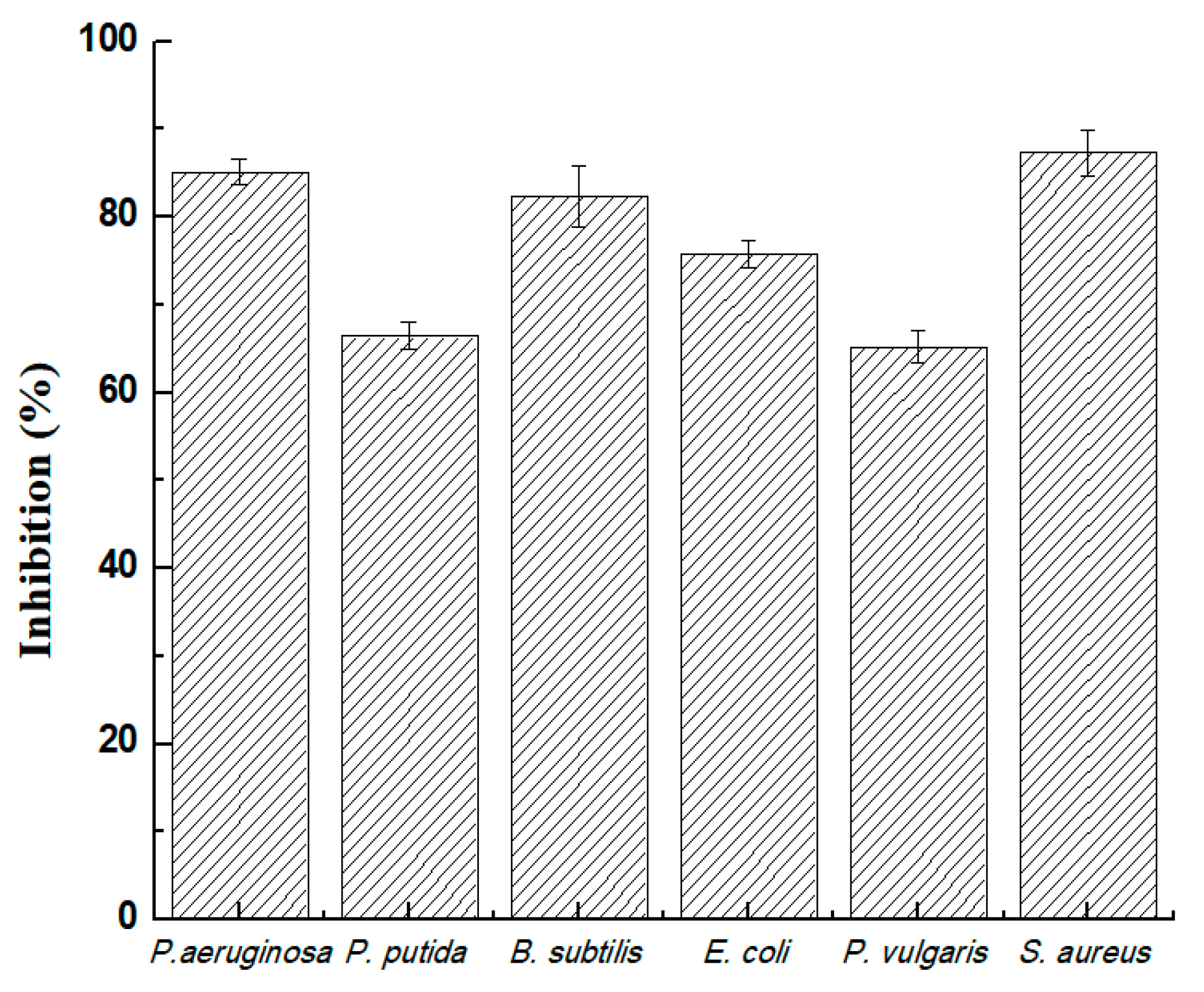
3.3. Determination of Antimicrobial Potential of Biosurfactant

Our studies showed good antimicrobial potential of CFBS. It could be clearly seen in Figure 7 that CFBS exhibited antimicrobial activity ranging between 65 and 87% against all bacterial strains used in the study. At the concentration of 625 µg/mL, it inhibited the growth of S. aureus NCIM 2079 (87%), Ps. aeruginosa MTCC 2297 (85%), B. subtilis MTCC 2423 (82%) and followed by E. coli NCIM 2065 (80%) and Ps. putida MTCC 2467 and P. valgaris NCIM 2027(70%).

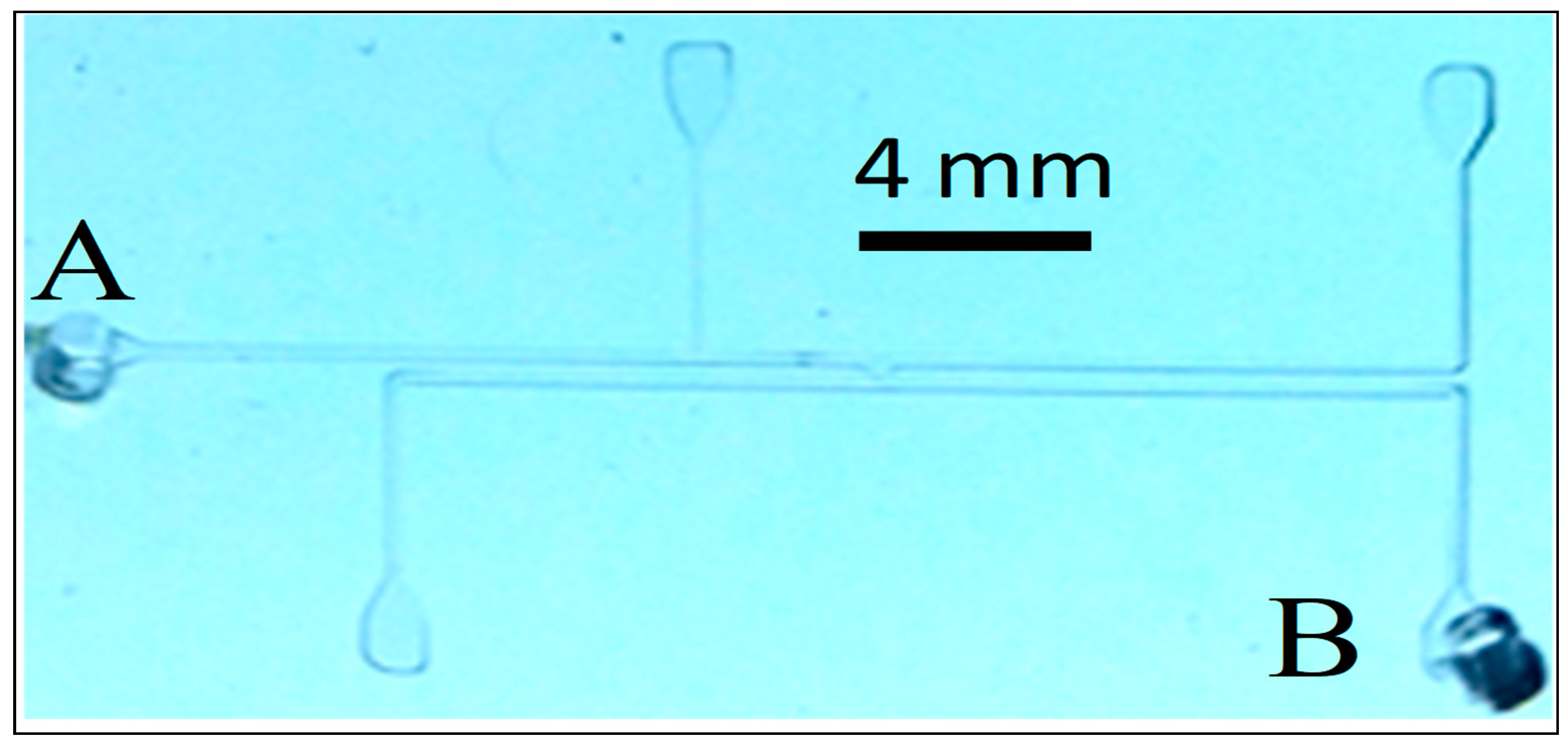
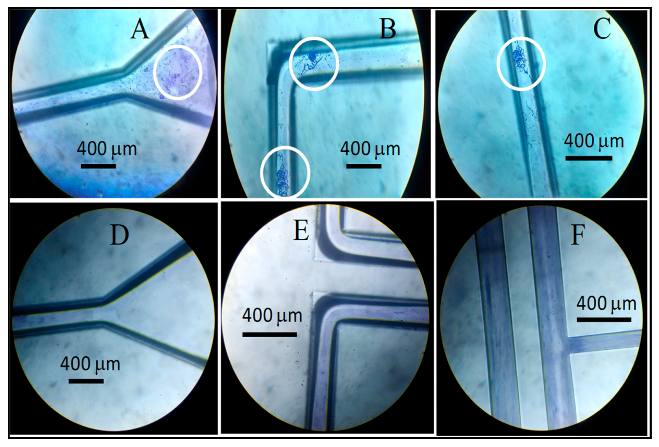
3.4. Investigation of Antiadhesive and Antibiofilm Potential of Biosurfactant viaMicrofluidic Approach

The MF assembly shown in Figure 1 was used to investigate the antiadhesive and antibiofilm effect of CFBS against B. subtilis. Biofilms were observed at different locations of the MF channels without any coating of CFBS. No noticeable effects were seen in the control treatments of PBS which was evident by the high growth and viability of the test culture (Figure 8A–C). The MF assembly coated with CFBS showed clear channels after staining with CV indicating that coating with CFBS inhibited the adhesion and biofilm formation on the surface (Figure 8D–F).

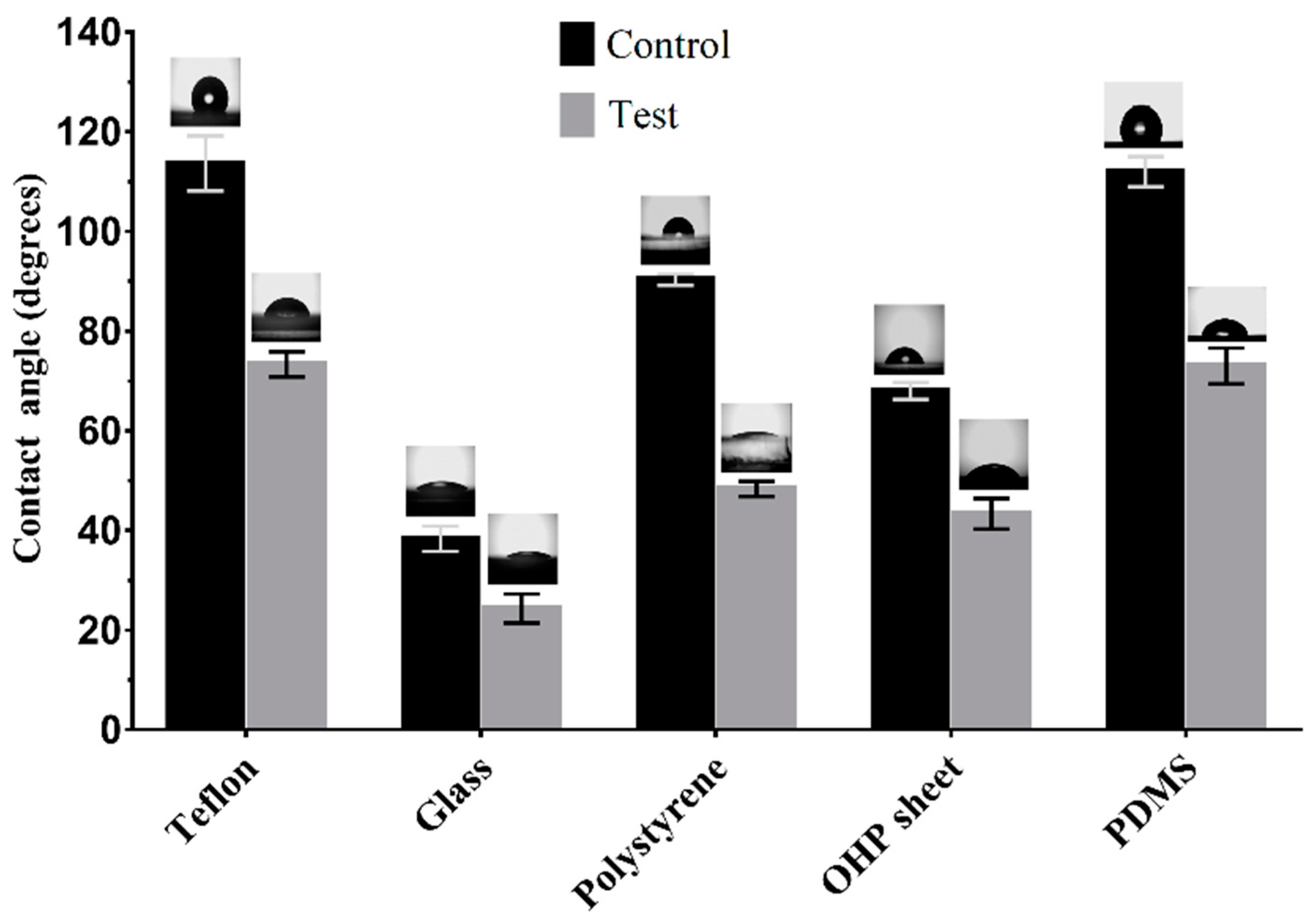
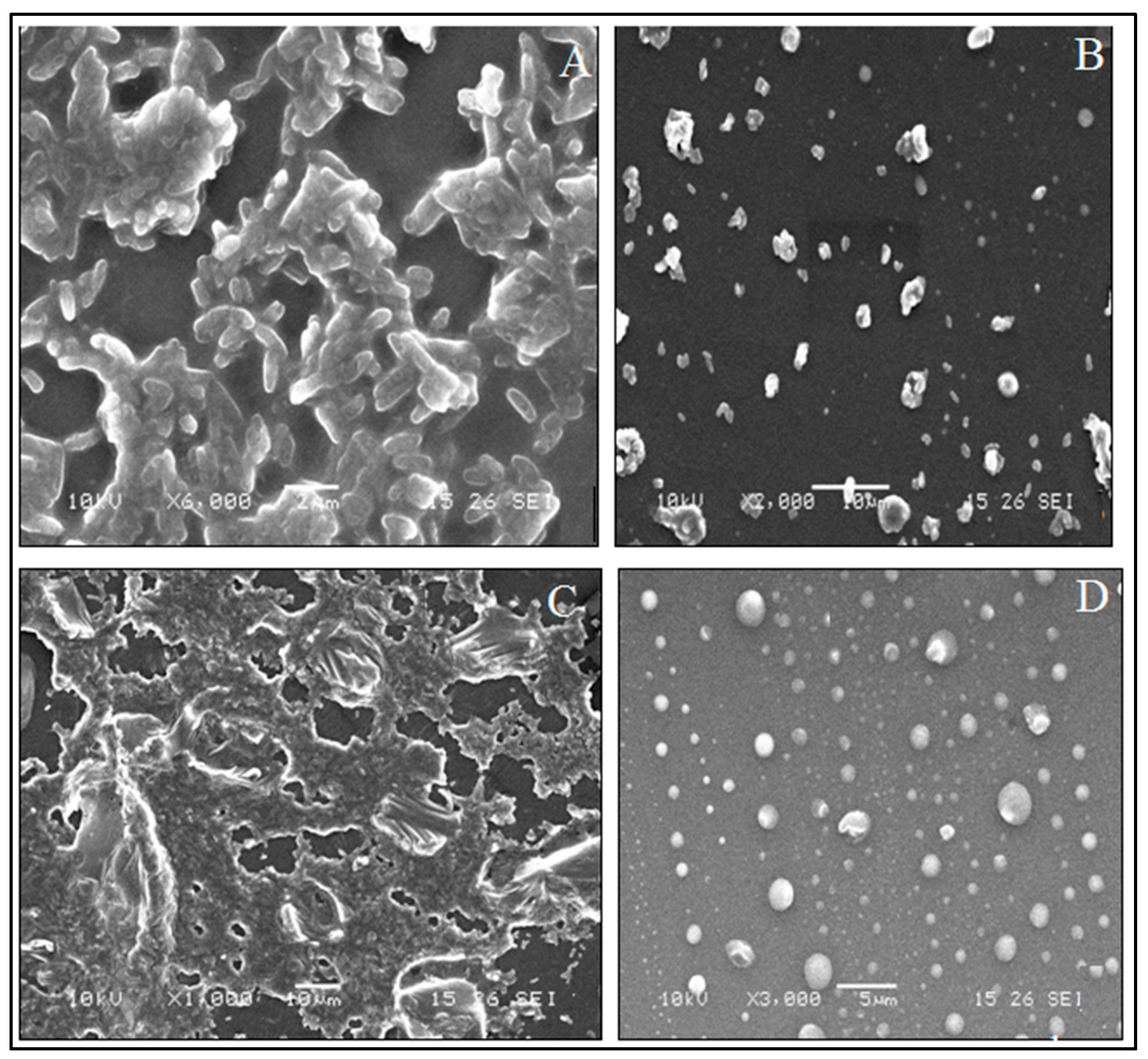
3.5. Evalution of Biofilm Inhibition Potential of Biosurfactant on Polydimethylsiloxane (PDMS) Surface

In addition to MF based approach, PDMS surfaces were also used to demonstrate the inhibiting effect of cell free biosurfactant (CFBS) against biofilms formed by B. subtilis MTCC 2423, P. valgaris NCIM 2027. We used SEM technique to distinguish the conglomerations of biofilms on PDMS surfaces. Under SEM, Figure 9A,C (Control) where confluent biofilms were clearly visualized than in Figure 9B,D (Test) representing the CFBS pre-coated Polydimethylsiloxane (PDMS) discs. Formation of microbial biofilms was restricted on surfaces of PDMS disc when they were previously coated with CFBS.

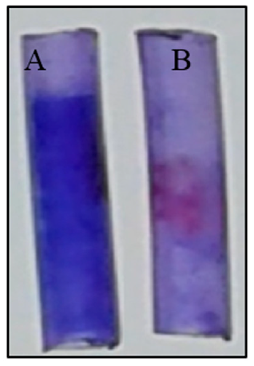
3.6. Evaluation of Antibiofilm Potential of Biosurfactant by Foley Catheter Assay

A commercially used medical grade catheter was also tested to explore the effect of CFBS on pathogenic bacterial biofilms. Figure 10A shows dark blue colouration due to the staining of biofilms formed by P. vulgaris NCIM 2027. Whereas Figure 10B did not show any blue colour development on catheter surface indicating that glycolipid CFBS inhibited the formation of biofilm of P. vulgaris NCIM 2027. The pre-coated of silicone urethral catheters with glycolipid CFBS completely inhibited the adherence and growth of P. vulgaris NCIM 2027.

4. Discussion

4.1. Production and Extraction of Biosurfactant

Most of the literature documented the use of de man Rogosa Sharpe (MRS) medium [33] for the synthesis BS from LAB including Lactobacilli spp. [1,14,34]. We have documented the lowest SFT value as 26 mN/m for FM after the growth of L. acidophilus NCIM 2903 [1].

4.2. Analysis of Physical and Chemical Properties of Biosurfactant

Physical properties viz., SFT, CMC, IFT, CA, emulsification clearly proved the potency of CFBS. We observed the maximum reduction in SFT of PBS from 72 to 27 mN/m at a CMC value of 625 µg/mL (Figure 2A,B). In addition, noticeable reduction in CA on various surfaces and IFT values against various hydrocarbons entirely proved the potency of CFBS isolated in the present studies (Figure 3 and Figure 4). It is important to note that the CA measurements of CFBS isolated from Lactobacillus sp. have hardly documented in literature. The emulsification with n-decane, and xylene was observed to be comparatively better. Our results are comparable with the Moldes et al. [13] for emulsions of octane/water stabilized by BS isolated from L. pentosus and SDS. Along with these results; the stability of BS at a wide physiological condition would contribute towards broadening its scope as antibiofilm agent. Our studies revealed the anionic nature of the CFBS. Similar results were reported by Sharma et al. [35] for the BS isolated from Enterococcus faecium. Structural examination of CFBS confirmed the glycolipid type. Closer observation in literature, we can say that our results are comparable with that of the work reported by Sharma et al. [5,6] who suggested the Xylolipidic type BS from Lactobacillus spp. In the literature, there are some instances, where researchers have claimed the production of glycolipid type BS from Lactobacillus sp. [5,6].

4.3. Antimicrobial Potential of Biosurfactant

Several microorganisms have been used for a wide range of BS production some of which with some antimicrobial properties [3,4,7,11]. However, limited number of reports documented antimicrobial activity of BS obtained from Lactobacilli spp. CFBS obtained from few Lactobacilli spp., displayed antimicrobial activity at low concentrations [14]. Antiadhesive and antibiofilm potential, however CABS are popular for their antiadhesive property rather than antimicrobial potential (concentration ranging between 4 and 50 mg/mL) [11]. In this work we have demonstrated antimicrobial activity of CFBS at a concentration of 625 µg/mL. More than 80% of antimicrobial effect of CFBS was achieved by the test organisms (S. aureus, Ps. aeruginosa, B. subtilis and E. coli) used in this study. Gudiña et al. [9] showed 76.8% antiadhesive activity at a 50 mg/mL of CABS isolated from L. paracasei spp. paracasei A20 against S. aureus.
The growth of bacteria like E. coli ATCC and Y. enterocolitica were inhibited at a concentration of 25 mg/mL for CABS isolated from L. plantarum CFR 2194. The same surfactant at both concentrations was found to be ineffective against S. typhi. The possible mode of action of CFBS needs thorough investigation. BS can act on microbial system through interfering the membrane functions and energy generating mechanisms. BS can reduce the cell surface hydrophobicity and microbial adherence to surfaces. Thus, microbial colonization can be prevented or reduced. BS can also increase the cell permeability and can cause the leakage of the metabolites. Alteration in physical membrane structure as well as interference of the protein conformations was also usually reported [4].

4.4. Investigation of Antiadhesive and Antibiofilm Potential via Microfluidic Approach

To the best of our knowledge, perhaps this is the first report of MF based studies to demonstrate the inhibitory effect of Lactobacillus derived glycolipid CFBS against bacterial biofilms. CFBS inhibiting the adherence, growth and biofilm formation of pathogens is significant. Similar observations have been put forward by De Rienzo et al. [36] indicating the effect of sophorolipid (SPL) on B. subtilis using BioFlux channel system. Authors also suggested that multidrug-resistant bacterial infections can be reduced with the help of combinations of RHL and SPL.

4.5. Inhibition of Bacterial Biofilms by Biosurfactant on Polydimethylsiloxane (PDMS)Surface

The BS strongly inhibited the biofilm formation on PDMS surface. Our results are comparable to studies carried out by Velraeds et al. [37] who showed inhibitory effect of CFBS produced by Lactobacillus sp. on E. faecalis biofilms on glass surfaces. Our work also gave a strong indication towards the high capability of CFBS to act as antiadhesive and antibiofilm effect against pathogens.

4.6. Foley Catheter Assay to Evaluate Inhibition of Bacterial Biofilms by Biosurfactant

Our results obtained for silicon based urinary catheter are comparable with Rivardo et al. [38], who showed that BSs of Bacillus spp. inhibits biofilm formed by E. coli and S. aureus. Similarly, Irie et al. [39] also displayed biofilm dispersion ability of Ps. aeruginosa originated RHL against Bordetella bronchiseptica. BS molecules can work excitingly in synergistic way (against pathogens) in association with antibiotics, NPs etc. Gómez-Graña et al. [40] suggested that the CFBS exhibit deep impact in reducing the metal precursor and also stabilizing the NPs. More importantly, BS stabilizes NPs and exhibits antimicrobial activity against various pathogens like E. coli, Ps. aeruginosa, S. aureus. Such evidence reflects the broad spectrum potential of BS mediated NPs for tackling the antimicrobial resistance. Promising experimental procedures are mandatory for synthesis of NPs where Green technology is the major driving force for the researchers. Currently nanotechnology is in an attempt to use clean, non-toxic and eco-friendly to synthesize nanomaterials in order to minimize the disposal of wastes. Microbes are popularly known for synthesis of inorganic molecules which can be deposited intra or outside the cells [15]. Use of nanomaterials amalgamated with extraordinary molecules like BS, antibiotics definitely provides opportunities to explore the innovative applications.

5. Conclusions

Glycolipid BS displayed virtuous antimicrobial, anti-adhesion and anti-biofilm potential against pathogenic bacteria on catheters and PDMS surfaces. MF confirms its ability to restrict the primary adhesion of biofilm forming pathogenic bacteria on surfaces. These studies are noteworthy to be widely used to examine biofilm cohesion under a variety of physiological like temperature and pH. CFBS explored by us has commercial applications for several medical devices. This would prolong the life of the biomaterials as well as reduce the possibilities of opportunistic infections. Possibly, for the first time we have utilized MF system to demonstrate antibiofilm effect CFBS derived from Lactobacillus spp. This can be exploited for testing the various antibiofilm and antiadhesive formulations against various pathogens at laboratory level.

Author Contributions

S.K.S., I.M.B. and A.G.B. conceived the research project and S.K.S. & A.G.B. achieved financial support from funding agency. S.K.S., I.M.B. and A.G.B. designed the study. S.K.S., N.S.M., P.D., A.G.B. performed the experiments and analyzed the results. All authors participated in writing the manuscript and approved the final manuscript for submission.

Funding

Department of Science and Technology (DST), Government of India, Ministry of Science and Technology {SR/WOS-A/LS-1076/2014(G)}.

Acknowledgments

Surekha K. Satpute, expresses special thanks of gratitude to Department of Science and Technology (DST), Government of India, Ministry of Science and Technology {SR/WOS-A/LS-1076/2014(G)} and Department Research and Development Programs (DRDP), Savitribai Phule Pune University (SPPU) for financial support. The help of Gauri R. Kulkarni, The Director, School of Basic medical Sciences, Department of Physics, SPPU is also acknowledged for providing the laboratory facilities to complete the current research.

Conflicts of Interest

The authors declare no conflicts of interest.

References

  1. Satpute, S.K.; Mone, N.S.; Patil, R.H.; Banat, I.M.; Banpurkar, A.G. Inhibition of pathogenic bacterial biofilms on PDMS based implants by L. acidophilus derived biosurfactant. BMC Microbiol. 2018. under revision; MCRO-D-17-00595R1. [Google Scholar]
  2. Satpute, S.K.; Zinjarde, S.Z.; Banat, I.M. Recent advances of biosurfactant in food industry. In Microbial Cell Factories: Recent Progress and Sustainable Trends; Sharma, D., Saharan, B.S., Eds.; CRC Press; Taylor and Francis: Boca Raton, FL, USA; London, UK; New York, NY, USA, 2018; pp. 1–20. ISBN 9781138061385. [Google Scholar]
  3. Satpute, S.K.; Płaza, G.A.; Banpurkar, A.G. Biosurfactants’ Production from Renewable Natural Resources: Example of Innovative and Smart Technology in Circular Bioeconomy. Manag. Syst. Prod. Eng. 2017, 25, 46–54. [Google Scholar] [CrossRef]
  4. Satpute, S.K.; Banpurkar, A.G.; Banat, I.M.; Sangshetti, J.N.; Patil, R.R.; Gade, W.N. Multiple roles of biosurfactants in biofilms. Curr. Pharm. Des. 2016, 22, 1429–1448. [Google Scholar] [CrossRef]
  5. Sharma, D.; Saharan, B.S.; Chauhan, N.; Bansal, A.; Procha, S. Production and structural characterization of Lactobacillus helveticus derived biosurfactant. Sci. World J. 2014, 2014, 493548. [Google Scholar] [CrossRef] [PubMed]
  6. Sharma, D.; Saharan, B.S. Simultaneous production of biosurfactants and bacteriocins by probiotic Lactobacillus casei MRTL3. Int. J. Microbiol. 2014, 2014, 698713. [Google Scholar] [CrossRef] [PubMed]
  7. Gomaa, E.Z. Antimicrobial and anti-adhesive properties of biosurfactant produced by Lactobacilli isolates, biofilm formation and aggregation ability. J. Gen. Appl. Microbiol. 2013, 59, 425–436. [Google Scholar] [CrossRef]
  8. Gudiña, E.J.; Fernandes, E.C.; Teixeira, J.A.; Rodrigues, L.R. Antimicrobial and anti-adhesive activities of cell-bound biosurfactant from Lactobacillus agilis CCUG31450. RSC Adv. 2015, 5, 90960–90968. [Google Scholar] [CrossRef]
  9. Gudiña, E.J.; Rocha, V.; Teixeira, J.A.; Rodrigues, L.R. Antimicrobial and antiadhesive properties of a biosurfactant isolated from Lactobacillus paracasei ssp. paracasei A20. Lett. Appl. Microbiol. 2010, 50, 419–424. [Google Scholar]
  10. Gudina, E.J.; Teixeira, J.A.; Rodrigues, L.R. Isolation and functional characterization of a biosurfactant produced by Lactobacillus paracasei. Colloids Surf. B Biointerfaces 2010, 76, 298–304. [Google Scholar] [CrossRef] [PubMed]
  11. Madhu, A.N.; Prapulla, S.G. Evaluation and functional characterization of a biosurfactant produced by Lactobacillus plantarum CFR 2194. Appl. Biochem. Biotechnol. 2014, 172, 1777–1789. [Google Scholar] [CrossRef] [PubMed]
  12. Vecino, X.; Barbosa-Pereira, L.; Devesa-Rey, R.; Cruz, J.M.; Moldes, A.B. Optimization of extraction conditions and fatty acid characterization of Lactobacillus pentosus cell-bound biosurfactant/bioemulsifier. J. Sci. Food Agric. 2015, 95, 313–320. [Google Scholar] [CrossRef] [PubMed]
  13. Moldes, A.B.; Paradelo, R.; Vecino, X.; Cruz, J.M.; Gudiña, E.; Rodrigues, L.; Teixeira, J.A.; Domínguez, J.M.; Barral, M.T. Partial characterization of biosurfactant from Lactobacillus pentosus and comparison with sodium dodecyl sulphate for the bioremediation of hydrocarbon contaminated soil. BioMed Res. Int. 2013, 2013, 961842. [Google Scholar] [CrossRef] [PubMed] [Green Version]
  14. Satpute, S.K.; Kulkarni, G.R.; Banpurkar, A.G.; Banat, I.M.; Mone, N.S.; Patil, R.H.; Cameotra, S.S. Biosurfactant/s from Lactobacilli species: Properties, challenges and potential biomedical applications. J. Basic Microbiol. 2016, 56, 1140–1158. [Google Scholar] [CrossRef] [PubMed]
  15. Płaza, G.A.; Chojniak, J.; Banat, I.M. Biosurfactant mediated biosynthesis of selected metallic nanoparticles. Int. J. Mol. Sci. 2014, 15, 13720–13737. [Google Scholar] [CrossRef] [PubMed]
  16. Mukherjee, P.; Senapati, S.; Mandal, D.; Ahmad, A.; Khan, M.I.; Kumar, R.; Sastry, M. Extracellular synthesis of gold nanoparticles by the fungus Fusarium oxysporum. ChemBioChem 2002, 3, 461–463. [Google Scholar] [CrossRef]
  17. Ahmad, A.; Senapati, S.; Khan, M.I.; Kumar, R.; Ramani, R.; Srinivas, V.; Sastry, M. Intracellular synthesis of gold nanoparticles by a novel alkalotolerant actinomycete, Rhodococcus species. Nanotechnology 2003, 4, 824. [Google Scholar] [CrossRef]
  18. Reddy, A.S.; Chen, C.Y.; Chen, C.C.; Jean, J.S.; Fan, C.W.; Chen, H.R.; Wang, J.C.; Nimje, V.R. Synthesis of gold nanoparticles via an environmentally benign route using a biosurfactant. J. Nanosci. Nanotechnol. 2009, 9, 6693–6699. [Google Scholar] [CrossRef] [PubMed]
  19. Reddy, A.S.; Chen, C.Y.; Baker, S.C.; Chen, C.C.; Jean, J.S.; Fan, C.W.; Chen, H.R.; Wang, J.C. Synthesis of silver nanoparticles using surfactin: A biosurfactant as stabilizing agent. Mater. Lett. 2009, 63, 1227–1230. [Google Scholar] [CrossRef]
  20. Rane, A.N.; Baikar, V.V.; Ravi Kumar, V.; Deopurkar, R.L. Agro-industrial wastes for production of biosurfactant by Bacillus subtilis ANR 88 and its application in synthesis of silver and gold nanoparticles. Front. Microbiol. 2017, 8, 492. [Google Scholar] [CrossRef] [PubMed]
  21. Zheng, B.; Tice, J.D.; Roach, L.S.; Ismagilov, R.F. A droplet-based, composite PDMS/glass capillary microfluidic system for evaluating protein crystallization conditions by microbatch and vapour-diffusion methods with on-chip X-ray diffraction. Angew. Chem. Int. Ed. 2004, 43, 2508–2511. [Google Scholar] [CrossRef] [PubMed]
  22. Kumaresan, P.; Yang, C.J.; Cronier, S.A.; Blazej, R.G.; Mathies, R.A. High-throughput single copy DNA amplification and cell analysis in engineered nanoliter droplets. Anal. Chem. 2008, 80, 3522–3529. [Google Scholar] [CrossRef] [PubMed]
  23. Huebner, A.; Sharma, S.; Srisa-Art, M.; Hollfelder, F.; Edel, J.B. Microdroplets: A sea of applications? Lab Chip 2008, 8, 1244–1254. [Google Scholar] [CrossRef] [PubMed]
  24. Srinivasan, V.; Pamula, V.K.; Fair, R.B. An integrated digital microfluidic lab-on-a-chip for clinical diagnostics on human physiological fluids. Lab Chip 2004, 4, 310–315. [Google Scholar] [CrossRef] [PubMed]
  25. Khor, S.Y.; Vu, M.U.; Pilkington, E.H.; Johnston, A.P.R.; Whittaker, M.R.; Quinn, J.F.; Truong, N.; Davis, T.P. Elucidating the influences of size, surface chemistry, and dynamic flow on cellular association of nanoparticles made by polymerization-Induced self-assembly. Small 2018, 14, e1801702. [Google Scholar] [CrossRef] [PubMed]
  26. Brouzes, E.; Medkova, M.; Savenelli, N.; Marran, D.; Twardowski, M.; Hutchison, J.B.; Rothberg, J.M.; Link, D.R.; Perrimon, N.; Samuels, M.L. Droplet microfluidic technology for single-cell high-throughput screening. Proc. Natl. Acad. Sci. USA 2009, 106, 14195–14200. [Google Scholar] [CrossRef] [PubMed] [Green Version]
  27. Satpute, S.K.; Banpurkar, A.G.; Dhakephalkar, P.K.; Banat, I.M.; Chopade, B.A. Methods for investigating biosurfactants and bioemulsifiers: A review. Crit. Rev. Biotechnol. 2010, 30, 127–144. [Google Scholar] [CrossRef] [PubMed]
  28. Hamza, F.; Satpute, S.; Banpurkar, A.; Kumar, A.R.; Zinjarde, S. Biosurfactant from a marine bacterium disrupts biofilms of pathogenic bacteria in a tropical aquaculture system. FEMS Microbiol. Ecol. 2017, 93, fix140. [Google Scholar] [CrossRef] [PubMed]
  29. Das, M.; Das, S.K.; Mukherjee, R.K. Surface active properties of the culture filtrates of a Micrococcus species grown on n-alkanes and sugars. Bioresour. Technol. 1998, 63, 231–235. [Google Scholar] [CrossRef]
  30. Meylheuc, T.; Van Oss, C.J.; Bellon-Fontaine, M.N. Adsorption of biosurfactant on solid surfaces and consequences regarding the bioadhesion of Listeria monocytogenes LO28. J. Appl. Microbiol. 2001, 91, 822–832. [Google Scholar] [CrossRef] [PubMed]
  31. Mireles, J.R.; Toguchi, A.; Harshey, R.M. Salmonella enterica serovar typhimurium swarming mutants with altered biofilm-forming abilities: Surfactin inhibits biofilm formation. J. Bacteriol. 2001, 183, 5848–5854. [Google Scholar] [CrossRef] [PubMed]
  32. McIntyre, R.A. Common nano-materials and their use in real world applications. Sci. Prog. 2012, 95, 1–22. [Google Scholar] [CrossRef] [PubMed]
  33. De Man, J.C.; Rogosa, D.; Sharpe, M.E. A medium for the cultivation of Lactobacilli. J. Appl. Bacteriol. 1960, 23, 130–135. [Google Scholar] [CrossRef]
  34. Banat, I.M.; Satpute, S.K.; Cameotra, S.S.; Patil, R.; Nyayanit, N.V. Cost effective technologies and renewable substrates for biosurfactants’ production. Front. Microbiol. 2014, 5, 697. [Google Scholar] [CrossRef] [PubMed]
  35. Sharma, D.; Saharan, B.S.; Chauhan, N.; Procha, S.; Lal, S. Isolation and functional characterization of novel biosurfactant produced by Enterococcus faecium. SpringerPlus 2015, 4, 4. [Google Scholar] [CrossRef] [PubMed]
  36. De Rienzo, M.D.; Stevenson, P.S.; Marchant, R.; Banat, I.M. Effect of biosurfactants on Pseudomonas aeruginosa and Staphylococcus aureus biofilms in a BioFlux channel. Appl. Microbiol. Biotechnol. 2016, 100, 5773–5779. [Google Scholar] [CrossRef] [PubMed]
  37. Velraeds, M.M.; Van der Mei, H.C.; Reid, G.; Busscher, H.J. Inhibition of initial adhesion of uropathogenic Enterococcus faecalis by biosurfactants from Lactobacillus isolates. Appl. Environ. Microbiol. 1996, 62, 1958–1963. [Google Scholar] [PubMed]
  38. Rivardo, F.; Turner, R.J.; Allegrone, G.; Ceri, H.; Martinotti, M.G. Anti-adhesion activity of two biosurfactants produced by Bacillus spp. prevents biofilm formation of human bacterial pathogens. Appl. Microbiol. Biotechnol. 2009, 83, 541–553. [Google Scholar] [CrossRef] [PubMed]
  39. Irie, Y.; O’toole, G.A.; Yuk, M.H. Pseudomonas aeruginosa rhamnolipids disperse Bordetella bronchiseptica biofilms. FEMS Microbiol. Lett. 2005, 250, 237–243. [Google Scholar] [CrossRef] [PubMed]
  40. Gómez-Graña, S.; Perez-Ameneiro, M.; Vecino, X.; Pastoriza-Santos, I.; Perez-Juste, J.; Cruz, J.M.; Moldes, A.B. Biogenic synthesis of metal nanoparticles using a biosurfactant extracted from corn and their antimicrobial properties. Nanomaterials 2017, 7, 139. [Google Scholar] [CrossRef] [PubMed]
Figure 1. Microfluidics (MF) model used to conduct Lab on Chip assay. (A) Sample loading point; (B) Sample exit point after filling the MF channels completely.
Applsci 08 01555 g001
Figure 2. (A) Variation in surface tension and (B) semi-logarithmic reflection of critical micelle concentration (CMC) value from surface tension (SFT) (mN/m) versus biosurfactant (BS) concentration.
Applsci 08 01555 g002
Figure 3. Measurement of interfacial tension (IFT) mN/m for biosurfactant obtained from L. acidophilus NCIM 2903 and synthetic surfactants against water insoluble liquids. Control: Phosphate buffer saline; CFBS: Cell free biosurfactant; AOT: Aerosol OT; CTAB: Cetyl trimethyl ammonium bromide; SDS: Sodium dodecyl sulphate.
Applsci 08 01555 g003
Figure 4. Measurement of contact angle (CA) (degrees) for biosurfactant (BS) obtained from L. acidophilus NCIM 2903 on different surfaces, OHP: Overhead Projector Transparent sheet; PDMS: polydimethlysiloxane.
Applsci 08 01555 g004
Figure 5. Double diffusion of cationic surfactant (B) (positively charged) Cetyl trimethyl ammonium bromide (CTAB) against biosurfactant (BS) (A) on agar plate indicating a line of precipitation due to formation of ionic pair.
Applsci 08 01555 g005
Figure 6. Fourier-transform infrared spectroscopy (FTIR) spectrum for biosurfactant obtained from L. acidophilus NCIM 2903.
Applsci 08 01555 g006
Figure 7. Antimicrobial potential of biosurfactant derived from NCIM 2903 against Ps. aeruginosa MTCC 2297, Ps. putida MTCC 2467, B. subtilis MTCC 2423, E. coli NCIM 2065, P. vulgaris NCIM 2027 and S. aureus NCIM 2079.
Applsci 08 01555 g007
Figure 8. Optical microscopic images of microfluidics (MF) assembly and biofilms. MF channels with (Test-Lower row DF) and without (Control-Upper row AC) biosurfactant (BS) coatings (625 µg/mL). In absence of biosurfactant (BS) coating, no inhibition of adhesion and therefore confluent biofilm formed by B. subtilis MTCC 2423 (AC). White coloured circles indicates the biofilm stained with crystal violet (1%) solution. In contrast, the test MF channels (DF) are seen clear without biofilms.
Applsci 08 01555 g008
Figure 9. Scanning electron microscope (SEM) images of polydimethylsiloxane (PDMS) disc with (A,C) and without biofilms (B,D) formed by bacterial cultures. The upper row (A,B) represents for P. valgaris NCIM 2027 and lower row (C,D) represents for B. subtilis MTCC 2423. Figure (A) and (C) represent the control surfaces indicating the PDMS disc without any biosurfactant (BS) coating where confluent growth of biofilms is seen. Whereas, Figure (B) and (D) represents the test surfaces indicating pre-coating with BS inhibiting not only the adherence but also the growth of bacteria; proving the antiadhesion and antibiofilm potential of BS.
Applsci 08 01555 g009
Figure 10. Foley catheters assay: Inhibition of biofilm formed by P. vulgaris NCIM 2027 by biosurfactant (BS) on urethral catheters. (A) The control where bacterial biofilm appears as a dark blue colour; (B) The test, showing anti-biofilm property of BS against the bacterial strain. Development of biofilm was diminished absolutely on test catheter tubing.
Applsci 08 01555 g010

