Review

Silver Nanowires: Synthesis, Antibacterial Activity and Biomedical Applications

1
School of Pharmacy and Biomedical Sciences, University of Portsmouth, White Swan Road, Portsmouth PO1 2DT, UK
2
Institute of Biological and Biomedical Science, University of Portsmouth, White Swan Road, Portsmouth PO1 2DT, UK
*
Authors to whom correspondence should be addressed.
Appl. Sci. 2018, 8(5), 673; https://doi.org/10.3390/app8050673
Submission received: 15 March 2018 / Revised: 18 April 2018 / Accepted: 20 April 2018 / Published: 26 April 2018
(This article belongs to the Special Issue Nano-systems for Antimicrobial Therapy)

Abstract

:
Silver is well known for its antibacterial properties and low toxicity, and it is currently widely used both in the form of ions and of nanoparticles in many diverse products. One-dimensional silver nanowires (AgNWs) have the potential to further enhance the properties of nanosilver-containing products, since they appear to have higher antimicrobial efficacy and lower cytotoxicity. While they are widely used in optics and electronics, more studies are required in order to better understand their behavior in the biological environment and to be able to advance their application in uses such as wound healing, surface coating and drug delivery.

1. Introduction

For several decades, silver has been used for its demonstrated antibacterial activity [1] and low toxicity to human cells [2], with successful applications in prophylactic burn treatment and water disinfection [3,4]. With the rising presence of antibiotic-resistant bacteria [5,6], and the increased development of nanoscale technology, scientific focus has shifted toward silver nanospecies, due to their unique electrical, optical, chemical and physical properties and their potential in a variety of fields from toothpastes to electrodes [7], including potential future biomedical applications [8,9]. Silver nanospecies have been synthesized via a variety of different methods, resulting in the production of different configurations, each possessing a discrete extent of antibacterial activity. Commonly synthesized silver nanospecies include: silver nanowires, silver nanorods, silver nanocubes and silver nanospheres [10,11,12], as well as more recent silver nanocoils [13]. Due to their greater surface area to volume ratio, silver nanospecies offer enhanced characteristics compared to bulk silver [14,15,16], and may therefore serve as an additional weapon in our current antibacterial arsenal. To date, these silver nanospecies have been shown to be effective against bacteria such as Escherichia coli [17], Staphylococcus aureus [17], Klebsiellia pneumoniae [18] and methicillin-resistant Staphylococcus aureus [19], among others.
Of these silver nanospecies, silver nanowires (AgNWs) are currently garnering major research interest. Due to their one-dimensional structure, they have a higher aspect ratio compared to other silver nanospecies, and therefore very different physicochemical properties. This review will consider the different synthesis methods used to generate silver nanowires of different lengths and diameters; address the proposed mechanisms of how AgNWs elicit powerful biocidal effects and how their toxicity potential is mitigated compared to other silver species; and finally, provide an up-to-date overview of AgNW-based applications in biomedicine.

2. Synthesis of AgNWs

AgNW synthesis has developed greatly over the last thirty years. Several solution methods have been studied and are extensively reviewed by Tang and Tsuji [20]; subsequent progress in the field has led to the development of two major methods by which AgNWs can be synthesized, referred to as hard- and soft-template methods. Some key findings are reported in Table 1.

2.1. Hard-Template Synthesis

The hard-template method is based on the use of nanoporous membranes, such as carbon nanotubes, nanoporous silica and alumina membranes, zeolites and track-etched polycarbonate, to direct the growth of AgNWs [19,20,21,43,44]. The greatest benefit of this method is that the synthesis can be carried out in a controlled manner, obtaining AgNWs that are uniform in size; furthermore, the obtained nanowires are already immobilized on a substrate, which is ideal in the production of nanodevices; on the other hand, this is not desirable when AgNWs are produced for biomedical applications [45,46]. Nanoporous membranes are commercially available, and easy-to-use hard templates with the wires synthesis occur within the cylindrical pores of the membrane, leading to the formation of silver nanowires with a controlled length and diameter. Furthermore, the production yield can be modified by choosing the desired membrane porosity [45]. Despite these advantages, the purification of AgNWs from the membrane requires dissolution of the nanoporous support in harsh conditions, which often results in damage to the nanowires [25].

2.2. Soft-Template Synthesis

To overcome the limitations associated with the hard-template method, the soft-template or polyol method was developed [47]. The soft-template, or silver cation reduction mediated by a polyol, is generally a faster process compared to the hard-template method, and it produces a greater yield of AgNWs, but most importantly, it is carried out in solution. This is advantageous when considering biomedical applications, as the resulting AgNWs can be successively readily dispersed in aqueous environments [26]. Sun et al. [25,26] were the first to demonstrate the synthesis of AgNWs with the use of poly(vinyl pyrrolidone) (PVP) as the soft template; this allowed to overcome limitations of previous methods that employed platinum (Pt) nanoparticle seeds to induce heterogeneous nucleation. The Pt-based process required a two-step reaction and the cost of Pt nanoparticles was prohibitory. Instead, the introduction of PVP allowed the development of a much simplified self-seeding method; PVP was initially dissolved in ethylene glycol, and AgNO3 was carefully injected by a two-syringe pump into the reaction mixture at a controlled rate, the rate of addition of the Ag+ source was crucial in the control of the size of the nanospecies produced [26]. Despite successful synthesis on a small scale, carefully and accurately controlling the injection rate of AgNO3 into the reaction mixture was not feasible on an industrial scale. Thus, Korte et al. [27] suggested that the addition of trace amounts of salts was able to slow the reaction rate and more finely control the growth of AgNWs. Initially, CuCl2 was added to the reaction mixture after the addition of an Ag+ salt. Cu2+ was found to remove oxygen atoms blocking growing sites of the AgNWs surface, favoring deposition of more silver, while also preventing oxidative etching. On the other hand, Cl- acted as the rate-determining step of the overall reaction, by forming temporary precipitates of AgCl, the slow degradation of which provided control over the reaction rate [28]. Choi et al. [29] exploited the same principle by adding trace amounts of NaCl in place of CuCl2, this resulted in similar size distribution (50 nm in diameter and 20 µm length) as assessed by SEM. Other factors able to control the size of the nanowires obtained are the molar ratio between PVP and AgNO3 and the length of the PVP molecule employed as soft template, as shown by Sun and Xia [24]. Bergin et al. [30] studied the effect of reaction time and temperature on the size of the obtained nanowires. They were able to control the length and diameter of the AgNWs produced by carefully selecting the temperature of the seeding and growth phases and by stopping the reaction at predefined time points.
Several studies have also been carried out in an attempt to elucidate the molecular mechanisms involved in the formation of silver nanowires; the complexities of this process are well explained in a review by Xia et al. [48]. The soft-template method involves the reduction of a silver salt to metallic silver in a reducing solvent, a polyol. At elevated temperatures, the polyol undergoes oxidation to an aldehyde that is the actual reducing agent [30]. The formed silver crystals assemble to form multiply twinned particles of decahedron structure (Figure 1a,b) [47]. Adsorption of further silver atoms to this structure was shown to force growth in both the axial and radial direction until a critical phase was reached where radial growth ceases and only axial growth continues over the {111} facets. In this case, PVP is thought to act as a capping agent, thanks to the ability of its carbonyl groups to bind silver atoms on the {100} facets (Figure 1c), which helps to drive the growth solely in the axial direction (Figure 1d) [48,49,50,51]. Controversial evidence is, however, available, as Choi et al. showed that the PVP coating is homogeneous and does not preferentially adsorb on the {100} facets [29]. Furthermore, subsequent work by Zhang et al. [31] showed that AgNWs could also be synthesized in the absence of PVP, using just a reducing agent such as glucose at elevated temperature (100 °C). This reaction lead to the formation of AgNWs of average diameters of between 30 and 50 nm; however, these were mixed with silver nanoparticles (AgNPs) as well, demonstrating that PVP is not essential for the formation of the nanowires but beneficial to the homogeneity of the product obtained. These results suggest that the exact mechanism of AgNW formation is still elusive, and that many as yet not understood factors may contribute to their formation.
Due to the exceptional electrical, optical and chemical characteristics of AgNWs, combined with their potential antimicrobial activity, there is an interest in developing a synthetic method that is suitable for future biomedical applications. Replacement of ethylene glycol with glycerol by Yang et al. [32] resulted in a rapid, scalable and green pathway for silver nanowire synthesis via the polyol soft-template method. In addition to being non-toxic to humans, glycerol also contains an extra hydroxyl group, facilitating faster silver ion reduction and stability at higher temperatures, allowing faster reaction kinetics [32]. Further optimizations, included dissolving PVP in glycerol at elevated temperatures, addition of AgNO3 upon rapid stirring, addition of a trace amount of water as a catalyst to help dissolve the NaCl before adding it to the reaction mixture. These changes successfully resulted in biomedical-ready silver nanowires with diameters between 60 and 90 nm and lengths between 6 and 12 µm [32].
This brief overview of the synthesis of AgNWs has shown that there is scope to further investigate the formation of AgNWs to better understand its molecular mechanisms. Furthermore, it is evident that the diameter and length distribution of AgNWs can be controlled by a number of different reaction conditions. The scalability of the most recent method, combined with the relatively low cost and toxicity of the starting materials, is advantageous for developing AgNWs for biomedical applications.

3. Mechanism of Antibacterial Activity

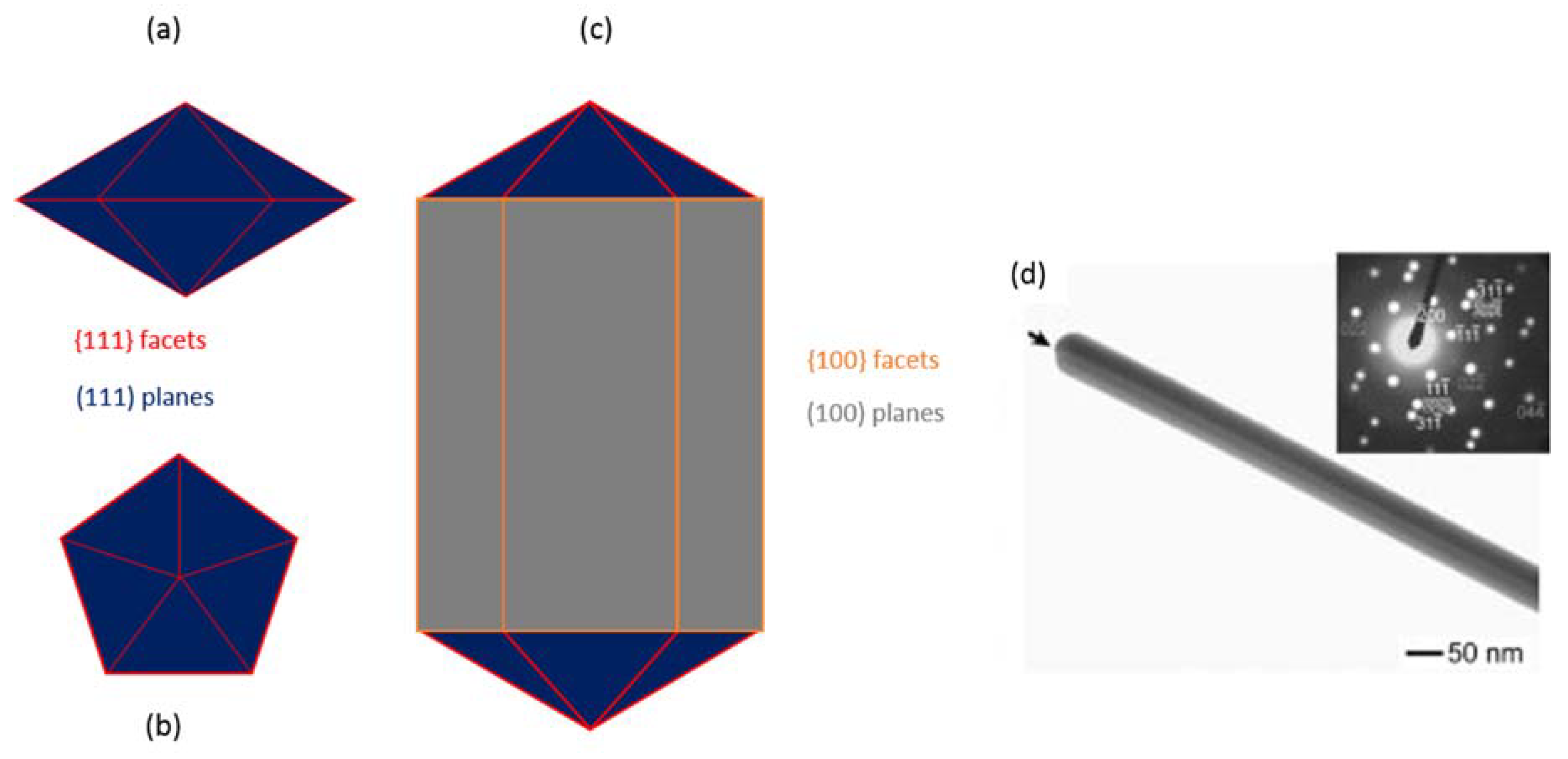
3.1. Silver Nanoparticles Mechanism of Action

Silver is currently present in a vast number of consumer products in the form of engineered materials and nanomaterials. Antimicrobial activity has been demonstrated for many different materials containing silver, but it has become evident that the mechanism of action of bulk silver may substantially differ from that of nanosilver [52], although the mechanism by which silver nanospecies elicit their antibacterial effects remains elusive. Three hypothesized mechanisms of action for silver nanomaterials have been reported in some depth in a review by Marambio-Jones and Hoek [52]. These three main mechanisms of antibacterial action, detailed below, are consistently hypothesized for AgNPs and are reported as directly causing or contributing also to the antibacterial activity of AgNWs.
The first commonly reported mechanism of antibacterial action is based on the generation of silver ions by oxidation of the metallic silver that forms AgNPs. The oxidation reaction has been speculated to be dependent on either a reaction with hydrogen peroxide in acidic medium in the bacterial cell membrane [53] or by reaction with oxygen [54]. Once released, the ions are free to interact with the bacterial cell membrane resulting in direct cell damage [19,55]. Furthermore, silver ions have been reported to have high affinity for the thiol groups of cysteine residues, and therefore favor interaction with respiration chain proteins [56]; this allows the ions to strongly bind to transport pumps leading to proton leakage and a decrease in proton motive force [57]. Silver ions can also inhibit uptake of phosphate resulting in efflux of extracellular phosphate [57,58] and cause extreme cytoplasmic shrinkage and detachment of the cell membrane, resulting in cell wall degradation and eventual leakage of intracellular contents [40,44].
A second proposed mechanism is centered upon the formation of reactive oxygen species (ROS) and their effect on bacterial cells [59,60,61]. It has been theorized that silver ions enhance the generation of ROS by interacting with the thiol groups on the respiratory chain enzymes [62] or by directly interacting and inhibiting the action of free radical scavenging enzymes, such as superoxide dismutase [63,64]. This results in an increased presence of ROS, which attack membrane lipids and DNA, resulting in faulty replication [65,66].
The final commonly suggested mechanism is direct physical damage by silver nanospecies and silver ions adhering to the surface of bacterial cells [53]. Various reports have suggested different mechanisms for adherence of silver to bacterial membranes. One potential mechanism is that the negatively charged bacterial surface attracts the positively charged Ag+. However, metallic silver nanocrystals are thought to possess a negative charge, in this case as the bacterial cell and the crystal become closer the repulsive forces are overcome and strong attractive forces are present [67]. It is important to note that this mechanism has been recently disputed by Hwang et al. [68], who discovered unusual protein damage on E. coli cells, which has led to the suggestion that silver nanospecies bind to sulfur-containing proteins within the cell membrane and result in direct membrane damage [69,70].

3.2. Silver Nanowires Mechanism of Action

As previously stated, AgNWs have been shown to exert their antimicrobial activity in ways similar to that of Ag+ ions and silver nanoparticles. This section will focus primarily on the differences of mechanism of action researchers have identified for AgNWs on various Gram-positive and Gram-negative bacteria, with an overview of the shape and size effect.
Visnapuu et al. [33] performed some of the earliest work that directly compared the antibacterial effects of AgNWs (70–150 nm diameter; 3–8 µm length) to silver nanospheres (98 nm diameter); in order to do this, they selected silver particles of similar diameter and surface charge and comparable Ag+ release rate. By using a bioluminescent E. coli strain, the bioluminescence of which decreased in response to disruption of cellular energy production and damage to membrane integrity, resulting in a decrease in overall cell viability; they demonstrated how the release of silver ions dictated toxicity to bacterial cells, and that particle shape had no significant effect. However, they propose that by engineering the size and shape of the particles, antibacterial activity can be controlled, as these factors will affect Ag+ release rate; they also suggest that in smaller particles, other factors might play a role.
Recent studies by Hong et al. [34] have examined the effect of the shape and size of silver nanospecies in relation to their activity against E. coli. By altering NaCl concentrations, Hong et al. successfully synthesized silver nanospheres (60 nm diameter), nanocubes (55 nm edge length) and nanowires (60 nm diameter, 2–4 µm length) via a soft-template microwave-assisted method. It was reported that the AgNWs effectively prevented growth at 1.5 µg/mL, which was not statistically different from silver nanospheres; however, silver nanocubes inhibited E. coli at concentrations as low as 0.12 µg/mL. TEM studies showed the higher interaction occurring between the bacterial cell membrane and spheres and cubes compared to wires (Figure 2); the authors suggest that this interaction is critical to the expression of antibacterial activity that also depends on the reactivity of the crystal facets. In fact, silver nanocubes with highly active {100} facets present higher activity than both spheres and wires with exposed {111} facets.
The relationship between rate of dissolution and antibacterial activity was demonstrated also by Helmlinger et al. [35] who studied silver nanoparticles (40–70 and 120–180 nm in diameter), nanowires (50–100 nm in diameter and 1–20 µm in length), nanocubes (140–180 nm in edge length) and nanoplates (20–60 nm in diameter), and found that the activity is directly related to the surface area, and therefore to the Ag+ release rate. They also observed that this was not the case in eukaryotic cells that are able to internalize the smaller particles as opposed to prokaryotic cells were only an interaction with the cell membrane is observed. They concluded that controlling the release of ions is the main way to control the antibacterial activity.
Despite the demonstration that AgNWs have strong antibacterial properties, further enhancement of their application has been pursued. Cui and Liu [36] were the first to load AgNWs onto graphene oxide in order to prevent nanowire aggregation in solution as well as enhance antibacterial activity. Graphene oxide, in a similar manner to AgNWs, has been shown to possess unique optical, chemical and thermal properties as well as antibacterial activity against Gram-positive and Gram-negative bacteria. Cui and Liu compared AgNWs alone, graphene oxide, AgNWs loaded onto graphene oxide, and ampicillin against E. coli. Graphene oxide or AgNWs alone were shown to delay and decrease E. coli growth overall, while ampicillin or AgNWs loaded onto graphene oxide completely inhibited E. coli. These results are particularly interesting, as AgNW-loaded graphene oxide produced similar results to a commonly prescribed antibacterial therapeutic.
Cui and Liu also demonstrated mechanisms in which AgNWs and graphene oxide may elicit their antibacterial effect. They found that AgNWs, graphene oxide and AgNW-loaded graphene oxide all drastically increase the concentration of ROS in solution, and this contributed to E. coli death. These results explain why the previous work by Visnapuu et al. [33] showed no significant difference between the ion-free-silver and silver ion-containing nanowire suspensions, because the process of bacterial cell death is produced via a combination of mechanisms as opposed to one single mechanism of action. Finally, the authors show that FESEM images of treated E. coli cells exhibit high levels of cell membrane disruption and overall lysis, which would be consistent with the elevated levels of DNA, RNA and protein leakage and the decreased bioluminescence observed by Visnapuu et al. [33]. It is also important to note that Cui and Liu [36] observed that similar results against S. aureus, although the data is not shown, suggesting that these results apply to both Gram-negative and Gram-positive bacteria.

4. Toxicity of Silver Nanowires

In contrast to the literature dedicated to the toxicity of silver nanoparticles to eukaryotic cells, animals and humans, scarce work is available for silver nanowires. Chen et al. [71] noted disagreement in literature about the mechanism of toxicity of silver nanospecies to eukaryotic cells in in vitro culture; data collected by different groups disagreed on whether these nanomaterials act by the sole effect of released Ag+ or by a combined effect of released ions and direct action of the nanoparticles. Therefore, they hypothesized that these differences might be due to a change in chemical properties of the elemental silver in contact with the cell culture media used for the in vitro investigations. They found that AgNWs, when incubated with protein-rich cell culture medium, underwent sulfidation, with formation of Ag2S crystals on their surface (Figure 3a,b). However, they were unable to demonstrate that the process was due to the presence of the proteins and suggested that, even though Ag+ have a high affinity for sulfur-rich proteins, the sulfidation reaction required the presence of other oxidizing agents in order to occur. This study highlighted the need to further study the chemistry of AgNWs in vitro and in vivo to better understand their antibacterial activity and eukaryotic toxicity mechanisms.
A limited number of investigations have reported in vitro cell toxicity studies, such as Polívková et al. [42], who tested a AgNW-treated polymeric surface (polyethylene naphthalate) on mouse embryonic fibroblasts. They found significantly reduced cell bioavailability after 24 h; however, they did not investigate AgNWs on their own and did not study this further, presenting little or no insight into the reason for the observed toxicity. Human dermal fibroblasts grown on another AgNW-loaded polymeric surface (polydimethylsiloxane) showed a concentration-dependent toxicity; however, it was possible to identify a AgNW concentration that had good compatibility and high antibacterial efficacy [41]. Additionally, in this case, no further study was conducted to elucidate the mechanism of toxicity. Kim et al. studied the toxicity of AgNWs on erythrocyte rheology and compared it to the effects of AgNPs [72]. They found that the hemolytic effect observed was not due to the release of Ag+ ions, but was dependent on size and surface area of the nanospecies, with AgNWs showing the least toxicity overall. Interestingly AgNWs caused the most evident deformation of red blood cells from biconcave discocytes to echinocytes associated with decreased cell deformability (Figure 2c,d); this would require further studies to better understand what process is involved in this morphological modification. Singh et al. [7] tested AgNWs (>10 µm in length) with fibroblasts and adenocarcinoma cells and were able to visualize internalized wires, but this was not associated with evident decrease in cell viability. They further observed that AgNWs had a different effect on the cell cycle of normal and cancer cells, further studies could investigate if these differences have potential to be exploited in specific tumor targeting for diagnostic and therapeutic purposes.
In vivo studies are even more limited that in vitro ones; an in vivo toxicity study carried out by Silva et al. evaluated the effect of AgNWs length on rats after intratecal instillation of a bolus dose [73]. They could not find a specific effect linked to length only, but they observed that both short (2 µm) and long (20 µm) AgNWs induced a dose-dependent foreign-body response. Differences in the inflammatory response were linked not only to the difference in length, but also to dissimilar doses (as a dose by mass of the two species corresponded to a different number of individual wires administered) and a different rate of Ag+ release due to the difference in surface area. Further in vitro studies looked at the mechanisms of defense present in the lungs that are able to respond to AgNWs exposure [74]. It was found that proteins, present in the pulmonary surfactant secretion, bind to AgNWs reducing their ability to be uptaken by alveolar cells. Furthermore, these proteins were also responsible for a reduced rate of Ag+ release that resulted in longer-term effects. Interestingly, those AgNWs that were uptaken, entered the cells either by membrane piercing or were observed inside endosome/lysosome vesicles and showed evidence of sulfidation, as previously described by Chen et al. [71]. These results highlight once more the complexity of the interaction between AgNWs and the biological environment reiterating the need for further studies to be conducted.
Some studies have evaluated the toxicity of AgNWs in aquatic species finding that again toxicity is linked to the rate of release of Ag+ form the nanomaterial with lower toxicity induced by AgNWs compared to AgNPs [75,76]. Ecotoxicity studies reported similar findings; in earthworms, longer AgNWs had a lower toxicity compared to shorter ones. The toxic activity of AgNWs was linked to a decrease in intracellular esterase activity combined with earlier finding about ROS production and the role of sulfur rich proteins [77]. Kwak et al. also demonstrated that the in vitro toxicity was due to cell adsorption, rather than internalization, and that in vivo toxicity in earthworms was elicited by oral assumption rather than topical contact. It would be interesting to compare these results in other animal models. Studies so far tend to show that AgNWs might present lower toxicity compared to free Ag+ and AgNPs. However, our current knowledge on the toxicity of AgNWs is extremely limited and both in vitro and in vivo studies must be performed to better understand the fate of AgNWs in contact with the biological environment.

5. Emerging Biomedical Applications of Silver Nanowires

The potential of using silver nanowires in biomedical applications has as yet been poorly explored. Recently, however, researchers have begun to demonstrate the promising applications of silver nanowires for textiles, surface coating and drug delivery, opening the possibility to further uses such as diagnostic and tissue regeneration.

5.1. Textiles and Fibrous Membranes

Due to the history of silver use in wound dressing there is interest in exploring ways of exploiting AgNWs in the design of textiles and other wound dressing materials. Nateghi et al. [37] produced silver nanowire-loaded cotton fabric, obtaining a multifunctional textile with antimicrobial properties coupled with electrical conductivity and UV protection. AgNWs, obtained via the soft-template polyol method, were loaded into cotton fibers by repeating iterations of submerging the cotton in an alcoholic suspension of AgNWs with drying at room temperature. This study demonstrated that AgNW-functionalized cotton induces complete killing of E. coli and S. aureus cultures, compared to the control material, proving that it is possible to effectively create a cellulosic textile with antibacterial properties [37]. These textiles have potential in wound healing applications, and it would be interesting to see more studies in this area to compare the effect of nanowires vs nanoparticles in this application. Zhang et al. [38] used electrospinning to form hybrid membranes of AgNWs and poly(vinyl alcohol) (PVA). The AgNWs arranged into coaxial nanocable structures and demonstrated high antibacterial efficiency, with a higher effect observed against gram positive compared to gram negative. This paper demonstrates a simple manufacturing process that can used for the incorporation of AgNWs into fibrous membranes that can have applications in wound dressing and water or air filtration.

5.2. Surface Coating of Medical Devices

In an attempt to reduce hospital-generated infections linked to contamination of work surfaces and use of invasive devices such as catheters, many researchers also explored the use of AgNWs as components of surface coating materials. For applications in flexible electronics, AgNWs have been dispersed in a variety of polymeric materials, with a view to future biomedical applications Shahzadi et al. investigated the effects of AgNWs dispersion in chitosan films [39]. Chitosan is a safe and biocompatible polysaccharide of renewable origin used in numerous biomedical applications; therefore, exploring its physicochemical compatibility with AgNWs could open new possibilities for the use of the metal nanostructures in drug delivery, wound healing, medical devices, etc. The bio-based hybrid films were formed by mixing soft-template synthesized AgNWs with chitosan solution followed by casting [39]. Disc diffusion experiments showed that these hybrid films were active against E. coli and B. subtilis, confirming that AgNWs can bestow antibacterial properties to biocompatible composite films. Environmentally friendly composite films have also been produced by blending AgNWs with konjac glucomannan (KGM) active against E. coli and S. aureus [40]. Taken together these two studies show activity against both Gram-positive and Gram-negative bacteria. Moreover, the zones of inhibition reported for silver nanowires-KGM films for E. coli were much greater than those for silver nanowire–chitosan-based hybrid films, suggesting that the different components of the films may change or alter the effectiveness of the silver nanowires against Gram-negative bacteria. Furthermore, in both studies, the mechanical properties of the hybrid films were significantly improved. This is of interest when considering applications such as wound dressing, where flexibility and tensile strength are crucial [78]; or in the regeneration of tissue such as cartilage and/or bone, where the mechanics of the environment plays a key role in the regeneration of the tissue [79,80]. Jiang and Teng [41] explored the use of AgNWs loaded on a polydimethylsiloxane (PDMS) films and their resultant antibacterial activity and human cell compatibility with the aim of developing effective antibacterial coatings to address the global problem of surface microbial contamination above all in secondary care settings. Synthesized AgNWs were drop-coated onto a silicon wafer before being embedded in PDMS to form a uniformly coated matrix. In the short term, the AgNW-coated PDMS films demonstrated very high rates of killing against E. coli and S. aureus compared to a PDMS-only control. Furthermore, the number of bacterial cells (live and dead) adhered to the AgNW-containing films was significantly greater than the PDMS control. This fact led the authors to hypothesize that the hydrophobic moiety of the PVP molecule, left on the AgNWs after synthesis, was able to establish a hydrophobic interaction with the bacterial cell wall. Therefore, a higher number of cells adhered to the wires with subsequent increase in bacterial cell death by contact with the film surface presenting a high concentration of released silver ions. Jiang and Teng [41] also analyzed long-term antibacterial effects over a period of 30 days and bacterial mortality was found not to decrease over time. The authors surmised that this was due to an initial release of silver ions on day one and that the intercalated network of AgNWs allowed for a steady release of silver ions over a longer period, providing effective killing of both E. coli and S. aureus over 30 days. These findings are vital to a better understanding of the long-term effects of silver nanowires on bacterial cell viability and shows the potential that AgNWs have in the development of future applications in antibacterial coatings for long term application as for example in food packaging, biomedical devices, catheters and implants. Similarly, Polívková et al. [42] developed a material with antibacterial properties to reduce the spread of hospital-generated infections by surface contamination of devices such as catheters. They obtained a highly organized material by combining laser pretreatment of polyethylene naphthalate (PEN) followed by metal vacuum evaporation that resulted in the formation of self-organized arrays of individual AgNWs. The material showed a higher antibacterial activity than that expected by comparing the levels of Ag+ released and the MIC value reported in literature for Ag+; indicating that release of oxidized ions is not the only responsible mechanism for the activity of the AgNWs impregnated material.

5.3. Drug Delivery

In the area of drug delivery, the constant need for improved safety and efficacy of therapies has fueled a lot of research in nanotechnology-based systems, AgNWs have the potential to provide new munitions in the fight against aggressive disease such as tumors. Singh et al. [7] investigated the cytocompatibility of AgNWs in view of their use as theranostic agents. They suggested that since AgNWs are amenable to chemical modification, they could be engineered for targeted delivery; once accumulated at the tumor site they could be exploited as: thermal ablation agents due to their thermal conductivity; tunable contrast agents thanks to their localized surface plasmon modes; and drug carriers by chemical conjugation.

6. Conclusions and Future Prospects

The present work looked at the state of the art in the development of AgNWs for biomedical applications mainly based on their positive antibacterial properties but also in conjunction with the unique physical properties bestowed to them by their one-dimensional nature. It is clear from an analysis of the current literature that our knowledge on the interaction of AgNWs with the biological environment is, thus far, too limited to allow a clear understanding of their antibacterial properties and toxic potential. However, encouraging data show that AgNWs might be more potent antibacterial and with lower eukaryotic cytotoxicity compared to other silver nanospecies that have so far been more extensively studied and used. They might therefore represent a paradigm shift in the use of silver. Current developments are focused on the production of materials for topical application such as wound dressings or coating of medical devices in the form of textiles, nanofibrous membranes and polymeric films. Potential uses in drug delivery are also under investigation.

Author Contributions

Conceptualization, R.S.J. and M.R.; Resources, R.R.D. and M.R.; Writing—Original Draft Preparation, R.S.J. and M.R.; Writing—Review & Editing, R.S.J., R.R.D. and M.R.; Supervision, R.R.D. and M.R.; Project Administration, R.R.D and M.R.; Funding Acquisition, R.R.D. and M.R.

Acknowledgments

The authors wish to thank the University of Portsmouth Research and Innovation Development Fund and the Institute of Biology and Biomedical Science (IBBS) for financial support.

Conflicts of Interest

The authors declare no conflict of interest.

References

  1. Kumar, S.S.D.; Houreld, N.N.; Kroukamp, E.M.; Abrahamse, H. Cellular imaging and bactericidal mechanism of green-synthesized silver nanoparticles against human pathogenic bacteria. J. Photochem. Photobiol. B 2018, 178, 259–269. [Google Scholar] [CrossRef] [PubMed]
  2. Korshed, P.; Li, L.; Liu, Z.; Mironov, A.; Wang, T. Antibacterial mechanisms of a novel type picosecond laser-generated silver-titanium nanoparticles and their toxicity to human cells. Int. J. Nanomed. 2018, 13, 89–101. [Google Scholar] [CrossRef] [PubMed]
  3. Atiyeh, B.S.; Costagliola, M.; Hayek, S.N.; Dibo, S.A. Effect of silver on burn wound infection control and healing: Review of the literature. Burns 2018, 33, 139–148. [Google Scholar] [CrossRef] [PubMed]
  4. Phuong Phong, N.T.; Ke Thanh, N.V.; Phuong, P.H. Fabrication of antibacterial water filter by coating silver nanoparticles on flexible polyurethane foams. J. Phys. Conf. Ser. 2009, 187. [Google Scholar] [CrossRef]
  5. Laxminarayan, R.; Duse, A.; Wattal, C.; Zaidi, A.K.M.; Wertheim, H.F.L.; Sumpradit, N.; Vlieghe, E.; Hara, G.L.; Gould, I.M.; Goossens, H.; et al. Antibiotic resistance-the need for global solutions. Lancet Infect. Dis. 2013, 13, 1057–1098. [Google Scholar] [CrossRef]
  6. Jenks, J. Antibiotic resistance needs global solutions. Lancet Infect. Dis. 2018, 14, 550. [Google Scholar] [CrossRef]
  7. Singh, M.; Movia, D.; Mahfoud Omar, K.; Volkov, Y.; Prina-Mello, A. Silver nanowires as prospective carriers for drug delivery in cancer treatment: An in vitro biocompatibility study on lung adenocarcinoma cells and fibroblasts. Eur. J. Nanomed. 2013, 5, 195. [Google Scholar] [CrossRef]
  8. Huang, Z.; Jiang, X.; Guo, D.; Gu, N. Controllable synthesis and biomedical applications of silver nanomaterials. J. Nanosci. Nanotechnol. 2011, 11, 9395–9408. [Google Scholar] [CrossRef] [PubMed]
  9. Xia, Y.; Yang, P.; Sun, Y.; Wu, Y.; Mayers, B.; Gates, B.; Yin, Y.; Kim, F.; Yan, H. One-Dimensional Nanostructures: Synthesis, Characterization, and Applications. Adv. Mater. 2003, 15, 353–389. [Google Scholar] [CrossRef]
  10. Bachenheimer, L.; Scherzer, R.; Elliott, P.; Stagon, S.; Gasparov, L.; Huang, H. Degradation Mechanism of Ag Nanorods for Surface Enhanced Raman Spectroscopy. Sci. Rep. 2017, 7, 4–7. [Google Scholar] [CrossRef] [PubMed]
  11. Liang, H.; Wang, W.; Huang, Y.; Zhang, S.; Wei, H.; Xu, H. Controlled Synthesis of Uniform Silver Nanospheres. J. Phys. Chem. C 2010, 114, 7427–7431. [Google Scholar] [CrossRef]
  12. Korte, K. Rapid Synthesis of Silver Nanowires. Natl. Nanotechnol. Infrastruct. Netw. 2007, 28–29. [Google Scholar] [CrossRef]
  13. Hoop, M.; Shen, Y.; Chen, X.Z.; Mushtaq, F.; Iuliano, L.M.; Sakar, M.S.; Petruska, A.; Loessner, M.J.; Nelson, B.J.; Pané, S. Magnetically Driven Silver-Coated Nanocoils for Efficient Bacterial Contact Killing. Adv. Funct. Mater. 2016, 26, 1063–1069. [Google Scholar] [CrossRef]
  14. Ge, L.; Li, Q.; Wang, M.; Ouyang, J.; Li, X.; Xing, M.M.Q. Nanosilver particles in medical applications: Synthesis, performance, and toxicity. Int. J. Nanomed. 2014, 9, 2399–2407. [Google Scholar] [CrossRef]
  15. Seil, J.T.; Webster, T.J. Antimicrobial applications of nanotechnology: Methods and literature. Int. J. Nanomed. 2012, 7, 2767–2781. [Google Scholar] [CrossRef]
  16. Dhapte, V.; Kadam, S.; Moghe, A.; Pokharkar, V. Probing the wound healing potential of biogenic silver nanoparticles. J. Wound Care 2014, 23, 431–432, 434, 436 passim. [Google Scholar] [CrossRef] [PubMed]
  17. Jung, W.K.; Koo, H.C.; Kim, K.W.; Shin, S.; Kim, S.H.; Park, Y.H. Antibacterial Activity and Mechanism of Action of the Silver Ion in Staphylococcus aureus and Escherichia coli. Appl. Environ. Microbiol. 2008, 74, 2171–2178. [Google Scholar] [CrossRef] [PubMed]
  18. Kalpana, D.; Lee, Y.S. Synthesis and characterization of bactericidal silver nanoparticles using cultural filtrate of simulated microgravity grown Klebsiella pneumoniae. Enzyme Microb. Technol. 2013, 52, 151–156. [Google Scholar] [CrossRef] [PubMed]
  19. Ovington, L.G. The truth about silver. Ostomy Wound Manag. 2004, 50, 1S–10S. [Google Scholar]
  20. Tang, X.; Tsuji, M. Syntheses of Silver Nanowires in Liquid Phase. In Nanowires Science and Technology; Lupu, N., Ed.; InTech: Rijeka, Croatia, 2010; pp. 25–42. ISBN 9789537619343. [Google Scholar]
  21. Han, Y.-J.; Kim, J.M.; Stucky, G.D. Preparation of Noble Metal Nanowires Using Hexagonal Mesoporous Silica SBA-15. Chem. Mater. 2000, 12, 2068–2069. [Google Scholar] [CrossRef]
  22. Takai, A.; Doi, Y.; Yamauchi, Y.; Kuroda, K. Soft-Chemical Approach of Noble Metal Nanowires Templated from Mesoporous Silica (SBA-15) through Vapor Infiltration of a Reducing Agent. J. Phys. Chem. C 2010, 114, 7586–7593. [Google Scholar] [CrossRef]
  23. Kim, K.-J.; Lee, E.-S.; Kwon, Y.-U. Syntheses of micrometer-long Pt and Ag nanowires through SBA-15 templating. J. Nanopart. Res. 2012, 14, 1270. [Google Scholar] [CrossRef]
  24. Sun, Y.; Mayers, B.; Herricks, T.; Xia, Y. Polyol Synthesis of Uniform Silver Nanowires:  A Plausible Growth Mechanism and the Supporting Evidence. Nano Lett. 2003, 3, 955–960. [Google Scholar] [CrossRef]
  25. Sun, Y.; Yin, Y.; Mayers, B.T.; Herricks, T.; Xia, Y. Uniform silver nanowires synthesis by reducing AgNO3 with ethylene glycol in the presence of seeds and poly(vinyl pyrrolidone). Chem. Mater. 2002, 14, 4736–4745. [Google Scholar] [CrossRef]
  26. Sun, Y.; Xia, Y. Large-scale synthesis of uniform silver nanowires through a soft, self-seeding, polyol process. Adv. Mater. 2002, 14, 833–837. [Google Scholar] [CrossRef]
  27. Korte, K.E.; Skrabalak, S.E.; Xia, Y. Rapid synthesis of silver nanowires through a CuCl- or CuCl2-mediated polyol process. J. Mater. Chem. 2008, 18, 437–441. [Google Scholar] [CrossRef]
  28. Chen, D.; Qiao, X.; Qiu, X.; Chen, J.; Jiang, R. Convenient synthesis of silver nanowires with adjustable diameters via a solvothermal method. J. Colloid Interface Sci. 2010, 344, 286–291. [Google Scholar] [CrossRef] [PubMed]
  29. Choi, Y.-H.; Chae, Y.-S.; Lee, J.-H.; Kwon, Y.; Kim, Y.-S. Mechanism of metal nanowire formation via the polyol process. Electron. Mater. Lett. 2015, 11, 735–740. [Google Scholar] [CrossRef]
  30. Bergin, S.M.; Chen, Y.-H.; Rathmell, A.R.; Charbonneau, P.; Li, Z.-Y.; Wiley, B.J. The effect of nanowire length and diameter on the properties of transparent, conducting nanowire films. Nanoscale 2012, 4, 1996. [Google Scholar] [CrossRef] [PubMed]
  31. Zhang, S.H.; Jiang, Z.Y.; Xie, Z.X.; Xu, X.; Huang, R.B.; Zheng, L.S. Growth of silver nanowires from solutions: A cyclic penta-twinned-crystal growth mechanism. J. Phys. Chem. B 2005, 109, 9416–9421. [Google Scholar] [CrossRef] [PubMed]
  32. Yang, C.; Tang, Y.; Su, Z.; Zhang, Z.; Fang, C. Preparation of Silver Nanowires via a Rapid, Scalable and Green Pathway. J. Mater. Sci. Technol. 2015, 31, 16–22. [Google Scholar] [CrossRef]
  33. Visnapuu, M.; Joost, U.; Juganson, K.; Künnis-Beres, K.; Kahru, A.; Kisand, V.; Ivask, A. Dissolution of silver nanowires and nanospheres dictates their toxicity to escherichia coli. Biomed. Res. Int. 2013, 2013. [Google Scholar] [CrossRef] [PubMed]
  34. Hong, X.; Wen, J.; Xiong, X.; Hu, Y. Shape effect on the antibacterial activity of silver nanoparticles synthesized via a microwave-assisted method. Environ. Sci. Pollut. Res. 2016, 23, 4489–4497. [Google Scholar] [CrossRef] [PubMed]
  35. Helmlinger, J.; Sengstock, C.; Mayer, C.; Schildhauer, T.A.; Epple, M. Silver nanoparticles with different size and shape: Equal cytotoxicity, but different antibacterial effect. RSC Adv. 2016, 6, 18490–18501. [Google Scholar] [CrossRef]
  36. Cui, J.; Liu, Y. Preparation of graphene oxide with silver nanowires to enhance antibacterial properties and cell compatibility. RSC Adv. 2015, 5, 85748–85755. [Google Scholar] [CrossRef]
  37. Nateghi, M.R.; Shateri-Khalilabad, M. Silver nanowire-functionalized cotton fabric. Carbohydr. Polym. 2015, 117, 160–168. [Google Scholar] [CrossRef] [PubMed]
  38. Zhang, Z.; Wu, Y.; Wang, Z.; Zhang, X.; Zhao, Y.; Sun, L. Electrospinning of Ag Nanowires/polyvinyl alcohol hybrid nanofibers for their antibacterial properties. Mater. Sci. Eng. C 2017, 78, 706–714. [Google Scholar] [CrossRef] [PubMed]
  39. Shahzadi, K.; Wu, L.; Ge, X.; Zhao, F.; Li, H.; Pang, S.; Jiang, Y.; Guan, J.; Mu, X. Preparation and characterization of bio-based hybrid film containing chitosan and silver nanowires. Carbohydr. Polym. 2016, 137, 732–738. [Google Scholar] [CrossRef] [PubMed]
  40. Lei, J.; Zhou, L.; Tang, Y.; Luo, Y.; Duan, T.; Zhu, W. High-strength konjac glucomannan/silver nanowires composite films with antibacterial properties. Materials 2017, 10, 524. [Google Scholar] [CrossRef] [PubMed]
  41. Jiang, S.; Teng, C.P. Fabrication of silver nanowires-loaded polydimethylsiloxane film with antimicrobial activities and cell compatibility. Mater. Sci. Eng. C 2017, 70, 1011–1017. [Google Scholar] [CrossRef] [PubMed]
  42. Polívková, M.; Štrublová, V.; Hubáček, T.; Rimpelová, S.; Švorčík, V.; Siegel, J. Surface characterization and antibacterial response of silver nanowire arrays supported on laser-treated polyethylene naphthalate. Mater. Sci. Eng. C 2017, 72, 512–518. [Google Scholar] [CrossRef] [PubMed]
  43. Ajayan, P.M.; lijima, S. Capillarity-induced filling of carbon nanotubes. Nature 1993, 361, 333. [Google Scholar] [CrossRef]
  44. Ugarte, D.; Chatelain, A.; de Heer, W.A. Nanocapillarity and Chemistry in Carbon Nanotubes. Science 1996, 274, 1897–1899. [Google Scholar] [CrossRef] [PubMed]
  45. Martin, C.R. Nanomaterials—A membrane-based synthetic approach. Science 1994, 266, 1961–1966. [Google Scholar] [CrossRef] [PubMed]
  46. Kawamura, G.; Muto, H.; Matsuda, A. Hard template synthesis of metal nanowires. Front. Chem. 2014, 2, 1–4. [Google Scholar] [CrossRef] [PubMed]
  47. Viau, G.; Fiévet-Vincent, F.; Fiévet, F. Nucleation and growth of bimetallic CoNi and FeNi monodisperse particles prepared in polyols. Solid State Ion. 1996, 84, 259–270. [Google Scholar] [CrossRef]
  48. Xia, Y.; Xiong, Y.; Lim, B.; Skrabalak, S.E. Shape-controlled synthesis of metal nanocrystals: Simple chemistry meets complex physics? Angew. Chem. Int. Ed. 2009, 48, 60–103. [Google Scholar] [CrossRef] [PubMed]
  49. Rycenga, M.; Cobley, C.M.; Zeng, J.; Li, W.; Moran, C.H.; Zhang, Q.; Qin, D.; Xia, Y. Controlling the synthesis and assembly of silver nanostructures for plasmonic applications. Chem. Rev. 2011, 111, 3669–3712. [Google Scholar] [CrossRef] [PubMed]
  50. Mao, H.; Feng, J.; Ma, X.; Wu, C.; Zhao, X. One-dimensional silver nanowires synthesized by self-seeding polyol process. J. Nanopart. Res. 2012, 14. [Google Scholar] [CrossRef]
  51. Gao, Y.; Jiang, P.; Song, L.; Liu, L.; Yan, X.; Zhou, Z.; Liu, D.; Wang, J.; Yuan, H.; Zhang, Z.; et al. Growth mechanism of silver nanowires synthesized by polyvinylpyrrolidone-assisted polyol reduction. J. Phys. D Appl. Phys. 2005, 38, 1061–1067. [Google Scholar] [CrossRef]
  52. Marambio-Jones, C.; Hoek, E.M.V. A review of the antibacterial effects of silver nanomaterials and potential implications for human health and the environment. J. Nanopart. Res. 2010, 12, 1531–1551. [Google Scholar] [CrossRef]
  53. AshaRani, P.V.; Low Kah Mun, G.; Hande, M.P.; Valiyaveettil, S. Cytotoxicity and Genotoxicity of Silver Nanoparticles in Human Cells. ACS Nano 2009, 3, 279–290. [Google Scholar] [CrossRef] [PubMed]
  54. Choi, O.; Deng, K.K.; Kim, N.J.; Ross, L.; Surampalli, R.Y.; Hu, Z. The inhibitory effects of silver nanoparticles, silver ions, and silver chloride colloids on microbial growth. Water Res. 2008, 42, 3066–3074. [Google Scholar] [CrossRef] [PubMed]
  55. Dakal, T.C.; Kumar, A.; Majumdar, R.S.; Yadav, V. Mechanistic Basis of Antimicrobial Actions of Silver Nanoparticles. Front. Microbiol. 2016, 7, 1831. [Google Scholar] [CrossRef] [PubMed]
  56. Ravindran, A.; Chandran, P.; Khan, S.S. Biofunctionalized silver nanoparticles: Advances and prospects. Colloids Surf. B Biointerfaces 2013, 105, 342–352. [Google Scholar] [CrossRef] [PubMed]
  57. Schreurs, W.J.A.; Rosenberg, H. Effect of silver ions on transport and retention of phosphate by Escherichia coli. J. Bacteriol. 1982, 152, 7–13. [Google Scholar] [PubMed]
  58. Ramalingam, B.; Parandhaman, T.; Das, S.K. Antibacterial Effects of Biosynthesized Silver Nanoparticles on Surface Ultrastructure and Nanomechanical Properties of Gram-Negative Bacteria viz. Escherichia coli and Pseudomonas aeruginosa. ACS Appl. Mater. Interfaces 2016, 8, 4963–4976. [Google Scholar] [CrossRef] [PubMed]
  59. Caro, A.A.; Cederbaum, A.I. Oxidative stress, toxicology, and pharmacology of CYP2E1. Annu. Rev. Pharmacol. Toxicol. 2004, 44, 27–42. [Google Scholar] [CrossRef] [PubMed]
  60. Apel, K.; Hirt, H. Reactive oxygen species: Metabolism, oxidative stress, and signal transduction. Annu. Rev. Plant Biol. 2004, 55, 373–399. [Google Scholar] [CrossRef] [PubMed]
  61. Holt, K.B.; Bard, A.J. Interaction of Silver(I) Ions with the Respiratory Chain of Escherichia coli:  An Electrochemical and Scanning Electrochemical Microscopy Study of the Antimicrobial Mechanism of Micromolar Ag+. Biochemistry 2005, 44, 13214–13223. [Google Scholar] [CrossRef] [PubMed]
  62. Park, H.-J.; Kim, J.Y.; Kim, J.; Lee, J.-H.; Hahn, J.-S.; Gu, M.B.; Yoon, J. Silver-ion-mediated reactive oxygen species generation affecting bactericidal activity. Water Res. 2009, 43, 1027–1032. [Google Scholar] [CrossRef] [PubMed]
  63. Rhoads, D.M.; Umbach, A.L.; Subbaiah, C.C.; Siedow, J.N. Mitochondrial Reactive Oxygen Species. Contribution to Oxidative Stress and Interorganellar Signaling. Plant Physiol. 2006, 141, 357–366. [Google Scholar] [CrossRef] [PubMed]
  64. Lobo, V.; Patil, A.; Phatak, A.; Chandra, N. Free radicals, antioxidants and functional foods: Impact on human health. Pharmacogn. Rev. 2010, 4, 118–126. [Google Scholar] [CrossRef] [PubMed]
  65. Imlay, J.A. The molecular mechanisms and physiological consequences of oxidative stress: Lessons from a model bacterium. Nat. Rev. Microbiol. 2013, 11, 443–454. [Google Scholar] [CrossRef] [PubMed]
  66. Lok, C.-N.; Ho, C.-M.; Chen, R.; He, Q.-Y.; Yu, W.-Y.; Sun, H.; Tam, P.K.-H.; Chiu, J.-F.; Che, C.-M. Proteomic analysis of the mode of antibacterial action of silver nanoparticles. J. Proteome Res. 2006, 5, 916–924. [Google Scholar] [CrossRef] [PubMed]
  67. Durán, N.; Durán, M.; de Jesus, M.B.; Seabra, A.B.; Fávaro, W.J.; Nakazato, G. Silver nanoparticles: A new view on mechanistic aspects on antimicrobial activity. Nanomed. Nanotechnol. Biol. Med. 2016, 12, 789–799. [Google Scholar] [CrossRef] [PubMed]
  68. Hwang, E.T.; Lee, J.H.; Chae, Y.J.; Kim, Y.S.; Kim, B.C.; Sang, B.-I.; Gu, M.B. Analysis of the toxic mode of action of silver nanoparticles using stress-specific bioluminescent bacteria. Small 2008, 4, 746–750. [Google Scholar] [CrossRef] [PubMed]
  69. Carlson, C.; Hussain, S.M.; Schrand, A.M.; Braydich-Stolle, L.K.; Hess, K.L.; Jones, R.L.; Schlager, J.J. Unique cellular interaction of silver nanoparticles: Size dependent generation of reactive oxygen species. J. Phys. Chem. B 2008, 112, 13608–13619. [Google Scholar] [CrossRef] [PubMed]
  70. Xu, F.F.; Imlay, J.A. Silver(I), mercury(II), cadmium(II), and zinc(II) target exposed enzymic iron-sulfur clusters when they toxify Escherichia coli. Appl. Environ. Microbiol. 2012, 78, 3614–3621. [Google Scholar] [CrossRef] [PubMed]
  71. Chen, S.; Theodorou, I.G.; Goode, A.E.; Gow, A.; Schwander, S.; Zhang, J.; Chung, K.F.; Tetley, T.D.; Shaffer, M.S.; Ryan, M.P.; et al. High-resolution analytical electron microscopy reveals cell culture media-induced changes to the chemistry of silver nanowires. Environ. Sci. Technol. 2013, 47, 13813–13821. [Google Scholar] [CrossRef] [PubMed]
  72. Kim, M.J.; Shin, S. Toxic effects of silver nanoparticles and nanowires on erythrocyte rheology. Food Chem. Toxicol. 2014, 67, 80–86. [Google Scholar] [CrossRef] [PubMed]
  73. Silva, R.M.; Xu, J.; Saiki, C.; Anderson, D.S.; Franzi, L.M.; Vulpe, C.D.; Gilbert, B.; Van Winkle, L.S.; Pinkerton, K.E. Short versus long silver nanowires: A comparison of in vivo pulmonary effects post instillation. Part. Fibre Toxicol. 2014, 11. [Google Scholar] [CrossRef] [PubMed]
  74. Sweeney, S.; Theodorou, I.G.; Zambianchi, M.; Chen, S.; Gow, A.; Schwander, S.; Zhang, J.; Chung, K.F.; Shaffer, M.S.P.; Ryan, M.P.; et al. Silver nanowire interactions with primary human alveolar type-II epithelial cell secretions: Contrasting bioreactivity with human alveolar type-I and type-II epithelial cells. Nanoscale 2015, 7, 10398–10409. [Google Scholar] [CrossRef] [PubMed]
  75. Cui, R.; Chae, Y.; An, Y.J. Dimension-dependent toxicity of silver nanomaterials on the cladocerans Daphnia magna and Daphnia galeata. Chemosphere 2017, 185, 205–212. [Google Scholar] [CrossRef] [PubMed]
  76. Sohn, E.K.; Johari, S.A.; Kim, T.G.; Kim, J.K.; Kim, E.; Lee, J.H.; Chung, Y.S.; Yu, I.J. Aquatic toxicity comparison of silver nanoparticles and silver nanowires. Biomed. Res. Int. 2015, 2015. [Google Scholar] [CrossRef] [PubMed]
  77. Kwak, J.I.; Park, J.W.; An, Y.J. Effects of silver nanowire length and exposure route on cytotoxicity to earthworms. Environ. Sci. Pollut. Res. 2017, 24, 14516–14524. [Google Scholar] [CrossRef] [PubMed]
  78. Zaman, H.U.; Islam, J.M.M.; Khan, M.A.; Khan, R.A. Physico-mechanical properties of wound dressing material and its biomedical application. J. Mech. Behav. Biomed. Mater. 2011, 4, 1369–1375. [Google Scholar] [CrossRef] [PubMed]
  79. De Mori, A.; Peña, M.; Blunn, G.; Tozzi, G.; Roldo, M. 3D Printing and Electrospinning of Composite Hydrogels for Cartilage and Bone Tissue Engineering. Polymers 2018, 10, 285. [Google Scholar] [CrossRef]
  80. Tozzi, G.; De Mori, A.; Oliveira, A.; Roldo, M. Composite Hydrogels for Bone Regeneration. Materials 2016, 9, 267. [Google Scholar] [CrossRef] [PubMed] [Green Version]
Figure 1. Crystal structure of AgNWs: (a) side and (b) top view of a multiply twinned particle of decahedral structure with {111} facets and (111) planes highlighted; (c) side view of an axially elongated crystal grown by passivation of the {100} facets; (d) typical TEM image of a single AgNW [Reproduced from [26] with permission. Copyright (2002) Wiley].
Applsci 08 00673 g001
Figure 2. TEM images of silver nanoparticles (a); silver nano cubes (b) and silver nanowires (c) and their respective interaction with E. coli cells (df). [Reproduced from [34] with permission. Copyright (2016) Springer].
Applsci 08 00673 g002
Figure 3. (a,b) Evidence of chemical modification of the AgNW surface in contact with cell culture medium; formation of Ag2S crystals [Reproduced from [71] with permission. Copyright (2013) ACS]. SEM images of (c) untreated erythrocytes with biconcave discocyte shape and (d) erythrocytes treated with AgNWs, presenting a echinocyte shape with evident spicules [Reproduced from [72] with permission. Copyright (2014) Elsevier].
Applsci 08 00673 g003
Table 1. Synopsis of synthesis methods, antibacterial activity and emerging applications found in this review.
Hard-Template Synthesis
AgNW DiameterAgNW LengthMicrobes ExaminedKey FindingsReference
7 nm50 nm—1 µmNot testedMicroporous silica (SBA-15) template
Length controlled via loading percentage, annealing temp/time
[21]
7–8 nmover 1 µmNot testedMicroporous silica (SBA-15) template
Length controlled by reduction time
[22]
7 nmup to 4 µmNot testedSurface modification of SBA-15
SBA-15 synthesis at low temp, AgNWs must be capped
[23]
Soft-Template Synthesis
AgNW DiameterAgNW LengthMicrobes ExaminedKey FindingsReference
30–40 nm2–50 µmNot testedTemperature and seed number important[24]
30–40 nmNot statedNot testedPt nanoparticle seeding
AgNO3 and PVP added dropwise
[25]
45–60 nm2–5 µmNot testedSelf-seeding with syringe pump; PVP to AgNO3 molar ratio critical; PVP molecular weight important[26]
100 nm10–50 µmNot testedAddition of CuCl2 or CuCl important[27]
170–310 nmNot statedNot testedSolvothermal method with H2S
Controllable diameters by H2S concentration
[28]
50 nm20 µmNot testedNaCl instead of CuCl2[29]
30–70 nm1–25 µmNot testedAltering temp and time controls size of generated AgNWs[30]
30–50 nm
80 nm
Not statedNot tested100° C w/o PVP
200° C w/o PVP
[31]
60–90 nm6–12 µmNot testedGlycerol substituted for ethylene glycol (green process)[32]
Antibacterial Activity of AgNWs Synthesised by Soft-Template Methods
AgNW DiameterAgNW LengthMicrobes ExaminedKey FindingsReference
70–150 nm3–8 µmE. coli MC1061 (pSLlux)Bioluminscent recombinant E. coli employed
No shape-dependent AgNP toxicity
[33]
60 nm2–4 µmE. coli ATCC 25922Shape of AgNPs dictates contact
Exposed facet type important
[34]
50–100 nm1–20 µmS. aureus DSMZ 1104Particle morphology dictates dissolution and inhibition[35]
50 nm10–100 µmE. coli K-12
S. aureus
AgNWs added to graphene oxide (GO) sheets
Enhanced activity when GO and AgNW combined
Slower release of Ag+/enhanced ROS
[36]
Emerging Applications for AgNWs
AgNW DiameterAgNW LengthMicrobes ExaminedKey FindingsReference
40–50 nmNot statedE. coli ATCC 1399
S. aureus ATCC 1431
Drip and dry coating of cotton fabric
Total bacterial elimination
[37]
70–100 nmup to 10 µmE. coli ATCC 23282
S. aureus ATCC 35696
Electrospun fibres of AgNW-PVA better than AgNW alone
Better against S. aureus than E. coli
[38]
60 nm7–12 µmE. coli
B. subtilis
Production of chitosan-AgNW hybrid films
Better against B. subtilis than E. coli
[39]
30–40 nmmore than 50 µmE. coli
S. aureus
Plant-based renewable polysaccharide, KGM-AgNW film
Better against S. aureus than E. coli
[40]
100 nm10–50 µmE. coli
S. aureus
AgNW-loaded PDMS films
Better against E. coli than S. aureus
[41]
Arrays 20 nm thickE. coli DBM 3138
S. aureus DBM 3179
AgNW-PEN arrays[42]

