Muscle Oxygenation During Exercise in Patients with Peripheral Artery Disease: A Systematic Review

 , ,
, , 
Abstract
1. Introduction
2. Materials and Methods
- Search Strategy
- Inclusion and exclusion
- Primary and secondary outcomes
- Quality control
3. Results
Descriptive Analysis
4. Discussion
4.1. Methodological Heterogeneity Across Included Studies
4.2. Clinical Translation of NIRS Findings
5. Conclusions
Author Contributions
Funding
Institutional Review Board Statement
Informed Consent Statement
Data Availability Statement
Acknowledgments
Conflicts of Interest
Abbreviations
| PAD | Peripheral artery disease |
| StO2 | Tissue oxygen saturation |
| SmO2 | Muscle oxygen saturation |
| TOI | Tissue oxygenation index |
| Mb | Myoglobin |
| tHb | Total hemoglobin |
| O2Hb | Oxyhemoglobin |
| Hb | Hemoglobin |
| HHb | Deoxyhemoglobin |
References
- Lakatta, E.G. Age-Associated Cardiovascular Changes in Health: Impact on Cardiovascular Disease in Older Persons. Heart Fail. Rev. 2002, 7, 29–49. [Google Scholar] [CrossRef] [Scilit]
- Strait, J.B.; Lakatta, E.G. Aging-Associated Cardiovascular Changes and Their Relationship to Heart Failure. Heart Fail. Clin. 2012, 8, 143. [Google Scholar] [CrossRef] [Scilit]
- Fallahtafti, F.; Salamifar, Z.; Hassan, M.; Rahman, H.; Pipinos, I.; Myers, S.A. Joint Angle Variability Is Altered in Patients with Peripheral Artery Disease after Six Months of Exercise Intervention. Entropy 2022, 24, 1422. [Google Scholar] [CrossRef] [Scilit]
- Fallahtafti, F.; Samson, K.; Salamifar, S.Z.; Johanning, J.; Pipinos, I.; Myers, S.A. Three Months of Wearing an Ankle Foot Orthosis Improves the Spatiotemporal Gait Characteristics in Patients with Peripheral Artery Disease. J. Appl. Biomech. 2025, 41, 200–206. [Google Scholar] [CrossRef] [Scilit]
- Ismaeel, A.; Miserlis, D.; Papoutsi, E.; Haynatzki, G.; Bohannon, W.T.; Smith, R.S.; Eidson, J.L.; Casale, G.P.; Pipinos, I.I.; Koutakis, P. Endothelial Cell-Derived pro-Fibrotic Factors Increase TGF-Β1 Expression by Smooth Muscle Cells in Response to Cycles of Hypoxia-Hyperoxia. Biochim. Biophys. Acta Mol. Basis Dis. 2022, 1868, 166278. [Google Scholar] [CrossRef] [Scilit]
- Mietus, C.J.; Lackner, T.J.; Karvelis, P.S.; Willcockson, G.T.; Shields, C.M.; Lambert, N.G.; Koutakis, P.; Fuglestad, M.A.; Hernandez, H.; Haynatzki, G.R.; et al. Abnormal Microvascular Architecture, Fibrosis, and Pericyte Characteristics in the Calf Muscle of Peripheral Artery Disease Patients with Claudication and Critical Limb Ischemia. J. Clin. Med. 2020, 9, 2575. [Google Scholar] [CrossRef] [Scilit]
- Duscha, B.D.; Kraus, W.E.; Jones, W.S.; Robbins, J.L.; Piner, L.W.; Huffman, K.M.; Allen, J.D.; Annex, B.H. Skeletal Muscle Capillary Density Is Related to Anaerobic Threshold and Claudication in Peripheral Artery Disease. Vasc. Med. 2020, 25, 411–418. [Google Scholar] [CrossRef] [Scilit] [PubMed]
- Baum, O.; Djonov, V.; Ganster, M.; Widmer, M.; Baumgartner, I. Arteriolization of Capillaries and FGF-2 Upregulation in Skeletal Muscles of Patients with Chronic Peripheral Arterial Disease. Microcirculation 2005, 12, 527–537. [Google Scholar] [CrossRef] [Scilit] [PubMed]
- Park, S.Y.; Pekas, E.J.; Anderson, C.P.; Kambis, T.N.; Mishra, P.K.; Schieber, M.N.; Wooden, T.S.K.; Thompson, J.R.; Kim, K.S.; Pipinos, I.I. Impaired Microcirculatory Function, Mitochondrial Respiration, and Oxygen Utilization in Skeletal Muscle of Claudicating Patients with Peripheral Artery Disease. Am. J. Physiol. Heart Circ. Physiol. 2022, 322, H867–H879. [Google Scholar] [CrossRef] [Scilit] [PubMed]
- Gardner, A.W.; Montgomery, P.S.; Wang, M.; Shen, B. Association between Calf Muscle Oxygen Saturation with Ambulatory Function and Quality of Life in Symptomatic Patients with Peripheral Artery Disease. J. Vasc. Surg. 2020, 72, 632–642. [Google Scholar] [CrossRef] [Scilit]
- McDermott, M.M.G. Lower Extremity Manifestations of Peripheral Artery Disease: The Pathophysiologic and Functional Implications of Leg Ischemia. Circ. Res. 2015, 116, 1540–1550. [Google Scholar] [CrossRef] [Scilit]
- Hiatt, W.R.; Armstrong, E.J.; Larson, C.J.; Brass, E.P. Pathogenesis of the Limb Manifestations and Exercise Limitations in Peripheral Artery Disease. Circ. Res. 2015, 116, 1527–1539. [Google Scholar] [CrossRef] [Scilit]
- Shahbad, R.; Pipinos, M.; Jadidi, M.; Desyatova, A.; Gamache, J.; MacTaggart, J.; Kamenskiy, A. Structural and Mechanical Properties of Human Superficial Femoral and Popliteal Arteries. Ann. Biomed. Eng. 2024, 52, 794–815. [Google Scholar] [CrossRef] [Scilit]
- McDermott, M.M.; Ferrucci, L.; Gonzalez-Freire, M.; Kosmac, K.; Leeuwenburgh, C.; Peterson, C.A.; Saini, S.; Sufit, R. Skeletal Muscle Pathology in Peripheral Artery Disease: A Brief Review. Arterioscler. Thromb. Vasc. Biol. 2020, 40, 2577–2585. [Google Scholar] [CrossRef] [Scilit] [PubMed]
- Murrow, J.R.; Brizendine, J.T.; Djire, B.; Young, H.J.; Rathbun, S.; Nilsson, K.R.; McCully, K.K. Near Infrared Spectroscopy-Guided Exercise Training for Claudication in Peripheral Arterial Disease. Eur. J. Prev. Cardiol. 2019, 26, 471–480. [Google Scholar] [CrossRef] [Scilit] [PubMed]
- Reiner, Ž.; De Sutter, J.; Ryden, L.; Mirrakhimov, E.; Pogosova, N.; Dolzhenko, M.; Fras, Z.; Kotseva, K.; Wood, D.; De Bacquer, D. Peripheral Arterial Disease and Intermittent Claudication in Coronary Heart Disease Patients. Int. J. Cardiol. 2021, 322, 227–232. [Google Scholar] [CrossRef] [Scilit]
- Matsui, S.; Tamura, N.; Hirakawa, T.; Kobayashi, S.; Takekoshi, N.; Murakami, E. Assessment of Working Skeletal Muscle Oxygenation in Patients with Chronic Heart Failure. Am. Heart J. 1995, 129, 690–695. [Google Scholar] [CrossRef] [Scilit] [PubMed]
- Jones, B.; Hesford, C.M.; Cooper, C.E. The Use of Portable NIRS to Measure Muscle Oxygenation and Haemodynamics during a Repeated Sprint Running Test. Adv. Exp. Med. Biol. 2013, 789, 185–191. [Google Scholar] [CrossRef] [Scilit]
- Nygren, A.; Rennerfelt, K.; Zhang, Q. Detection of Changes in Muscle Oxygen Saturation in the Human Leg: A Comparison of Two near-Infrared Spectroscopy Devices. J. Clin. Monit. Comput. 2014, 28, 57–62. [Google Scholar] [CrossRef] [Scilit]
- Bauer, T.A.; Brass, E.P.; Hiatt, W.R. Impaired Muscle Oxygen Use at Onset of Exercise in Peripheral Arterial Disease. J. Vasc. Surg. 2004, 40, 488–493. [Google Scholar] [CrossRef] [Scilit]
- Nagasawa, T. Slower Recovery Rate of Muscle Oxygenation after Sprint Exercise in Long-Distance Runners Compared with That in Sprinters and Healthy Controls. J. Strength Cond. Res. 2013, 27, 3360–3366. [Google Scholar] [CrossRef] [Scilit]
- Fu, T.C.; Wang, C.H.; Lin, P.S.; Hsu, C.C.; Cherng, W.J.; Huang, S.C.; Liu, M.H.; Chiang, C.L.; Wang, J.S. Aerobic Interval Training Improves Oxygen Uptake Efficiency by Enhancing Cerebral and Muscular Hemodynamics in Patients with Heart Failure. Int. J. Cardiol. 2013, 167, 41–50. [Google Scholar] [CrossRef] [Scilit]
- Lanzi, S.; Pousaz, A.; Fresa, M.; Besson, C.; Desgraz, B.; Gremeaux-Bader, V.; Mazzolai, L. Short-Duration Aerobic High-Intensity Intervals versus Moderate Exercise Training Intensity in Patients with Peripheral Artery Disease: Study Protocol for a Randomised Controlled Trial (the Angiof-HIIT Study). BMJ Open 2024, 14, e081883. [Google Scholar] [CrossRef] [Scilit]
- McCully, K.; Posner, J. Measuring Exercise-Induced Adaptations and Injury with Magnetic Resonance Spectroscopy. Int. J. Sports Med. 1992, 13, S147–S149. [Google Scholar] [CrossRef] [Scilit]
- Komiyama, T.; Shigematsu, H.; Yasuhara, H.; Muto, T. Near-Infrared Spectroscopy Grades the Severity of Intermittent Claudication in Diabetics More Accurately than Ankle Pressure Measurement. Br. J. Surg. 2000, 87, 459–466. [Google Scholar] [CrossRef] [Scilit]
- Yamamoto, K.; Miyata, T.; Onozuka, A.; Koyama, H.; Ohtsu, H.; Nagawa, H. Plantar Flexion as an Alternative to Treadmill Exercise for Evaluating Patients with Intermittent Claudication. Eur. J. Vasc. Endovasc. Surg. 2007, 33, 325–329. [Google Scholar] [CrossRef] [Scilit]
- Mohler, E.R.; Lech, G.; Supple, G.E.; Wang, H.; Chance, B. Impaired Exercise-Induced Blood Volume in Type 2 Diabetes with or without Peripheral Arterial Disease Measured by Continuous-Wave near-Infrared Spectroscopy. Diabetes Care 2006, 29, 1856–1859. [Google Scholar] [CrossRef] [Scilit]
- Komiyama, T.; Shigematsu, H.; Yasuhara, H.; Muto, T. An Objective Assessment of Intermittent Claudication by Near-Infrared Spectroscopy. Eur. J. Vasc. Surg. 1994, 8, 294–296. [Google Scholar] [CrossRef] [Scilit] [PubMed]
- Murakami, M.; Katsumura, T.; Hamaoka, T.; Osada, T.; Sako, T.; Higuchi, H.; Esaki, K.; Kime, R.; Shimomitsu, T. Effects of Epinephrine and Lactate on the Increase in Oxygen Consumption of Nonexercising Skeletal Muscle after Aerobic Exercise. J. Biomed. Opt. 2000, 5, 406. [Google Scholar] [CrossRef] [Scilit] [PubMed]
- Hamaoka, T.; McCully, K.K.; Quaresima, V.; Yamamoto, K.; Chance, B. Near-Infrared Spectroscopy/Imaging for Monitoring Muscle Oxygenation and Oxidative Metabolism in Healthy and Diseased Humans. J. Biomed. Opt. 2007, 12, 062105. [Google Scholar] [CrossRef] [Scilit] [PubMed]
- Sanni, A.A.; McCully, K.K. Interpretation of Near-Infrared Spectroscopy (NIRS) Signals in Skeletal Muscle. J. Funct. Morphol. Kinesiol. 2019, 4, 28. [Google Scholar] [CrossRef] [Scilit]
- Fuglestad, M.A.; Hernandez, H.; Gao, Y.; Ybay, H.; Schieber, M.N.; Brunette, K.E.; Myers, S.A.; Casale, G.P.; Pipinos, I.I. A Low-Cost, Wireless near-Infrared Spectroscopy Device Detects the Presence of Lower Extremity Atherosclerosis as Measured by Computed Tomographic Angiography and Characterizes Walking Impairment in Peripheral Artery Disease. J. Vasc. Surg. 2020, 71, 946–957. [Google Scholar] [CrossRef] [Scilit]
- Kawaguchi, K.; Hayashi, Y.; Sekikawa, K.; Tabusadani, M.; Inamizu, T.; Onari, K.; Bhambhani, Y. Vastus Lateralis Oxygenation during Prolonged Cycling in Healthy Males. Appl. Physiol. Nutr. Metab. 2006, 31, 48–55. [Google Scholar] [CrossRef] [Scilit]
- Feldmann, A.M.; Erlacher, D.; Pfister, S.; Lehmann, R. Muscle Oxygen Dynamics in Elite Climbers during Finger-Hang Tests at Varying Intensities. Sci. Rep. 2020, 10, 3040. [Google Scholar] [CrossRef] [Scilit] [PubMed]
- Shapiro, N.I.; Arnold, R.; Sherwin, R.; O’Connor, J.; Najarro, G.; Singh, S.; Lundy, D.; Nelson, T.; Trzeciak, S.W.; Jones, A.E. The Association of Near-Infrared Spectroscopy-Derived Tissue Oxygenation Measurements with Sepsis Syndromes, Organ Dysfunction and Mortality in Emergency Department Patients with Sepsis. Crit. Care 2011, 15, R223. [Google Scholar] [CrossRef] [Scilit]
- Varis, E.; Pettilä, V.; Wilkman, E. Near-Infrared Spectroscopy in Adult Circulatory Shock: A Systematic Review. J. Intensiv. Care Med. 2020, 35, 943–962. [Google Scholar] [CrossRef] [Scilit]
- Hampton, D.A.; Schreiber, M.A. Near Infrared Spectroscopy: Clinical and Research Uses. Transfusion 2013, 53, 52S–58S. [Google Scholar] [CrossRef] [Scilit]
- Coelho, R.; Tavares, J.; Marinheiro, C.; Costa, C.; Ferreira, S.; Gregório, T. The Effectiveness of NIRS Technology to the Early Diagnosis of Lower Limb Ischemia in Patients on Peripheral VA ECMO: A Systematic Review and Meta-Analysis. Intensiv. Crit. Care Nurs. 2025, 89, 104039. [Google Scholar] [CrossRef] [Scilit] [PubMed]
- Gardner, A.W.; Montgomery, P.S.; Wang, M.; Liang, M. Effects of Long-Term Home Exercise in Participants with Peripheral Artery Disease. J. Am. Heart Assoc. Cardiovasc. Cerebrovasc. Dis. 2023, 12, 29755. [Google Scholar] [CrossRef] [Scilit]
- Barstow, T.J. Understanding near Infrared Spectroscopy and Its Application to Skeletal Muscle Research. J. Appl. Physiol. 2019, 126, 1360–1376. [Google Scholar] [CrossRef] [Scilit] [PubMed]
- Al-Rawi, P.G.; Smielewski, P.; Kirkpatrick, P.J. Evaluation of a Near-Infrared Spectrometer (NIRO 300) for the Detection of Intracranial Oxygenation Changes in the Adult Head. Stroke 2001, 32, 2492–2499. [Google Scholar] [CrossRef] [Scilit]
- McManus, C.J.; Collison, J.; Cooper, C.E. Performance Comparison of the MOXY and PortaMon Near-Infrared Spectroscopy Muscle Oximeters at Rest and during Exercise. J. Biomed. Opt. 2018, 23, 015007. [Google Scholar] [CrossRef] [Scilit] [PubMed]
- El Masry, M.; Li, R.; Balasubramani, G.K.; Roy, S.; Sen, C.K.; Gnyawali, S.C. Comparative Assessment of Healthy Tissue Oxygenation Using Near-Infrared Imaging, Transcutaneous Oxygen Measurement, and Plethysmography. Sci. Rep. 2025, 15, 30424. [Google Scholar] [CrossRef] [Scilit]
- Feldmann, A.; Schmitz, R.; Erlacher, D. Near-Infrared Spectroscopy-Derived Muscle Oxygen Saturation on a 0% to 100% Scale: Reliability and Validity of the Moxy Monitor. J. Biomed. Opt. 2019, 24, 115001. [Google Scholar] [CrossRef] [Scilit] [PubMed]
- Page, M.J.; McKenzie, J.E.; Bossuyt, P.M.; Boutron, I.; Hoffmann, T.C.; Mulrow, C.D.; Shamseer, L.; Tetzlaff, J.M.; Akl, E.A.; Brennan, S.E.; et al. The PRISMA 2020 Statement: An Updated Guideline for Reporting Systematic Reviews. BMJ 2021, 372, n71. [Google Scholar] [CrossRef] [Scilit]
- Manfredini, F.; Malagoni, A.M.; Felisatti, M.; Mandini, S.; Mascoli, F.; Manfredini, R.; Basaglia, N.; Zamboni, P. A Dynamic Objective Evaluation of Peripheral Arterial Disease by Near-Infrared Spectroscopy. Eur. J. Vasc. Endovasc. Surg. 2009, 38, 441–448. [Google Scholar] [CrossRef] [Scilit]
- Bae, S.Y.; Hamaoka, T.; Katsumura, T.; Shiga, T.; Ohno, H.; Haga, S. Comparison of Muscle Oxygen Consumption Measured by near Infrared Continuous Wave Spectroscopy during Supramaximal and Intermittent Pedalling Exercise. Int. J. Sports Med. 2000, 21, 168–174. [Google Scholar] [CrossRef] [Scilit] [PubMed]
- Bhambhani, Y.; Maikala, R.; Esmail, S. Oxygenation Trends in Vastus Lateralis Muscle during Incremental and Intense Anaerobic Cycle Exercise in Young Men and Women. Eur. J. Appl. Physiol. 2001, 84, 547–556. [Google Scholar] [CrossRef] [Scilit]
- Azuma, K.; Homma, S.; Kagaya, A. Oxygen Supply-Consumption Balance in the Thigh Muscles during Exhausting Knee-Extension Exercise. J. Biomed. Opt. 2000, 5, 97. [Google Scholar] [CrossRef] [Scilit]
- Kemp, G.J.; Hands, L.J.; Ramaswami, G.; Taylor, D.J.; Nicolaides, A.; Amato, A.; Radda, G.K. Calf Muscle Mitochondrial and Glycogenolytic ATP Synthesis in Patients with Claudication Due to Peripheral Vascular Disease Analysed Using 31P Magnetic Resonance Spectroscopy. Clin. Sci. 1995, 89, 581–590. [Google Scholar] [CrossRef] [Scilit]
- Takaishi, T.; Ishida, K.; Katayama, K.; Yamazaki, K.; Yamamoto, T.; Moritani, T. Effect of Cycling Experience and Pedal Cadence on the Near-Infrared Spectroscopy Parameters. Med. Sci. Sports Exerc. 2002, 34, 2062–2071. [Google Scholar] [CrossRef] [Scilit]
- Neary, J.P.; McKenzie, D.C.; Bhambhani, Y.N. Effects of Short-Term Endurance Training on Muscle Deoxygenation Trends Using NIRS. Med. Sci. Sports Exerc. 2002, 34, 1725–1732. [Google Scholar] [CrossRef] [Scilit]
- Marwood, S.; Bowtell, J.L. Effects of Glutamine and Hyperoxia on Pulmonary Oxygen Uptake and Muscle Deoxygenation Kinetics. Eur. J. Appl. Physiol. 2007, 99, 149–161. [Google Scholar] [CrossRef] [Scilit] [PubMed]
- Ding, H.; Wang, G.; Lei, W.; Wang, R.; Huang, L.; Xia, Q.; Wu, J. Non-Invasive Quantitative Assessment of Oxidative Metabolism in Quadriceps Muscles by near Infrared Spectroscopy. Br. J. Sports Med. 2001, 35, 441. [Google Scholar] [CrossRef] [Scilit]
- Legrand, R.; Ahmaidi, S.; Moalla, W.; Chocquet, D.; Marles, A.; Prieur, F.; Mucci, P. O2 Arterial Desaturation in Endurance Athletes Increases Muscle Deoxygenation. Med. Sci. Sports Exerc. 2005, 37, 782–788. [Google Scholar] [CrossRef] [Scilit]
- Watanabe, T.; Matsushita, M.; Nishikimi, N.; Sakurai, T.; Komori, K.; Nimura, Y. Near-Infrared Spectroscopy with Treadmill Exercise to Assess Lower Limb Ischemia in Patients with Atherosclerotic Occlusive Disease. Surg. Today 2004, 34, 849–854. [Google Scholar] [CrossRef] [Scilit] [PubMed]
- Watanabe, S.; Ishii, C.; Takeyasu, N.; Ajisaka, R.; Nishina, H.; Morimoto, T.; Sakamoto, K.; Eda, K.; Ishiyama, M.; Saito, T.; et al. Assessing Muscle Vasodilation Using Near-Infrared Spectroscopy in Cardiac Patients. Circ. J. 2005, 69, 802–814. [Google Scholar] [CrossRef] [Scilit] [PubMed]
- Kankaanpää, M.; Colier, W.N.; Taimela, S.; Anders, C.; Airaksinen, O.; Kokko-Aro, S.M.; Hänninen, O. Back Extensor Muscle Oxygenation and Fatigability in Healthy Subjects and Low Back Pain Patients during Dynamic Back Extension Exertion. Pathophysiology 2005, 12, 267–273. [Google Scholar] [CrossRef] [Scilit]
- Kell, R.T.; Bhambhani, Y. Relationship between Erector Spinae Static Endurance and Muscle Oxygenation-Blood Volume Changes in Healthy and Low Back Pain Subjects. Eur. J. Appl. Physiol. 2006, 96, 241–248. [Google Scholar] [CrossRef] [Scilit]
- Maikala, R.V.; Bhambhani, Y.N. In Vivo Lumbar Erector Spinae Oxygenation and Blood Volume Measurements in Healthy Men during Seated Whole-Body Vibration. Exp. Physiol. 2006, 91, 853–866. [Google Scholar] [CrossRef] [Scilit]
- Yoshitake, Y.; Ue, H.; Miyazaki, M.; Moritani, T. Assessment of Lower-Back Muscle Fatigue Using Electromyography, Mechanomyography, and near-Infrared Spectroscopy. Eur. J. Appl. Physiol. 2001, 84, 174–179. [Google Scholar] [CrossRef] [Scilit]
- Inuzuka, K.; Unno, N.; Mitsuoka, H.; Yamamoto, N.; Ishimaru, K.; Sagara, D.; Suzuki, M.; Konno, H. Intraoperative Monitoring of Penile and Buttock Blood Flow during Endovascular Abdominal Aortic Aneurysm Repair. Eur. J. Vasc. Endovasc. Surg. 2006, 31, 359–365. [Google Scholar] [CrossRef] [Scilit]
- McKeon, M.D.; Albert, W.J.; Neary, J.P. Assessment of Neuromuscular and Haemodynamic Activity in Individuals with and without Chronic Low Back Pain. Dyn. Med. 2006, 5, 6. [Google Scholar] [CrossRef] [Scilit]
- Legrand, R.; Marles, A.; Prieur, F.; Lazzari, S.; Blondel, N.; Mucci, P. Related Trends in Locomotor and Respiratory Muscle Oxygenation during Exercise. Med. Sci. Sports Exerc. 2007, 39, 91–100. [Google Scholar] [CrossRef] [Scilit] [PubMed]
- Esaki, K.; Hamaoka, T.; Rådegran, G.; Boushel, R.; Hansen, J.; Katsumura, T.; Haga, S.; Mizuno, M. Association between Regional Quadriceps Oxygenation and Blood Oxygen Saturation during Normoxic One-Legged Dynamic Knee Extension. Eur. J. Appl. Physiol. 2005, 95, 361–370. [Google Scholar] [CrossRef] [Scilit]
- Tamaki, T.; Uchiyama, S.; Tamura, T.; Nakano, S. Changes in Muscle Oxygenation during Weight-Lifting Exercise. Eur. J. Appl. Physiol. Occup. Physiol. 1994, 68, 465–469. [Google Scholar] [CrossRef] [Scilit] [PubMed]
- de Oliveira, G.V.; Volino-Souza, M.; Barros-Santos, E.; Conte-Junior, C.A.; Alvares, T.S. The Influence of Cardiovascular Risk Factors on Near-Infrared Spectroscopy-Derived Muscle Oxygen Saturation during Exercise Recovery in Older Adults. Sport Sci. Health 2022, 18, 1221–1228. [Google Scholar] [CrossRef] [Scilit]
- Ichimura, S.; Murase, N.; Osada, T.; Kime, R.; Homma, T.; Ueda, C.; Nagasawa, T.; Motobe, M.; Hamaoka, T.; Katsumura, T. Age and Activity Status Affect Muscle Reoxygenation Time after Maximal Cycling Exercise. Med. Sci. Sports Exerc. 2006, 38, 1277–1281. [Google Scholar] [CrossRef] [Scilit]
- Kutsuzawa, T.; Shioya, S.; Kurita, D.; Haida, M.; Yamabayashi, H. Effects of Age on Muscle Energy Metabolism and Oxygenation in the Forearm Muscles. Med. Sci. Sports Exerc. 2001, 33, 901–906. [Google Scholar] [CrossRef] [Scilit]
- Figoni, S.F.; Kunkel, C.F.; Scremin, A.M.E.; Asher, A.; Banks, N.L.; Rivera, A.; Tin, J.K.; Cohen, B. Effects of Exercise Training on Calf Tissue Oxygenation in Men with Intermittent Claudication. PM&R 2009, 1, 932–940. [Google Scholar] [CrossRef] [Scilit]
- Almushayt, S.J.; Hussain, S.; Wilkinson, D.J.; Selby, N.M. A Systematic Review of the Acute Effects of Hemodialysis on Skeletal Muscle Perfusion, Metabolism, and Function. Kidney Int. Rep. 2020, 5, 307–317. [Google Scholar] [CrossRef] [Scilit] [PubMed]
- Akobeng, A.K. Principles of Evidence Based Medicine. Arch. Dis. Child. 2005, 90, 837–840. [Google Scholar] [CrossRef] [Scilit]
- Lockwood, C.; Porritt, K.; Munn, Z.; Rittenmeyer, L.; Salmond, S.; Bjerrum, M.; Loveday, H.; Carrier, J.; Stannard, D. Chapter 2: Systematic Reviews of Qualitative Evidence. JBI Rev. Man. 2019. [Google Scholar] [CrossRef] [Scilit]
- Swinnen, E.; Kerckhofs, E. Compliance of Patients Wearing an Orthotic Device or Orthopedic Shoes: A Systematic Review. J. Bodyw. Mov. Ther. 2015, 19, 759–770. [Google Scholar] [CrossRef] [Scilit]
- Barclay, L.; McDonald, R.; Lentin, P. Social and Community Participation Following Spinal Cord Injury: A Critical Review. Int. J. Rehabil. Res. 2015, 38, 1–19. [Google Scholar] [CrossRef] [Scilit]
- Egun, A.; Farooq, V.; Torella, F.; Cowley, R.; Thorniley, M.S.; McCollum, C.N. The Severity of Muscle Ischemia during Intermittent Claudication. J. Vasc. Surg. 2002, 36, 89–93. [Google Scholar] [CrossRef] [Scilit]
- Comerota, A.J.; Throm, R.C.; Kelly, P.; Jaff, M. Tissue (Muscle) Oxygen Saturation (StO2): A New Measure of Symptomatic Lower-Extremity Arterial Disease. J. Vasc. Surg. 2003, 38, 724–729. [Google Scholar] [CrossRef] [Scilit]
- Kooijman, H.M.; Hopman, M.T.E.; Colier, W.N.J.M.; Van Der Vliet, J.A.; Oeseburg, B. Near Infrared Spectroscopy for Noninvasive Assessment of Claudication. J. Surg. Res. 1997, 72, 1–7. [Google Scholar] [CrossRef] [Scilit] [PubMed]
- Luck, J.C.; Miller, A.J.; Aziz, F.; Radtka, J.F.; Proctor, D.N.; Leuenberger, U.A.; Sinoway, L.I.; Muller, X.M.D. Blood Pressure and Calf Muscle Oxygen Extraction during Plantar Flexion Exercise in Peripheral Artery Disease. J. Appl. Physiol. 2017, 123, 2–10. [Google Scholar] [CrossRef] [Scilit]
- Manfredini, F.; Lamberti, N.; Rossi, T.; Mascoli, F.; Basaglia, N.; Zamboni, P. A Toe Flexion NIRS Assisted Test for Rapid Assessment of Foot Perfusion in Peripheral Arterial Disease: Feasibility, Validity, and Diagnostic Accuracy. Eur. J. Vasc. Endovasc. Surg. 2017, 54, 187–194. [Google Scholar] [CrossRef] [Scilit]
- Ubbink, D.T.; Koopman, B. Near-Infrared Spectroscopy in the Routine Diagnostic Work-up of Patients with Leg Ischaemia. Eur. J. Vasc. Endovasc. Surg. 2006, 31, 394–400. [Google Scholar] [CrossRef] [Scilit]
- Kemp, G.J.; Roberts, N.; Bimson, W.E.; Bakran, A.; Harris, P.L.; Gilling-Smith, G.L.; Brennan, J.; Rankin, A.; Frostick, S.P. Mitochondrial Function and Oxygen Supply in Normal and in Chronically Ischemic Muscle: A Combined 31P Magnetic Resonance Spectroscopy and near Infrared Spectroscopy Study in Vivo. J. Vasc. Surg. 2001, 34, 1103–1110. [Google Scholar] [CrossRef] [Scilit]
- McCully, K.K.; Halber, C.; Posner, J.D. Exercise-Induced Changes in Oxygen Saturation in the Calf Muscles of Elderly Subjects with Peripheral Vascular Disease. J. Gerontol. 1994, 49, B128–B134. [Google Scholar] [CrossRef] [Scilit]
- Koutakis, P.; Miserlis, D.; Myers, S.A.; Kim, J.K.S.; Zhu, Z.; Papoutsi, E.; Swanson, S.A.; Haynatzki, G.; Ha, D.M.; Carpenter, L.A.; et al. Abnormal Accumulation of Desmin in Gastrocnemius Myofibers of Patients with Peripheral Artery Disease: Associations with Altered Myofiber Morphology and Density, Mitochondrial Dysfunction and Impaired Limb Function. J. Histochem. Cytochem. 2015, 63, 256–269. [Google Scholar] [CrossRef] [Scilit]
- Ha, D.M.; Carpenter, L.C.; Koutakis, P.; Swanson, S.A.; Zhu, Z.; Hanna, M.; DeSpiegelaere, H.K.; Pipinos, I.I.; Casale, G.P. Transforming Growth Factor-Beta 1 Produced by Vascular Smooth Muscle Cells Predicts Fibrosis in the Gastrocnemius of Patients with Peripheral Artery Disease. J. Transl. Med. 2016, 14, 39. [Google Scholar] [CrossRef] [Scilit]
- Zahner, G.J.; Spaulding, K.A.; Ramirez, J.L.; Schaller, M.S.; Walker, S.C.; Hills, N.K.; Gasper, W.J.; Grenon, S.M. Characterizing the Relationship between Flow-Mediated Vasodilation and Radial Artery Tonometry in Peripheral Artery Disease. J. Surg. Res. 2018, 224, 121–131. [Google Scholar] [CrossRef] [Scilit]
- Makris, K.I.; Nella, A.A.; Zhu, Z.; Swanson, S.A.; Casale, G.P.; Gutti, T.L.; Judge, A.R.; Pipinos, I.I. Mitochondriopathy of Peripheral Arterial Disease. Vascular 2007, 15, 336–343. [Google Scholar] [CrossRef] [Scilit] [PubMed]
- Pipinos, I.I.; Judge, A.R.; Zhu, Z.; Selsby, J.T.; Swanson, S.A.; Johanning, J.M.; Baxter, B.T.; Lynch, T.G.; Dodd, S.L. Mitochondrial Defects and Oxidative Damage in Patients with Peripheral Arterial Disease. Free Radic. Biol. Med. 2006, 41, 262–269. [Google Scholar] [CrossRef] [Scilit] [PubMed]
- Pipinos, I.I.; Judge, A.R.; Selsby, J.T.; Zhu, Z.; Swanson, S.A.; Nella, A.A.; Dodd, S.L. The Myopathy of Peripheral Arterial Occlusive Disease: Part 1. Functional and Histomorphological Changes and Evidence for Mitochondrial Dysfunction. Vasc. Endovasc. Surg. 2007, 41, 481–489. [Google Scholar] [CrossRef] [Scilit]
- Pipinos, I.I.; Judge, A.R.; Selsby, J.T.; Zhu, Z.; Swanson, S.A.; Nella, A.A.; Dodd, S.L. The Myopathy of Peripheral Arterial Occlusive Disease: Part 2. Oxidative Stress, Neuropathy, and Shift in Muscle Fiber Type. Vasc. Endovasc. Surg. 2008, 42, 101–112. [Google Scholar] [CrossRef] [Scilit] [PubMed]
- Allen, M.F.; Park, S.Y.; Kwak, Y. Oxidative Stress and Vascular Dysfunction: Potential Therapeutic Targets and Therapies in Peripheral Artery Disease. Microvasc. Res. 2024, 155, 104713. [Google Scholar] [CrossRef] [Scilit]
- Kim, K.; Anderson, E.M.; Scali, S.T.; Ryan, T.E. Skeletal Muscle Mitochondrial Dysfunction and Oxidative Stress in Peripheral Arterial Disease: A Unifying Mechanism and Therapeutic Target. Antioxidants 2020, 9, 1304. [Google Scholar] [CrossRef] [Scilit]
- Gratl, A.; Wipper, S.; Frese, J.P.; Raude, B.; Greiner, A.; Pesta, D. The Role of Mitochondrial Function in Peripheral Arterial Disease: Insights from Translational Studies. Int. J. Mol. Sci. 2021, 22, 8478. [Google Scholar] [CrossRef] [Scilit]
- Signorelli, S.S.; Marino, E.; Scuto, S.; Di Raimondo, D. Pathophysiology of Peripheral Arterial Disease (PAD): A Review on Oxidative Disorders. Int. J. Mol. Sci. 2020, 21, 4393. [Google Scholar] [CrossRef] [Scilit]
- Brass, E.P.; Hiatt, W.R.; Green, S. Skeletal Muscle Metabolic Changes in Peripheral Arterial Disease Contribute to Exercise Intolerance: A Point-Counterpoint Discussion. Vasc. Med. 2004, 9, 293–301. [Google Scholar] [CrossRef] [Scilit]
- Gardner, A.W.; Montgomery, P.S.; Wang, M.; Shen, B.; Afaq, A.; Khurana, A. Light and Moderate Intensity Physical Activity Are Associated with Better Ambulation, Quality of Life, and Vascular Measurements in Patients with Claudication. J. Vasc. Surg. 2022, 75, 1739–1749. [Google Scholar] [CrossRef] [Scilit]
- Yu, G.; Durduran, T.; Lech, G.; Zhou, C.; Chance, B.; Mohler, E.R.; Yodh, A.G. Time-Dependent Blood Flow and Oxygenation in Human Skeletal Muscles Measured with Noninvasive near-Infrared Diffuse Optical Spectroscopies. J. Biomed. Opt. 2005, 10, 024027. [Google Scholar] [CrossRef] [Scilit]
- Yasunobu, Y.; Oudiz, R.J.; Sun, X.G.; Hansen, J.E.; Wasserman, K. End-Tidal Pco2 Abnormality and Exercise Limitation in Patients with Primary Pulmonary Hypertension. Chest 2005, 127, 1637–1646. [Google Scholar] [CrossRef] [Scilit] [PubMed]
- Hart, C.R.; Layec, G.; Trinity, J.D.; Le Fur, Y.; Gifford, J.R.; Clifton, H.L.; Richardson, R.S. Oxygen Availability and Skeletal Muscle Oxidative Capacity in Patients with Peripheral Artery Disease: Implications from In Vivo and In Vitro Assessments. Am. J. Physiol. Heart Circ. Physiol. 2018, 315, H897–H909. [Google Scholar] [CrossRef] [Scilit] [PubMed]
- Baker, W.B.; Li, Z.; Schenkel, S.S.; Chandra, M.; Busch, D.R.; Englund, E.K.; Schmitz, K.H.; Yodh, A.G.; Floyd, T.F.; Mohler, E.R. Effects of Exercise Training on Calf Muscle Oxygen Extraction and Blood Flow in Patients with Peripheral Artery Disease. J. Appl. Physiol. 2017, 123, 1599–1609. [Google Scholar] [CrossRef] [Scilit] [PubMed]
- Paulino Geisel, P.; Pantuso Monteiro, D.; de Oliveira Nascimento, I.; Gomes Pereira, D.A. Evaluation of Functional Capacity and Muscle Metabolism in Individuals with Peripheral Arterial Disease with and without Diabetes. J. Vasc. Surg. 2022, 75, 671–679. [Google Scholar] [CrossRef] [Scilit]
- Cornelis, N.; Chatzinikolaou, P.N.; Buys, R.; De Wilde, C.; Fourneau, I.; Claes, J.; Goetschalckx, K.; Cornelissen, V. Near Infrared Spectroscopy to Evaluate the Effect of a Hybrid Exercise Programme on Peripheral Muscle Metabolism in Patients with Intermittent Claudication: An Exploratory PROSECO-IC Sub Study. J. Sports Sci. 2022, 40, 1031–1041. [Google Scholar] [CrossRef] [Scilit]
- Prior, S.J.; Chrencik, M.T.; Christensen, E.; Kundi, R.; Ryan, A.S.; Addison, O.; Lal, B.K. An Exercise Stress Test for Contrast-Enhanced Duplex Ultrasound Assessment of Lower Limb Muscle Perfusion in Patients with Peripheral Arterial Disease. J. Vasc. Surg. 2024, 79, 397–404. [Google Scholar] [CrossRef] [Scilit]
- Davidson, B.P.; Hodovan, J.; Mason, O.R.; Moccetti, F.; Gupta, A.; Muller, M.; Belcik, J.T.; Annex, B.H.; Lindner, J.R. Limb Perfusion During Exercise Assessed by Contrast Ultrasound Varies According to Symptom Severity in Patients with Peripheral Artery Disease. J. Am. Soc. Echocardiogr. 2019, 32, 1086–1094.e3. [Google Scholar] [CrossRef] [Scilit] [PubMed]
- Meneses, A.L.; Nam, M.C.Y.; Bailey, T.G.; Magee, R.; Golledge, J.; Hellsten, Y.; Keske, M.A.; Greaves, K.; Askew, C.D. Leg Blood Flow and Skeletal Muscle Microvascular Perfusion Responses to Submaximal Exercise in Peripheral Arterial Disease. Am. J. Physiol. Heart Circ. Physiol. 2018, 315, H1425–H1433. [Google Scholar] [CrossRef] [Scilit]
- Davidson, B.P.; Belcik, J.T.; Landry, G.; Linden, J.; Lindner, J.R. Exercise versus Vasodilator Stress Limb Perfusion Imaging for the Assessment of Peripheral Artery Disease. Echocardiography 2017, 34, 1187–1194. [Google Scholar] [CrossRef] [Scilit]
- Bakermans, A.J.; Wessel, C.H.; Zheng, K.H.; Groot, P.F.C.; Stroes, E.S.G.; Nederveen, A.J. Dynamic Magnetic Resonance Measurements of Calf Muscle Oxygenation and Energy Metabolism in Peripheral Artery Disease. J. Magn. Reson. Imaging 2020, 51, 98–107. [Google Scholar] [CrossRef] [Scilit]
- Anderson, J.D.; Epstein, F.H.; Meyer, C.H.; Hagspiel, K.D.; Wang, H.; Berr, S.S.; Harthun, N.L.; Weltman, A.; DiMaria, J.M.; West, A.M.; et al. Multifactorial Determinants of Functional Capacity in Peripheral Arterial Disease. Uncoupling of Calf Muscle Perfusion and Metabolism. J. Am. Coll. Cardiol. 2009, 54, 628–635. [Google Scholar] [CrossRef] [Scilit]
- Sporkin, H.L.; Patel, T.R.; Betz, Y.; Mathew, R.; Schumann, C.L.; Meyer, C.H.; Kramer, C.M. Chemical Exchange Saturation Transfer Magnetic Resonance Imaging Identifies Abnormal Calf Muscle-Specific Energetics in Peripheral Artery Disease. Circ. Cardiovasc. Imaging 2022, 15, 511–519. [Google Scholar] [CrossRef] [Scilit]
- Kriel, Y.; Kwintowski, A.; Feka, K.; Windsor, M.; Young, G.; Walker, M.; Askew, C.D. Near-Infrared Spectroscopy During Reactive Hyperemia for the Assessment of Lower Limb Vascular Function. J. Vis. Exp. 2024, 2024, e66511. [Google Scholar] [CrossRef] [Scilit]
- Grassi, B.; Quaresima, V. Near-Infrared Spectroscopy and Skeletal Muscle Oxidative Function in Vivo in Health and Disease: A Review from an Exercise Physiology Perspective. J. Biomed. Opt. 2016, 21, 091313. [Google Scholar] [CrossRef] [Scilit]
- Jones, S.; Chiesa, S.T.; Chaturvedi, N.; Hughes, A.D. Recent Developments in Near-Infrared Spectroscopy (NIRS) for the Assessment of Local Skeletal Muscle Microvascular Function and Capacity to Utilise Oxygen. Artery Res. 2016, 16, 25. [Google Scholar] [CrossRef] [Scilit]
- Moudgil, S.; Kalra, N.; Prabhakar, N.; Dhiman, R.K.; Behera, A.; Chawla, Y.K.; Khandelwal, N. Comparison of Contrast Enhanced Ultrasound with Contrast Enhanced Computed Tomography for the Diagnosis of Hepatocellular Carcinoma. J. Clin. Exp. Hepatol. 2017, 7, 222. [Google Scholar] [CrossRef] [Scilit]
- Bajwa, A.; Wesolowski, R.; Patel, A.; Saha, P.; Ludwinski, F.; Smith, A.; Nagel, E.; Modarai, B. Assessment of Tissue Perfusion in the Lower Limb Current Methods and Techniques under Development. Circ. Cardiovasc. Imaging 2014, 7, 836–843. [Google Scholar] [CrossRef] [Scilit]
- Jones, E.J.; Atherton, P.J.; Piasecki, M.; Phillips, B.E. Contrast-Enhanced Ultrasound Repeatability for the Measurement of Skeletal Muscle Microvascular Blood Flow. Exp. Physiol. 2023, 108, 549–553. [Google Scholar] [CrossRef] [Scilit]
- Galanakis, N.; Maris, T.G.; Kontopodis, N.; Tsetis, K.; Kehagias, E.; Tsetis, D. Perfusion Imaging Techniques in Lower Extremity Peripheral Arterial Disease. Br. J. Radiol. 2022, 95, 20211203. [Google Scholar] [CrossRef] [Scilit]
- Elsaid, N.M.H.; Peters, D.C.; Galiana, G.; Sinusas, A.J. Clinical Physiology: The Crucial Role of MRI in Evaluation of Peripheral Artery Disease. Am. J. Physiol. Circ. Physiol. 2024, 326, H1304–H1323. [Google Scholar] [CrossRef] [Scilit]
- Ghadimi, M.; Thomas, A. Magnetic Resonance Imaging Contraindications. In StatPearls [Internet]; StatPearls Publishing: Tampa/St. Petersburg, FL, USA, 2025. [Google Scholar] [CrossRef] [Scilit]
- Tafti, D.; Singh, V.; Cheung, M.E.; Firstenberg, M.S. Duplex Ultrasound. In Epidemiology of Peripheral Vascular Disease; Springer: Berlin/Heidelberg, Germany, 2025; pp. 41–54. [Google Scholar] [CrossRef] [Scilit]
- Cosgrove, D.; Lassau, N. Imaging of Perfusion Using Ultrasound. Eur. J. Nucl. Med. Mol. Imaging 2010, 37, 65–85. [Google Scholar] [CrossRef] [Scilit] [PubMed]
- Nguyen, T.; Davidson, B.P. Contrast Enhanced Ultrasound Perfusion Imaging in Skeletal Muscle. J. Cardiovasc. Imaging 2019, 27, 163. [Google Scholar] [CrossRef] [Scilit] [PubMed]
- Tuesta, M.; Yáñez-Sepúlveda, R.; Verdugo-Marchese, H.; Mateluna, C.; Alvear-Ordenes, I. Near-Infrared Spectroscopy Used to Assess Physiological Muscle Adaptations in Exercise Clinical Trials: A Systematic Review. Biology 2022, 11, 1073. [Google Scholar] [CrossRef] [Scilit] [PubMed]
- Miranda-Fuentes, C.; Chirosa-Ríos, L.J.; Guisado-Requena, I.M.; Delgado-Floody, P.; Jerez-Mayorga, D. Changes in Muscle Oxygen Saturation Measured Using Wireless Near-Infrared Spectroscopy in Resistance Training: A Systematic Review. Int. J. Environ. Res. Public Health 2021, 18, 4293. [Google Scholar] [CrossRef] [Scilit]
- Hoffman, J.R.; Im, J.; Rundell, K.W.; Kang, J.; Nioka, S.; Speiring, B.A.; Kime, R.; Chance, B. Effect of Muscle Oxygenation during Resistance Exercise on Anabolic Hormone Response. Med. Sci. Sports Exerc. 2003, 35, 1929–1934. [Google Scholar] [CrossRef] [Scilit]
- da Fonseca Cerqueira, M.M.B.; Bastos, N.S.S.G.; da Silva, D.A.R.; Gregori, D.; Magalhães, L.B.N.C.; Pimentel, M.M.W. Accuracy of Ankle-Brachial Index in Screening for Peripheral Arterial Disease in People with Diabetes. PLoS ONE 2024, 19, e0309083. [Google Scholar] [CrossRef] [Scilit]
- Maruhashi, T.; Matsui, S.; Yusoff, F.M.; Kishimoto, S.; Kajikawa, M.; Higashi, Y. Falsely Normalized Ankle-Brachial Index despite the Presence of Lower-Extremity Peripheral Artery Disease: Two Case Reports. J. Med. Case Rep. 2021, 15, 622. [Google Scholar] [CrossRef] [Scilit] [PubMed]
- Abouhamda, A.; Alturkstani, M.; Jan, Y. Lower Sensitivity of Ankle-Brachial Index Measurements among People Suffering with Diabetes-Associated Vascular Disorders: A Systematic Review. SAGE Open Med. 2019, 7, 2050312119835038. [Google Scholar] [CrossRef] [Scilit]
- Van Beekvelt, M.C.P.; Borghuis, M.S.; Van Engelen, B.G.; Wevers, R.A.; Colier, W.N. Adipose Tissue Thickness Affects In Vivo Quantitative Near-IR Spectroscopy in Human Skeletal Muscle. Clin. Sci. 2001, 101, 21–28. [Google Scholar] [CrossRef] [Scilit]
- Bopp, C.M.; Townsend, D.K.; Barstow, T.J. Characterizing Near-Infrared Spectroscopy Responses to Forearm Post-Occlusive Reactive Hyperemia in Healthy Subjects. Eur. J. Appl. Physiol. 2011, 111, 2753–2761. [Google Scholar] [CrossRef] [Scilit] [PubMed]
- Craig, J.C.; Broxterman, R.M.; Wilcox, S.L.; Chen, C.; Barstow, T.J. Effect of Adipose Tissue Thickness, Muscle Site, and Sex on near-Infrared Spectroscopy Derived Total-[Hemoglobin + Myoglobin]. J. Appl. Physiol. 2017, 123, 1571–1578. [Google Scholar] [CrossRef] [Scilit] [PubMed]



| NIRS-Derived Variables | Frequent Terms and Equations |
|---|---|
| Total hemoglobin | Total [hem]; total [Hb + Mb]; [tHb] [40] |
| Oxyhemoglobin | Oxy [hem]; oxy [Hb + Mb]; O2Hb; HbO2 [40] |
| Deoxyhemoglobin | Deoxy [hem]; deoxy [Hb + Mb]; HHb [40] |
| Muscle Oxygenation | Tissue oxygen saturation (StO2) [43]; StO2% = (O2Hb/O2Hb + HHb) × 100 Muscle oxygen saturation (SmO2) [44]; SmO2% = (O2Hb/O2Hb + HHb) × 100 Tissue oxygenation index (TOI) [41]; TOI% = (O2Hb/O2Hb + Hb) × 100 Tissue saturation index (TSI) [42,45]; TSI% = (O2Hb/O2Hb + HHb) × 100 |
| Acronym | Definition | Description |
|---|---|---|
| P | Population | Patients with PAD. |
| I | Intervention | Exercise (typically this means an acute exercise session). |
| C | Comparison | Baseline (resting) versus exercise and post-exercise (recovery) values, and healthy versus PAD. |
| O | Outcomes | NIRS muscle oxygenation measurements. |
| Authors | 1 | 2 | 3 | 4 | 5 | 6 | 7 | 8 | 9 |
|---|---|---|---|---|---|---|---|---|---|
| Song-Young Park, et al. 2022 [9]. | Yes | Yes | Yes | N/A | Yes | Yes | Yes | N/A | Yes |
| Matthew Fuglestad, et al. 2020 [32]. | Yes | Yes | Yes | N/A | Yes | Yes | Yes | Yes | Yes |
| J. Carter Luck, et al. 2017 [79]. | Yes | Yes | Yes | Yes | Yes | Yes | Yes | N/A | Yes |
| F Manfredini, et al., 2017 [80]. | Yes | Yes | Yes | Yes | Yes | Yes | Yes | N/A | Yes |
| F Manfredini, et al. 2009 [46]. | Yes | Yes | Yes | N/A | Yes | Yes | Yes | N/A | Yes |
| DT Ubbink, et al. 2006 [81]. | Yes | Yes | Yes | N/A | Yes | Yes | Yes | Yes | Yes |
| Anthony J. Comerota, et al. 2003 [77]. | Yes | Yes | Yes | N/A | Yes | Yes | Yes | N/A | Yes |
| Anselm Egun, et al. 2002 [76]. | Yes | Yes | Yes | N/A | Yes | Yes | Yes | Yes | Yes |
| G.J. Kemp, et al. 2001 [82]. | Yes | Yes | Yes | N/A | Yes | Yes | Yes | Yes | Yes |
| H. Miriam Kooijman, et al. 1997 [78]. | Yes | Yes | Yes | N/A | Yes | Yes | Yes | N/A | Yes |
| Kevin K. McCully, et al. 1994 [83]. | Yes | Yes | Yes | N/A | Yes | Yes | Yes | Yes | Yes |
| Article | Sample (n) | Study | Study Objective | Outcome Measurements | Results |
|---|---|---|---|---|---|
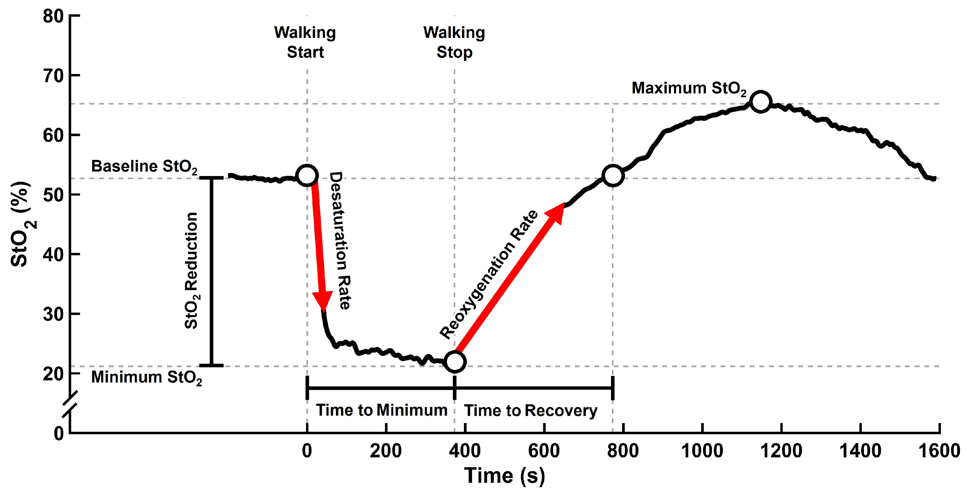
| Song-Young Park, et al. 2022 [9]. | 10 patients with claudication pain and 11 healthy subjects | Observational study | (1) Determine the effect of chronic ischemia on skeletal microcirculatory function. (2) Determine the correlation between leg ischemia and muscle mitochondrial function and StO2. | An NIRS device was attached to the calf muscle to measure StO2 at baseline and during walking and recovery using the Gardner–Skinner protocol. | Patients with PAD and healthy subjects showed no significant difference between baseline StO2 prior to exercise and maximum StO2 after exercise. However, patients with PAD showed greater StO2 reduction during walking and slower StO2 recovery compared with healthy subjects. Minimum StO2 during walking did not differ between patients with PAD and healthy subjects. |
| Matthew Fuglestad, et al. 2020 [32]. | 40 patients with PAD and 10 control subjects | Observational study | (1) Examine NIRS profile differences in patients with PAD vs. healthy subjects. (2) Determine how walking limitations relate to NIRS parameters during exercise training. | Calf muscle StO2 was measured for 3 min at baseline/rest, during the graded treadmill test, and 30 min after the test. Healthy control subjects performed a 540 s treadmill test. Additionally, all subjects performed computerized angiography and a 6 min walking test. | Patients with PAD and healthy subjects showed similar baseline StO2 levels. However, patients with PAD showed faster StO2 drop, greater StO2 fluctuation, greater decrease in StO2 during, and longer recovery after treadmill walking compared with healthy subjects. In patients with PAD, a strong correlation between calf muscle hypoxia and walking capacity during the 6 min walking test was observed. |
| J. Carter Luck, et al. 2017 [79] | Experiment 1: 8 patients with PAD and 8 healthy subjects. Experiment 2: 7 patients with PAD and 7 healthy subjects. | Controlled experimental study | Compare blood pressure (BP) and StO2 responses to different exercise intensities during plantar flexion exercise in patients with PAD vs. healthy subjects. | NIRS assessed calf muscle StO2 in the most symptomatic leg of patients with PAD. Experiment 1: After a 3 min baseline, subjects performed supine single-leg contraction (most symptomatic leg) at 30 contractions/min with progressive weights (0.5–7kg) for up to 14 min, stopping if fatigue or pain exceed 5/10. Experiment 2: The same exercise was performed but at 20 contraction/min, short pauses during exercise, and constant weights for 14 min. | Experiment 1: Patients with PAD showed significantly greater StO2 reduction during exercise compared with healthy subjects. At the time of fatigue, patients with PAD showed threefold greater StO2 reduction compared with healthy subjects. Experiment 2: Due to the lower StO2 level at baseline in patients with PAD compared with healthy subjects, StO2 was measured as the percentage relative changes with respect to the baseline at 1 min and 14 min. No significant changes were observed at 1 min, while a significant change was observed at 14 min during exercise. |
| F. Manfredini, et al. 2017 [80] | 80 patients with PAD and 13 healthy subjects | Observational study | Assess NIRS feasibility, validity, and diagnostic accuracy during dynamic ambulatory test to diagnose foot perfusion in PAD. | Subjects performed 10 toe flexion repetitions with NIRS attached to the dorsum of their feet. Healthy subjects repeated the test with increased tight blood flow restriction. ABI and the area under the oxygenated hemoglobin curve were measured. Reliability was assessed by repeating the measurement in two trials, and validity was evaluated by finding the association among toe flex curve area, oxygen delivery, ABI, and ankle pressure. | Higher levels of blood flow restriction in healthy subjects resulted in larger O2Hb deficit during toe flexion, as shown by a larger area under the curve (more negative). In patients with PAD, the most symptomatic legs showed significantly greater area under curve of O2Hb compared with healthy legs, with a weak correlation with dorsal pedis artery pressure. Toe flexion is an effective test to evaluate foot perfusion and PAD even in the presence or absence of ABI measurements. |
| F Manfredini, et al. 2009 [46] | 67 patients with PAD and 28 healthy subjects | Observational study | Assess if NIRS can determine calf muscle deoxygenation during treadmill walking in patients with PAD. | NIRS was attached to the calf muscle to measure the variation in oxygenation, deoxygenation, tHB and differential oxygenation. The test included 1 min warm-up, followed by treadmill walking at 1.5 km/h, increasing the speed every 10 min by 0.1 km/h. The test was stopped when the patients were unable to continue due to fatigue, dyspnea, or claudication. | The areas under curves for Hb, deoxygenation, and differential oxygenation were significantly differed in symptomatic and non-symptomatic legs. However, no significant differences in tHb were observed between symptomatic and non-symptomatic legs. Patients with PAD showed higher compensatory heart rate during exercise compared with healthy subjects. NIRS measurements were used to effectively quantify muscle metabolic response in PAD. |
| DT Ubbink, et al. 2006 [81] | 45 patients with different stages of leg ischemia and 20 healthy subjects | Prospective comparative study | Determine the reproducibility and clinical applicability of NIRS in patients with leg ischemia. | Reproductivity of various diagnostic tests, leg perfusion, and blood pressure were measured at rest, during exercise, and after changing posture. NIRS was attached to the calf muscle’s lateral side. NIRS was used to measure StO2 for 5 min while subjects walked on a treadmill at 3.1km/h speed and 8% incline. | NIRS could not detect resting StO2 differences across various PAD stages or correlate with ABI. However, lower ABI after exercise was correlated with the StO2 reduction measured by NIRS. Although ABI was able to detect leg ischemia at rest, NIRS could not detect it due to its inability to differentiate between the presence or absence of vascular disease. NIRS is a reliable measurement due to its ability to detect greater Hb desaturation during exercise in patients with PAD compared with controls. NIRS outcomes did not correlate with macro- and microcirculatory measurements. |
| Anthony J. Comerota, et al. 2003 [77] | 14 patients with PAD and 35 healthy subjects | Prospective cross-sectional study | (1) Identify calf StO2 and ABI correlation in patients with PAD. (2) Identify the sensitivity of muscle oxygenation to identify PAD. (3) Identify the relationship between StO2 and claudication symptoms. (4) Assess the safety of NIRS in screening for PAD. | Patients rested before the progressive treadmill test. The ABI was measured before and at 5 min intervals following the test. NIRS was attached to the calf muscle to record StO2 5 min before, during, and 20 min after the test. Outcomes included baseline StO2, peak exercise StO2, absolute percentage changes between baseline and peak exercise, StO2 values at ICT and ACT, and StO2 at 50% (T50) and 100% (T100) after stopping the exercise. | ABI was significantly different between PAD and healthy subjects, while no significant differences in StO2 were observed. Patients with PAD showed significantly lower StO2 at peak exercise, greater absolute difference between baseline and peak exercise, greater percentage changes from baseline to peak exercise, and longer recovery time at (T50) and (T100) compared with healthy subjects. A significant correlation was observed between T50 and ABI. |
| Anselm Egun, et al. 2002 [76] | 16 patients with PAD, and 7 age-matched and 7 young healthy control subjects. | Comparative study | Determine exercise-induced calf muscle ischemia in patients with PAD using NIRS. | NIRS equipment was attached to the patients’ most affected calf and the right calf of the healthy subjects. StO2 was recorded 10 min before, during, and 10 min after treadmill walking. Subjects first walked for 1 min at 3.2 kph and 10° incline. After a 20 min rest, the maximum walking capacity was assessed at the same speed and incline. In this test, patients with PAD walked until pain forced them to stop, while healthy subjects walked for 7 min. | During treadmill walking, all subjects showed O2Hb reduction. Patients showed greater HHb during exercise compared with healthy age-matched and young subjects; the differences were not significant. O2Hb and tHb were lower during the maximum-exercise test in patients with PAD vs. healthy subjects. In recovery, patients showed significantly increased O2Hb, tHb, and oxygen index, while healthy subjects showed no significant changes. ABI at rest was correlated with HHb but not with tHb or the oxygenated index during a maximum-walking test. |
| G.J. Kemp, et al. 2001 [82] | 11 patients with PAD and 9 healthy subjects | Case–control study | Measure calf muscle oxygenation and adenosine triphosphate (ATP) in patients with PAD by using magnetic resonance spectroscopy (MRS) and NIRS. | NIRS and MRS measured calf muscle ATP level and StO2 during isometric plantar flexion at 50% and 75% of maximum voluntary contraction for 2–4 min, following 5 min of recovery. Each subject repeated the plantar flexion test three times, and the results were averaged. | Compared with healthy subjects, patients with PAD showed faster and greater deoxygenation during exercise and slower reoxygenation after exercise. |
| H. Miriam Kooijman, et al. 1997 [78] | 11 patients with PAD and 15 healthy subjects | Experimental study | Evaluate the utility of NIRS to assess muscle oxygenation hemodynamics in patients with PAD compared with healthy subjects. | NIRS equipment was attached to the left calf of healthy subjects and the patients’ most symptomatic calf muscle. Part 1: Venous occlusion was performed at rest using a cuff, followed by at least 30 min of rest. Part 2: Subjects performed standard treadmill walking for 4 min at 3.2 km/h speed and 6° incline. Subjects reported ICT and walked until pain forced them to stop. Part 3: The same walking protocol in part 1 was repeated to measure StO2 during and after treadmill walking. | Patients with PAD showed significantly greater deoxygenation during walking compared to healthy subjects. Following the walking exercise, no significant difference was observed in blood flow between patients with PAD and healthy subjects. Patients with PAD showed significantly slower resaturation rates, longer recovery time measured by NIRS, and longer recovery time measured by ABI compared with healthy subjects. |
| Kevin K. McCully, et al. 1994 [83] | 8 patients with PAD, 20 healthy older adult, and 6 healthy young subjects. | Comparative study | (1) Compare post-reoxygenation rate in PAD vs. healthy older adults. (2) Compare post-reoxygenation rate in healthy older vs. young adults. (3) Compare calf StO2 depletion in PAD vs. healthy subjects. | ABI was measured before exercise. NIRS equipment was attached to the lateral soleus while subjects performed one-leg plantar flexion exercise every 5 s for one minute. Recovery time and StO2 were recorded for 3–6 min after exercise (one leg in healthy subjects and both legs in patients with PAD). StO2 was measured for 5 min in the standing position before and during the progressive treadmill test. During the treadmill test, ICT and ACT were recorded. | There was a significant difference in the rate of muscle oxygen saturation after plantar flexion exercise between the most symptomatic and less symptomatic legs of patients with PAD. Compared with healthy young and old subjects, patients with PAD showed greater and faster deoxygenation during walking and longer recovery after the walking test. The recovery time after exercise for the most symptomatic leg was longer compared with the less symptomatic leg. |
Disclaimer/Publisher’s Note: The statements, opinions and data contained in all publications are solely those of the individual author(s) and contributor(s) and not of MDPI and/or the editor(s). MDPI and/or the editor(s) disclaim responsibility for any injury to people or property resulting from any ideas, methods, instructions or products referred to in the content. |
© 2026 by the authors. Licensee MDPI, Basel, Switzerland. This article is an open access article distributed under the terms and conditions of the Creative Commons Attribution (CC BY) license.
Share and Cite
Salamifar, Z.; Fallahtafti, F.; Pipinos, I.I.; Anderson, C.P.; Park, S.-Y.; Myers, S.A. Muscle Oxygenation During Exercise in Patients with Peripheral Artery Disease: A Systematic Review. Appl. Sci. 2026, 16, 4348. https://doi.org/10.3390/app16094348
Salamifar Z, Fallahtafti F, Pipinos II, Anderson CP, Park S-Y, Myers SA. Muscle Oxygenation During Exercise in Patients with Peripheral Artery Disease: A Systematic Review. Applied Sciences. 2026; 16(9):4348. https://doi.org/10.3390/app16094348
Chicago/Turabian StyleSalamifar, Zahra, Farahnaz Fallahtafti, Iraklis I. Pipinos, Cody P. Anderson, Song-Young Park, and Sara A. Myers. 2026. "Muscle Oxygenation During Exercise in Patients with Peripheral Artery Disease: A Systematic Review" Applied Sciences 16, no. 9: 4348. https://doi.org/10.3390/app16094348
APA StyleSalamifar, Z., Fallahtafti, F., Pipinos, I. I., Anderson, C. P., Park, S.-Y., & Myers, S. A. (2026). Muscle Oxygenation During Exercise in Patients with Peripheral Artery Disease: A Systematic Review. Applied Sciences, 16(9), 4348. https://doi.org/10.3390/app16094348