1. Introduction
Maxillary transverse deficiency represents a frequent condition during the deciduous and mixed dentitions and is commonly associated with unilateral or bilateral posterior crossbite [
1,
2]. Posterior crossbite is defined as an abnormal transverse relationship between the maxillary and mandibular posterior teeth and has been reported to affect a considerable proportion of the pediatric population [
3,
4]. The etiology of this malocclusion is multifactorial and includes dental, skeletal, and neuromuscular components leading to a reduced transverse dimension of the maxillary arch [
5,
6]. If left untreated, maxillary transverse deficiency may induce a functional mandibular shift, potentially resulting in asymmetric mandibular growth and long-term alterations in craniofacial development and function [
7,
8].
Early diagnosis and interceptive treatment of transverse discrepancies are therefore widely advocated. Maxillary expansion is a well-established orthopedic and orthodontic procedure aimed at correcting posterior crossbite and restoring adequate transverse maxillary width in growing patients [
9,
10,
11]. By promoting separation of the midpalatal suture, palatal expansion allows an increase in maxillary transverse dimensions while reducing the risk of future crowding and the need for extraction-based treatment strategies [
12].
Several appliances and activation protocols have been proposed to achieve maxillary expansion, leading to the distinction between rapid maxillary expansion (RME), slow maxillary expansion (SME), and semi-rapid protocols [
13,
14]. RME is typically characterized by heavy, intermittent forces delivered over a short period of time through fixed tooth-borne or tissue-borne expanders, whereas SME relies on lower and more continuous forces applied over an extended duration [
3]. Both approaches have been shown to produce transverse maxillary enlargement; however, the relative balance between skeletal and dentoalveolar effects, as well as the magnitude of undesirable side effects, remains a matter of debate. Current evidence does not clearly identify a superior protocol or appliance capable of maximizing skeletal expansion while minimizing dental tipping and periodontal risks.
Regardless of the activation protocol, maxillary expansion may induce lateral bending of the alveolar processes and buccal displacement of the anchorage teeth, often accompanied by varying degrees of dental inclination [
15,
16,
17,
18]. These changes may result in reductions in buccal bone thickness and potential displacement of teeth beyond the alveolar envelope, raising concerns regarding periodontal health and long-term tooth stability [
18]. While early investigations relied on conventional two-dimensional radiographic techniques, the advent of cone-beam computed tomography has enabled more accurate three-dimensional assessment of skeletal, dentoalveolar, and periodontal changes associated with expansion procedures [
19]. Nevertheless, the heterogeneity of appliances, force systems, and measurement methods has led to inconsistent and sometimes conflicting results in the literature.
In recent years, growing interest has focused on compliance-free expansion devices capable of delivering controlled and biologically favorable forces. Among these, a palatal expander incorporating nickel–titanium leaf springs (Leaf Expander
®, Leone, Italy) has been introduced [
20]. The appliance resembles a conventional Hyrax expander in design but differs in its active mechanism, as calibrated Ni–Ti leaf springs deliver low, continuous, and predetermined lateral forces. This system was developed to achieve slow maxillary expansion without home screw activation, thereby reducing reliance on patient or parental compliance and simplifying clinical management [
20]. Preliminary reports suggest that the Leaf Expander can provide transverse expansion comparable to conventional rapid expanders, with reduced pain perception and improved patient comfort [
21,
22,
23]. However, the extent to which laboratory-derived expansion reflects the clinical dentoalveolar response with this device remains insufficiently clarified.
Accordingly, the aim of this study was to compare in vitro and in vivo expansion outcomes obtained with a Leaf Expander delivering 900 g of force and allowing a maximum expansion of 6 mm, and to test whether a correlation exists between laboratory measurements and clinical findings. Accordingly, the present study explored the relationship between protocol-declared activation and the in vivo transverse response, and whether laboratory-derived behavior can help interpret the clinical expansion timeline.
2. Materials and Methods
2.1. Trial Design
This retrospective study was conducted partly at the Department of Orthodontics, University of Genoa, and partly at the University of Milan, and was designed to compare the expansion produced by the Leaf Expander® under controlled in vitro conditions with the expansion observed in vivo during clinical treatment. The clinical component was based on diagnostic records of consecutively treated patients, whereas the laboratory component reproduced the device activation under standardized experimental conditions. The study protocol received approval from the Ethics Committee of Fondazione IRCCS Ca’ Granda, Ospedale Maggiore Policlinico, Milan, Italy (No. 51/2021; 18 May 2021). All procedures involving human participants were performed in accordance with institutional ethical standards and the Declaration of Helsinki and its subsequent amendments. As part of routine clinical practice, written informed consent for the anonymous use of diagnostic records for scientific purposes was obtained from the parents/legal guardians of all patients prior to treatment.
2.2. Sample Recruitment and Eligibility Criteria
Patients were recruited between November 2021 and November 2023 at two centers: the Department of Surgical and Dental Sciences, IRCCS Cà Granda Foundation, University of Milan, and the Orthodontics Department, University of Genoa. A total of 15 patients (8 females, 7 males) aged 7–10 years (mean age, 8.8 years) were retrospectively included.
All subjects presented with transverse maxillary deficiency associated with unilateral posterior crossbite.
The inclusion criteria were (a) transverse maxillary deficiency, calculated as the difference between the mandibular intermolar width measured at the distobuccal (or midbuccal) cusp tips of the lower first permanent molars and the maxillary intermolar width measured between the central fossae of the upper first permanent molars [
24,
25]; (b) mixed dentition with cervical vertebral maturation stage CS1–CS3 [
26]; (c) fully erupted maxillary and mandibular first permanent molars; (d) good general health, based on medical history and clinical judgment.
Exclusion criteria were skeletal Class III pattern, craniofacial malformations (including cleft lip/palate), history of dental trauma, oral neoformations or other oral cavity pathologies, clinically evident oral and/or periodontal disease, and previous or concurrent orthodontic treatment.
2.3. Intervention with Leaf Expander Appliance and Activation Protocol
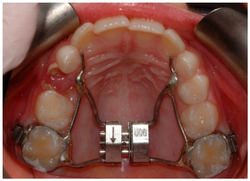
Maxillary expansion was performed with a Leaf Expander
® (Leone, Sesto Fiorentino, Italy) cemented on bands adapted to the maxillary molars (
Figure 1).
The appliance incorporates double nickel–titanium leaf springs that are loaded by activation of a central cobalt–chromium screw, which compresses the springs and generates a continuous force system.
For this study, the 900 g Leaf Expander with a maximum expansion capacity of 6 mm was used. The planned amount of expansion was determined according to each patient’s transverse discrepancy. Each activation of the central screw produced 0.1 mm of expansion (i.e., 10 activations = 1 mm), with a maximum of 30 activations for the 6 mm device.
At placement, the appliance was delivered in a preactivated configuration that allowed an initial phase of expansion to occur spontaneously after removal of the safety ligature/locking mechanism. This initial phase corresponded to 3 mm of expansion. Thereafter, the device was reactivated in-office by turning the screw to re-compress the leaf springs; thus, no home activation and no parental collaboration were required. In the present protocol, the first in-office reactivation was performed 6 weeks after cementation, delivering 10 turns with the activation key (equivalent to 1 mm). Additional reactivations were performed at 4-week intervals, each consisting of 10 turns, until the planned expansion was achieved. Overall, a total expansion of 6 mm was obtained over approximately 12 weeks, consisting of 3 mm of initial preactivation plus 3 mm delivered through in-office reactivations.
The screw unit was connected to the anchorage bands through a 0.9 mm stainless-steel wire framework, extended palatally and adapted to the deciduous canine area, and soldered to the bands. All appliances were fabricated by an experienced laboratory technician and cemented using an orthodontic band composite (Transbond™ Plus Light Cure Band Adhesive, 3M Unitek, Monrovia, CA, USA). Polymerization was performed with a halogen curing unit (Optilux, Kerr, Orange, CA, USA) for 20 s per tooth.
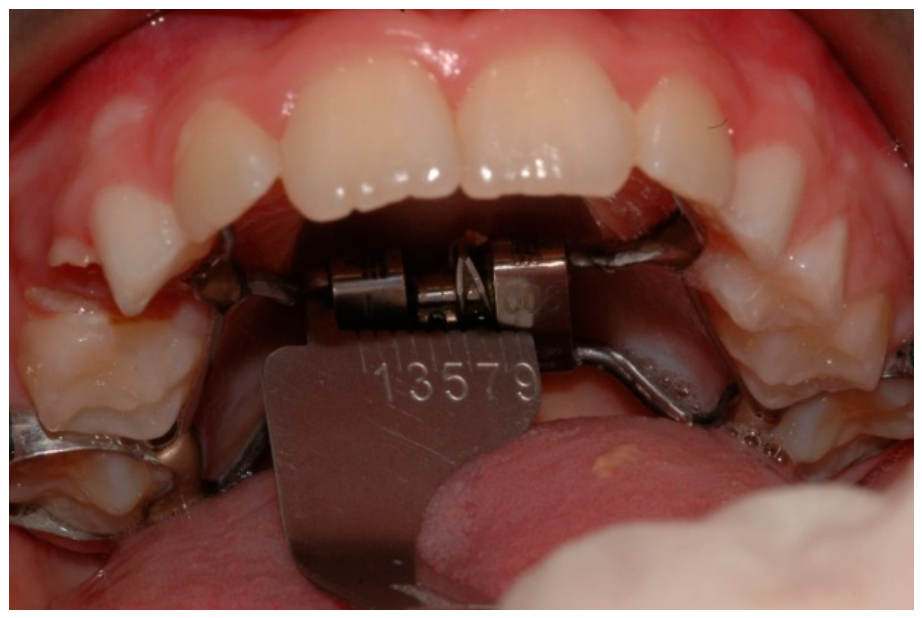
At each follow-up visit and reactivation appointment, clinical records were collected to monitor device effectiveness. Intraoral transverse measurements were obtained with a caliper to quantify the incremental expansion between consecutive visits (
Figure 2). In addition, the manufacturer’s Leaf Expander expansion gauge was used to verify the amount of expansion achieved and to assess the need for further screw reactivation (spring recompression).
2.4. Laboratory Analysis
The force delivered by commercially available Leaf Expander® reactivatable units was quantified through systematic compression testing performed at the Leone BioResearch Center (Sesto Fiorentino, Italy). The purpose of these tests was to reproduce the clinical reactivation protocol of the Leaf Expander 6 mm/900 g device and to characterize the force released at each phase of the activation sequence.
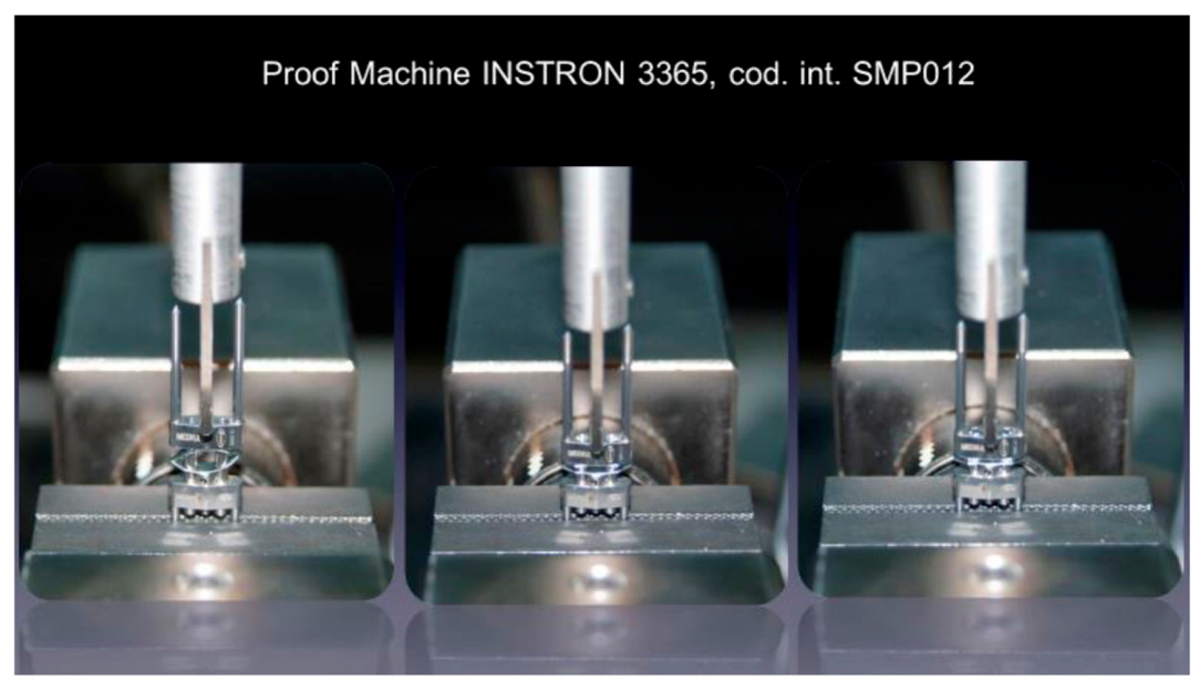
Reactivatable Leaf Expander 6 mm–900 g expanders were tested using a universal testing machine (Instron Model 3365, Instron Industrial Products Group, Grove City, PA, USA) equipped with a 100-N load cell (Instron Industrial Products Group, Grove City, PA, USA). The test method was implemented, and data were acquired using Bluehill software 3.1 version (Instron) (
Figure 3). Each tested expander underwent four complete compression–release cycles according to the planned activation protocol. However, because of the retrospective nature of the study and the limited availability of the original laboratory documentation, detailed information regarding calibration records, environmental conditions, and full repeatability assessment could not be comprehensively retrieved and should therefore be considered a limitation of the in vitro analysis.
For testing, the expander was secured by its arms in the lower fixed grip. To ensure that the measurements reflected the mechanical response of the expander body and spring system rather than the flexibility of the arms, the arms on the opposite side were sectioned, allowing the compressive load to be applied directly to the expander body. Compression was applied using a custom-made loading device mounted to the upper crosshead, specifically designed and manufactured by Leone S.p.A. (Firenze, Italy) for this experimental setup. The Bluehill protocol consisted of distinct phases under force- and displacement-control, reproducing the planned reactivation sequence.
2.5. Outcomes
Based on the clinical assessments performed directly on the patients and on the corresponding measurements recorded and tabulated, three transverse parameters were selected as reference outcomes: intermolar distance at the second primary molars (E–E), intermolar distance at the maxillary first permanent molars (6–6), and intercanine distance (C–C).
For each patient, transverse measurements were collected according to the activation and follow-up protocol at the following time points:
T0: baseline measurements obtained from pretreatment digital dental models.
T1 (6 weeks): intraoral measurements obtained with a millimetric caliper, 6 weeks after appliance cementation.
T2 (10 weeks): measurements obtained 4 weeks after T1.
T3 (14 weeks): measurements obtained 4 weeks after T2.
T4 (18 weeks): measurements obtained 4 weeks after T3.
T5 (22 weeks): final assessment of expansion, performed 8 weeks after T3.
To quantify the amount of expansion achieved over time, incremental changes were calculated between consecutive recordings (T1–T0, T2–T1, T3–T2, T4–T3, and T5–T4). In addition, the overall change from baseline to the final observation (T5–T0) was computed.
In summary, patients were evaluated at 6, 10, 14, 18, and 22 weeks, following the protocol for a 6 mm/900 g Leaf Expander. All measurements were expressed in millimeters. After data collection, means and standard deviations were calculated for each parameter and time interval.
For the in vitro assessment, force release was characterized through a standardized mechanical testing protocol consisting of three sequential phases:
Preload: the crosshead moved downward until a load of 0.1 N was detected; data acquisition was initiated at this point.
Compression: the crosshead continued moving downward until the springs were fully compressed. This phase was included to ensure consistent spring loading and to minimize operator-related variability.
Release: the crosshead motion was reversed until the load returned to 0 N, at a constant speed of 0.01 mm/s.
Each expander underwent the complete testing sequence four times. The first cycle was performed with the screw fully closed. At the end of each complete cycle, the screw was activated in accordance with the clinical protocol. Because the device had a 6 mm expansion capacity, the screw was advanced by 10 turns, corresponding to 1 mm of activation, before repeating the subsequent test cycle.
Raw data were exported in .csv format and analyzed using spreadsheet-based statistical procedures (Microsoft Excel, Microsoft, 2010). The primary outcome for the laboratory analysis was the force released during the Release phase (Phase 3). Force values were extracted across the displacement interval from 0.1 mm to 1.0 mm for the 6 mm device. A post-processing procedure was applied to reduce the influence of friction between expander components and residual mechanical play in the experimental setup, thereby improving the reliability of the force–displacement data.
2.6. Statistical Analysis
Statistical analyses were performed by evaluating the association between the clinical (in vivo) measurements and the laboratory (in vitro)force/displacement data. Specifically, the analysis included (1) transverse measurements collected at each patient visit according to the activation protocol, and (2) the values obtained from the mechanical compression tests.
All statistical comparisons were carried out using IBM SPSS Statistics (version 25.0, IBM Corp., Armonk, NY, USA). Data normality was assessed with the Shapiro–Wilk test; because the data were normally distributed, parametric tests were used for both within- and between-condition comparisons. Descriptive statistics were reported as mean ± standard deviation. Because this was a retrospective study based on all eligible patients with complete records during the recruitment period, an a priori sample size calculation was not performed.
Pearson correlation coefficients (r) were used to explore the linear association between protocol-declared activation (expected expansion according to the activation schedule) and the clinically observed in vivo transverse changes. Correlation was interpreted as a measure of association and not as agreement. Descriptive statistics are reported as mean ± standard deviation and 95% confidence intervals (CI) Correlation strength was interpreted as weak for 0 < r < 0.30, moderate for 0.30 ≤ r < 0.70, and strong for r ≥ 0.70. The purpose of this analysis was to determine whether a measurable agreement exists between the declared activation protocol (laboratory-derived values) and the observed in vivo outcomes. To contextualize the interpretation of correlation analyses, a sensitivity analysis was conducted using the Fisher z-transformation for Pearson correlation. With n = 15 and two-sided α = 0.05, the study has approximately 80% power to detect correlations of about r ≥ 0.71; therefore, smaller associations may not have been detectable. Correlation coefficients are reported as effect sizes with 95% confidence intervals.
To assess intra-observer reliability, a random subset of records was remeasured by the same examiner after a 2-week interval under identical conditions. Reliability was quantified using the intraclass correlation coefficient (ICC; two-way mixed-effects model, absolute agreement). Method error was also calculated according to Dahlberg’s formula: , where d is the difference between the first and second measurements and n is the number of double measurements.
Intra-observer reliability was excellent for all transverse measurements, with ICC values ranging from 0.92 to 0.94. Method error according to Dahlberg’s formula was low, ranging from 0.18 to 0.27 mm, indicating minimal measurement discrepancy and good repeatability of the adopted protocol.
3. Results
Table 1 summarizes the mean changes (mm) in transverse dimensions—E–E, 6–6, and C–C, recorded on the patients between consecutive time points (T1–T0 to T5–T4), as well as the overall change from baseline to the end of observation (T0–T5).
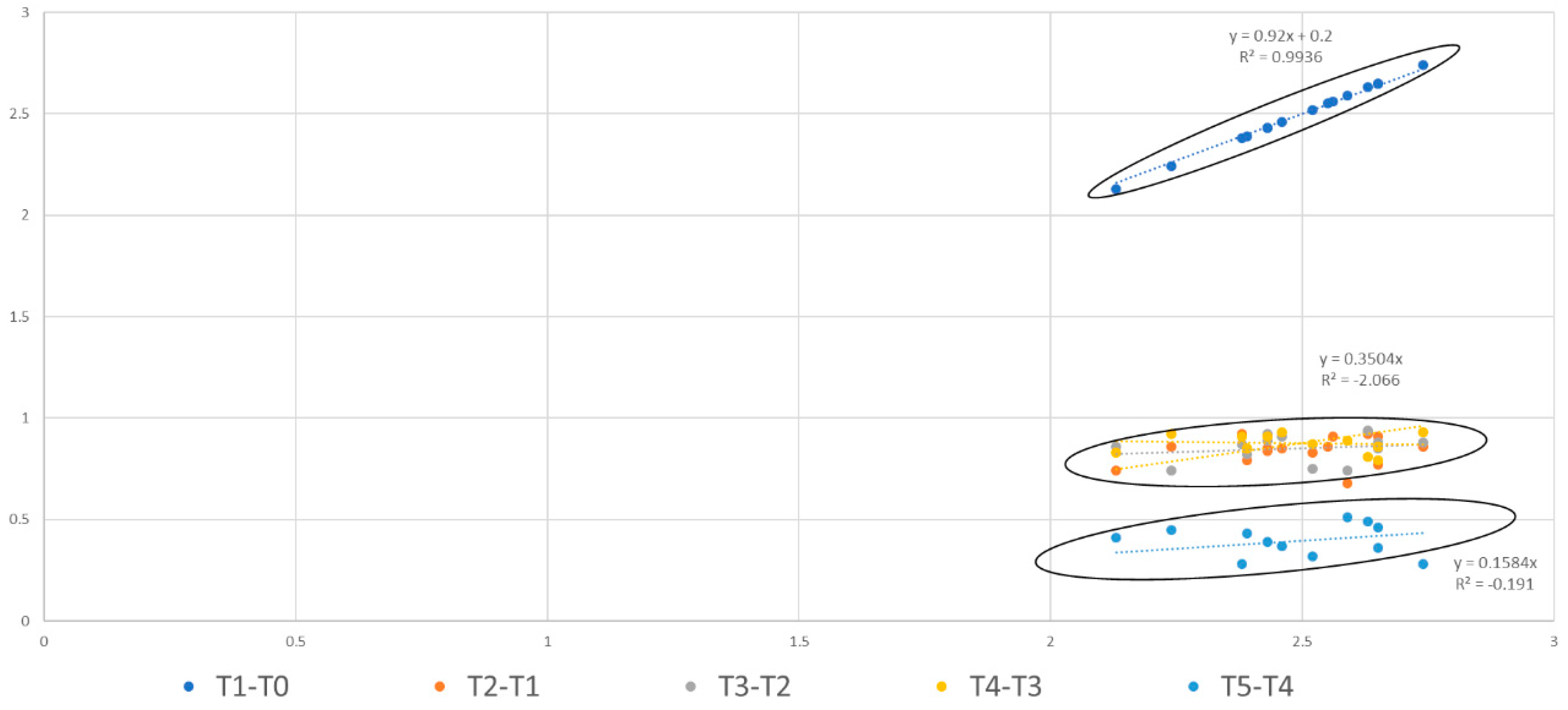
In light of the data described above, the Pearson correlation coefficient (r) was subsequently calculated to assess the presence and direction of a linear association between the variables, and specifically to evaluate the agreement between the declared device activation and the in vivo clinical outcome. The results of the Pearson correlation analysis are summarized in the table and figure below (
Table 2).
Pearson correlation coefficients (r) describe the linear association between the declared activation/expansion recorded at each follow-up visit (Leaf Expander gauge/screw opening) and the clinically measured transverse change obtained with a caliper for each outcome (E–E, 6–6, C–C). Correlation is reported as association and does not represent a formal agreement analysis. Correlations were calculated at the patient level (n = 15) at each time point.
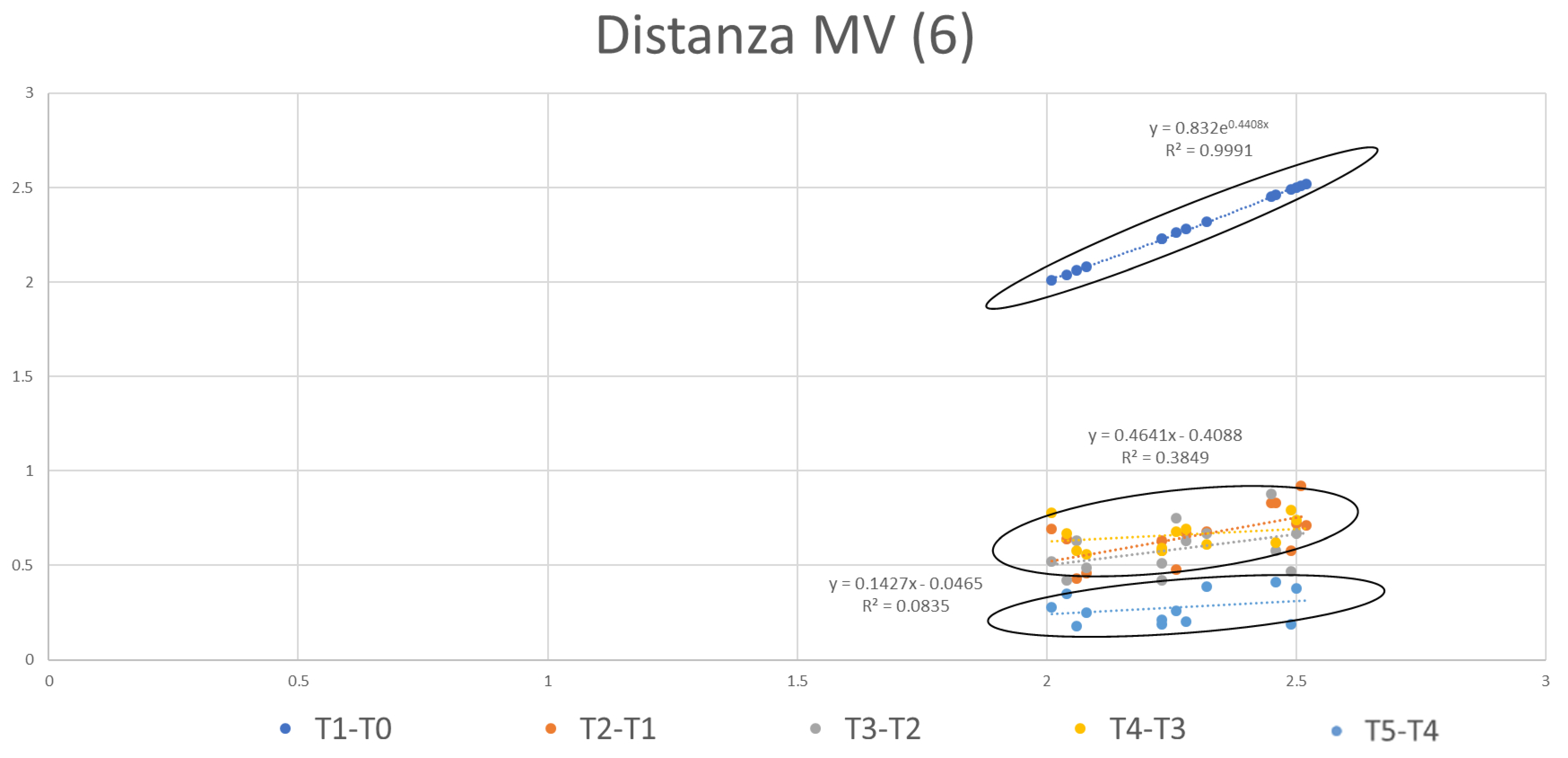
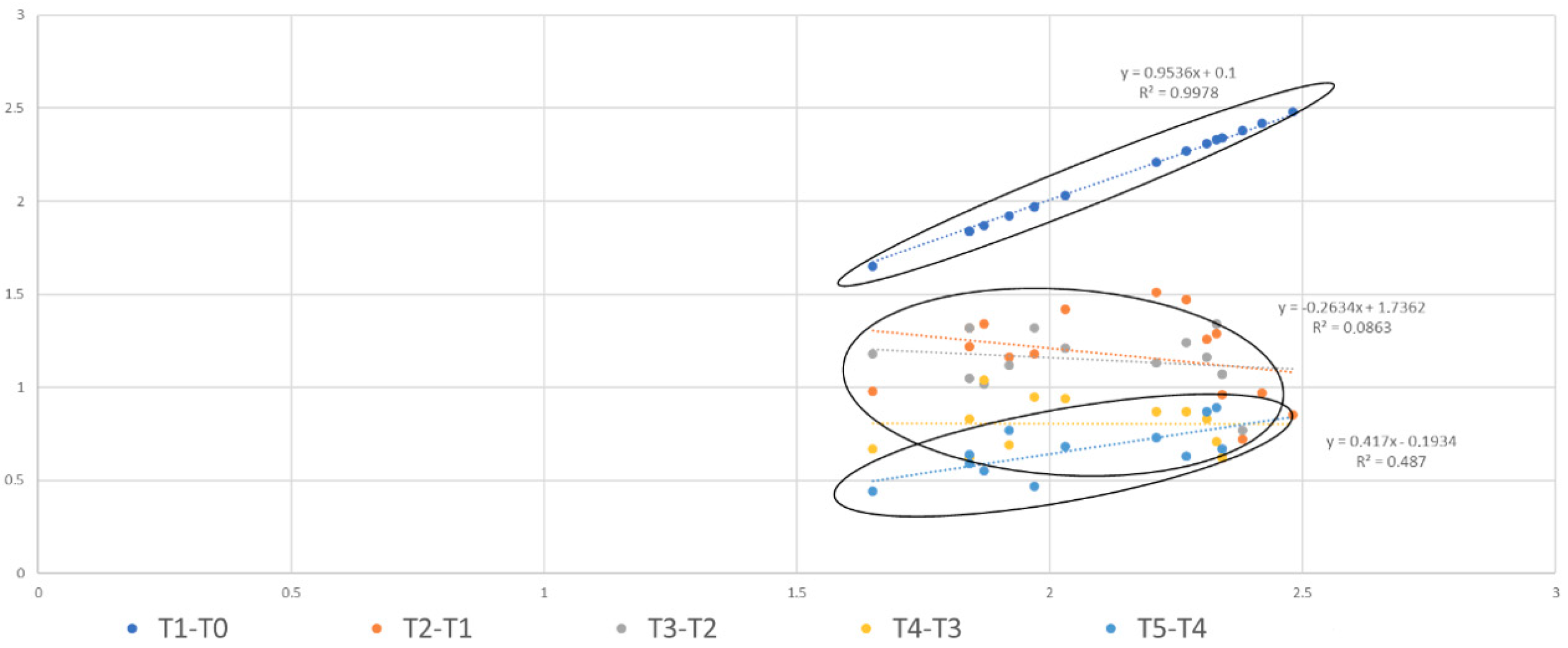
By comparing these findings from the clinical measurements collected at each activation visit, it was inferred that smaller and more compact ellipses correspond to higher agreement (
Figure 4,
Figure 5 and
Figure 6). Accordingly, the association between laboratory-derived activation metrics and clinically observed transverse response was good for the E–E and 6–6 distances, as their ellipses appeared short and narrow, whereas agreement was lower for the C–C distance, which showed longer and wider ellipses. Consistently, inspection of the box plot depicting overall expansion indicated a greater variability for the intercanine distance (C–C), reflected by a larger standard deviation compared with E–E and 6–6. These findings mirror the results of the Pearson correlation analysis, which showed higher agreement for E–E and 6–6 than for C–C.
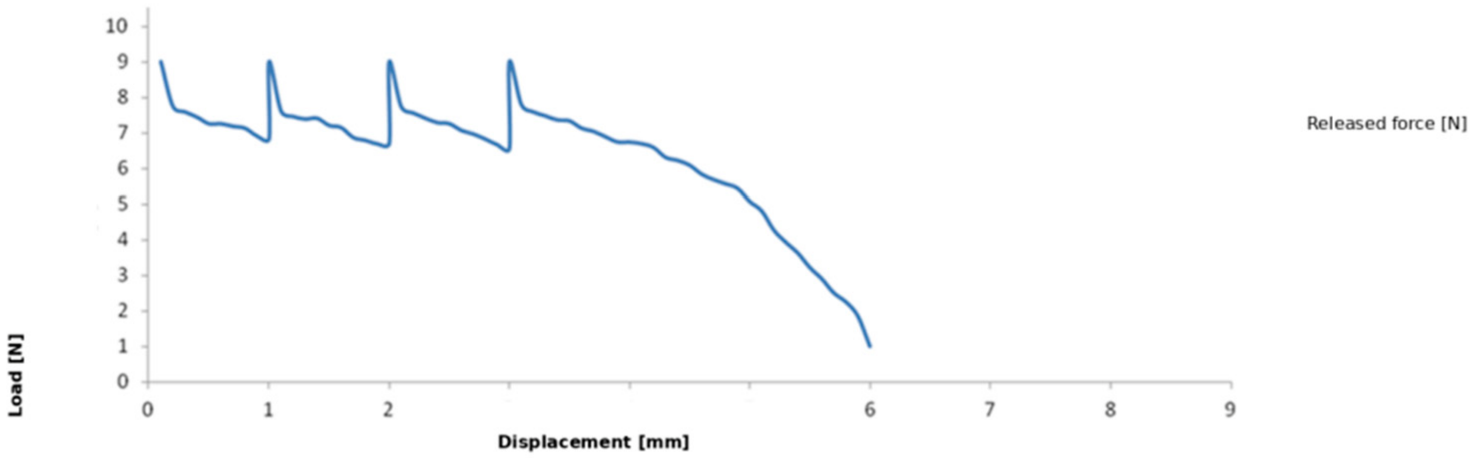
Based on the laboratory tests performed on the Leaf Expander, the force–displacement data shown in the following graphs were obtained (
Figure 7).
Figure 7 illustrates the mean force released by the expander during the full 6 mm activation sequence. In each plot, displacement (mm) is reported on the x-axis and load (N) on the y-axis. The mean standard deviation across the entire test was <2%, indicating high measurement consistency.
4. Discussion
The present study investigated the clinical and laboratory performance of a nickel–titanium leaf-spring expander, integrating in vivo transverse changes with in vitro force–displacement behavior. Clinically, maxillary expansion with the Leaf Expander resulted in successful transverse correction in the treated sample, with posterior crossbite resolved in all completed cases. Overall, these outcomes are consistent with previous reports describing predictable transverse increases and favorable patient acceptance with compliance-free slow expansion systems [
3,
21,
27,
28].
A key contribution of this work lies in the laboratory characterization of the device behavior across the activation protocol and its comparison with clinical findings. The in vitro compression tests, designed to reproduce the reactivation sequence of the reactivatable Leaf Expander (6 mm screw, 900 g force), revealed a reproducible force–displacement pattern: an initial activation peak followed by a plateau phase during which the released force remained continuous and relatively constant, and a subsequent phase characterized by residual load after completion of the programmed activations. This sustained force profile is plausibly related to the mechanical behavior of the NiTi leaf springs, which progressively recover their original configuration and thereby maintain a continuous force delivery over time [
29].
Importantly, variability in the released forces observed in the laboratory should be interpreted in light of the inherent differences between a mechanical test and the oral environment. First, the clinical expression of expansion occurs over weeks, whereas the laboratory protocol applied displacement at a controlled rate (0.01 mm/s), which cannot replicate the biologic time scale of sutural and dentoalveolar adaptation. Second, the test was conducted in dry conditions; intraorally, saliva is expected to reduce friction between components, whereas dry testing may increase frictional losses and energy dissipation, potentially affecting the effective force recorded. Third, tests were performed at approximately room temperature (~22 °C), while the intraoral environment is warmer and may influence NiTi mechanical response. Finally, the experimental setup applied displacement essentially along a single axis, whereas clinical expansion is a three-dimensional process influenced by midpalatal suture compliance, circummaxillary articulations, and dentoalveolar biomechanics. These factors can reasonably account for dispersion in force values without contradicting the overall evidence of continuous force release.
The comparison between laboratory-derived activation behavior and clinical measurements provides a clinically relevant interpretation of the expansion timeline. During the early period after cementation (T1–T0), the mean clinical increase in the E–E distance was slightly below the nominal 3 mm preactivation expected by protocol. Similarly, the incremental gain after the first in-office reactivation was marginally lower than the theoretical 1 mm displacement. These findings suggest that, at intermediate time points, the expansion expressed clinically may not mirror the nominal mechanical activation in a strictly “immediate” manner. However, longitudinal observation indicated that the device continues to work beyond reactivation visits, with force progressively decreasing over time. This is consistent with the presence of a residual load and delayed expression of expansion, a phenomenon that has been classically described for maxillary expansion mechanics, in which force persists between activations and contributes to additional transverse change over time. Clinically, this implies that an apparent shortfall at an early checkpoint does not necessarily indicate insufficient final expansion.
The concordance analyses further highlighted that agreement between declared activation and observed clinical response was higher at the posterior segments than at the canine region. In particular, agreement was better for E–E and 6–6, whereas the intercanine distance (C–C) exhibited lower concordance and greater variability, also reflected by larger dispersion in the corresponding box plots. This pattern is biologically plausible: changes at the canine region are more susceptible to local factors such as eruption stage, archform adaptation, rotation or tipping components, and variability in anterior dentoalveolar response. In addition, anterior transverse measurements may be affected by tooth position changes that are not purely translational, resulting in greater measurement variability compared with the posterior segments, where anchorage and appliance design can provide more stable reference points.
From a clinical perspective, the laboratory evidence of a continuous low-force plateau supports the rationale for the high tolerability reported with NiTi leaf-spring expanders. Compared with high intermittent forces typical of rapid maxillary expansion protocols, a more constant and lower magnitude force system is closer to sustained physiologic loading, which may contribute to improved patient comfort and potentially reduce the intensity of pain experiences during the active phase. This aspect is clinically relevant because discomfort remains a frequent concern during maxillary expansion, particularly in younger patients, and can influence acceptance and adherence to follow-up schedules.
An additional practical implication relates to retention. The device is not fully passive immediately after completion of activation; rather, the presence of residual spring activity suggests that the expander can provide a period of active retention, potentially counteracting early relapse tendencies during consolidation [
30,
31]. While this study was not designed to quantify relapse or long-term stability, the combined laboratory and clinical observations support the concept that residual load may contribute both to completion of the planned expansion and to short-term stabilization. The discrepancy between protocol-declared activation and clinically observed expansion may not depend exclusively on the delayed biological expression of force. Part of the transverse increase may reflect dentoalveolar tipping and alveolar adaptation rather than pure skeletal expansion, particularly with a tooth-borne appliance in mixed dentition. Moreover, linear transverse measurements may be influenced by measurement artifacts, tooth inclination, and local eruptive variability, especially at the canine level.
Some limitations should be acknowledged. Its retrospective design inherently exposes the analysis to potential selection bias, as only patients with complete records and adequate follow-up could be included. Although the clinical protocol was consistent with routine practice, retrospective data collection does not allow the same level of control over standardization of treatment conditions that can be achieved in a prospective design. Third, the use of digital models at baseline and intraoral caliper measurements at follow-up may have introduced a degree of measurement bias, despite the use of identical anatomical reference points and the reliability assessment performed. The in vitro model did not replicate intraoral humidity, temperature, or complex 3D constraints, and thus cannot fully capture the biomechanical interaction between the expander and the craniofacial structures. The clinical sample size was limited, and clinical measurements—although standardized—may be influenced by intraoral measurement error, eruption changes, and dentoalveolar remodeling over time. Moreover, given the limited sample size, the correlation analyses should be considered exploratory and primarily sensitive to large effects; larger prospective studies are needed to confirm these findings and provide more precise estimates. Future studies with a larger sample size should consider controlled environmental testing (humidity and temperature regulation), more comprehensive 3D clinical assessments, and larger samples to refine the laboratory–clinical relationship, particularly for the anterior transverse dimension.
Because no control or comparative group was included, the present findings cannot be directly extrapolated to other maxillary expansion appliances. Comparative prospective studies including different expander designs would be necessary to determine whether similar laboratory–clinical relationships are observed across devices.
Within these limits, the present findings support the effectiveness of the Leaf Expander in producing clinically meaningful transverse increases and demonstrate a force–displacement profile characterized by continuous, sustained loading with residual force expression. The integration of in vitro and in vivo data suggests that delayed force release contributes to the completion of expansion over time and that laboratory testing can provide clinically informative insight into device behavior, especially at the posterior segments where concordance appears higher.




 ,
,