Article

Electromyographic Assessment of Masticatory Muscle Function After Short-Term Vertical Dimension Increase in Class II Division 2 Malocclusion: A Pilot Clinical Study


by
Tatiana-Maria Coman
1,2,
Zsuzsanna Bardocz-Veres
3,*,
Liana-Claudia Dobreci
4,
Sorin Popșor
4 and
Mariana Păcurar
2
1
Doctoral School of Medicine and Pharmacy, George Emil Palade University of Medicine, Pharmacy, Science and Technology of Targu Mures, 38 Gheorghe Marinescu Street, 540142 Targu Mures, Romania
2
Department of Orthodontics, Faculty of Dental Medicine, George Emil Palade University of Medicine, Pharmacy, Science and Technology of Targu Mures, 38 Gheorghe Marinescu Street, 540142 Targu Mures, Romania
3
Department of Oral Rehabilitation and Occlusology, Faculty of Dental Medicine, George Emil Palade University of Medicine, Pharmacy, Science and Technology of Targu Mures, 38 Gheorghe Marinescu Street, 540142 Targu Mures, Romania
4
Department of Removable Prosthodontics, Faculty of Dental Medicine, George Emil Palade University of Medicine, Pharmacy, Science and Technology of Targu Mures, 38 Gheorghe Marinescu Street, 540142 Targu Mures, Romania
*
Author to whom correspondence should be addressed.
Appl. Sci. 2026, 16(9), 4216; https://doi.org/10.3390/app16094216
Submission received: 2 April 2026 / Revised: 19 April 2026 / Accepted: 23 April 2026 / Published: 25 April 2026
(This article belongs to the Special Issue Biosignal and Motion Measurements)

Abstract

Background: Alterations of the vertical dimension of occlusion may affect masticatory muscle function, which is critical in pre-prosthetic planning, especially in Class II division 2 malocclusions. This study aimed to evaluate the impact of increasing VDO on the myoelectric activity of masticatory muscles using surface electromyography (EMG). The null hypothesis was that a 2–4 mm increase in VDO does not significantly influence muscle activity. Methods: Nine patients with Class II division 2 malocclusion were evaluated. EMG recordings of the masseter and anterior digastric muscles were obtained using the BioEMG II system (BioResearch Asoc., Milwaukee, WI, USA) and Biopak™ software (AcqKnowledge 4.x). VDO was increased using the Dupas universal jig. EMG was recorded for 30 s under six conditions: resting posture, intercuspal position (IM), swallowing, resting posture after VDO increase, IM with increased VDO (IM2), and swallowing with increased VDO (swallowing 2). Results: Most EMG variables showed no statistically significant differences after short-term VDO increase. Significant differences were observed only in the resting activity of both masseter muscles and in the right masseter during maximum intercuspation. No significant changes were identified during swallowing. Conclusions: Within the limitations of this pilot study, a 2–4 mm increase in VDO appears to produce minimal short-term changes in masticatory muscle activity in Class II division 2 patients. These findings should be interpreted with caution due to the small sample size and short observation period, and further studies are required to evaluate long-term neuromuscular adaptation.

1. Introduction

The pre-prosthetic assessment of the occlusal plane and vertical occlusal dimension is paramount from an aesthetic and functional perspective. This is particularly crucial when issues related to potential prosthetic space are apparent in patients with class II division 2 relationships. The potential to alter the vertical occlusal dimension, especially when linked to excessive tooth abrasion, remains a contentious issue among specialists [1].
Class II Division 2 malocclusion is characterized by a distinctive dentoskeletal and neuromuscular pattern that may differentiate it from other sagittal skeletal relationships. Typical features include a deep overbite, retruded maxillary incisors, and a reduced lower anterior facial height, often accompanied by increased chin prominence [2]. These morphological characteristics are not purely dental in nature but are closely associated with functional adaptations of the orofacial musculature [3].
In particular, altered incisor inclination and increased vertical overlap may influence mandibular posture and restrict anterior mandibular movement during functional activities. As a consequence, compensatory changes in the activity of the masticatory muscles (especially the masseter and temporalis muscles) have been suggested in previous electromyographic (EMG) studies. These adaptations may reflect an attempt to maintain functional efficiency under altered occlusal and skeletal constraints [4,5].
Furthermore, individuals with Class II Division 2 malocclusion have been reported to exhibit different neuromuscular recruitment strategies compared with other skeletal classes, potentially resulting in distinct baseline EMG activity patterns [6]. However, the existing literature remains limited and somewhat inconsistent regarding the magnitude and direction of these differences [7].
Therefore, a better understanding of the neuromuscular characteristics associated with Class II Division 2 malocclusion is essential to clarify how dentoskeletal morphology influences functional muscle activity and to improve diagnostic and therapeutic approaches [8].
Vertical skeletal parameters in patients with a Class II division 2 relationship clearly show a hypodivergent developmental pattern, with a shrinking of the lower facial floor. The absence of incisal support may cause a decrease in the vertical dimension, which signifies the mandible’s upward and forward growth. On the contrary, an increase in vertical dimension may indicate the mandible’s downward and backward growth [9,10].
The patient with low facial height and mandibular retrognathia usually has an overactive mental muscle and poorly defined chin projection, which results in a tendency towards deep occlusion [11,12].
Swallowing is the most significant functional test for assessing the vertical dimension of occlusion. The mandible achieves the optimal ratio to the maxilla horizontally and vertically during swallowing. A study that involved raising the occlusion by 3 to 8 mm highlighted that increasing the vertical dimension of occlusion alters the size of the mandibular trajectory during swallowing, particularly when this augmentation exceeds 3 mm [13].
This study, using electromyography, aimed to evaluate the impact of altering the vertical dimension of occlusion on the neuromuscular behavior at the level of the elevator musculature of the mandible. The results of this study have significant practical implications for dental practice, especially when its augmentation is necessary to achieve the potential prosthetic space and for aesthetic reasons in patients with class II division 2 relationships.
The study’s null hypothesis was that raising the vertical dimension of occlusion by 2–4 mm does not significantly influence the myoelectric activity at the level of the mobilizing musculature of the mandible. This hypothesis was formulated to test the common belief in the dental field and provide a basis for comparing the study’s findings.

2. Materials and Methods

2.1. Study Participants

This study, which included nine patients undergoing orthodontic treatment at the Faculty of Dental Medicine, “George Emil Palade” University of Medicine, Pharmacy, Science, and Technology of Târgu-Mureș (UMFST), was conducted rigorously. The patients, aged between 19 and 25 years, consisting of seven females and two males, were thoroughly examined during routine clinical and paraclinical cephalometric and electromyographic examinations. The study protocol was reviewed and approved by the Ethics Committee of the George Emil Palade University of Medicine, Pharmacy, Science and Technology of Târgu Mureș (approval number: 3874/06.10.2025). Participation was voluntary, and informed consent was acquired to evaluate and include the results in the study, ensuring the validity and reliability of the research.
Given the exploratory nature of this investigation, no a priori sample size calculation was performed. The study was designed as a pilot study to identify preliminary trends and inform future research.

2.2. Cephalometric Analysis

Cephalometric analysis was conducted to evaluate the vertical dimension of the lower face. Following Slavicek’s method within the CADIAS computer analysis system, the angle defined by the ANS-Xi-Pm landmarks was measured, representing lower facial height [14,15]. Normal reference values vary according to craniofacial type: the overall accredited range is 47 ± 4°, with subgroups defined as 43 ± 3° for brachycephalic, 45 ± 3° for mesocephalic, and 48 ± 3° for dolichocephalic subjects [16]. For comparison, Ricketts’ analysis defines the lower facial angle (ANS-Xi-Pog) with an average of 45 ± 4°.

2.3. Electromyographic Recording

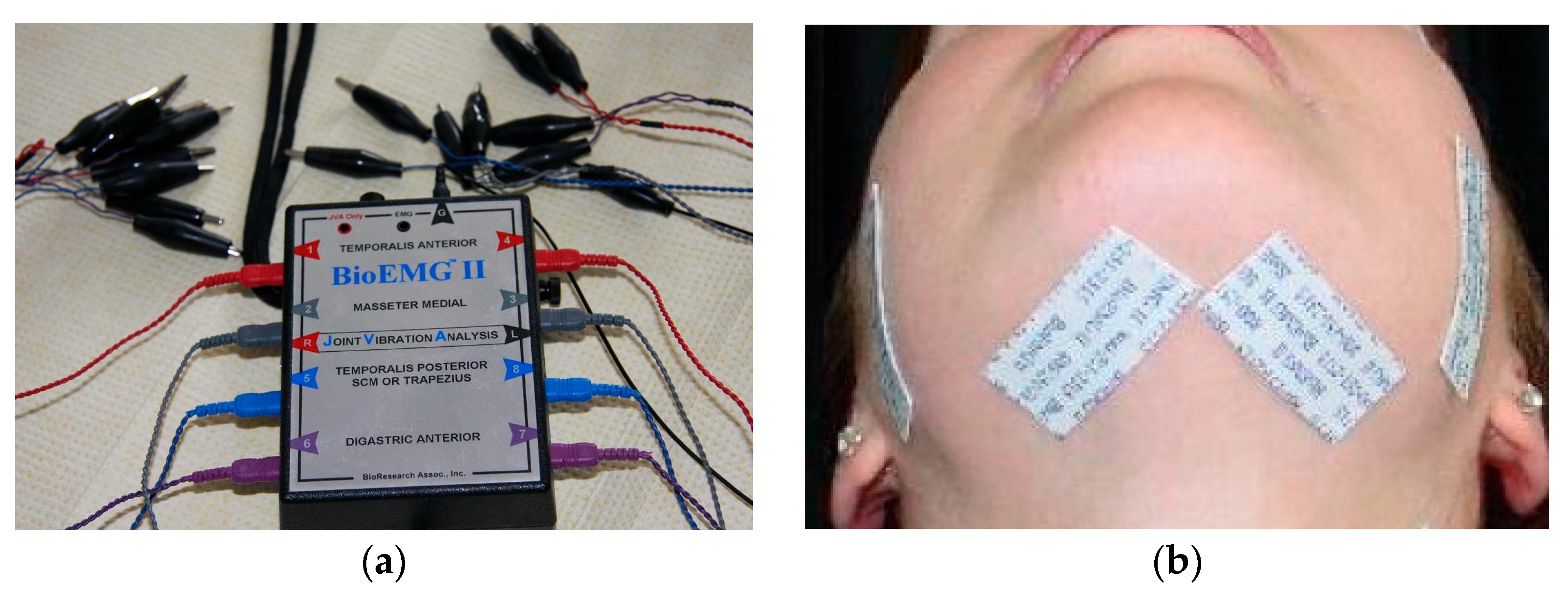
We recorded the myoelectric activity in the masseter and anterior digastric muscles through the BioEMG II computerised global electromyography system (BioResearch Asoc. Inc., Milwaukee, WI, USA) and its corresponding software, Biopak™, in nine patients with Class II division 2 (Figure 1a). The system allows for the simultaneous recording of bioelectric potentials from eight muscles, with the investigator able to select specific muscle combinations for analysis [17]. The muscles examined electromyographically were the masseter and the anterior belly of the digastric muscle, bilaterally. Surface electromyographic measurements were obtained using bipolar electrodes with conductive adhesive gel, attached to the skin above the muscle mass and oriented parallel to the muscle fibres. A TENS grounding electrode placed on the skin in the scapular region eliminates the artefacts.
Electrodes were positioned above the muscle mass parallel to the muscle fibers for the masseter, on the line connecting the angle of the mandible to the external angle of the eye; for the anterior belly of the digastric, they were parallel to the lower edge of the mandible and 2 cm from the mental protuberance (Figure 1b). The bioelectric signals collected by the electrodes were sent to a compatible computer (Intel processor), which analyzed the signal activity levels of individual muscles through a USB interface at a rate of 1000 to 6000 samples per second, with a resolution of 12 or 16 bits, using specific software.
The raw, filtered myoelectric signals were amplified 5000 times before analysis. The signals were shown offline as frequency domains and average levels of muscular contraction activity, and in real time as time-delayed waves on the computer screen. After recording, the data were digitally filtered to remove noise content (interference) from the electromyographic signals; a 40 dB reduction on a logarithmic scale corresponds to a 99% reduction in amplitude noise. The duration of the electromyographic recordings was for a single 30 s collection (AllEMG) under the following six conditions: posture (rest); IM; swallowing; posture 2; IM with increased vertical occlusion (IM2); swallowing at increased vertical occlusion (swallowing 2). We increased the vertical dimension using Dupas universal jig [18]. The silicone material used to stabilize the mandible had an approximate thickness of 2–3 mm, approximately corresponding to the induced disocclusion, and was adapted individually to each subject to ensure stable occlusal support during recordings. Due to its boomerang-shaped spatial configuration, the jig can be adjusted to any occlusal pattern and is fixed to the upper incisors with an impression material. This adjustment allows for a vertical dimension increase in the lateral areas of the arches (disocclusion space) of 2–4 mm, depending on the intercuspation contact pattern at the level of the central incisors. To ensure the stability of the mandible when raised using the jig in maximum intercuspation and swallowing, we placed a silicone material in the disocclusion space at the level of the lateral teeth. Once set, this material has substantial consistency and can withstand the forces exerted by the masticatory muscles on the dental arches.

2.4. Data Analysis

To compare the parameters of myoelectric activity established during the recording process before and after increasing the vertical occlusion dimension, the EMG data were organized in tables and analyzed using GraphPad InStat v3.1. Data distribution was assessed using the Shapiro–Wilk test. For normally distributed paired data, a paired t-test was applied. When normality assumptions were not met, the non-parametric Wilcoxon signed-rank test was used. The Mann–Whitney U test was applied for independent comparisons where appropriate. A significance level of p < 0.05 was considered statistically significant.

3. Results

Table 1, Table 2 and Table 3 present the amplitude of the electromyographic (EMG) signals, measured in microvolts (µV), from the masseter (RM, LM) and anterior digastric (RAD, LAD) muscles at rest, during maximum intercuspation (IM), and while swallowing, before and after increasing the vertical dimension of occlusion (VDO).
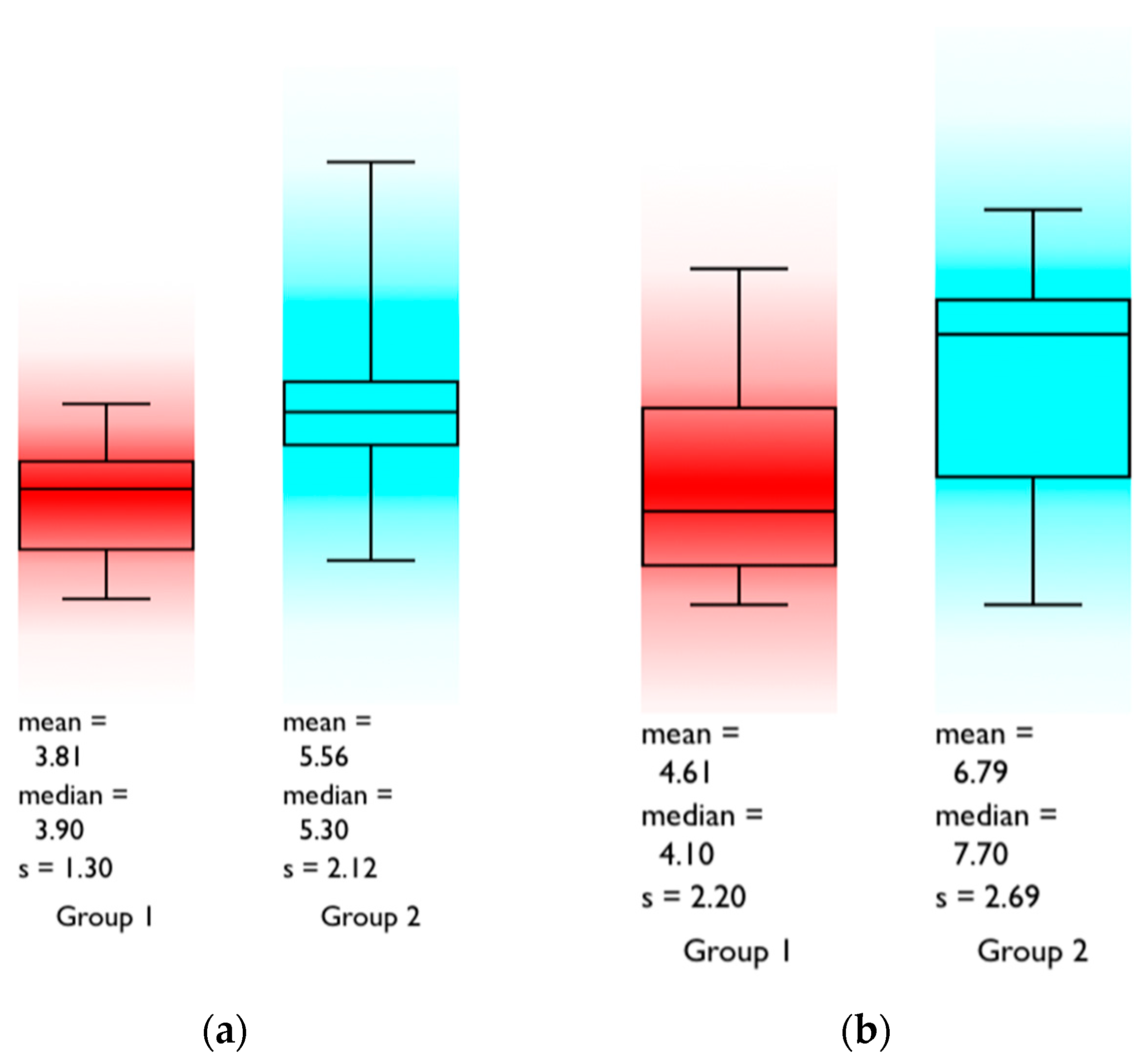
The mean EMG activity at rest slightly increased after VDO elevation. RM increased from 4.0 ± 3.6 µV to 5.1 ± 3.8 µV, LM from 4.8 ± 2.5 µV to 6.0 ± 3.2 µV, RAD from 9.9 ± 3.1 µV to 10.7 ± 3.3 µV, and LAD from 14.0 ± 4.0 µV to 15.4 ± 4.1 µV. Statistically significant differences were observed only for the masseter muscles (RM: p = 0.0166; LM: p = 0.015), whereas the digastric muscles showed no significant changes (Table 1).
EMG activity during swallowing showed variability among participants but remained largely unaffected by VDO elevation. Mean activity for RM was 15.6 ± 28.2 µV before and 18.9 ± 24.9 µV after VDO increase, LM was 16.5 ± 14.0 µV vs. 19.6 ± 17.5 µV, RAD 9.7 ± 0.5 µV vs. 10.0 ± 4.6 µV, and LAD 13.6 ± 1.4 µV vs. 15.3 ± 5.0 µV. No statistically significant differences were observed for any muscle (Table 2).
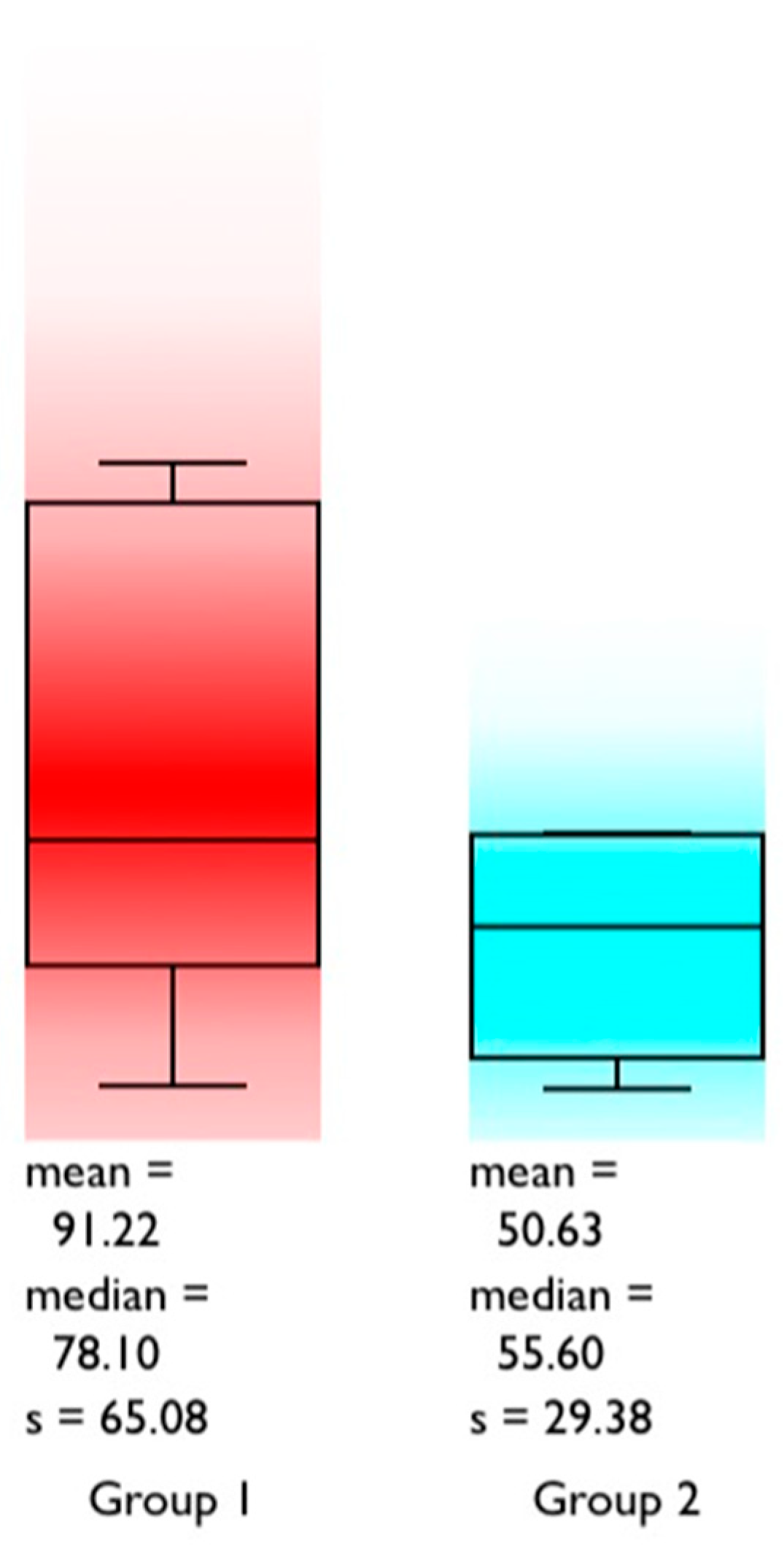
EMG activity during maximum intercuspation showed a statistically significant difference only in the right masseter (RM: 78.1 ± 64.7 µV vs. 65.5 ± 64.0 µV; p = 0.0399), while all other muscles remained stable: LM 111.1 ± 119.6 µV vs. 108.1 ± 86.1 µV, RAD 10.7 ± 3.0 µV vs. 10.9 ± 4.1 µV, and LAD 15.2 ± 3.4 µV vs. 14.8 ± 3.6 µV (Table 3).
Only statistically significant differences were found between resting muscle activity levels in the masseter muscles (p = 0.0166 for RM and p = 0.015 for LM) and maximum intercuspation in the right masseter muscle (p = 0.0399). All other comparative assessments proved statistically insignificant (Figure 2 and Figure 3).
Overall, these results indicate that a moderate increase in VDO of 2–4 mm in Class II/2 patients produces minimal alterations in masticatory muscle EMG activity, with only localized differences observed in the masseter muscles, suggesting neuromuscular adaptability to vertical occlusal changes.

4. Discussion

The present pilot study evaluated the short-term effect of a moderate increase in the vertical dimension of occlusion (VDO) on masticatory muscle activity in patients with Class II Division 2 malocclusion. Overall, most variables showed no statistically significant differences after VDO increase, supporting the concept of short-term neuromuscular adaptability to controlled vertical changes. Resting muscle hyperactivity was observed in most of the investigated muscles in subjects with Class II Division 2 malocclusion. Only a limited number of recordings obtained in the mandibular postural position were within the physiological range generally considered normal for the masseter and anterior digastric muscles. In addition, electromyographic activity of the anterior digastric muscle during swallowing did not differ significantly from postural activity, either before or after VDO increase. These findings suggest that Class II Division 2 patients may present a characteristic functional background with elevated resting muscle activity and relative short-term adaptability to moderate vertical modification. Significant differences were observed only in the resting activity of both masseter muscles and in the right masseter during maximum intercuspation.
The electromyographic recordings also showed that, during swallowing, the masseter muscles appear to contribute to arch stabilization, while the digastric muscles preserve their characteristic functional role in this physiological act. The absence of significant changes in signal amplitude in the postural position and during swallowing after VDO increase further supports the adaptability of the masticatory neuromuscular system to short-term clinical modification of the vertical dimension of occlusion. Similar findings have been reported by other authors, who observed that short-term increases in VDO do not significantly influence the electrical activity of the masticatory muscles. In contrast, Satygo et al. [19] found that after 12 months of pre-orthodontic treatment with a functional pre-orthodontic Trainer appliance, the amplitude of electromyographic signals in the temporalis and masseter muscles during maximum intercuspation increased in patients with Class II Division 1 malocclusion.
In the present study, we found statistically significant differences in the amplitude of myoelectric signals in the masseter muscles after the elevation of occlusion compared to the initial situation, with the values being lower after the elevation of occlusion. Smaglyuk et al. report increased myoelectric activity in the masseters and temporalis in maximum intercuspation in patients with class II relationships, with values of electromyographic signal amplitudes of over 2000 microvolts 4. Within our group, the maximum value recorded by the recordings was 356 microvolts before the elevation of occlusion; after the elevation of the vertical dimension of occlusion, the recorded values were lower, probably due to the relatively resilient occlusal support, represented by the elastic silicone-based addition material of chitosan consistency interposed between the arches in the lateral areas. The possibility of raising the occlusion in class II division 2 patients should be considered, especially regarding the reassessment of the position of the maxillary anterior teeth and their relationships with the antagonists in deep occlusions, essential not only from an aesthetic point of view but also functionally in eccentric movements of the mandible. In patients with class II division 2 relationships with pronounced dental abrasion of the anterior teeth, a decrease in the vertical dimension of occlusion, a reduction in the height of the dental crowns, and the possibility of the appearance of end-to-end incisal relationships can be observed, with the consequent impairment of both physiognomy and anterior guidance. Raising the vertical dimension corrects the occlusal relationship of the anterior teeth. Restoring the overjet and the overbite makes it possible to perform dental guidance that protects the posterior teeth in eccentric mandible movements [20]. Conducting a study on 80 patients from the point of view of an essential parameter for masticatory and occlusal muscle functionality, namely the disocclusion time in eccentric movements of the mandible (typically below 0.4 s), Kerstein statistically analyzed its average value according to the Angle skeletal classes, finding that in class I subjects this parameter was 1.2 s, in class II subjects it was 1.7 s, in class III subjects it was 1.3 s, and in open occlusion situations it was over 1.8 s. In the case of class II anomalies, the slight increase in the vertical dimension of occlusion could reduce the duration of the disocclusion time [17].
Interest in electromyographic investigation of the behavior of the masticatory musculature during augmentations of the vertical dimension of occlusion is all the greater when a craniomandibular dysfunction is associated with a class II division 2 anomaly, a frequent possibility in cases of these craniomandibular relationships [21,22,23]. One of the electromyographic approaches is the LEARRETA TMJ decompression test, which, among other objectives, aims to determine whether a clinically established VDO, in fact, a three-dimensional position of the mandible in occlusion, favors the best recruitment of the elevator mandibular muscle fibers. In this test, different degrees of occlusal elevation can be obtained with cotton rolls interposed between the two dental arches [24]. Ma Y et al. have performed an excellent systematic review of the literature, based on the analysis of 74 publications, regarding therapeutic strategies in class II division 2 malocclusions, especially in conditions associated with severe tooth wear [1]. They suggest a multidisciplinary approach to these patients with class II division 2 relationships, considering pre-prosthetic orthodontic treatment essential. Routine cephalometric analysis should include evaluations of the anterior teeth (interincisal angle, cervical angle). Increasing the VDO above 3 mm intrinsically can be readily accepted by the patient if a test period of between 8 weeks and 6 months is possible with temporary prosthetic devices, preferably on the entire arch. To elevate the occlusion below 3 mm, temporary restorations may or may not be considered, depending on the clinical situation, but the literature generally suggests it. However, current occlusal concepts consider using occlusal splints or provisional restorations to test patient tolerance as unnecessary and error-generating in determining the vertical dimension of occlusion, since this is unrelated to comfort. The elimination of occlusal interferences towards the centric relationship (the orthopedically stable position of the mandible) and in the eccentric trajectories of the mandible is essential.
Peter Dawson maintains that the key element for understanding the vertical dimension of occlusion is the recognition of the fact that “the teeth adapt to the vertical position of the mandible as dictated by the musculature and not vice versa”, the correct determinant of the maxillo-mandibular relationship in maximum intercuspation being the result of the length of the muscle fibers of the elevator muscles of the mandible in repetitive contractions [25]. The renowned author warns practitioners that unnecessary augmentation of the VDO is considered excessive treatment (overtreatment) because the increase in vertical dimension cannot be maintained over time. Suppose there are dental-dental contacts of equal intensity and simultaneous in the CR at the level of the arches. In that case, the relationship between the dentin-enamel junction of the teeth and the alveolar bone remains unchanged as the VDO decreases or increases, according to the need for muscle contraction (contraction length of muscle fibers), which supports the fact that changes in vertical dimension occur either consecutively to elongation or regressive remodeling of the dentoalveolar processes.
The alteration of the vertical dimension of occlusion is allowed in the case of class II division 2 patients to obtain a more stable interdental relationship, since the changes in the vertical dimension of occlusion of the position of the lower teeth in the horizontal and vertical planes are on the closing arch of the mandible. The objective of achieving functional anterior dental contact is often the determining factor in choosing the most advantageous vertical dimension. Still, the only predictable way to evaluate the effect of the alteration of the vertical dimension in making the prosthetic therapeutic decision is to mount the models in an Arcon-type semi-adjustable articulator in centric relation, with a transfer face-bow, in association with the cephalometric examination of the vertical dimension. Based on the cephalometric examination of the angle of the lower floor of the face, it can be modified in one direction or another if we accept that a 1-degree change in the angle can be achieved by raising or lowering the graded incisal pin of the Arcon articulator by 2 mm. The increase in vertical dimension by placing the universal jig to achieve a spacing between the incisors of 3–4 mm did not produce significant changes at the level of the muscles, and even less is this elevation unlikely to cause overload of the temporomandibular joint. One mm interincisal elevation of the occlusion determines a 1-degree rotation of the condyles in the joint, which cannot induce direct articular stress, the condylar rotation being a perfectly physiological movement for this joint [26]. For the cephalometric examination of the VDO, some authors also measure the distance from the anterior nasal spine to the chin [9]. If the exact intercondylar axis is obtained Axiographically, it is possible to evaluate the potential effects of the reassessment of the vertical dimension on the anterior and posterior teeth. Digital workflows allow virtual articulators with remarkable results, such as digital jaw tracking systems (e.g., Modjaw technology).
The study has several limitations: First, the small number of subjects is an issue. Future studies should include larger groups, possibly recording myoelectric activity at the level of the anterior fascicles of the temporalis muscle, given that it is an important postural muscle and easier to approach in patients with excessive hairiness in the masseter area. Surface EMG examination also has its limits because not all fibers of a muscle contract at the same time. Some muscle spindles contract in a coordinated manner, under the control of the central nervous system, while others remain at rest without detectable electromyographic activity. Surface electrodes placed over the masseter or temporalis can sometimes generate false negative responses (weaker activity or even lack of activity), which can lead to erroneous interpretations due to the resistance of the skin (the skin must be perfectly cleaned and degreased with alcohol before placing the electrode), the presence of hairiness, and the instability of the electrodes during recording.
Our findings indicate that increasing the vertical dimension of occlusion (VDO) by 2–4 mm in patients with Class II division 2 malocclusion does not significantly alter the electromyographic (EMG) activity of the masseter and anterior digastric muscles, suggesting an intrinsic adaptability of the masticatory system. This is consistent with previous reports showing minimal EMG changes during short-term VDO alterations in both dentate and edentulous subjects [27,28,29,30].
De-Freitas et al. [29] and Poggio et al. [21] observed that moderate VDO increases in centric relation did not significantly affect masseter EMG activity, supporting the notion that masticatory muscles can accommodate clinically relevant occlusal changes without functional compromise. They observed that short-term VDO elevations in centric relation do not significantly modify EMG amplitude, emphasizing that the masticatory muscles can accommodate occlusal modifications without functional compromise. Similarly, Matos et al. [30] and Lyons [31] reported that minor vertical adjustments, whether in prosthodontic or TMJ decompression contexts, resulted in negligible alterations in muscle electrical activity, emphasizing the neuromuscular system’s adaptability.
Valdés et al. [32] further demonstrated that even changes in tongue posture affecting vertical dimension produced limited EMG responses, highlighting the stability of masticatory muscle function under minor occlusal modifications. These converging findings reinforce the clinical relevance of cautiously increasing VDO to achieve aesthetic or functional goals, such as improved anterior dental contact, without compromising muscle activity or temporomandibular joint function.
In addition to studies directly assessing masticatory muscle EMG changes with occlusal vertical dimension modifications, other research demonstrates that functional behaviors such as swallowing patterns and perioral muscle activity are also altered with changes in vertical height, indicating broader neuromuscular adaptation [31,33,34]. Furthermore, investigations in edentulous populations show that bite force and muscle efficiency vary with vertical jaw position, suggesting the existence of optimal VDO ranges for efficient muscle activity, a concept that complements our finding of muscular adaptability under moderate VDO increases [29,30,31,32,35].
Lyons [31] and Olthoff et al. [36] demonstrated that even prosthetic VDO increases produce only slight variations in masticatory muscle performance, supporting the idea that moderate vertical adjustments are well-tolerated by the neuromuscular system. Furthermore, Constantinescu et al. [37] and Goldstein et al. [38] emphasize that careful clinical planning of VDO adjustments preserves muscular coordination and temporomandibular joint stability, corroborating the practical applicability of our findings. Collectively, these studies suggest that controlled VDO elevation can be implemented safely in Class II division 2 patients to improve anterior dental contact, optimize prosthetic space, and enhance esthetic outcomes without compromising muscle function.
From a biomechanical perspective, changes in vertical dimension influence load distribution across the dentoalveolar structures and temporomandibular joint. Insights from biomaterials and tissue mechanics suggest that even minor occlusal modifications may alter force transmission patterns, potentially contributing to adaptive neuromuscular responses. This interdisciplinary perspective supports the interpretation of subtle EMG variations observed in the present study.
The absence of statistically significant differences in several parameters should be interpreted with caution. Given the limited sample size, the study may be underpowered, increasing the risk of a Type II error. Therefore, the lack of statistical significance does not necessarily indicate the absence of a biological effect but rather may reflect insufficient statistical sensitivity.
Furthermore, the present investigation evaluated only the immediate neuromuscular response over a short recording period (30 s), which does not allow conclusions regarding long-term adaptation. Neuromuscular responses to occlusal changes are known to evolve, and longitudinal studies are required.

5. Conclusions

Within the limitations of this pilot study, a short-term increase in vertical dimension of occlusion (2–4 mm) produced only minor changes in masticatory muscle electromyographic activity in Class II Division 2 patients.
These findings suggest a degree of short-term neuromuscular adaptability; however, they should be interpreted with caution due to the limited sample size, gender imbalance, and short observation period.
The absence of statistically significant differences in most parameters does not exclude the possibility of clinically relevant effects, and future studies with larger samples and long-term follow-up are necessary.
Clinical recommendations regarding VDO modification should be based on comprehensive assessment and supported by the existing literature rather than based solely on the findings of this study.
The null hypothesis is supported by the fact that, within the parameters of this investigation, increasing the vertical dimension of occlusion (VDO) by 2–4 mm does not significantly change electromyographic activity in the investigated masticatory muscles.
EMG results indicate that the mandible’s postural position in Class II/2 patients represents a range of postural myoelectric activity rather than a single fixed position.
In Class II/2 patients, VDO elevation can be clinically applied to achieve a more stable interdental relationship. When the mandible is in an orthopedically stable position relative to the maxilla (centric relation), changes in VDO primarily affect the closing path of the mandible, provided appropriate articulator mounting and face-bow transfer are used. Achieving functional anterior dental contact, along with aesthetic considerations and prosthetic space creation, often guides the selection of the optimal VDO.
The previous literature suggests that occlusal elevation may be evaluated using provisional restorations over a period of 2–6 months; however, this recommendation is not directly derived from the present study. The predominance of female participants (7 out of 9) may limit the generalizability of the findings, as sex-related differences in muscle activity cannot be excluded.

Author Contributions

Conceptualization S.P. and T.-M.C.; methodology, S.P.; software, L.-C.D.; validation, S.P., L.-C.D. and Z.B.-V.; formal analysis, T.-M.C.; investigation, T.-M.C.; resources, S.P.; data curation, Z.B.-V.; writing—original draft preparation, T.-M.C.; writing—review and editing, Z.B.-V. and M.P.; visualization, T.-M.C.; supervision, S.P. and M.P.; project administration, Z.B.-V.; funding acquisition, T.-M.C. All authors have read and agreed to the published version of the manuscript.

Funding

This research received no external funding.

Institutional Review Board Statement

The study protocol was reviewed and approved by the Ethics Committee of the George Emil Palade University of Medicine, Pharmacy, Science and Technology of Târgu Mureș (approval number: 3874/06.10.2025). The study was conducted in accordance with the Declaration of Helsinki (2013 revision) and in compliance with Regulation (EU) 2016/679 (GDPR).

Informed Consent Statement

Informed consent was obtained from all subjects involved in the study. Written informed consent has been obtained from the patient(s) to publish this paper.

Data Availability Statement

The data supporting the findings of this study are contained within the article.

Acknowledgments

The authors thank all study participants for their cooperation throughout the research.

Conflicts of Interest

The authors declare no conflicts of interest.

Abbreviations

The following abbreviations are used in this manuscript:
RMRight Masseter Muscle
LMLeft Masseter Muscle
RADRight Anterior Digastric Muscle
LADLeft Anterior Digastric Muscle
IMIntercuspal Position
EMGElectromyography
VDOVertical Dimension of Occlusion

References

  1. Ma, Y.; Zhao, W.; Zhiang, S.; Jin, X.; Xu, J.; Fu, B.; Shi, Y. Treatment decisions of patients with Class II Division 2 malocclusion and severe tooth wear: A systematic review. BDJ Open 2024, 10, 65–72. [Google Scholar] [CrossRef] [PubMed]
  2. Ferrario, V.F.; Sforza, C.; Serrao, G. Electromyographic activity of masticatory muscles in normal young people. J. Oral Rehabil. 2000, 27, 715–723. [Google Scholar]
  3. Monaco, A.; Cattaneo, R.; Spadaro, A.; Giannoni, M.; Sgolastra, F. Surface electromyography pattern of human masticatory muscles in jaw clenching. Eur. J. Paediatr. Dent. 2013, 14, 21–26. [Google Scholar]
  4. de Felício, C.M.; Sidequersky, F.V.; Tartaglia, G.M.; Sforza, C. Masticatory muscle activity in Class II malocclusion. J. Oral Rehabil. 2010, 37, 760–767. [Google Scholar]
  5. Tuxen, A.; Bakke, M. Functional and EMG characteristics of masticatory muscles in different occlusal conditions. Acta Odontol. Scand. 1995, 53, 235–240. [Google Scholar]
  6. Ingervall, B.; Thüer, U. Activity of temporal and masseter muscles in subjects with different malocclusions. Am. J. Orthod. 1980, 77, 198–206. [Google Scholar]
  7. Manns, A.; Diaz, G. Masticatory function and muscle adaptation in malocclusion. J. Dent. Res. 1988, 67, 1203–1207. [Google Scholar]
  8. Sforza, C.; Rosati, R.; De Menezes, M.; Musto, F.; Toma, M. EMG analysis of trapezius and masticatory muscles: Experimental protocol and data reproducibility. J. Oral Rehabil. 2011, 38, 648–654. [Google Scholar] [CrossRef] [PubMed]
  9. Prasad, S.A.; Indukuri, R.R.; Singh, R.; Noong, A.; Palagri, F.B.; Nasayana, V. Pathognomonic features of Angle’s Class II division malocclusion: A comparative cephalometric and arch width study. J. Int. Soc. Prevent. Community Dent. 2014, 4, 105–109. [Google Scholar] [CrossRef] [PubMed]
  10. Ngan, P.; Tai, S.K. Orthopedic treatment of Class II malocclusion with mandibular deficiency: A clinical practice review. Front. Oral Maxillofac. Med. 2024, 6, 27–31. [Google Scholar] [CrossRef]
  11. Smaglyuk, L.V.; Liakhovska, A.V. EMG characteristics of masticatory muscles in patients with Class II malocclusion and temporomandibular disorders. Wiad. Lek. 2019, 72, 1043–1047. [Google Scholar] [CrossRef]
  12. Bratu, D.C.; Popa, G.; Popa, N.S.; Matichescu, A.; Petrescu, P.H.; Pop, S.I. Surface electromyographic study in patients with Angle Class II malocclusion. Med. Evol. 2019, 25, 44–50. [Google Scholar]
  13. Shen, Y.-F.; Wei, M.-C.; Li, H.-P.; Pan, Y.-H.; Hong, H.-H.; Chen, C.-C.; Kuo, S.-B.; Ho, C.-Y.; Chang, C.-T.; Huang, Y.-F. Vertical dimension of occlusion related to mandibular movement during swallowing. Biomed. J. 2021, 44, 217–222. [Google Scholar] [CrossRef] [PubMed]
  14. Slavicek, R. Concepts in Oral Medicine—Concipio; GAMMA Medizinisch-wissenschaftliche Fortbildungs-GmbH: Klosterneuburg, Austria, 2015; pp. 301–322. [Google Scholar]
  15. Morais, E.C.C.; Ornaghi, B.P.; Zielak, A.P.S.J.C.; da Costa, R.G.; Bindo, M.J.F.; de Campos, E.A. Determination of final occlusal vertical dimension by cephalometric analysis. Rev. Sul-Bras. Odontol. 2015, 12, 143–150. [Google Scholar]
  16. Dawson, P.E. Functional Occlusion: From TMJ to Smile Design; Mosby: St. Louis, MO, USA, 2007. [Google Scholar]
  17. Kerstein, R. Employing T-Scan synchronized with electromyography to treat chronic occluso-muscle disorder. In Handbook of Researches in Computerized Occlusal Analysis Technology Applications in Dental Medicine; IGI Global: Hershey, PA, USA, 2015; pp. 269–357. [Google Scholar]
  18. Dupas, I.P.; Picart, H.; Lefevre, C.; Graux, F. Centric relation and programming semiadjustable articulators with the universal jig. Part I: Technique. J. Prosthet. Dent. 1990, 63, 134–138. [Google Scholar] [CrossRef] [PubMed]
  19. Satygo, E.A.; Silin, A.V.; Ramirez-Yañez, G.O. Electromyographic muscular activity improvement treated with the pre-orthodontic trainer. J. Clin. Pediatr. Dent. 2014, 38, 380–384. [Google Scholar] [CrossRef] [PubMed]
  20. Abduci, J.; Lyons, K. Clinical considerations for increasing occlusal vertical dimensions: A review. Aust. Dent. J. 2012, 57, 2–10. [Google Scholar] [CrossRef] [PubMed]
  21. Sadrabad, M.J.; Ameli, N.; Kianpour, M.; Ghorbani, R.; Sohanian, S. The relationship of temporomandibular disorders with Class II malocclusion as a risk factor. APOS Trends Orthod. 2021, 11, 41–47. [Google Scholar] [CrossRef]
  22. Rathi, S.; Gilan, R.; Kamble, R.; Bhandwalkar, S. Temporomandibular joint disorders and airway in Class II malocclusions: A review. Cureus 2022, 14, e30515. [Google Scholar] [CrossRef]
  23. Berar, A.; Constantiniuc, M.; Kui, A.; Fluerasu, M.; Iacob, S.M.; Manziuc, M.; Mițaru, L.; Stricatu, M.; Buduru, S. Temporomandibular dysfunction and Angle Class II/2 malocclusion. Int. J. Med. Dent. 2020, 24, 434–441. [Google Scholar]
  24. Matos, M.F.; Durst, S.A.C.; Matos, J.L.F.; Learreta, J.A. Electromyographic evaluation of the ‘vertical’ dimension: The Learreta TMJ decompression test. Cranio 2011, 29, 255–260. [Google Scholar] [CrossRef]
  25. Dawson, P.E. Changing Vertical Dimension: A Solution or Problem? CDE World Courses, Dawson Academy. Available online: https://cdeworld.com (accessed on 22 April 2026).
  26. Bellester, J. Apport au concept d’intervalle de dimension verticale. Coll. Natl. Occlusodontol. 1997, 6, 3–7. [Google Scholar]
  27. Elsokkary, M.M. Evaluating the impact of vertical dimension on masticatory muscle activity during clenching in complete denture wearers: An electromyographic (EMG) analysis. Al-Azhar Assiut Dent. J. 2024, 7, 159–165. [Google Scholar] [CrossRef]
  28. Poggio, C.E.; Manfredini, D. Does increasing vertical dimension of occlusion in centric relation affect muscular activity? An electromyographic study. J. Esthet. Restor. Dent. 2023, 36, 231–238. [Google Scholar] [CrossRef]
  29. De-Freitas, E.F.; Guarda-Nardini, L.; Florencio, L.S.; Conti, P.C.R. Effect of short-term increase in occlusal vertical dimension on masticatory muscle electrical activities and pressure-to-pain threshold: A crossover clinical study. J. Prosthet. Dent. 2022, 128, 970–976. [Google Scholar]
  30. Coelho-Ferraz, M.J.; Berzin, F.; Amorim, C.F.; Romano, F.L.; de Paula Queluz, D.P. Electromyographic and cephalo-metric correlation with the predominant masticatory movement. Stomatologija 2010, 12, 51–55. [Google Scholar] [PubMed]
  31. Lyons, M.F. An electromyographic study of masticatory muscle activity at increased occlusal vertical dimension in complete denture wearers. J. Prosthet. Dent. 1988, 60, 346–348. [Google Scholar] [CrossRef] [PubMed]
  32. Valdés, C.; Gutiérrez, M.; Falace, D.; Astaburuaga, F.; Manns, A. The effect of tongue position and resulting vertical dimension on masticatory muscle activity: A cross-sectional study. J. Oral Rehabil. 2013, 40, 650–656. [Google Scholar] [CrossRef]
  33. MacAvoy, S.K.; Jack, H.C.; Kieser, J.; Farella, M. Effect of occlusal vertical dimension on swallowing patterns and perioral electromyographic activity. J. Oral Rehabil. 2016, 43, 481–487. [Google Scholar] [CrossRef]
  34. de Caxias, F.P.; Leal Túrcio, K.H.; Melo Neto, C.L.M.; de Athayde, F.R.F.; Goiato, M.C.; Dos Santos, D.M. Effects of rehabilitation with complete dentures on bite force and electromyography of jaw and neck muscles and correlations with occlusal vertical dimension. Clin. Oral Investig. 2021, 25, 4691–4698. [Google Scholar] [CrossRef]
  35. Morimoto, T.; Abekura, H.; Tokuyama, H.; Hamada, T. Alteration in the bite force and EMG activity with changes in the vertical dimension of occlusion in edentulous subjects. J. Oral Rehabil. 1996, 23, 336–341. [Google Scholar] [CrossRef] [PubMed]
  36. Olthoff, L.W.; van der Glas, H.W.; van der Bilt, A. Influence of occlusal vertical dimension on masticatory performance during chewing with maxillary splints. J. Oral Rehabil. 2007, 34, 560–565. [Google Scholar] [CrossRef] [PubMed]
  37. Constantinescu, F.E.; Savastano, F.; Perlea, P.; Constantinescu, M.-V. Complete morphofunctional oral rehabilitation by physiological increase of occlusal vertical dimension according to computerized mandibular scanner: A case report. Rom. J. Morphol. Embryol. 2022, 63, 245–251. [Google Scholar] [CrossRef]
  38. Goldstein, G.; Goodacre, C.; MacGregor, K. Occlusal vertical dimension: Best evidence consensus statement. J. Prosthodont. 2021, 30, 12–19. [Google Scholar] [CrossRef] [PubMed]
Figure 1. (a) The BioEMG II system used for surface electromyography recording. (b) Electrode placement: electrodes positioned parallel to the muscle fibers on the masseter and anterior belly of the digastric muscles.
Applsci 16 04216 g001
Figure 2. Comparison of postural myoelectric activity (µV) in the (a) right and (b) left masseter muscles before (red) and after (blue) the vertical dimension of occlusion (VDO) increase.
Applsci 16 04216 g002
Figure 3. Comparison of myoelectric activity (µV) in the right masseter muscle during maximum intercuspation before (red) and after (blue) the vertical dimension of occlusion (VDO) increase.
Applsci 16 04216 g003
Table 1. Amplitude of myoelectric signals (µV) in the resting position, before and after vertical dimension (VDO) elevation in the four investigated muscles.
SubjectSwallowing (µV)After VDO Elevation (µV)
RMLMRADLADRMLMRADLAD
15.53.09.714.36.14.814.220.8
22.22.80.92.03.46.01.73.6
34.44.69.214.37.17.78.915.1
43.84.18.813.09.98.48.814.0
51.96.29.713.85.610.39.813.9
64.33.311.416.24.78.114.619.7
73.56.210.114.35.34.410.114.9
83.99.110.514.85.39.59.013.6
92.82.29.614.22.62.79.515.2
Table 2. Amplitude of myoelectric signals (µV) during swallowing, before and after vertical dimension (VDO) elevation.
SubjectSwallowing (µV)After VDO Elevation (µV)
RMLMRADLADRMLMRADLAD
186.819.29.915.315.18.819.426.3
26.65.98.212.65.04.26.08.7
34.611.41013.612.217.18.913.6
420.419.78.713.314.316.29.213.8
59.813.59.613.810.219.99.714.5
610.616.910.314.76.416.614.120.3
75.08.510.915.87.67.89.814.0
86.79.49.914.010.012.210.014.7
916.243.39.413.763.250.69.814.6
Table 3. Amplitude of myoelectric signals (µV) in maximum intercuspation, before and after VDO elevation.
SubjectSwallowing (µV)After VDO Elevation (µV)
RMLMRADLADRMLMRADLAD
1165.8169.811.015.380.7125.519.026.5
245.565.84.79.321.522.02.03.5
318.837.98.711.913.423.79.614.0
478.183.28.813.379.579.110.113.2
5177.5356.413.319.455.6114.110.413.8
6172.011713.217.881.272.914.220.3
714.215.99.914.819.815.410.214.0
887.956.910.314.829.46.29.814.2
961.2105.310.714.574.675.410.415.0
Disclaimer/Publisher’s Note: The statements, opinions and data contained in all publications are solely those of the individual author(s) and contributor(s) and not of MDPI and/or the editor(s). MDPI and/or the editor(s) disclaim responsibility for any injury to people or property resulting from any ideas, methods, instructions or products referred to in the content.