1. Introduction
Extraction of third molars is one of the most commonly performed procedures in maxillofacial surgery [
1]. Third-molar impaction is multifactorial. Factors such as insufficient space, eruption sequencing, tooth position, mandibular growth trends, and genetic components all play a role [
2]. Clinicians utilize preoperative classifications to estimate the complexity of the procedure and the likelihood of postoperative complications. For instance, the Pell and Gregory system assesses the tooth’s spatial relationship to the mandibular ramus and the occlusal plane, whereas Winter’s classification categorizes the specific angulation of the impaction [
3].
Indications for removal are mainly symptom-driven. Common reasons include pain, recurrent pericoronitis, caries, or periodontal breakdown on the distal aspect of the second molar, and associated pathology such as cystic change [
4]. Extraction may also be required for orthodontic or prosthetic purposes to prevent progression of local disease [
5]. In patients with systemic risk factors, timing and perioperative management are important to reduce infectious or bleeding complications [
6].
Despite being routine, impacted third-molar surgery is followed by predictable postoperative outcomes. Pain, facial swelling, and trismus are the most common early outcomes and usually peak within the first postoperative days [
7]. Pain often increases after local anesthesia resolves and may be clinically significant in the first 24–48 h. Swelling peaks around 48–72 h due to inflammatory exudation and tissue edema. Trismus is influenced by inflammation, muscle guarding, and surgical trauma in the masticatory region [
8]. These outcomes affect oral hygiene, diet, sleep, work, and patient satisfaction [
9].
Corticosteroids are among the most studied adjuncts in third-molar surgery. They exert anti-inflammatory effects through glucocorticoid receptor–mediated pathways, downregulating inflammatory mediators, decreasing capillary permeability, and limiting edema formation [
10]. Clinical trials and reviews generally report reduced swelling and improved postoperative comfort, although effects on pain and trismus vary according to dose, timing, and route of administration [
11]. Short perioperative regimens are typically considered safe in appropriately selected patients, but outcomes are not uniform across protocols [
12]. Dexamethasone and methylprednisolone are widely used in this setting: dexamethasone is highly potent, with prolonged biological activity and negligible mineralocorticoid effect, whereas methylprednisolone has intermediate duration and minimal mineralocorticoid activity [
13]. Based on standard glucocorticoid equivalence tables for systemic dosing (0.75 mg dexamethasone ≈ 4 mg methylprednisolone), the doses used in the present trial—8 mg dexamethasone (Decort
®) and 40 mg methylprednisolone (Prednol
®)—can be considered approximately systemically equivalent anti-inflammatory doses, while acknowledging that local intra-alveolar pharmacokinetics are not established [
14].
Most evidence on perioperative corticosteroids in third-molar surgery is based on systemic regimens or submucosal injection protocols [
10,
12]. Intra-alveolar delivery via an absorbable gelatin sponge offers a pragmatic chairside alternative: it avoids an additional needle injection, can be applied immediately after extraction, and places the drug directly at the surgical site during early clot formation and the peak inflammatory window. However, intra-socket delivery may be affected by bleeding-related dilution, saliva washout, and sponge-retention variability, and the local retention/systemic absorption profile remains incompletely characterized. Consequently, comparative evidence for this carrier-based intra-alveolar approach—particularly direct comparisons between dexamethasone and methylprednisolone—is limited, leaving an important route- and carrier-specific evidence gap [
10,
12,
15].
This randomized, controlled, three-arm parallel trial compared intra-alveolar methylprednisolone and dexamethasone delivered via a gelatin sponge with a saline control. Postoperative pain, swelling, trismus, and analgesic use were assessed at standardized intervals to determine which protocol improves early postoperative recovery after impacted mandibular third-molar surgery.
2. Materials and Methods
The study was designed and reported in accordance with the principles of the Declaration of Helsinki and the CONSORT guidelines for randomized clinical trials to ensure methodological transparency and reproducibility. Ethical approval was granted by the Cyprus Health and Social Sciences University Research Ethics Committee (Approval No. KSTU//2025/118). Participant recruitment and data collection were conducted from November 2025 to January 2026 at the Department of Oral and Maxillofacial Surgery.
2.1. Study Design
This study was designed as a prospective, randomized, controlled, three-arm parallel clinical trial. Participants were randomly assigned to one of three groups:
Outcomes were postoperative pain (VAS), facial swelling (edema), trismus (maximum interincisal opening), and analgesic consumption during follow-up. To control multiplicity from multiple endpoints and repeated postoperative assessments, we prespecified an outcome hierarchy: the primary endpoint for inference (powering target) was Day 1 VAS pain; key secondary endpoints were Day 1 analgesic consumption and Day 3 facial swelling (preselected because edema typically peaks at 48–72 h); trismus across postoperative Days 1, 2, 3, and 7 was exploratory. Swelling was captured using three linear facial distances (tragus–pogonion, tragus–labial commissure, and angulus–canthus), which were considered correlated subdomains and interpreted conservatively. For between-group comparisons at each postoperative day, p-values were adjusted across days within each outcome using the Holm procedure.
Systemically healthy adults aged 18–35 years (ASA I), non-smokers, and not using regular medication were eligible if they had a partially impacted mandibular third molar classified as Pell and Gregory Class I, Position B, without associated pathology (e.g., active pericoronitis, cystic lesion, or tumor), provided written informed consent, and agreed to attend follow-up visits. Exclusion criteria were ASA II–IV status; smoking; incomplete documentation or refusal to participate; acute or chronic infection at the surgical site; systemic disease or medication use that could affect wound healing, bleeding time, or platelet function; or noncompliance with postoperative instructions or follow-up appointments. All surgical procedures within the randomized cohort were successfully completed within 30 min and without any deviation from the operative protocol, strictly preserving the initial randomization balance.
After enrollment, participants were assigned a unique study ID. The randomization sequence was generated by an independent staff member not involved in surgery or outcome assessment. Group assignments were placed into sequentially numbered, opaque, sealed envelopes and stored by the same independent staff member in a secure location. Envelopes were provided to the operating room in numerical order and were opened by the surgeon only after tooth removal and socket irrigation, immediately prior to sponge placement, to ensure allocation concealment until the point of intervention.
Participants were randomized into MP, DEX, or Control groups. Patients and outcome assessors were blinded to group allocation. The surgeon was aware of the group assignment due to the nature of the intervention but did not take part in postoperative measurements.
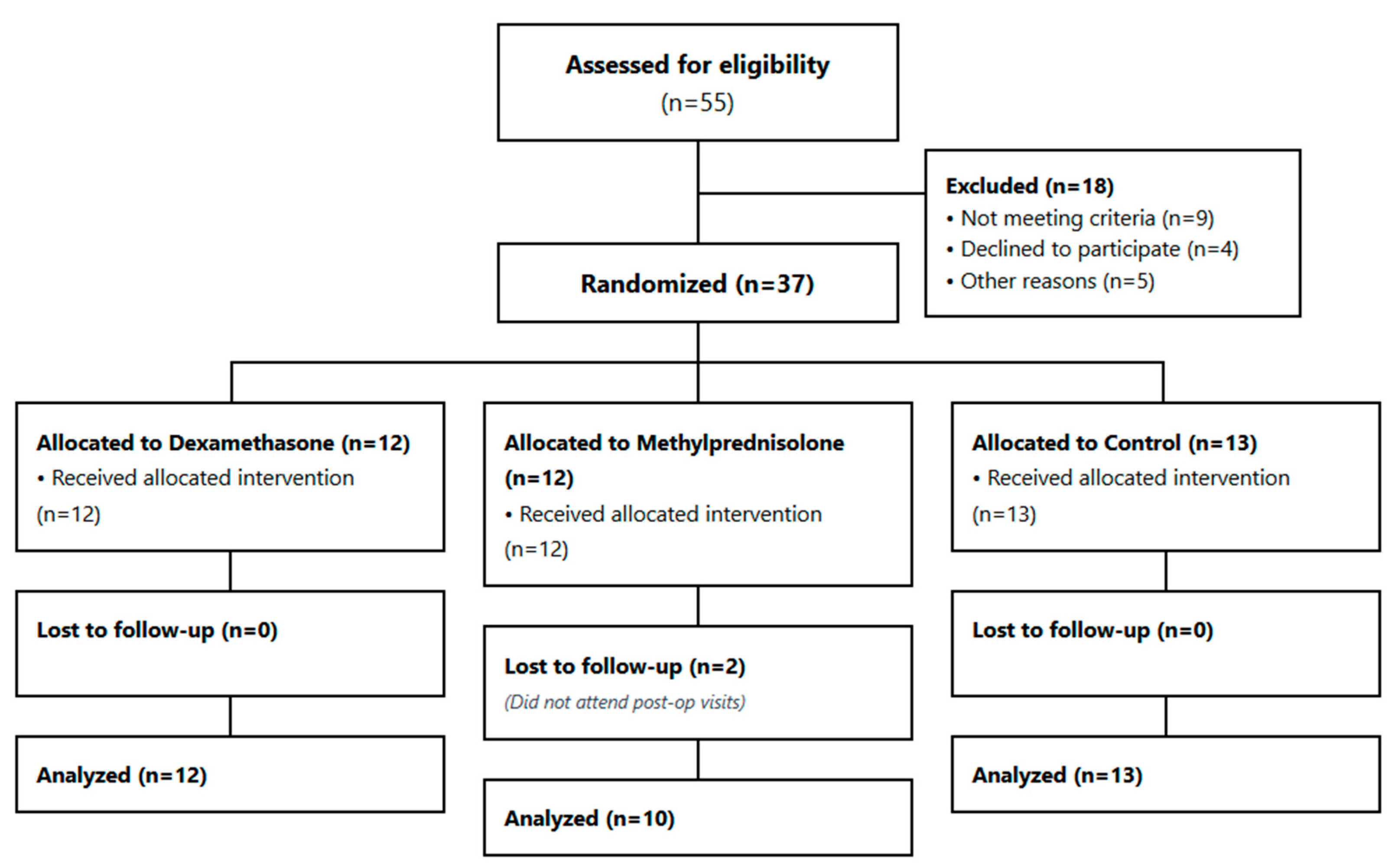
An a priori sample size calculation was performed using G*Power 3.1.9.4 (one-way ANOVA, three independent groups, α = 0.05, power = 0.80, allocation ratio 1:1:1). The calculation was based on detecting a clinically meaningful between-group difference in early postoperative morbidity, with postoperative Day 1 pain (VAS) prespecified as the powering target. A large standardized effect (Cohen’s f = 0.60) was selected to reflect the magnitude of early postoperative differences reported in third-molar corticosteroid RCTs during the first postoperative day (clinically meaningful reductions in pain and/or early morbidity in steroid arms compared with control) and to support a pragmatic pilot-sized trial design [
16,
17,
18,
19]. This yielded a minimum required sample size of 30 participants (10 per group). To account for potential exclusions or incomplete follow-up, we enrolled 37 participants (
Figure 1). We acknowledge that smaller true effects—particularly for swelling and trismus and at later time points—may not be detectable with this sample size; therefore, secondary and exploratory outcomes were interpreted cautiously. Because distributions were not normal and group sizes were small, primary inference used nonparametric tests; the ANOVA-based calculation was used as a pragmatic approximation for three-group comparisons.
2.2. Surgery Protocol
All procedures were performed under strict aseptic and antiseptic conditions. To minimize variability, surgeries were carried out by the same surgeon with the same assistant using a standardized protocol.
Local anesthesia was achieved using a 2 mL ampoule of Ultracain® D-S forte (Sanofi Aventis, Petaling Jaya, Malaysia), containing 40 mg/mL articaine HCl and 0.012 mg/mL epinephrine HCl. Inferior alveolar nerve block and buccal nerve block were administered, and the procedure was initiated only after adequate anesthesia was confirmed.
A horizontal incision with a buccal releasing incision was made using a No. 15 scalpel blade, followed by elevation of a triangular mucoperiosteal flap to expose the surgical field. Ostectomy was performed using a 1.6 mm steel round bur and a 1.4 mm fissure bur mounted on a surgical handpiece operating at 40,000 rpm. Continuous irrigation was maintained throughout bone removal with sterile saline cooled to +4 °C.
After adequate buccal and occlusal bone removal, the tooth was mobilized and extracted using a Bein elevator. The socket was curetted to remove residual debris and soft-tissue remnants and irrigated thoroughly with sterile saline to support debridement and hemostasis.
An absorbable gelatin hemostatic sponge was used to carry the assigned solution into the extraction socket (Spongostan®, Ethicon/J&J, Raritan, NJ, USA; sterile absorbable gelatin sponge). The sponge is a sterile gelatin hemostatic material and does not contain added active drugs such as thrombin or antibiotics. Because features such as pore size and absorption capacity can vary between batches and are not measured during routine clinical use, we reduced variability by standardizing how the sponge was prepared and used: each sponge was 10 × 10 × 5 mm, soaked with 1.0 mL of the allocated solution for 30 s, and then placed intra-alveolarly immediately after preparation.
In the methylprednisolone (MP) group, the sponge was impregnated with methylprednisolone (40 mg; Prednol®) and placed in the socket. In the dexamethasone (DEX) group, the sponge was impregnated with dexamethasone (8 mg; Decort®) and placed similarly. In the control group, an identical sponge was moistened with an equivalent volume of sterile saline and placed in the socket. The mucoperiosteal flap was repositioned, and primary closure was achieved with 3/0 silk sutures (Doğsan®, Istanbul, Turkey; 18 mm, 1/2-circle needle).
Postoperative medications were standardized across groups: amoxicillin–clavulanic acid 1000 mg (Augmentin®) twice daily for 7 days, chlorhexidine mouthwash (Kloroben®) twice daily for 7 days, and paracetamol 500 mg (Parol®) as needed (PRN) for pain, not exceeding 4 g/day. Standardized postoperative instructions were also provided regarding cold application, oral hygiene, and wound care.
Adverse events were assessed at each postoperative visit using structured questioning and clinical examination by the blinded evaluator. Events of interest included infection/alveolitis, delayed bleeding, allergic reactions, delayed wound healing, and any unexpected symptoms reported by participants.
2.3. Data Collection and Evaluation
Postoperative data were collected by a blinded evaluator and encompassed assessments of pain, swelling, and trismus.
2.3.1. Edema Assessment
Edema was assessed using the method described by Gabka and Matsumura [
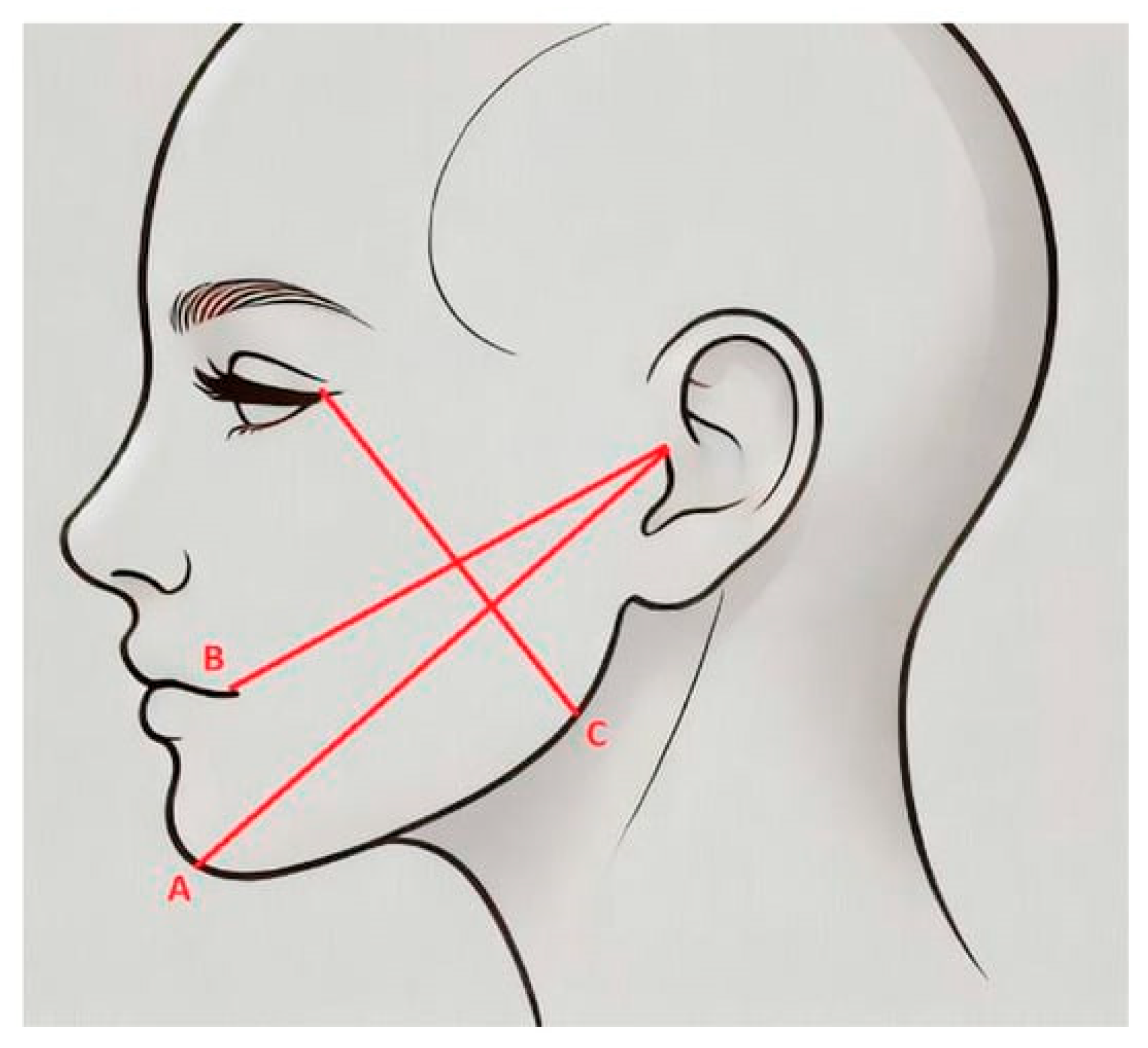
15]. Linear facial distances (tragus–pogonion, tragus–labial commissure, and angulus–canthus) were measured preoperatively and on postoperative Days 1, 2, 3, and 7 using a tape measure (cm) (
Figure 2). To reduce measurement variability, all assessments were performed by a single blinded evaluator who received standardized training in landmark identification and tape-measure positioning before the study. At each visit, participants were seated upright with a neutral head position, and landmarks (tragus, pogonion, labial commissure, mandibular angle, lateral canthus) were identified according to a predefined written protocol. The tape measure was aligned along the same anatomic path with consistent tension. For each facial line, two consecutive measurements were recorded, and the mean value was used for analysis. Intra-examiner reliability was assessed using the Intraclass Correlation Coefficient (ICC) based on the duplicate recordings, yielding an ICC of 0.94 (95% CI: 0.89–0.97), indicating excellent measurement reliability.
2.3.2. Pain and Analgesic Assessment
Postoperative pain typically begins after the local anesthetic effect subsides, which we observed to occur approximately 2–4 h after surgery. Pain intensity was assessed over the first postoperative week using a 10 cm visual analog scale (VAS) at standardized time points on postoperative Days 1, 2, 3, and 7. Participants recorded VAS scores at the same time of day at each time point; values were converted to a 0–10 scale for analysis, yielding four postoperative pain measurements (
Figure 3).
Paracetamol 500 mg was prescribed as needed (PRN) for postoperative pain, and patients were instructed not to exceed the maximum daily dose recommended on the product label. Analgesic consumption was documented in a daily diary as the number of paracetamol tablets taken per day on postoperative days 1, 2, 3, and 7. Diaries were checked at each follow-up visit by the blinded evaluator for completeness and internal consistency, and any missing or ambiguous entries were resolved through clarification with the participant.
2.3.3. Trismus Assessment
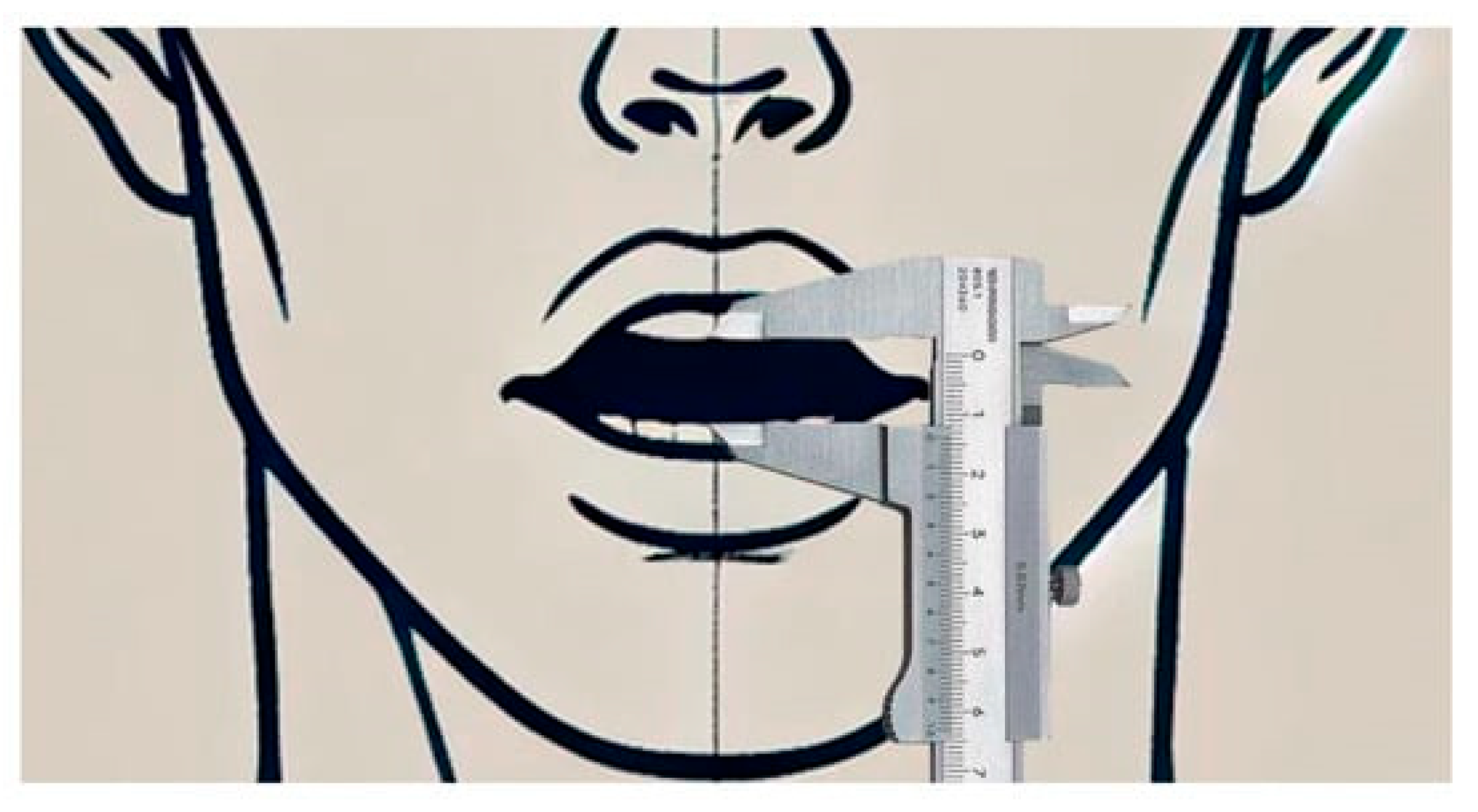
Trismus was measured as the maximum interincisal distance (mm) using electronic calipers between the mesial corners of the central incisors. Measurements were taken at baseline (pre-surgery) and again on days 1, 2, 3, and 7 to observe changes in mouth opening over time (
Figure 4).
2.4. Statistical Analysis
All analyses were prespecified and performed using IBM SPSS Statistics v27 (IBM Corp., Armonk, NY, USA). Continuous variables are presented as median (IQR), and categorical variables as n (%). Normality was assessed using the Shapiro–Wilk test and Q–Q plots; because distributions were non-normal and group sizes were small, nonparametric methods were used. All tests were two-sided with α = 0.05 unless otherwise adjusted.
The primary analyses were initially conducted on an available-case basis. However, to address potential attrition bias from the two participants in the methylprednisolone group who were lost to follow-up, an Intention-to-Treat (ITT) sensitivity analysis was subsequently performed. Missing data for these two participants were imputed using a conservative worst-case scenario by assigning them the median outcome values of the control group. Furthermore, a global False Discovery Rate (FDR, Benjamini–Hochberg) correction was applied across primary and key secondary endpoint domains to strictly control for multiplicity.
Between-group differences were evaluated at each standardized postoperative time point (Days 1, 2, 3, and 7) using the Kruskal–Wallis test. To limit Type I error arising from repeated postoperative testing, p-values were adjusted across postoperative days within each outcome using the Holm procedure. When the overall between-group test remained significant after Holm adjustment, post hoc pairwise comparisons were performed using Dunn’s test with Holm-corrected p-values.
Within-group changes over time were assessed using the Friedman test; when significant, post hoc comparisons were conducted using the Wilcoxon signed-rank test with Holm adjustment. Effect sizes were reported as epsilon-squared (ε2) for Kruskal–Wallis analyses and Kendall’s W for Friedman analyses. For the primary endpoint (Day 1 VAS), we additionally report the Hodges–Lehmann median difference (steroid − control) with 95% CI based on the Mann–Whitney (Wilcoxon rank-sum) method. All analyzed participants completed assessments on postoperative Days 1, 2, 3, and 7.
4. Discussion
This randomized controlled trial evaluated the effects of intra-alveolar methylprednisolone and dexamethasone, delivered via a gelatin sponge, on early postoperative sequelae after impacted mandibular third-molar surgery. Three main patterns were observed in this study:
- (1)
Both steroid groups were associated with lower Day 1 pain than control, and analgesic consumption was also lower on Day 1, particularly in the methylprednisolone group.
- (2)
Lower Day 3 swelling values were observed in the methylprednisolone group than in the control for two facial measurements (tragus–pogonion and angulus–canthus).
- (3)
Trismus improved over time in all groups, without persistent between-group significance after multiplicity adjustment.
Regarding pain and early analgesic use, our Day 1 results in both steroid groups agree with previous evidence that perioperative corticosteroids reduce early postoperative morbidity after third-molar surgery. Our findings are also in line with direct MP–DEX comparisons reporting similar early pain trends under certain protocols [
10,
11,
12]. However, not all meta-analyses show clear MP superiority over control at all time points [
20]. In our study, lower Day 1 analgesic intake suggests that the Day 1 VAS difference was clinically meaningful.
The principal between-group finding in this study was lower Day 3 swelling values in the methylprednisolone group with intra-alveolar sponge delivery. This observation differs from some reports based on systemic or injection-based protocols, where dexamethasone was associated with more favorable swelling control. Alcântara et al. and Darawade et al., both using oral regimens (MP 40 mg vs. DEX 8 mg), reported greater swelling reduction with DEX [
16,
21]. Chugh et al., using submucosal injection, also found DEX superior for swelling outcomes [
17]. This pattern is supported by reviews showing that DEX often provides better edema and trismus control in conventional perioperative protocols [
22,
23,
24]. However, findings are not fully consistent. Lim and Ngeow (2017) reported similar efficacy between MP and DEX in a submucosal protocol (MP 40 mg vs. DEX 4 mg), suggesting that route and dose can change relative performance [
18]. One possible explanation is a route–carrier effect: the gelatin sponge may modify local retention and washout dynamics during the 48–72 h inflammatory peak; however, we did not measure intra-socket drug concentrations or systemic exposure. Because postoperative outcomes vary by administration route, route–carrier interactions may have contributed to the observed pattern, although this remains speculative because local retention and exposure were not measured [
25,
26].
For trismus, we found significant recovery over time within each group, but no significant between-group difference after multiplicity correction. This differs from trials reporting better mouth opening with dexamethasone at specific time points [
16,
17,
19] and from meta-analyses that favor dexamethasone for trismus outcomes [
20,
22]. A likely reason is the site of action: intra-alveolar delivery may affect socket and periosteal inflammation more than inflammation in deeper masticatory muscles (masseter, medial pterygoid), which strongly influences postoperative mouth opening.
Clinical relevance should be interpreted alongside statistical significance. On Day 1, both steroid groups reported lower pain than the control group, and this was accompanied by reduced rescue analgesic use, supporting a meaningful reduction in early discomfort. For trismus, between-group differences did not remain significant after correction and were modest, so any functional benefit in mouth opening is likely limited under this intra-alveolar protocol. Compared with submucosal injection, intra-alveolar gelatin sponge impregnation offers practical advantages: it is simple, avoids an additional needle injection (potentially improving patient comfort), is applied directly at the surgical site after debridement, and may provide localized exposure during early clot formation. Potential disadvantages include uncertain local pharmacokinetics (retention and washout within the socket), variability related to sponge size and absorption characteristics, socket bleeding, and a less extensive evidence base than injection-based protocols. Accordingly, we chose this approach as a pragmatic, reproducible chairside method that standardizes carrier-based delivery across participants while minimizing injection-related discomfort.
In this trial, both steroid groups were associated with lower Day 1 pain than control, while methylprednisolone showed lower Day 3 swelling values than control for two facial measurements. However, these findings should be interpreted cautiously, given the small sample size, available-case analysis, and the limited precision of linear swelling assessment. These results do not contradict the broader literature favoring dexamethasone in many systemic or submucosal regimens but instead suggest that relative corticosteroid performance may depend on the specific protocol, including drug, dose, route, and carrier.
The main contribution of the present study is not to re-establish the general anti-inflammatory role of corticosteroids, but to evaluate a pragmatic intra-alveolar delivery strategy using a gelatin sponge carrier and to suggest that the relative performance of methylprednisolone and dexamethasone may differ under this local delivery model compared with systemic or submucosal injection protocols.
This study has several limitations that should be explicitly acknowledged. First, the trial may have been underpowered to detect smaller clinical differences. Second, because two participants in the methylprednisolone group were lost to follow-up, an ITT sensitivity analysis with conservative imputation was required to confirm the robustness of the primary findings against attrition bias. Third, although we applied a global FDR correction, the risk of Type I error inflation in small sample sizes must still be considered. Furthermore, because pain assessments began on postoperative Day 1, the immediate early postoperative pain trajectory (the first 12 h) was not captured. In addition, although patients and outcome assessors were blinded, no formal assessment of blinding success was performed, which introduces a potential risk of detection bias. Finally, swelling was assessed using linear tape measurements rather than more precise volumetric methods. Therefore, the Day 3 swelling differences, while statistically robust under sensitivity analyses, should be strictly interpreted as preliminary findings requiring validation in larger, 3D-volumetrically assessed clinical trials.
Future studies should include larger, more diverse cohorts, employ high-precision 3D or volumetric swelling assessments, and incorporate pharmacokinetic and retention measurements to better define exposure-response relationships for intra-socket corticosteroid delivery. Overall, within this pragmatic chairside protocol, intra-alveolar corticosteroid application was associated with improved early postoperative outcomes in some domains; however, these findings should be interpreted cautiously and confirmed in larger trials.





 ,
,