Review

Presurgical Orthopedic Interventions in Cleft Lip and Palate: A Scoping Review of Current Approaches and Evidence Distribution

by
Ana Catarina Machado
1,
Inês Francisco
1,2,3,4,5,6,7,*,
Carlos Miguel Marto
1,3,4,5,6,7,8,9,
Raquel Travassos
1,2,3,4,5,6,7,
Catarina Nunes
1,2,3,4,5,6,7,
Catarina Oliveira
1,2,
Anabela Baptista Paula
1,2,3,4,5,6,7,9,* and
Francisco Vale
1,2,3,4,5,6,7
1
Faculty of Medicine, University of Coimbra, 3000-075 Coimbra, Portugal
2
Institute of Orthodontics, Faculty of Medicine, University of Coimbra, 3000-075 Coimbra, Portugal
3
Coimbra Institute for Clinical and Biomedical Research (iCBR), Area of Environment Genetics and Oncobiology (CIMAGO), Faculty of Medicine, University of Coimbra, 3000-075 Coimbra, Portugal
4
Laboratory for Evidence-Based Sciences and Precision Dentistry, University of Coimbra, 3000-075 Coimbra, Portugal
5
Clinical Academic Center of Coimbra (CACC), Hospitais da Universidade de Coimbra, 3004-561 Coimbra, Portugal
6
Centre for Mechanical Engineering, Materials and Processes (CEMMPRE), Advanced Production and Intelligent Systems (ARISE), University of Coimbra, 3030-788 Coimbra, Portugal
7
Centre for Innovative Biomedicine and Biotechnology (CIBB), University of Coimbra, 3000-075 Coimbra, Portugal
8
Institute of Experimental Pathology, Faculty of Medicine, University of Coimbra, 3004-531 Coimbra, Portugal
9
Institute of Integrated Clinical Practice, Faculty of Medicine, University of Coimbra, 3004-531 Coimbra, Portugal
*
Authors to whom correspondence should be addressed.
Appl. Sci. 2026, 16(7), 3542; https://doi.org/10.3390/app16073542
Submission received: 22 February 2026 / Revised: 1 April 2026 / Accepted: 3 April 2026 / Published: 4 April 2026
(This article belongs to the Special Issue Innovative Materials and Technologies in Orthodontics)

Abstract

Background: Cleft lip and/or palate (CLP) is a common craniofacial malformation with aesthetic, functional, and psychosocial impacts. Although surgical repair is performed early in life, scar tissue formation may intensify maxillary deformities. Presurgical orthopedic interventions have therefore been introduced to optimize anatomical conditions prior to surgery. This scoping review aimed to systematically map presurgical orthopedic approaches described in the literature for patients with CLP. Methods: A Scoping Review was conducted in accordance with PRISMA-ScR guidelines. The protocol was registered in the Open Science Framework. Searches were performed in PubMed, Embase, Web of Science, and Cochrane databases without language or date restrictions. Two independent reviewers assessed the articles and extracted data. Results: A total of 207 studies were included, with a predominance of case series, case reports, and cohort studies, reflecting a generally low level of evidence. Nasoalveolar molding (NAM) was the most frequently reported intervention, while other appliances such as the Hotz plate and Latham device were considerably less represented. Across studies, reported outcomes included reduction of the alveolar cleft, improved nasal symmetry, and facilitation of feeding; however, variability in protocols and outcome measures limited comparability. Conclusions: The available evidence is heterogeneous and largely based on observational designs, which restricts definitive conclusions regarding the comparative effectiveness of presurgical orthopedic approaches. The predominance of NAM in the literature may reflect clinical preference rather than superior evidence, highlighting the need for standardized protocols and higher-quality studies.

1. Introduction

Cleft lip and palate (CLP) is one of the most prevalent congenital craniofacial malformations, with a global incidence of approximately 14 per 10,000 live births [1]. This malformation results from disturbances in the embryonic development of the craniofacial region, which begins around the fourth week of gestation. During this period, neural crest cells migrate and are responsible for the formation of the frontonasal, maxillary and mandibular processes, which give rise to the main facial structures.
The etiology of cleft lip and palate is not fully understood. However, the literature recognises the involvement of multiple genetic and epigenetic factors. Although heredity plays a significant role, this condition is not linked to a single gene, as physical, chemical or biological agents may interfere with the differentiation and migration of neural crest cells, thereby increasing the risk of occurrence [2,3]. Prevalence varies according to phenotype, being higher in cleft lip and palate than in isolated cleft lip or isolated cleft palate. CLP is approximately twice as frequent in males, whereas isolated cleft palate presents an incidence approximately 1.5 times higher in females [4]. Regarding laterality, 52% of cases are unilateral on the left side, 24% on the right side and the remaining 24% are bilateral [5]. Cleft lip and palate may be classified as complete or incomplete, depending on the extent of tissue separation. In complete clefts, the disruption involves the entire lip, alveolar ridge, and palate, frequently extending to the nasal floor, resulting in greater anatomical and functional impairment. In contrast, incomplete clefts present partial separation, with preservation of some anatomical continuity, typically associated with less severe deformities. This distinction is clinically relevant, as it influences both the indication and expected outcomes of presurgical orthopedic interventions.
This condition encompasses a wide range of needs, with the most evident manifestation being the presence of a distinct facial appearance. In cases of unilateral cleft lip, separation of the lip into two segments is observed, frequently accompanied by a widened and flattened nasal base, because of the absence of structural support. In situations where cleft lip is associated with cleft palate, deviation of the nasal septum towards the side opposite the cleft is commonly observed, as well as alterations to the nasal base [6,7,8]. In addition to morphological alterations, CLP entails functional implications affecting speech, feeding, cognitive development and psychosocial well-being. For this reason, the management of these cases requires early and coordinated intervention, based on a multidisciplinary team composed of specialists in maxillofacial surgery, otorhinolaryngology, speech and language therapy, genetics, orthodontics, psychiatry and paediatrics [9,10]. This multidisciplinary approach allows the achievement of objectives such as facial reconstruction, appropriate growth of the affected structures, restoration of the integrity of the primary and secondary palate, maintenance of patent airways, preservation of auditory and speech function, and the promotion of balanced psychosocial development [11,12].
In the neonatal period, the primary therapeutic objective is the closure of the lip and/or the palate. However, even when surgical techniques considered most appropriate are applied, aesthetic outcomes do not always meet expectations, as these defects involve multiple anatomical structures. Within this context, presurgical maxillary orthopaedics, applied during the first weeks of life, has been proposed with the aim of minimising soft tissue, skeletal and cartilaginous defects prior to cheiloplasty, palatoplasty and/or rhinoplasty, thereby improving the aesthetic outcomes achieved by these procedures [13,14,15,16,17]. Since its introduction by McNeil in the 1950s, presurgical maxillary orthopaedics has undergone several modifications in appliance design and treatment protocols, with clinical outcomes remaining controversial. Following McNeil’s contributions, several appliance designs were proposed, such as facial tapes and Latham-type appliances, recognised for their ability to retract the premaxilla and expand the posterior alveolar segments. However, these appliances proved to be limited in correcting the nasal deformities frequently associated with CLP. To overcome this limitation, Grayson and colleagues developed nasoalveolar molding (NAM) in 1993, resulting from the evolution of pre-prosthetic palatal plates.
Presurgical maxillary orthopaedics initiated before the first month of life demonstrates superior outcomes in terms of nasal symmetry, with stable long-term effects. However, the current literature does not reach a consensus regarding the effectiveness of this therapeutic approach. In addition, several complications associated with this appliance have been reported, namely mucosal ulceration, haemorrhage, fungal infections of the tissues and asymmetric arch form [18].
Although several systematic reviews have evaluated presurgical orthopedic interventions in patients with cleft lip and palate, most have focused on specific techniques—particularly NAM—or on selected clinical outcomes, often within restricted timeframes. Furthermore, these reviews typically adopt a comparative or effectiveness-based approach, which may limit the inclusion of heterogeneous study designs and reduce the breadth of evidence considered.
As a result, a comprehensive overview of the full spectrum of presurgical orthopedic approaches, including different appliance types, variations in clinical protocols, and their distribution across the literature, remains lacking. In particular, there is limited synthesis addressing how these interventions have evolved, how they are applied across different clinical settings, and what patterns or inconsistencies exist in their use. Therefore, the present scoping review aims to provide a broad and systematic mapping of all presurgical orthopedic interventions in patients with cleft lip and palate, encompassing diverse study designs and time periods. By doing so, this review seeks to identify trends in clinical practice, highlight gaps in the existing evidence, and support the development of more consistent and evidence-informed treatment protocols.

2. Materials and Methods

2.1. Protocol

The protocol of the present study was duly registered on the OSF Registries platform and approved under the identifier https://doi.org/10.17605/OSF.IO/C5V6U. The adopted methodology followed the guidelines established by the Preferred Reporting Items for Systematic Reviews and Meta-Analyses extension for Scoping Reviews (PRISMA-ScR) [19] and the recommendations of the Joanna Briggs Institute [20].
The following research question was formulated based on the PCC strategy (Population, Concept, Context): “Which orthodontic appliances are used prior to cheiloplasty in patients with cleft lip and palate?”. The population comprised patients with cleft lip and palate, the concept referred to the type of presurgical maxillary orthopaedics, and the context concerned presurgical application.

2.2. Search Strategy

The literature search was conducted using the following databases: MEDLINE (via PubMed), Cochrane, Embase and Web of Science. A search strategy combining MeSH terms and relevant keywords was developed for PubMed and subsequently adapted to the other databases in order to respect their specific characteristics (Appendix A Table A1). No language filters or publication date restrictions were applied. The last search was carried out on 30 December 2024. Additionally, the reference lists of the included studies were analysed to identify other potentially relevant articles.

2.3. Eligibility Criteria

The following inclusion criteria were defined: studies describing the use of presurgical orthopaedics in patients with cleft lip and/or palate; study design including case reports, case series, randomised controlled trials, non-randomised controlled trials, cohort studies and case-control studies. This approach allowed the identification and characterisation of the different appliances and clinical protocols reported in the literature. Given the characteristics of the available evidence in this field, which includes a limited number of controlled studies, the inclusion of multiple study designs enabled a more comprehensive representation of current clinical practices and reported interventions.
The exclusion criteria were: editorials; academic books; opinion articles; review articles; studies that did not evaluate presurgical orthopaedic treatments; and studies involving individuals with syndromes, craniofacial malformations other than cleft lip and/or palate, or metabolic and endocrine diseases.
The primary outcome was defined as the identification of all presurgical orthopaedic methods used in the treatment of patients with cleft lip and/or palate. Secondary outcomes included alveolar alignment, nasal symmetry, columella projection, and closure of alveolar and labial clefts.

2.4. Selection Process

To ensure consistency and reproducibility of the study selection process, a calibration exercise was conducted prior to the formal screening phase. A random sample of studies was independently assessed by the two reviewers (R.T. and C.N.) to align the interpretation of the inclusion and exclusion criteria. Discrepancies identified during this preliminary phase were discussed and resolved, and the eligibility criteria were further refined where necessary to ensure a shared understanding.
All records were imported into the Rayyan software (Rayyan Systems Inc., Cambridge, MA, USA), which facilitated the organization, screening, and management of references, as well as the blinding of reviewers’ decisions during the initial stages. The screening process was conducted independently by the same two reviewers in two stages (title/abstract screening and full-text assessment).
The level of agreement between reviewers was monitored throughout the process. In cases of disagreement regarding study eligibility, the reviewers discussed the article in question to reach consensus. When consensus could not be achieved, a third reviewer (C.O.) was consulted to make the final decision. This approach ensured methodological rigor and minimized potential selection bias.

2.5. Data Extraction

Data extraction was performed independently by two investigators (A.C.M. and I.F.), and in cases of disagreement, a third investigator (A.B.P.) was consulted. During the first phase of data extraction, information was collected regarding study design, type of presurgical dentofacial orthopaedic appliance, first author’s name, year of publication, journal of publication and orthodontic protocol. Subsequently, studies were organised according to the type of orthodontic appliance.
In the second phase, all included studies were considered for data extraction and analysis. Data were extracted regarding sample size and characteristics, age at initiation of presurgical orthopedics, appliance design, study protocol, and treatment outcomes. All studies contributed to the overall synthesis, which is presented in tabular and figure format. For the descriptive analysis of the most frequently reported applications and clinical approaches, emphasis was placed on the most relevant and influential studies in the field to provide a contemporary overview of clinical protocols while maintaining feasibility.

2.6. Quality Assessment of the Studies

Considering that the primary objective of this scoping review was to map the current state of knowledge on a given topic rather than to assess bias or methodological quality of the included studies, no critical appraisal of the risk of bias of the analysed sources of evidence was performed.

3. Results

3.1. Study Selection

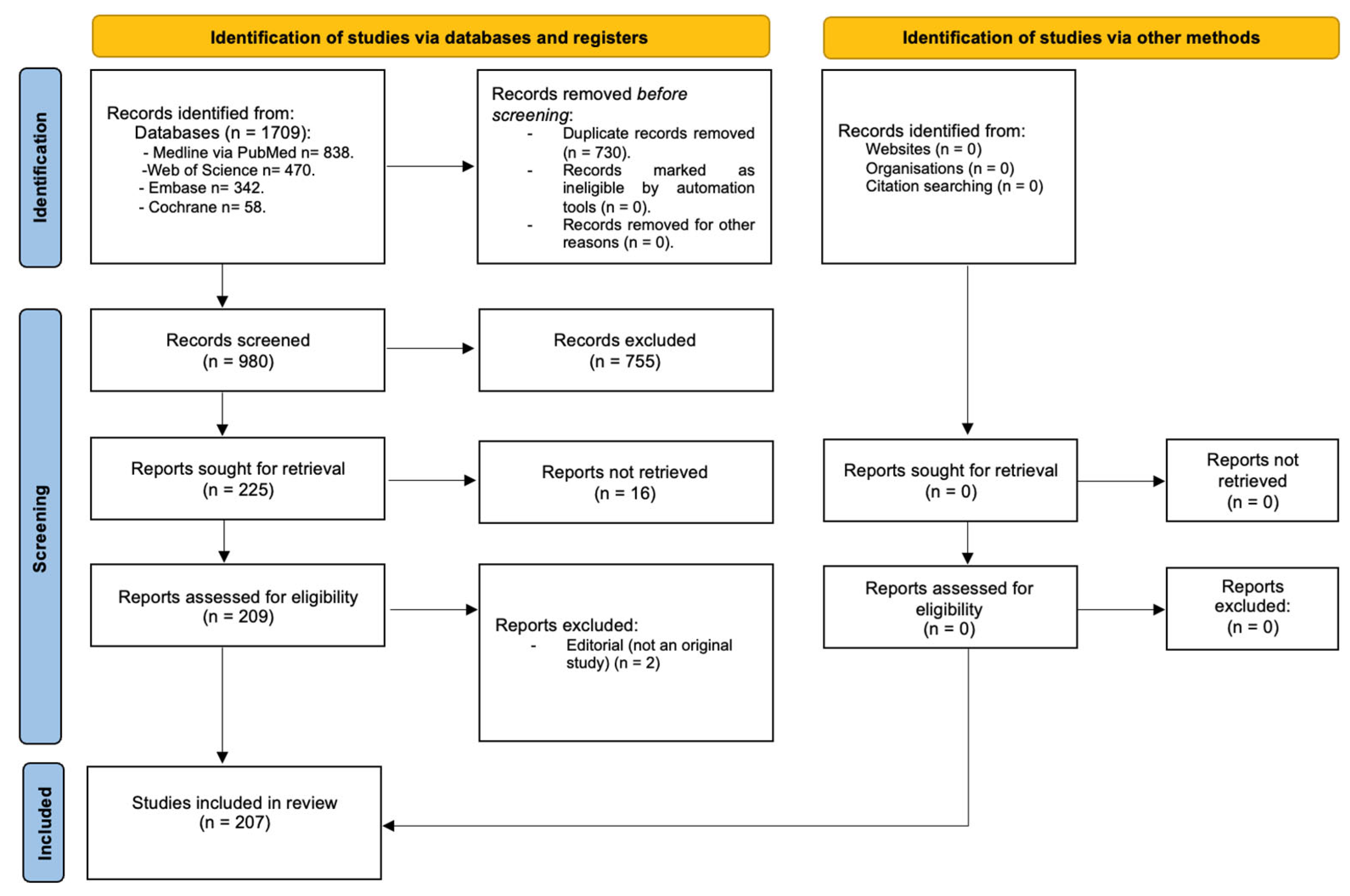
The initial search across the various databases yielded 1709 studies. After identification and removal of 730 duplicates, the titles and abstracts of 980 studies were screened. Of these, 755 studies were excluded for not meeting the eligibility criteria. Subsequently, 225 potentially relevant references were retrieved and assessed in full text. The identification, screening and eligibility processes are presented in the PRISMA flow diagram (Figure 1).

3.2. Characteristics of the Included Studies

To describe the information included in this review, a quantitative analysis was conducted based on the following parameters: type of presurgical orthopaedic appliance used, study design, the top 10 journals in which the studies were published, and the number of publications per year (Table 1).
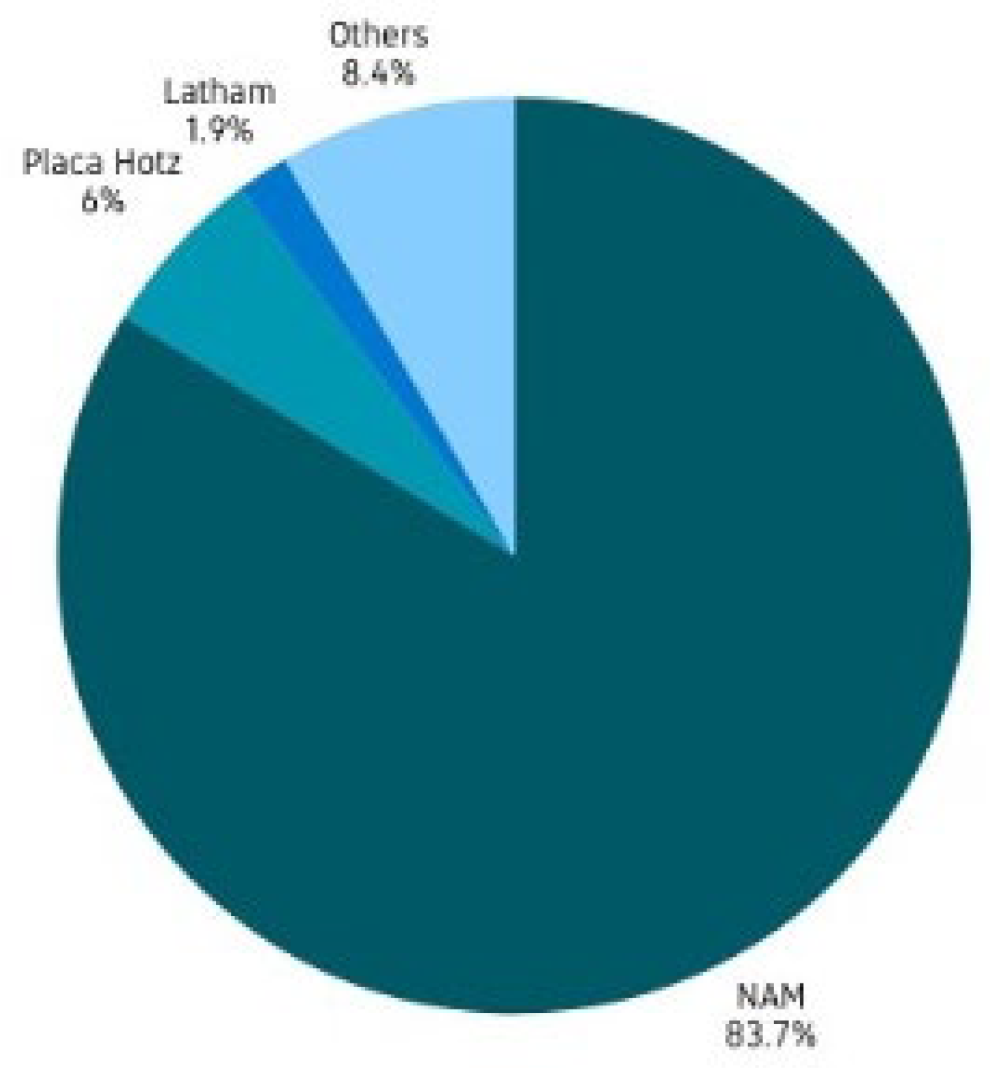
Regarding the types of appliances used, NAM was the most frequently reported, followed by the Hotz plate and the Latham appliance. Other appliances, such as DynaCleft®, as well as NAM derivatives including D-NAM, CAD-NAM and A-NAM, were also described. Figure 2 illustrates the percentage of references for each type of appliance reported in the literature.
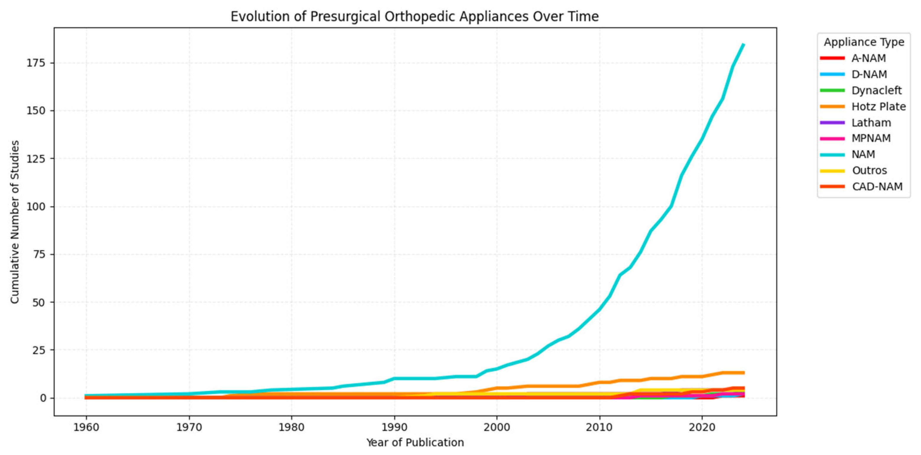
The temporal analysis of presurgical orthopedic appliances demonstrates a progressive increase in the number of publications over time, particularly after the year 2000 (Figure 3). Among the different techniques, nasoalveolar molding (NAM) emerges as the most frequently reported appliance.
Regarding study design, the following types of studies were identified: case reports (n = 56), case–control studies (n = 17), cross-sectional studies (n = 3), case series (n = 58), randomised controlled trials (n = 10), non-randomised controlled trials (n = 2) and cohort studies (n = 61). The included studies were also interpreted according to their level of evidence, with case reports and case series considered as lower-level evidence, cohort and case–control studies as intermediate-level evidence, and randomized controlled trials as higher-level evidence.
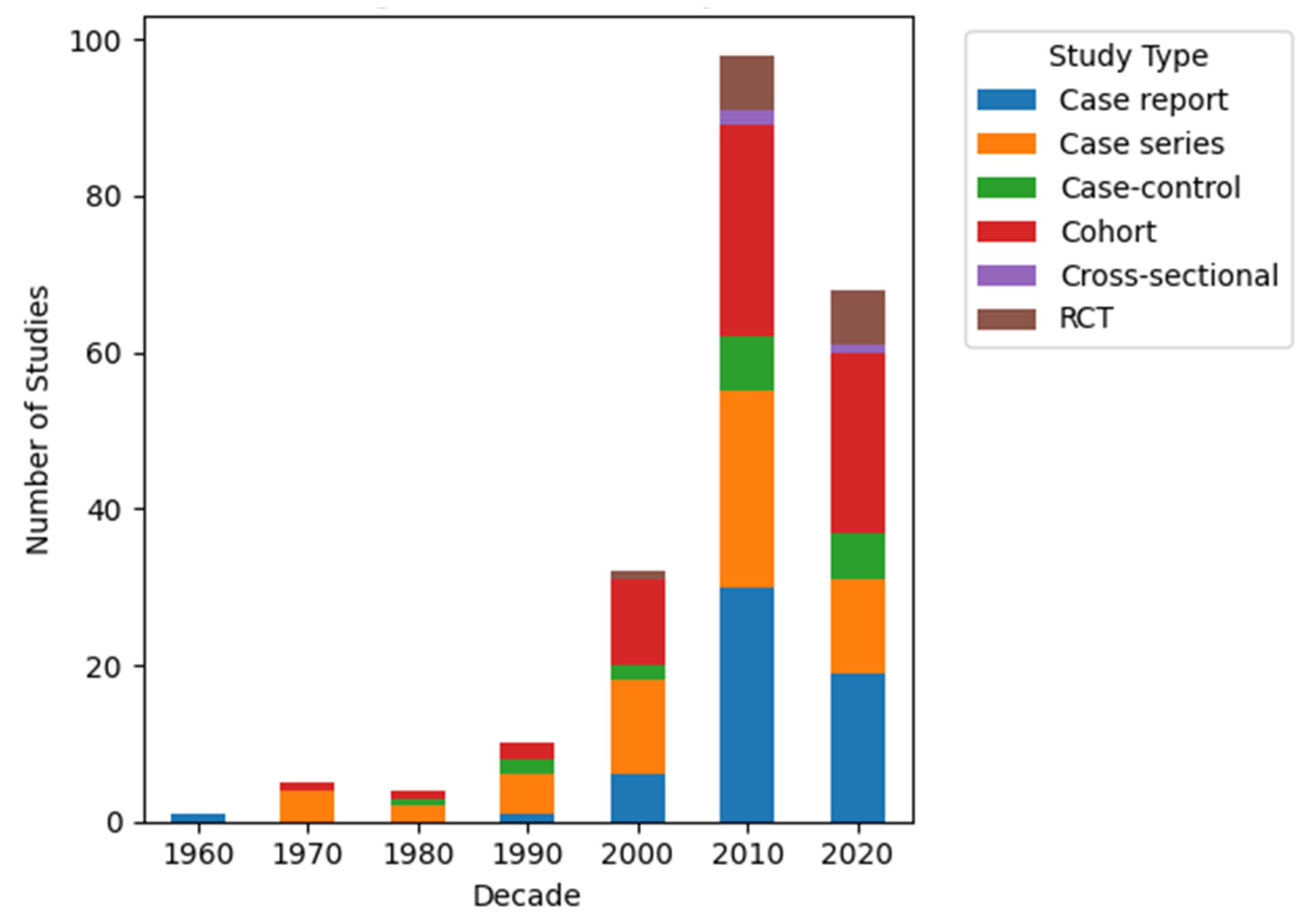
A decade-based analysis was subsequently performed to explore the temporal distribution of the included studies (Figure 4). Overall, a progressive increase in publications was observed, with the highest number of studies identified in the 2010s (n = 98), followed by the 2020s (n = 68), whereas earlier decades showed considerably lower scientific output. An increase in publication rates on this topic has been observed since 2012, with the peak occurring in 2023 (n = 18), representing 8.70% of the included studies.
This increase was mainly driven by observational study designs. In the most recent decades (2010s–2020s), cohort studies (n = 50), case reports (n = 49), and case series (n = 37) were the most frequently reported designs, while randomised controlled trials remained limited (n = 14). These findings indicate that, despite the growth in publication volume, the evidence base continues to be predominantly composed of lower-level study designs. Of the 207 articles included, The Cleft Palate–Craniofacial Journal was the journal with the highest number of publications (n = 39).
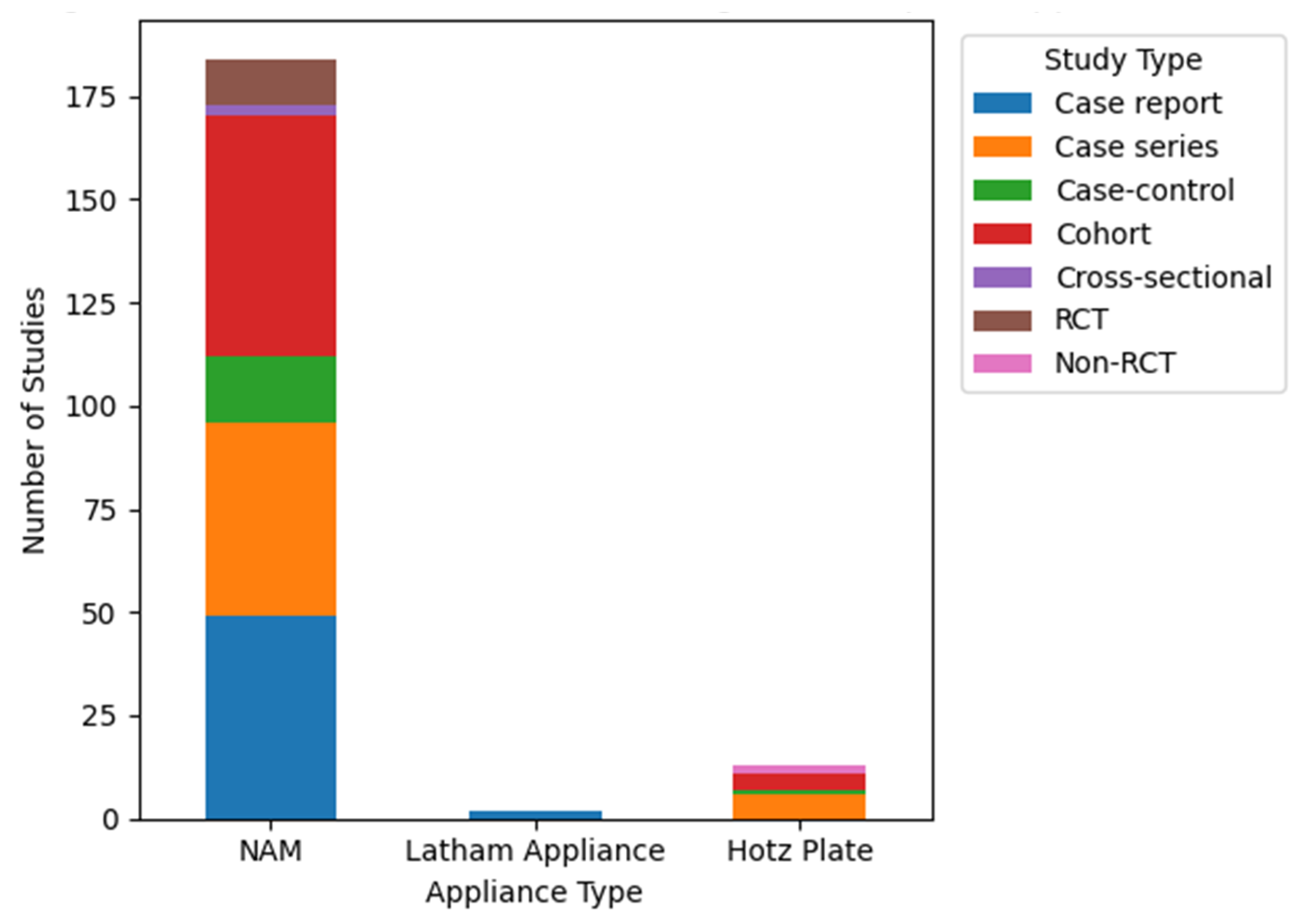
The analysis of study design distribution across the three most frequently reported presurgical orthopedic appliances—NAM, Latham appliance, and Hotz plate—revealed distinct patterns in the type of evidence supporting each technique (Figure 5). NAM was associated with the highest number of studies (n = 184) and the greatest diversity of study designs, including case reports (n = 49), case series (n = 47), case–control studies (n = 16), cohort studies (n = 58), cross-sectional studies (n = 3), and randomized controlled trials (n = 11). In contrast, the Latham appliance (n = 4) and Hotz plate (n = 13) were supported by fewer studies overall and a more limited range of study designs. For the Latham appliance, the evidence was primarily based on case reports (n = 2), with minimal representation of other study types. Similarly, the Hotz plate was mainly supported by case series (n = 6) and case–control studies (n = 1), with only a small number of cohort studies (n = 4) and non-randomized controlled trials (n = 2), and no cross-sectional studies identified.

3.2.1. Nasoalveolar Molding

NAM was reported in 184 of the included articles, which described a variety of protocols adopted by different research centres. The sample of 184 articles comprised 49 case reports, 58 cohort studies, 47 case series, 16 case–control studies, 9 randomised controlled trials, 2 non-randomised controlled trials and 3 cross-sectional studies. NAM consists of a passive palatal plate used for alignment of the maxillary arch and guidance of growth, to which a nasal stent may be attached to allow active modulation of the nasal cartilage [21]. Although the passive acrylic plate base was similar in most studies, following the Grayson protocol, greater variability was observed in active nasal moulding. The nasal stent, intended to improve nasal symmetry, elongate the columella and facilitate nasal projection, is attached to the appliance via a retention button positioned on the most anterior portion of the palatal plate, on the cleft side, in order to direct the molding effects towards the collapsed nostril. In cases of bilateral cleft lip and palate, Magyar et al. [22], Meazzini et al. [23] and Altug [24] suggested the use of two nasal stents.
The literature reports a wide range of adaptations to the design of NAM appliances. In bilateral clefts, particular variants include MPNAM [25] (modified NAM with a traction screw), which incorporates an active component for retraction and centralisation of the premaxilla, and A-NAM [26], which integrates a three-dimensional expansion screw allowing controlled retraction of the premaxilla and simultaneous expansion of the collapsed segments. In response to ongoing advances in the fabrication of medical and dental devices, CAD-NAM [27,28,29] has emerged as a NAM appliance manufactured using CAD–CAM (Computer-Aided Design) technology. Although production is facilitated by this technology, it may involve either conventional techniques or the use of scanners for data acquisition. In most of the studies analysed, palatal plates and nasal stents were progressively modified through the addition or removal of resin.
Another approach described in the literature includes D-NAM, reported by El-Ghafour et al. [30], and iDNAM, described by Parakarn et al. [31], in which models are digitised, and the entire planning process is performed using software. This enables model segmentation and virtual construction of the appliance, which is subsequently fabricated by 3D printing using biocompatible resin for each treatment phase. In the D-NAM protocol, three consecutive plates are used, with nasal stents manually attached to the passive palatal plates during the second and third phases of treatment. In iDNAM, two plates are employed, with the nasal stent attached during the second phase to promote active moulding of the nasal cartilage [31].
The complications most frequently associated with NAM use, as described in the literature, include irritation of the oral mucosa, gingival tissue and nasal mucosa, possibly resulting from the forces generated by the active components of the appliance [32]. In addition, the development of fungal infections has been reported, often related to inadequate hygiene practices or continuous appliance use. To minimise this complication, caregivers should be instructed to remove the appliance daily for cleaning [33,34].
To reduce complications associated with the NAM protocol, regular clinical follow-up is recommended, with review appointments conducted weekly or fortnightly, depending on individual clinical progression. These appointments aim to allow careful inspection of the mucosae, adjustment of the forces applied by the appliance, and selective grinding to enable controlled movement of the segments, as well as resin additions to guide the desired growth [32,35,36]. It should also be noted that NAM application should preferably occur before three months of age, a period during which tissue plasticity is increased due to the influence of maternal oestrogen, thereby enhancing treatment effectiveness [37,38,39].

3.2.2. Latham Appliance

Regarding the Latham appliance, four studies were identified in the literature, including one case–control study, two case reports and one cohort study. The Latham appliance is a fixed, intraoral device with active mechanics [40,41,42], used in newborns with cleft lip and palate, particularly in cases of bilateral clefts with premaxillary protrusion. Its application takes place prior to primary lip surgery, with the main objective of aligning the maxillary segments in order to optimise anatomical conditions for surgical repair. Structurally, the appliance consists of acrylic pads supported on the maxillary segments and connected by a transverse expansion screw, which allows controlled mobilisation of the osseous segments [40,43].
The premaxilla is retracted posteriorly by means of elastics connected to a metal pin previously fixed to the bone located posterior to the premaxilla [44]. The Latham appliance has been shown to be effective in repositioning the premaxilla and improving initial facial aesthetics, even in more severe cases. However, although the application of active forces to the maxillary segments may be beneficial for cleft closure, the generation of tissue tension may interfere with maxillary growth; therefore, its indication should be carefully evaluated [40,44]. Due to its active nature, the Latham appliance requires rigorous clinical follow-up with preferably weekly assessments [41,42]. Activation of the expansion screw is performed daily by caregivers on an outpatient basis, with continuous monitoring to ensure appropriate progression of expansion. In the presence of signs of irritation, ulceration or excessive premaxillary retraction, the clinician should reduce the applied forces [45].

3.2.3. Hotz Plate

With respect to the Hotz plate, thirteen studies were identified in the literature, including six case series, four cohort studies, two non-randomised controlled studies and one case–control study. The Hotz plate is a presurgical orthopaedic device applied during the first days to weeks of life. It consists of a rigid acrylic structure which, in certain modified protocols, may include areas of soft acrylic in order to improve comfort and intraoral adaptation [46]. By fully covering the palate, the plate prevents interposition of the tongue between the cleft segments, thereby promoting their spontaneous approximation [47]. It is characterised by a passive mechanism of action, functioning exclusively through containment and guidance of the physiological growth of the maxillary structures.
In 1976, Hotz and Gnoinski demonstrated that use of the plate could reduce the width of the alveolar and palatal clefts, facilitate repositioning of the premaxilla, preserve transverse maxillary growth until the first corrective surgery, and improve feeding in the newborn [48].
Follow-up of this appliance is typically performed on a weekly basis to assess device adaptation and detect the presence of soft tissue lesions [49,50]. In parallel, functional evaluation of the appliance should be carried out, particularly by observing tongue position, which should be prevented from interposing between the palatal segments, as well as by assessing improvements in sucking and swallowing with the plate in place [49,51]. Every 4 to 6 weeks, selective grinding of the acrylic structure is performed, including relief of pressure areas, with the aim of promoting alignment of the maxillary segments and appropriately guiding bone growth [52,53]. The plate should be worn continuously, 24 h per day, and removed only after each feeding for cleaning, which should be performed by gentle brushing using warm water [48].
Compared with nasoalveolar moulding, the Hotz plate has a simpler structural design, as it does not incorporate nasal stents nor apply traction forces to the soft tissues or nasal cartilages. Consequently, it is considered a less invasive and more technically accessible approach, although with limited effectiveness in correcting nasal asymmetry [54].

3.3. Comparative Analysis of Presurgical Orthopedic Protocols and Clinical Variability

A comparative analysis of the most frequently reported presurgical orthopedic appliances—NAM, Latham appliance, and Hotz plate—revealed substantial differences in treatment protocols, reflecting the heterogeneity identified in the extracted data. NAM was consistently described as a combined approach involving both alveolar molding and active nasal cartilage modulation, typically initiated within the first weeks of life and requiring frequent adjustments. In contrast, the Hotz plate was predominantly used as a passive appliance aimed at guiding natural maxillary growth, with fewer modifications over time and limited emphasis on nasal correction. The Latham appliance, on the other hand, was characterised by the application of semi-active mechanical forces through expansion systems, often involving a more invasive approach [40,41,42,43,44]. These differences extended to treatment duration, activation protocols, and follow-up schedules, highlighting the absence of standardized clinical pathways across appliances.
Variability was also evident in the outcomes reported across studies. While most investigations focused on morphological changes—such as reduction of alveolar cleft width, improvement in nasal symmetry, and premaxillary repositioning—the selection and measurement of these outcomes were inconsistent. Studies involving NAM more frequently reported combined nasal and alveolar improvements [21] whereas those assessing the Hotz plate tended to emphasise maxillary growth guidance [46,47,48], and Latham appliance studies focused primarily on premaxillary repositioning and arch alignment [40,44]. The lack of uniform outcome definitions and assessment methods limited direct comparability between appliances and constrained the ability to synthesise consistent patterns of effectiveness.
In addition, notable inconsistencies were identified in clinical indications and decision-making criteria. Although early intervention was generally recommended, the timing of treatment initiation, criteria for appliance selection, and use of adjunctive components—such as nasal stents—varied considerably between studies. For example, treatment initiation ranged from the first days of life to up to three months of age [37,38,39], while nasal stents were introduced either immediately at appliance insertion or delayed until the alveolar cleft width was reduced to approximately 5 mm [55,56]. Similarly, appliance selection was variably based on cleft severity, presence of premaxillary protrusion, or institutional preference [40,44], rather than standardized criteria. Follow-up frequency also differed across protocols, ranging from weekly to fortnightly adjustments [32,35,36,41,42]. Differences in cleft severity, patient selection, and institutional protocols further contributed to this variability. Taken together, these findings suggest that the observed differences across appliances are not solely related to their mechanical characteristics, but also reflect broader disparities in clinical practice, highlighting the need for greater standardization in both treatment protocols and outcome reporting.

4. Discussion

The present investigation aimed to identify, distinguish and analyse the presurgical orthopaedic treatment options applied in patients with CLP through the development of a scoping review. The results demonstrate the existence of multiple presurgical orthopaedic protocols, including devices such as NAM and its variations, the Latham appliance, the Hotz plate, and DynaCleft®, as well as institutional approaches developed based on the clinical experience of different centres. This methodological diversity reflects the absence of a universally accepted protocol, thereby reinforcing the relevance of the present review. Among the identified devices, NAM is the most widely used and documented, being consistently associated with improved aesthetic and functional outcomes, which justifies its predominance in current clinical practice.
The literature reviewed demonstrates consensus regarding the importance of early appliance initiation, ideally within the first weeks of life, to maximise its effects. When this is not feasible, application up to three months of age is recommended, a period during which the high plasticity of the nasal tissues—resulting from maternal oestrogen levels and the consequent increase in hyaluronic acid in neonatal cartilages—allows effective modulation of anatomical structures [37,38,39].
Regarding the introduction of the nasal stent for modulation of the nasal cartilages, most studies recommend its application after reduction of the alveolar cleft to a width of 5 mm or less [55]. However, some authors advocate immediate introduction of the nasal stent concomitantly with the initiation of NAM, within the first days of life. Jahanbin et al., in a case–control study, reported improved outcomes in reducing nostril width on the cleft side using this early approach [56]. Conversely, Altug et al. and Rau et al. warned of the risk of nostril widening when nasal modulation is initiated prior to approximation of the alveolar segments, emphasising the need for protocol individualisation according to the initial morphology of the defect [24,57].
The primary benefit of NAM treatment lies in the reduction of the alveolar cleft prior to cheiloplasty, allowing minimisation of tissue tension and, consequently, the formation of visible scarring [58,59,60,61,62,63,64,65]. Additional benefits described include columella elongation, improved nasal symmetry and projection [66,67,68], retraction and rotation of the premaxilla, and modulation and alignment of the maxillary arch [22,57,69,70,71]. In cases of bilateral CLP, centralisation of the premaxilla is observed. Studies such as those by Saad et al. and Haddad et al. reported reductions in alveolar cleft width of 57% and 70%, respectively, associated with significant increases in columella length [60,72]. As a relatively low-cost approach, NAM is particularly valuable in countries with a high incidence of clefts, where geographical distance and financial constraints may limit access to specialised care [73]. In addition to functional and aesthetic benefits, the appliance contributes to the psychological well-being of the child and family by improving perception of treatment from an early stage [73,74].
Several comparative studies have demonstrated that NAM, when compared with control groups without any presurgical intervention prior to cheiloplasty, provides superior functional and aesthetic outcomes, namely in maxillary arch alignment, reduction of alveolar and labial clefts, premaxillary rotation, correction of the facial midline and, consequently, nasal symmetry, as well as a reduction in scarring and the need for future surgical interventions [75,76,77,78,79,80].
In comparison with other presurgical orthopaedic appliances, such as the Hotz plate, a passive device, NAM has been shown to be more effective in maxillary alignment, modulation of the nasal cartilages, columella projection and correction of the collapsed nostril [46,47,54,81]. The Latham appliance, as a semi-active device, demonstrates favourable outcomes in maxillary arch alignment, premaxillary retraction and centralisation; however, it presents significant limitations, including the need for anaesthesia for placement [40,44,82], its invasive nature, and the potential negative impact on maxillary growth, which may increase the need for future orthognathic surgery [56]. Beyond these technical differences, broader variability in clinical practice was also observed. The variability identified across treatment protocols, outcome measures, and clinical indications further highlights the challenges in comparing presurgical orthopedic approaches. This heterogeneity suggests that current clinical practices are largely influenced by institutional experience and practitioner preference rather than standardized, evidence-based guidelines.
Despite the benefits, NAM presents certain limitations. Its effectiveness depends on parental cooperation, requiring frequent clinic visits and strict adherence to daily instructions [36,47,83]. Achieving satisfactory outcomes requires the involvement of a specialised team, as application errors, such as excessive or asymmetric forces, may induce severe distortions, including nasal asymmetries, columellar deviation, excessive premaxillary retraction and impairment of maxillary growth, potentially aggravating the initial condition [47,53]. An important aspect influencing treatment outcomes is the magnitude and distribution of forces applied by different presurgical orthopedic appliances. Passive devices, such as the Hotz plate, rely primarily on guiding natural growth with minimal mechanical stress, whereas NAM combines passive alveolar molding with controlled active nasal cartilage shaping. In contrast, the Latham appliance generates continuous active forces through expansion screws and elastics, which may induce higher levels of force in the maxillary segments.
Although no standardized quantitative thresholds for “optimal force” are defined in the literature, appropriate force application is generally characterised by gradual tissue adaptation without signs of ischemia, ulceration, or excessive segment displacement. Excessive or asymmetric forces, particularly in NAM or Latham protocols, may lead to complications such as tissue distortion, premaxillary over-retraction, or impaired maxillary growth. Therefore, careful clinical monitoring and progressive adjustment remain essential to ensure safe and effective treatment.
The available literature on presurgical orthopaedics is predominantly composed of case reports and case series, which may be explained by the ethical and logistical limitations associated with conducting controlled clinical trials in neonatal populations. A progressive increase in publications has been observed since the year 2000, reflecting growing scientific interest in this therapeutic approach. Nevertheless, there remains a scarcity of multicenter studies with representative samples and robust methodologies. The concentration of publications in specialised journals, such as The Cleft Palate–Craniofacial Journal, indicates that this is a specialised field, albeit one with expanding impact and visibility. This trend highlights the need to strengthen scientific evidence through prospective, controlled studies with a lower risk of bias. These trends may be explained by several underlying factors. Cleft lip and palate, as a relatively low-prevalence craniofacial condition, often results in limited patient numbers per center, thereby restricting the feasibility of studies with large sample sizes and adequate statistical power. Additionally, the early timing of intervention in neonatal patients introduces important ethical constraints, particularly regarding the use of randomized controlled designs or untreated control groups. Evidence from European cross-sectional surveys, further highlights considerable variability in clinical protocols, resource availability, and multidisciplinary team organization across centers. This heterogeneity, combined with differences in clinical expertise and infrastructure, poses significant challenges to standardization and limits the implementation of multicenter or controlled studies [84,85].
Among the limitations of this study, the heterogeneity of the included studies stands out, as evidenced by variability in applied protocols, ages at treatment initiation and criteria used to evaluate outcomes. Additionally, the predominance of studies with low levels of evidence limits the generalisability of the findings and restricts the robustness of the conclusions. Another limiting factor is the scarcity of multicenter and randomised studies, which hinders objective validation and comparison of the protocols used. Despite these limitations, this review provides a comprehensive overview of current practices, contributing to the justification for adopting more uniform and rigorous protocols. Most available studies do not include a control group, representing a significant limitation, as it complicates clear distinction between the effects of orthopaedic appliances and changes resulting from natural growth. However, withholding treatment from newborns with CLP may be considered unethical, which justifies the limited number of controlled studies in this population. The main clinical implication lies in recognising NAM as a safe tool for presurgical orthopaedics in newborns with CLP. Early application and individualised appliance design are decisive factors in achieving improved aesthetic and functional outcomes. The predominance of NAM in the literature should be interpreted with caution, as it may reflect publication bias, institutional expertise, and historical adoption patterns rather than clear evidence of superiority over other appliances. Additionally, differences in training and resource availability across centres may influence the selection of specific techniques.
Although a formal risk-of-bias assessment was not conducted, the included studies can be broadly interpreted according to their level of evidence. The predominance of case reports and case series, compared to the relatively limited number of randomized controlled trials, indicates that the current evidence base is weighted toward lower levels of evidence, with fewer studies providing higher-level support. Therefore, the results of this review should be interpreted with caution, as they reflect the distribution and characteristics of the existing literature rather than the effectiveness or quality of the evidence.
It should be noted that the descriptive nature of the presented data, including the graphical representations, reflects the primary objective of this scoping review, which is to map the distribution of presurgical orthopedic approaches rather than to assess clinical outcomes or the strength of evidence. The predominance of case reports, case series, and observational studies within the included literature has important implications for the interpretation of findings. These study designs, while valuable for describing clinical approaches and generating hypotheses, are inherently limited in their ability to establish causal relationships or provide high-level evidence on treatment effectiveness. Consequently, the apparent benefits associated with presurgical orthopedic interventions, particularly NAM, should be interpreted with caution, as they may reflect descriptive trends rather than robust comparative evidence. This limitation underscores the need for well-designed prospective and controlled studies to support more definitive clinical recommendations.
Future research should focus on conducting multicenter studies with representative samples and standardised protocols, allowing more robust evaluation of short- and long-term outcomes.

5. Conclusions

This scoping review maps current presurgical orthopedic practices and identifies consistent trends toward improved morphological outcomes with early intervention. The predominance of NAM reflects research activity and clinical adoption rather than clear evidence of superiority. The evidence base is largely composed of lower-level study designs, limiting robust comparisons and definitive conclusions. These findings highlight substantial variability across protocols and underscore the need for standardized approaches and higher-quality prospective research.

Author Contributions

Conceptualization, I.F. and F.V.; methodology, R.T., C.M.M. and A.B.P.; software, C.M.M.; validation, R.T., C.O. and C.N.; formal analysis, A.B.P. and C.M.M.; investigation, A.C.M. and I.F.; resources, A.B.P.; data curation, R.T. and C.N.; writing—original draft preparation, A.C.M., C.N. and C.O.; writing—review and editing, I.F., F.V. and A.B.P.; visualization, A.C.M.; supervision, F.V. and I.F. All authors have read and agreed to the published version of the manuscript.

Funding

This research is sponsored by national funds through FCT—Fundação para a Ciência e a Tecnologia, under projects UID/00285/2025 and LA/P/0112/2020.

Institutional Review Board Statement

Not applicable.

Informed Consent Statement

Not applicable.

Data Availability Statement

No new data were created or analyzed in this study. Data sharing is not applicable to this article.

Conflicts of Interest

The authors declare no conflicts of interest.

Abbreviations

The following abbreviations are used in this manuscript:
CAD-CAMComputer-Aided Design technology
CLPCleft lip and/or palate
NAMNasoalveolar molding
PCCPopulation, Concept, Context
PRISMA-ScRPreferred Reporting Items for Systematic Reviews and Meta-Analyses extension for Scoping Reviews

Appendix A

Table A1. Search strategy for data bases.
Data BaseSearch Strategy
Medline via PubMed (“Cleft Palate”[MeSH] OR palatischisis OR palatoschisis OR palatoschizis OR “Cleft* Palat*” OR “Palat*, Cleft*” OR “Cleft Lip”[MeSH] OR cheiloschisis OR labioschisis OR labioschizis OR “Lip cleft*” OR “Lips Cleft*” OR “Cleft* Lip” OR “Cleft* Lips” OR “Lip, Cleft*” OR “Lips, Cleft*” OR Harelip* OR “hare lip” OR “hare lips” OR “oral cleft*” OR “orofacial cleft*” OR “cleft* maxilla palate” OR “palatal cleft*” OR “palatum fissum”) AND (“Orthopedic Procedures”[MeSH] OR “orthopedic procedure*” OR “procedure*, orthopedic” OR “palatal obturators”[MeSH] OR “Obturator*, Palat*” OR “Palat* Obturator*” OR “Palat* Prosthes*” OR “speech aid prosthes*” OR “Molding Device*, Nasoalveolar” OR “Nasoalveolar Moding”[MeSH] OR “Nasoalveolar Molding” OR “Naso-alveolar Molding” OR “Molding, Nasoalveolar” OR “NAM Therap*” OR “Therap*, NAM” OR “Nasal Alveolar Molding” OR “Molding, Nasal Alveolar” OR “nasoalveolar (NAM) technique”) AND (presurg* OR pre-surg* OR “Preoperative Period”[Mesh] OR “Preoperative Care”[Mesh] OR preoperative OR pre-operative)
Embase(‘cleft palate’/exp OR ‘palatischisis’ OR ‘palatoschisis’ OR ‘palatoschizis’ OR ‘cleft* palat*’ OR ‘palat*, cleft*’ OR ‘cleft lip’/exp OR ‘cheiloschisis’ OR ‘labioschisis’ OR ‘labioschizis’ OR ‘lip* cleft’ OR ‘cleft* lip*’ OR ‘lip*, cleft*’ OR ‘harelip*’ OR ‘hare lip*’ OR ‘oral cleft*’ OR ‘orofacial cleft*’ OR ‘cleft* maxilla palate’ OR ‘palatal cleft*’ OR ‘palatum fissum’) AND (‘orthopedic procedure*’ OR ‘procedure*, orthopedic’ OR ‘palatal obturator’/exp OR ‘obturator*, palat*’ OR ‘palat* obturator*’ OR ‘palat* prosthes*’ OR ‘speech aid prosthes*’ OR ‘molding device*, nasoalveolar’ OR ‘nasoalveolar molding’/exp OR ‘naso-alveolar molding’ OR ‘nasoalveolar molding’ OR ‘molding, nasoalveolar’ OR ‘nam therap*’ OR ‘therap*, nam’ OR ‘nasal alveolar molding’ OR ‘molding, nasal alveolar’ OR ‘nasoalveolar (nam) technique’) AND (‘preoperative period’/exp OR ‘presurg*’ OR ‘pre-surg*’ OR ‘preoperative care’/exp OR ‘preoperative treatment’/exp OR ‘preoperative’ OR ‘pre-operative’) AND ([article]/lim OR [article in press]/lim OR [data papers]/lim OR [letter]/lim OR [review]/lim OR [short survey]/lim)
Cochrane#1 MeSH descriptor: [Cleft Palate] explode all trees
#2 palatischisis
#3 palatoschisis
#4 palatoschizis
#5 (cleft* NEXT palat*)
#6 (palat* NEXT cleft*)
#7 MeSH descriptor: [Cleft Lip] explode all trees
#8 cheiloschisis
#9 labioschisis
#10 labioschizis
#11 (lip* NEXT cleft*)
#12 (cleft* NEXT lip*)
#13 harelip*
#14 (hare NEXT lip*)
#15 (oral NEXT cleft*)
#16 (orofacial NEXT cleft*)
#17 (cleft* NEXT maxilla palate)
#18 “palatum fissum”
#19 MeSH descriptor: [Orthopedic Procedures] explode all trees
#20 (orthopedic NEXT procedure*)
#21 (procedure* NEXT orthopedic)
#22 MeSH descriptor: [Palatal Obturators] explode all trees
#23 (obturator* NEXT palat*)
#24 (palat* NEXT obturator*)
#25 (palat* NEXT prosthes*)
#26 (speech aid NEXT prosthes*)
#27 MeSH descriptor: [Nasoalveolar Molding] explode all trees
#28 (molding device* NEXT nasoalveolar)
#29 “nasoalveolar molding”
#30 “naso-alveolar molding”
#31 “molding, nasoalveolar”
#32 (NAM NEXT therap*)
#33 (therap* NEXT NAM)
#34 “nasal alveolar molding”
#35 “molding, nasal alveolar”
#36 “nasoalveolar (NAM) technique”
#37 MeSH descriptor: [Preoperative Period] explode all trees
#38 MeSH descriptor: [Preoperative Care] explode all trees
#39 presurg*
#40 pre-surg*
#41 preoperative
#42 pre-operative
#43 #1 OR #2 OR #3 OR #4 OR #5 OR #6 OR #7 OR #8 OR #9 OR #10 OR #11 OR #12 OR #13 OR #14 OR #15 OR #16 OR #17 OR #18
#44 #19 OR #20 OR #21 OR #22 OR #23 OR #24 OR #25 OR #26 OR #27 OR #28 OR #29 OR #30 OR #31 OR #32 OR #33 OR #34 OR #35 OR #36
#45 #37 OR #38 OR #39 OR #40 OR #41 OR #42
#46 #43 AND #44 AND #45
Web of Science All Databases(palatischisis OR palatoschisis OR palatoschizis OR “Cleft* Palat*” OR “Palat*, Cleft*” OR cheiloschisis OR labioschisis OR labioschizis OR “Lip cleft*” OR “Lips Cleft*” OR “Cleft* Lip” OR “Cleft* Lips” OR “Lip, Cleft*” OR “Lips, Cleft*” OR Harelip* OR “hare lip” OR “hare lips” OR “oral cleft*” OR “orofacial cleft*” OR “cleft* maxilla palate” OR “palatal cleft*” OR “palatum fissum”) AND (“orthopedic procedure*” OR “procedure*, orthopedic” OR “Obturator*, Palat*” OR “Palat* Obturator*” OR “Palat* Prosthes*” OR “speech aid prosthes*” OR “Molding Device*, Nasoalveolar” OR “Nasoalveolar Molding” OR “Naso-alveolar Molding” OR “Molding, Nasoalveolar” OR “NAM Therap*” OR “Therap*, NAM” OR “Nasal Alveolar Molding” OR “Molding, Nasal Alveolar” OR “nasoalveolar (NAM) technique”) AND (presurg* OR pre-surg* OR preoperative OR pre-operative)

References

  1. Vieira, A.R.; Orioli, I.M. Birth order and oral clefts: A meta analysis. Teratology 2002, 66, 209–216. [Google Scholar] [CrossRef] [PubMed]
  2. Ma, Q.; Conley, R.; Wu, T.; Li, H. Interdisciplinary treatment for an adult with a unilateral cleft lip and palate. Am. J. Orthod. Dentofac. Orthop. 2014, 146, 238–248. [Google Scholar] [CrossRef]
  3. Tolarová, M.M.; Cervenka, J. Classification and birth prevalence of orofacial clefts. Am. J. Med. Genet. 1998, 75, 126–137. [Google Scholar] [CrossRef]
  4. Mossey, P.A.; Little, J.; Munger, R.G.; Dixon, M.J.; Shaw, W.C. Cleft lip and palate. Lancet 2009, 374, 1773–1785. [Google Scholar] [CrossRef]
  5. Gundlach, K.K.H.; Maus, C. Epidemiological studies on the frequency of clefts in Europe and world-wide. J. Craniomaxillofac. Surg. 2006, 34, 1–2. [Google Scholar] [CrossRef]
  6. Kamble, V.D.; Parkhedkar, R.D.; Sarin, S.P.; Patil, P.G. Presurgical nasoalveolar molding (PNAM) for a unilateral cleft lip and palate: A clinical report. J. Prosthodont. 2013, 22, 74–80. [Google Scholar] [CrossRef]
  7. Thair, A.; Abid, M.; Dziedzic, A. Caregivers’ experience with lip taping as a presurgical orthopedic treatment for cleft lip and palate defects. Children 2024, 11, 332. [Google Scholar] [CrossRef]
  8. Bahaa, A.; El-Bagoury, N.; Khaled, N.; Mohamed, S.; Bahaa, A.; Ibrahim, A.M.; Taha, K.M.; Abdarrazik, M.A. The nasoalveolar molding technique versus DynaCleft nasal elevator application in infants with unilateral cleft lip and palate. Arch. Craniofac. Surg. 2024, 25, 123–132. [Google Scholar] [CrossRef]
  9. Artuza-Rosado, G.; Argueta-Figueroa, L.; Bautista-Hernández, M.A.; Torres-Rosas, R. Evidence of the effectiveness of the use of presurgical orthopedic appliances in patients with cleft lip and palate: A systematic review. Investig. Clin. 2023, 64, 81–107. [Google Scholar] [CrossRef]
  10. Weismann, C.; Xepapadeas, A.B.; Bockstedte, M.; Koos, B.; Krimmel, M.; Poets, C.F.; Aretxabaleta, M. Complete digital workflow for manufacturing presurgical orthodontic palatal plates in newborns and infants with cleft lip and/or palate. J. Funct. Biomater. 2024, 15, 301. [Google Scholar] [CrossRef] [PubMed]
  11. Celikoglu, M.; Buyuk, S.; Sekerci, A.; Ersoz, M.; Celik, S.; Sisman, Y. Facial soft-tissue thickness in patients affected by bilateral cleft lip and palate: A retrospective cone-beam computed tomography study. Am. J. Orthod. Dentofac. Orthop. 2014, 146, 573–578. [Google Scholar] [CrossRef]
  12. Wermker, K.; Jung, S.; Joos, U.; Kleinheinz, J. Dental implants in cleft lip, alveolus, and palate patients: A systematic review. Int. J. Oral Maxillofac. Implant. 2014, 29, 384–390. [Google Scholar] [CrossRef]
  13. Paranaiba, L.M.R.; Coletta, R.D.; Swerts, M.S.O.; Quintino, R.P.; de Barros, L.M.; Martelli-Junior, H. Prevalence of dental anomalies in patients with nonsyndromic cleft lip and/or palate in a Brazilian population. Cleft Palate Craniofac. J. 2013, 50, 400–405. [Google Scholar] [CrossRef]
  14. Allareddy, V.; Shusterman, S.; Ross, E.; Palermo, V.; Ricalde, P. Dentofacial orthopedics for the cleft patient: The Latham approach. Oral Maxillofac. Surg. Clin. N. Am. 2020, 32, 187–196. [Google Scholar] [CrossRef]
  15. Grayson, B.H.; Maull, D. Nasoalveolar molding for infants born with clefts of the lip, alveolus, and palate. Clin. Plast. Surg. 2004, 31, 149–158. [Google Scholar] [CrossRef]
  16. Liceras Liceras, E.; Cano, F.M.; Díaz Moreno, E.; García Gómez, M.; López, A.E.; Muñoz Miguelsanz, M.A.; Plaza, A.M.; Morales, J.G.; Valadés, R.F. Moldeamiento nasoalveolar prequirúrgico em el niño com fissura labiopalatina. Cir. Pediatr. 2012, 25, 109–115. [Google Scholar]
  17. Bhutiani, N.; Tripathi, T.; Verma, M.; Bhandari, P.S.; Rai, P. Assessment of treatment outcome of presurgical nasoalveolar molding in patients with cleft lip and palate and its postsurgical stability. Cleft Palate Craniofac. J. 2020, 57, 700–706. [Google Scholar] [CrossRef]
  18. Van der Heijden, P.; Dijkstra, P.U.; Stellingsma, C.; van der Laan, B.F.; Korsten-Meijer, A.G.; Goorhuis-Brouwer, S.M. Limited evidence for the effect of presurgical nasoalveolar molding in unilateral cleft on nasal symmetry: A call for unified research. Plast. Reconstr. Surg. 2013, 131, 62e–71e. [Google Scholar] [CrossRef]
  19. Tricco, A.C.; Lillie, E.; Zarin, W.; O’Brien, K.K.; Colquhoun, H.; Levac, D.; Moher, D.; Peters, M.D.J.; Horsley, T.; Weeks, L.; et al. PRISMA extension for scoping reviews (PRISMA-ScR): Checklist and explanation. Ann. Intern. Med. 2018, 169, 467–473. [Google Scholar] [CrossRef]
  20. Peters, M.D.J.; Godfrey, C.; McInerney, P.; Munn, Z.; Tricco, A.C.; Khalil, H. Chapter 11: Scoping reviews. In JBI Manual for Evidence Synthesis; Aromataris, E., Munn, Z., Eds.; JBI: Adelaide, Australia, 2020. [Google Scholar] [CrossRef]
  21. Grayson, B.H.; Shetye, P.R. Presurgical nasoalveolar moulding treatment in cleft lip and palate patients. Indian J. Plast. Surg. 2009, 42, S56–S61. [Google Scholar]
  22. Magyar, D.; Pálvölgyi, L.; Swennen, G.; Shujaat, S.; Nagy, K. Alteration in maxillary arch dimensions following preoperative nasoalveolar moulding therapy in bilateral cleft lip and palate patients. Clin. Investig. Orthod. 2024, 83, 112–118. [Google Scholar] [CrossRef]
  23. Meazzini, M.C.; Parravicini, F.; Cohen, N.; Rossetti, G.; Autelitano, L. Nasoalveolar molding and skeletal development in patients with bilateral cleft lip and palate: A retrospective cephalometric study at the completion of growth. J. Craniomaxillofac. Surg. 2022, 50, 400–405. [Google Scholar] [CrossRef]
  24. Altuğ, A.T. Presurgical nasoalveolar molding of bilateral cleft lip and palate infants: An orthodontist’s point of view. Turk. J. Orthod. 2017, 30, 118–125. [Google Scholar] [CrossRef]
  25. Hao, J.; Wan, Q.; Liu, J.; Wu, W.; Liu, J.; Luo, C.; Gao, Z.; Li, F.; Chen, Y.; Lu, Z. Modified presurgical nasoalveolar molding for patients with neonatal complete bilateral cleft lip and palate having a severely malpositioned premaxilla. Am. J. Transl. Res. 2022, 14, 3988–3994. [Google Scholar]
  26. Bhutiani, N.; Tripathi, T.; Rai, P. Active nasoalveolar molding using 3-directional expansion screw for arch alignment in bilateral cleft lip and palate. Am. J. Orthod. Dentofac. Orthop. Clin. Companion 2022, 2, 20–25. [Google Scholar] [CrossRef]
  27. Zheng, J.; He, H.; Kuang, W.; Yuan, W. Presurgical nasoalveolar molding with 3D printing for a patient with unilateral cleft lip, alveolus, and palate. Am. J. Orthod. Dentofac. Orthop. 2019, 156, 412–419. [Google Scholar] [CrossRef]
  28. Yu, Q.; Gong, X.; Shen, G. CAD presurgical nasoalveolar molding effects on the maxillary morphology in infants with UCLP. Oral Surg. Oral Med. Oral Pathol. Oral Radiol. 2013, 116, 418–426. [Google Scholar] [CrossRef]
  29. Mejia, M.J.; Gomes, J.P.; Moon, S.L.; Wolfe, E.M.; Perlyn, C.A.; Wolfe, S.A.; Steinberg, J.P. 3D Infant Orthopedic Nasal Molding System for Improved Outcomes in Cleft Nasal Deformity. Face 2023, 4, 141–147. [Google Scholar] [CrossRef]
  30. Abd El-Ghafour, M.; Aboulhassan, M.A.; Salah Fayed, M.M.; El-Beialy, A.R.; Kamel Eid, F.H.; El-Din Hegab, S.; El-Gendi, M.; Emara, D. Effectiveness of a novel 3D-printed nasoalveolar molding appliance (D-NAM) on improving the maxillary arch dimensions in unilateral cleft lip and palate infants: A randomized controlled trial. Cleft Palate Craniofac. J. 2020, 57, 1370–1381. [Google Scholar] [CrossRef]
  31. Parakarn, H.; Pisek, P.; Wangsrimongkol, B. Individualized digital nasoalveolar molding: A proof of concept. J. Craniofac. Surg. 2024, 35, e584–e586. [Google Scholar] [CrossRef]
  32. Grayson, B.H.; Santiago, P.E.; Brecht, L.E.; Cutting, C.B. Presurgical nasoalveolar molding in infants with cleft lip and palate. Cleft Palate Craniofac. J. 1999, 36, 486–498. [Google Scholar] [CrossRef]
  33. Kalantar-Hormozi, A.; Ghalambor, A.; Tolouei, M. Investigating the surgical outcomes of nasoalveolar molding (NAM) approach in cleft lip and palate repairing operations. Int. J. Pharm. Res. Allied Sci. 2016, 5, 412–416. [Google Scholar]
  34. Murthy, P.S.; Venkatapathy, A.L.; Deshmukh, S. Effect of selective trimming of nasoalveolar molding on maxillary arch growth in patients with unilateral cleft lip and palate. Spec. Care Dent. 2024, 44, 1627–1632. [Google Scholar] [CrossRef]
  35. Rubin, M.S.; Clouston, S.A.P.; Esenlik, E.; Shetye, P.R.; Flores, R.L.; Grayson, B.H. Midface growth in patients with unilateral cleft lip and palate treated with a nasoalveolar molding protocol. J. Craniofac. Surg. 2019, 30, 1640–1643. [Google Scholar] [CrossRef]
  36. Abhinav, B.A.; Batra, P.; Sood, S.C.; Sharma, K.; Srivastava, A.; Raghavan, S. Comparative Study of Presurgical Infant Orthopedics by Modified Grayson Method and Dynacleft with Nasal Elevators in Patients with Unilateral Cleft Lip and Palate—A Clinical Prospective Study. Cleft Palate Craniofac. J. 2021, 58, 189–201. [Google Scholar] [CrossRef] [PubMed]
  37. Barillas, I.; Dec, W.; Warren, S.M.; Cutting, C.B.; Grayson, B.H. Nasoalveolar molding improves long-term nasal symmetry in complete unilateral cleft lip–cleft palate patients. Plast. Reconstr. Surg. 2009, 123, 1002–1006. [Google Scholar] [CrossRef]
  38. Sabir, S.; Azhari, M.; Rokhssi, H.; Merzouk, N.; Bentahar, O. Presurgical nasoalveolar molding: An advantageous adjunctive neonatal therapy for cleft lip and palate defects in 2 clinical cases. J. Pediatr. Neonatal Individ. Med. 2021, 10, e100249. [Google Scholar]
  39. Bhagat, R.; Vandekar, M.; Vaid, N.R.; Park, J.H.; Kook, Y.A. A staged expansion screw protocol to retract the premaxilla in BCLP infants with delayed nasoalveolar molding. J. Clin. Pediatr. Dent. 2021, 45, 123–128. [Google Scholar] [CrossRef] [PubMed]
  40. Romero, M.; Latham, R.; Romance, A.; Salvan, R. Treatment of an infant with a rare cleft resolved with use of an orthopedic appliance. Cleft Palate Craniofac. J. 2003, 40, 642–644. [Google Scholar] [CrossRef] [PubMed]
  41. Lim, J.S.; Park, J.H.; Jung, H.D.; Jung, Y.S. Sequential Use of Latham device and Presurgical Nasoalveolar Molding (PNAM) device in Unilateral Complete Cleft Lip and Palate: A Case Report. J. Korean Assoc. Cleft Lip Palate 2017, 20, 21–27. [Google Scholar]
  42. Cruz, C. Presurgical Orthopedics Appliance: The Latham Technique. Oral Maxillofac. Surg. Clin. N. Am. 2016, 28, 161–168. [Google Scholar] [CrossRef]
  43. Burak, M.Y.; Hernández, S.B.; Roa, M.L.S. Use of the modified Latham appliance prior to cheiloplasty. Rev. Mex. Ortod. 2014, 2, 231–238. [Google Scholar] [CrossRef]
  44. Schwenzer, K.; Holberg, C.; Hagenmaier, C.; Bacher, M.; Kunz, B.; Cornelius, C.P.; Schwenzer, N.; Ehrenfeld, M. Positioning of the premaxilla in cases of bilateral cleft lip, alveolus and palate using Latham’s appliance. Mund- Kiefer- Gesichtschir. 2003, 7, 25–30. [Google Scholar] [CrossRef] [PubMed]
  45. Ross, R.B. Treatment variables affecting facial growth in complete unilateral cleft lip and palate. Cleft Palate J. 1987, 24, 5–77. [Google Scholar] [PubMed]
  46. Cerón-Zapata, A.M.; López-Palacio, A.M.; Rodriguez-Ardila, M.J.; Berrío-Gutiérrez, L.M.; De Menezes, M.; Sforza, C. 3D evaluation of maxillary arches in unilateral cleft lip and palate patients treated with nasoalveolar moulding vs. Hotz’s plate. J. Oral Rehabil. 2016, 43, 111–118. [Google Scholar] [CrossRef]
  47. Isogawa, N.; Ochiai, S.; Mito, T.; Kindaichi, J.; Ishibashi, N.; Takagi, Y.; Ishikawa, M. Three-dimensional comparison in palatal forms between modified presurgical nasoalveolar molding plate and Hotz’s plate applied to the infants with unilateral cleft lip and palate. Singap. Dent. J. 2010, 31, 36–42. [Google Scholar] [CrossRef]
  48. Hotz, M.; Gnoinski, W. Comprehensive care of cleft lip and palate children at Zürich University: A preliminary report. Am. J. Orthod. 1976, 70, 481–504. [Google Scholar] [CrossRef]
  49. Hotz, M.; Gnoinski, W.; Perko, M.; Wieser, G.; Ziegler, W. Early treatment of cleft lip and palate. Lancet 1969, 1, 544. [Google Scholar]
  50. Berkowitz, S. Cleft Lip and Palate: Diagnosis and Management, 2nd ed.; Springer: Berlin/Heidelberg, Germany, 2005. [Google Scholar]
  51. Yamada, T.; Mori, Y.; Mishima, K.; Sugahara, T. Nasolabial and alveolar morphology following presurgical orthopaedic treatment in complete unilateral clefts of lip, alveolus and palate. J. Craniomaxillofac. Surg. 2003, 31, 343–347. [Google Scholar] [CrossRef]
  52. Mishima, K.; Mori, Y.; Sugahara, T.; Minami, K.; Sakuda, M. Comparison between palatal configurations in UCLP infants with and without a Hotz plate until four years of age. Cleft Palate Craniofac. J. 2000, 37, 185–190. [Google Scholar] [CrossRef]
  53. Mishima, K.; Sugahara, T.; Mori, Y.; Minami, K.; Sakuda, M. Effects of presurgical orthopedic treatment in infants with complete bilateral cleft lip and palate. Cleft Palate Craniofac. J. 1998, 35, 227–232. [Google Scholar] [CrossRef]
  54. Aihara, Y.; Yanagawa, T.; Sasaki, M.; Sasaki, K.; Shibuya, Y.; Adachi, K.; Togashi, S.; Takaoka, S.; Tabuchi, K.; Bukawa, H.; et al. Nasal molding prevents relapse of nasal deformity after primary rhinoplasty in patients with unilateral complete cleft lip: An outcomes-based comparative study of palatal plate alone versus nasoalveolar molding. Clin. Exp. Dent. Res. 2022, 8, 197–208. [Google Scholar] [CrossRef] [PubMed]
  55. Patil, P.G.; Nimbalkar-Patil, S.P. Modified activation technique for nasal stent of nasoalveolar molding appliance for columellar lengthening in bilateral cleft lip/palate. J. Prosthodont. 2018, 27, 94–97. [Google Scholar] [CrossRef] [PubMed]
  56. Jahanbin, A.; Jamalinasab, A.; Ramazanzadeh, B.A.; Hoseini Zarch, S.H.; Shafaee, H.; Shojaeian, R. The effect of immediate versus delayed addition of the nasal stent to the nasoalveolar molding plate on nostrils shape and cleft width in infants with unilateral cleft lip and palate. J. Craniofac. Surg. 2020, 31, 1633–1636. [Google Scholar] [CrossRef]
  57. Rau, A.; Ritschl, L.M.; Mücke, T.; Wolff, K.-D.; Loeffelbein, D.J. Nasoalveolar molding in cleft care—Experience in 40 patients from a single centre in Germany. PLoS ONE 2015, 10, e0118103. [Google Scholar] [CrossRef]
  58. Shetty, V.; Vyas, H.J.; Sharma, S.M.; Sailer, H.F. A comparison of results using nasoalveolar moulding in cleft infants treated within 1 month of life versus those treated after this period: Development of a new protocol. Int. J. Oral Maxillofac. Surg. 2012, 41, 28–36. [Google Scholar] [CrossRef]
  59. Yadav, L.; Mattu, N.; Yadav, N.; Deo, J.K. Enhancing surgical outcomes: Presurgical nasoalveolar molding for unilateral cleft lip, alveolus, and palate in infants—A progressive clinical report. Int. J. Clin. Pediatr. Dent. 2024, 17, 951–954. [Google Scholar] [CrossRef]
  60. Haddad, R.; Saadeh, M.; Genno, P.; Abou Chebel, N. Comparison of post-surgical soft tissue changes between bilateral cleft patients treated with and without a modified nasoalveolar molding appliance: A cohort study. Int. Orthod. 2023, 21, 100728. [Google Scholar] [CrossRef]
  61. Kumar, P. A hospital-based study to assess the presurgical nasoalveolar moulding in unilateral cleft lip and palate. Int. J. Curr. Pharm. Rev. Res. 2023, 15, 530–534. [Google Scholar]
  62. Baek, S.H.; Son, W.S. Difference in alveolar molding effect and growth in the cleft segments: 3-dimensional analysis of unilateral cleft lip and palate patients. Oral Surg. Oral Med. Oral Pathol. Oral Radiol. Endod. 2006, 102, 160–168. [Google Scholar] [CrossRef] [PubMed]
  63. Doruk, C.; Kılıç, B. Extraoral nasal molding in a newborn with unilateral cleft lip and palate: A case report. Cleft Palate Craniofac. J. 2005, 42, 699–703. [Google Scholar] [CrossRef]
  64. Ogino, S.; Kawanabe, H.; Fukui, K.; Sone, R.; Oyama, A. Effect of pre-surgical orthopedic treatment on hard and soft tissue morphology in infants with cleft lip and palate. Diagnostics 2023, 13, 1444. [Google Scholar] [CrossRef]
  65. Bonanthaya, K.; Nayak, T.; Bitra, S.; Rachwalski, M.; Shetty, P.N. An assessment and comparison of nasolabial aesthetics in bilateral clefts using the anatomical subunit-based scale: A nasoalveolar moulding versus non-nasoalveolar moulding study. Int. J. Oral Maxillofac. Surg. 2019, 48, 298–301. [Google Scholar] [CrossRef]
  66. Dinh, T.T.N.; Nguyen, D.V.; Dien, V.H.A.; Dong, T.K. Effectiveness of presurgical nasoalveolar molding appliance in infants with complete unilateral cleft lip and palate. Cleft Palate Craniofac. J. 2022, 59, 995–1000. [Google Scholar] [CrossRef]
  67. Gomez, D.F.; Donohue, S.T.; Figueroa, A.A.; Polley, J.W. Nasal Changes After Presurgical Nasoalveolar Molding (PNAM) in the Unilateral Cleft Lip Nose. Cleft Palate Craniofac. J. 2012, 49, 689–700. [Google Scholar] [CrossRef]
  68. Suri, S.; Tompson, B.D. A modified muscle-activated maxillary orthopedic appliance for presurgical nasoalveolar molding in infants with unilateral cleft lip and palate. Cleft Palate Craniofac. J. 2004, 41, 225–229. [Google Scholar] [CrossRef] [PubMed]
  69. Sullivan, P.G. Early pre-surgical treatment of the cleft palate patient. J. R. Soc. Med. 1990, 83, 90–93. [Google Scholar] [CrossRef] [PubMed]
  70. Vura, N.; Gaddipati, R.; Palla, Y.; Kumar, P. An intraoral appliance to retract the protrusive premaxilla in bilateral cleft lip patients presenting late for primary lip repair. Cleft Palate Craniofac. J. 2018, 55, 622–625. [Google Scholar] [CrossRef]
  71. Vachiramon, T.; Groper, J.N.; Gamer, S. A rapid solution to align the severely malpositioned premaxilla in bilateral cleft lip and palate patients. Cleft Palate Craniofac. J. 2008, 45, 229–231. [Google Scholar] [CrossRef]
  72. Saad, M.S.; Fata, M.; Farouk, A.; Habib, A.M.A.; Gad, M.; Tayel, M.B.; Marei, M. Early progressive maxillary changes with nasoalveolar molding: Randomized controlled clinical trial. JDR Clin. Trans. Res. 2020, 5, 319–331. [Google Scholar] [CrossRef] [PubMed]
  73. Subramanian, C.S.; Prasad, N.K.; Chitharanjan, A.B.; Liou, E.J.W. A modified presurgical orthopedic (nasoalveolar molding) device in the treatment of unilateral cleft lip and palate. Eur. J. Dent. 2016, 10, 435–438. [Google Scholar] [CrossRef]
  74. Esmael, S.K.; Alsunbuli, M.B.M.; Zebun, M.A. Naso-Alveolar Mold (NAM) Appliance in Treating Seven Cleft Lip and Palate Neonates. J. Res. Med. Dent. Sci. 2022, 10, 339–341. [Google Scholar]
  75. Fedeles, J., Jr.; Ziak, P.; Fedeles, J. Nasoalveolar molding in complete cleft lip nasal deformity patients. Bratisl. Lek. Listy 2012, 113, 293–297. [Google Scholar] [CrossRef]
  76. Koželj, V. Changes produced by presurgical orthopedic treatment before cheiloplasty in cleft lip and palate patients. Cleft Palate Craniofac. J. 1999, 36, 515–523. [Google Scholar] [CrossRef][Green Version]
  77. Massie, J.P.; Bruckman, K.; Rifkin, W.J.; Runyan, C.M.; Shetye, P.R.; Grayson, B.H.; Flores, R.L. The effect of nasoalveolar molding on nasal airway anatomy: A 9-year follow-up of patients with unilateral cleft lip and palate. Cleft Palate Craniofac. J. 2018, 55, 596–601. [Google Scholar] [CrossRef]
  78. Akarsu-Guven, B.; Arisan, A.; Ozgur, F.; Aksu, M. Influence of nasoalveolar molding on skeletal development in patients with unilateral cleft lip and palate at 5 years of age. Am. J. Orthod. Dentofac. Orthop. 2018, 153, 489–495. [Google Scholar] [CrossRef] [PubMed]
  79. Kinouchi, N.; Horiuchi, S.; Yasue, A.; Kuroda, Y.; Kawai, N.; Watanabe, K.; Izawa, T.; Hashimoto, I.; Hassan, A.H.; Tanaka, E. Effectiveness of presurgical nasoalveolar molding therapy on unilateral cleft lip nasal deformity. Saudi Med. J. 2018, 39, 169–178. [Google Scholar] [CrossRef] [PubMed]
  80. Lautner, N.; Raith, S.; Ooms, M.; Peters, F.; Hölzle, F.; Modabber, A. Three-dimensional evaluation of the effect of nasoalveolar molding on the volume of the alveolar gap in unilateral clefts. J. Craniomaxillofac. Surg. 2020, 48, 141–147. [Google Scholar] [CrossRef] [PubMed]
  81. Fuchigami, T.; Kimura, N.; Tezuka, M.; Kibe, T.; Amir, M.S.; Tsujii, T.; Suga, H.; Hashiguchi, M.; Maeda-Iino, A.; Nakamura, N. Comparison of short-term effects of presurgical nasoalveolar molding and Hotz’s plate on maxillary arch form in unilateral cleft lip and palate. J. Oral Maxillofac. Surg. Med. Pathol. 2019, 31, 25–30. [Google Scholar] [CrossRef]
  82. Kornbluth, M.; Campbell, R.E.; Daskalogiannakis, J.; Ross, E.J.; Glick, P.H.; Russell, K.A.; Doucet, J.-C.; Hathaway, R.R.; Long, R.E., Jr.; Sitzman, T.J. Active presurgical infant orthopedics for unilateral cleft lip and palate: Inter-center outcome comparison of Latham, modified McNeil, and nasoalveolar molding. Cleft Palate Craniofac. J. 2018, 55, 639–648. [Google Scholar] [CrossRef]
  83. Rubin, M.S.; Clouston, S.; Ahmed, M.M.; Lowe, K.M.; Shetye, P.R.; Broder, H.L.; Warren, S.M.; Grayson, B.H.D. Assessment of presurgical clefts and predicted surgical outcome in patients treated with and without nasoalveolar molding. J. Craniofac. Surg. 2015, 26, 71–75. [Google Scholar] [CrossRef] [PubMed]
  84. Francisco, I.; Antonarakis, G.S.; Caramelo, F.; Baptista Paula, A.; Marto, C.M.; Carrilho, E.; Fernandes, M.H.; Vale, F. Current treatment of cleft patients in Europe from a provider perspective: A cross-sectional survey. Int. J. Environ. Res. Public Health 2022, 19, 10638. [Google Scholar] [CrossRef] [PubMed]
  85. Francisco, I.; Antonarakis, G.S.; Caramelo, F.; Fernandes, M.H.; Vale, F. Cleft orthodontic care in Europe: A cross-sectional survey. Healthcare 2022, 10, 1555. [Google Scholar] [CrossRef] [PubMed]
Figure 1. PRISMA flowchart.
Applsci 16 03542 g001
Figure 2. Types of appliances described in included studies and their respective percentages.
Applsci 16 03542 g002
Figure 3. Cumulative evolution of presurgical orthopedic appliances over time. A marked increase in publications is observed after 2000, with a clear predominance of NAM.
Applsci 16 03542 g003
Figure 4. Methodological distribution of included studies by decade.
Applsci 16 03542 g004
Figure 5. Study design profile of the most common presurgical orthopedic appliances.
Applsci 16 03542 g005
Table 1. Characteristics of the included studies.
Study Design n %
Case reports5627.05%
Case-control studies178.21%
Cross-sectional studies31.45%
Case series5828.02%
Randomised controlled trials104.83%
Non-randomised controlled trials20.97%
Cohort studies6129.47%
Total207100%
Top 5 Journalsn%
The Cleft Palate–Craniofacial Journal3919.69%
Plastic and Reconstructive Surgery126.06%
Journal of Craniofacial Surgery105.05%
International Journal of Clinical Pediatric Dentistry105.05%
Journal of Cranio-Maxillofacial Surgery42.02%
Year of Publicationn%
2024125.80%
2023188.70%
2022115.31%
2021136.28%
2020104.83%
2019115.31%
2018167.73%
201783.86%
201662.90%
2015115.31%
2014115.31%
201352.42%
2012136.28%
201173.38%
201052.42%
200952.42%
200841.93%
200720.97%
200631.45%
200541.93%
200431.45%
200362.90%
200120.97%
200020.97%
199931.45%
199810.48%
199610.48%
199410.48%
199310.48%
199020.97%
198920.97%
198510.48%
198410.48%
197810.48%
197610.48%
197410.48%
197310.48%
197010.48%
196010.48%
Total207100%
Disclaimer/Publisher’s Note: The statements, opinions and data contained in all publications are solely those of the individual author(s) and contributor(s) and not of MDPI and/or the editor(s). MDPI and/or the editor(s) disclaim responsibility for any injury to people or property resulting from any ideas, methods, instructions or products referred to in the content.