Article

Urtica dioica L. and Parietaria judaica L. subsp. judaica (Urticaceae): A Comparative Screening of the Phytochemical Profile and In Vitro Biological Potential


1
Department of Pharmacy, Health and Nutritional Sciences, University of Calabria, 87036 Cosenza, Italy
2
Laboratoire de la Barrière Hémato-Encéphalique (LBHE), UR 2465, Université d’Artois, F-62300 Lens, France
*
Author to whom correspondence should be addressed.
Appl. Sci. 2026, 16(7), 3155; https://doi.org/10.3390/app16073155
Submission received: 27 January 2026 / Revised: 16 March 2026 / Accepted: 24 March 2026 / Published: 25 March 2026

Abstract

Urtica dioica L. and Parietaria judaica L. subsp. judaica are two members of the Urticaceae family well-known for their sensitization and allergenic potential. Nevertheless, both species were used in traditional medicine, and various pharmacological properties have been described so far. Here, the dried aerial parts from Southern Italy were extracted through maceration, and a preliminary comparative study focused on the in vitro antioxidant and anti-denaturation properties, as well as on the pancreatic lipase inhibitory effects, was carried out. The most apolar constituents were extracted by means of liquid–liquid extraction, and the phytochemical profile of the two species was verified with gas chromatography–mass spectrometry (GC–MS) analyses and compared using principal component analysis (PCA). The correlation between detected phytochemicals and observed biological properties was also verified. A positive correlation was observed between lipase inhibition and the compounds mainly abundant in the U. dioica dichloromethane fraction, which showed the best activity (IC50 = 1.67 ± 0.01 mg/mL). The two P. judaica fractions instead showed the best anti-denaturation effects (IC50 = 321.6 ± 3.3 and 424.4 ± 1.8 μg/mL). Further studies are needed to optimize the solvent partitioning method and to deeply study the interesting biological properties of investigated species.

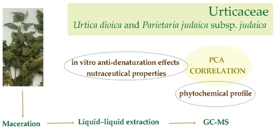
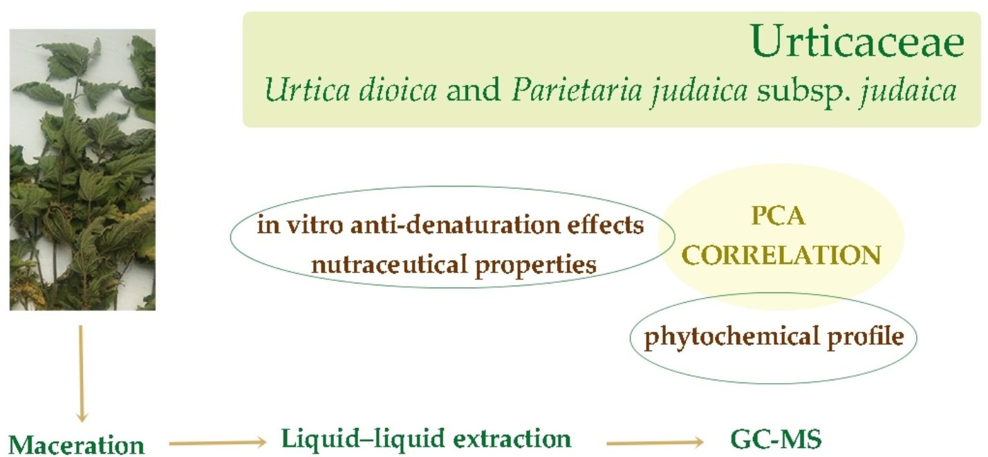
Graphical Abstract

1. Introduction

Given the well-established traditional use of the two common Urtucaecae, Urtica dioica L. and Parietaria judaica L. subsp. Judaica, in the treatment of inflammatory conditions and joint pain, as well as the culinary role of nettle, there is a clear need to scientifically validate their therapeutic potential. Accordingly, the aim of this study was to investigate in vitro the antioxidant, anti-denaturation, and nutraceutical properties of the hydroalcoholic extracts obtained from the aerial parts of these species. By evaluating their ability to neutralize free radicals, inhibit lipid peroxidation, and suppress the key inflammatory mediator nitric oxide, this research aims to contribute to providing a biochemical basis for their traditional medicinal reputation. According to The World Flora Online (WFO) database [1], Parietaria judaica L. subsp. judaica is an accepted infraspecific taxon of the species Parietaria judaica L. (pellitory), and it is also a synonym of Parietaria diffusa Mert. and W.D.J. Koch. This perennial plant primarily grows in the temperate biome: it is native to Northern Africa, Europe, Central Asia, the Central Himalaya, and Macaronesia, but it has been introduced into Denmark, Finland, Sweden, Western Australia, and a few states in North and South America [2]. Species belonging to the Parietaria genus are a major cause of allergy in the European regions and in the whole Mediterranean area, as they contain allergens recognized by IgE and IgG serum antibodies in patients with allergy. The allergens of Parietaria pollen are highly immunologically cross-reactive [3]. However, several traditional uses in folk medicine have been reported for P. judaica’s aerial parts, which were used for their anti-inflammatory and wound-healing properties to treat abdominal pain, hemorrhoids, rheumatism, skin ulcers, and burns [4]. This species, together with U. dioica, was also traditionally used in popular medicine of central Italy to treat Herpes zoster [5]. Some recent studies also pointed out interesting biological properties of P. judaica, such as anticancer [4] and antibacterial activities [6].
Urtica dioica L., commonly known as nettle, [7], is a widespread Urticaceae perennial herb native to Europe, Siberia, Western China, and Northwest Africa [1,2]. Even if the plant is commonly known as stinging, it becomes unharmful through boiling or if the leaves are consumed after drying [8]. Urtica dioica is edible and its nutritional properties are recognized. The young leaves are used in culinary applications for curries and soups. This species is characterized by an impressive nutritional value, particularly regarding its protein content, which accounts for 30% of its dry matter and confers to this plant a superior amino acid profile compared to other leafy vegetables. Additionally, nettle is also a rich source of polyunsaturated fatty acids (60%), particularly linoleic acid [7]. Beyond its nutritional value, Urtica dioica has a long history in traditional Chinese medicine as a remedy for joint pain [9]. At present, different biological and pharmacological properties have been demonstrated for the leaf and root extracts, such as anti-inflammatory, analgesic, antirheumatic, anti-proliferative, antioxidant, antibacterial, hypolipemic, hypoglycemic, and antiviral activities [10,11]. A wide range of phytochemicals have been detected, including phenolic compounds, flavonoids, lignans, sterols, terpenoids, and alkaloids [12].
Here, the aerial parts were extracted via maceration: the resulting hydroalcoholic extracts were evaluated for their radical scavenging and lipid peroxidation inhibitory properties. The antioxidant activity was measured using DPPH and the β-carotene bleaching assays. To tentatively investigate the anti-arthritic potential, the extracts were tested for their ability to inhibit protein denaturation and the production of nitric oxide. Additionally, the nutraceutical value and the potential benefits for obesity management were explored by assessing the inhibitory effects on pancreatic lipase. Finally, the phytochemical profiles of the two species were compared by means of principal component analysis (PCA), with the correlation between the detected constituents and observed biological activities further analyzed.

2. Materials and Methods

2.1. Chemicals

Analytical grade solvents and qualitative paper (10–20 µm particle retention) were purchased from VWR International (Milan, Italy). The following reagents were supplied by Sigma-Aldrich (Milan, Italy): phenolic and flavonoid assay reagents (Folin–Ciocalteu’s reagent, sodium carbonate, and aluminum chloride); antioxidant assay reagents (DPPH, ascorbic acid, β-carotene, linoleic acid, and propyl gallate); biological and cell culture chemicals (Griess reagent, indomethacin, L-NAME, PBS, DMEM, L-glutamine, fetal bovine serum, penicillin/streptomycin solution, LPS from Escherichia coli, and MTT, BSA); inorganic salts (NaCl, KCl, Na2HPO4, and KH2PO4); enzymes and drugs (diclofenac, orlistat, type II crude porcine pancreatic lipase, and 4-NPC).

2.2. Plant Material and Extraction Procedure

The specimens of Urtica dioica L. and Parietaria judaica L. subsp. judaica were gathered in Calabria (Southern Italy) in April. These samples are currently held at the Herbarium of the University of Calabria under accession numbers CLU-26251 and CLU-26246, leg. and det. F. Conforti. To obtain the raw extracts, the dried aerial parts were subjected to maceration at room temperature (3 extractions, 48 h each) using a 70% ethanol solution (1:10 g/mL plant to solvent ratio). After filtration (10–20 µm particle retention paper), the obtained solutions were concentrated via rotary evaporation IKA® RV 10 (VWR International, Milan, Italy) at 40 °C under vacuum to protect thermolabile compounds. This process resulted in extraction yields of 11.8% for P. judaica and 19.8% for U. dioica. Subsequently, a liquid–liquid partition was performed: dried raw extracts were first reconstituted in a 9:1 MeOH/H2O mixture and partitioned with n-hexane. The remaining solutions were then suspended in water and extracted with dichloromethane [13]. The final yields (relative to dry biomass) were 0.9% (n-hexane) and 0.8% (dichloromethane) for P. Judaica and 0.6% (n-hexane) and 1.0% (dichloromethane) for U. dioica.

2.3. Gas Chromatography–Mass Spectrometry (GC–MS) Analyses

A Hewlett-Packard gas chromatograph (Model 6890, Milan, Italy) equipped with a SE-30 capillary column (100% dimethylpolysiloxane; 30 m × 0.25 mm, 0.25 μm film thickness) was employed for GC–MS characterization. The system was coupled to a Hewlett-Packard 5973 mass selective detector (Milan, Italy). Helium served as the carrier gas at a linear velocity of 0.00167 cm/s, and 1 µL of each sample was injected using a Hamilton micro-syringe (Bonaduz, Switzerland). The oven temperature was programmed to rise from 60 °C to 280 °C at a constant rate of 16°/min. The operating conditions included an inlet temperature of 250 °C; an ionization energy of 70 eV; and a source temperature of 230 °C. Mass spectra, scanned at 1 scan/s over a range of 40–800 amu, were compared against the Wiley Mass Spectral Database per preliminary compound identification [13].

2.4. Total Phenolic and Flavonoid Content

The well-known and established Folin–Ciocalteu and aluminum chloride methods were used to estimate the total phenolic content (TPC) and the flavonoid content (TFC) of the extracts [14,15,16].
For the TPC analysis (first assay), 200 μL of each extract (2 mg/mL in an acetone/methanol/water/acetic acid mixture, pre-incubated at 60° C for 1 h), was combined with 1 mL of Folin–Ciocalteu reagent and 1 mL of 7.5% sodium carbonate. Following a 2 h incubation at room temperature, the absorbance was recorded at 726 nm using a Jenway 6300 spectrophotometer (Cole Parmer, St Neots, UK).
To evaluate the TFC, 1 mL of each extract solution (2 mg/mL in 80% ethanol) was mixed with 1 mL of 2% aluminum chloride. After 15 min of dark incubation, the absorbance was measured at 430 nm. The results were calculated via chlorogenic acid and quercetin calibration curves and expressed as mg of standard equivalents per gram of dry weight.

2.5. In Vitro Antioxidant Activity

The in vitro antioxidant properties were investigated through DPPH [17] and β-carotene bleaching [18] assays. In the first assay, 0.2 mL of sample solutions (5–1000 μg/mL) was mixed with 0.8 mL of 0.1 mM 2,2-diphenyl-1-picrylhidrazyl (DPPH) radical solution. Following a 30 min incubation period, the scavenging effect was determined spectrophotometrically (Jenway 6300, Cole Parmer, UK) at 517 nm.
Regarding the β-carotene bleaching test aimed at detecting the anti-lipid peroxidation activity, sample concentrations ranging from 0.25 to 100 μg/mL were assayed. The reaction mixture consisting of 0.2 mL of sample and 5 mL of β-carotene-linoleic acid emulsion was incubated in a 45 °C water bath. Absorbance at 470 nm was followed for 1 h.
Ascorbic acid (in the DPPH test) and propyl gallate (β-carotene bleaching assay) were used as positive controls.

2.6. In Vitro Evaluation of Inhibitory Effect on Nitric Oxide (NO) Production

The inhibitory effects on NO production were evaluated using the RAW 264.7 murine macrophage line (ATCC, UK, No. TIB-71) [13]. The cells were maintained in DMEM, supplemented with 10% FBS, 1% L-glutamine, and 1% penicillin/streptomycin) and seeded into 96-well plates at a density of 1 × 10⁵ cells/well after mechanical detachment via scraping. Following overnight incubation, the cells were stimulated with 1 μg/mL of Escherichia coli LPS and co-treated with various sample concentrations (3–1000 μg/mL, final ratio DMSO to medium 0.5% v/v). After 24 h, NO levels were determined by mixing 100 of the cell culture supernatants with an equal volume of Griess reagent, followed by absorbance measurement at 550 nm (Stat fax 3200, Awareness Technology Inc., Palm City, FL, USA). Indomethacin and L-NAME served as positive controls.
To ensure that the observed NO inhibition was not due to cellular damage, an MTT assay was performed. Briefly, 100 μL of 0.5% MTT in PBS were added to each well for 4 h; subsequently, the medium was replaced with 100 μL of DMSO, and the absorbance was recorded at 550 nm [13].

2.7. In Vitro Anti-Denaturation of Heat-Treated Protein

To verify the anti-arthritic potential of the raw extracts and their fractions, the in vitro protein denaturation assay was performed, employing bovine serum albumin (BSA) as the substrate [13]. Briefly, 2.40 mL of a 3.5% aqueous BSA solution were mixed with 0.1 mL of each sample (concentrations: 50–1000 μg/mL). The pH was then adjusted at 6.3 with 1 N HCl. Following a 20 min incubation at 37 °C, the mixtures were heated at 71 °C for 1 min to induce denaturation. After cooling, 2.5 mL of phosphate-buffered saline (pH 6.3) was added to each sample, and the resulting turbidity was quantified spectrophotometrically at 660 nm. Diclofenac sodium served as a positive control.

2.8. In Vitro Inhibition of Pancreatic Lipase

To evaluate the anti-obesity potential of the investigated samples, a pancreatic lipase inhibition assay was conducted [19]. The enzymatic activity was determined by monitoring the hydrolysis of 4-nitrophenyl caprylate (4-NPC) into 4-nitrophenol. The two extracts and their fractions (25 μL), tested at concentrations from 0.063 to 5 mg/mL, were mixed with 25 μL of type II crude porcine pancreatic lipase (1 mg/mL in water). The reaction mixture was completed with 1 mL of tris-HCl buffer (pH 8.5) and 25 μL of a 5 mM NPC solution, followed by a 25 min incubation at 37 °C. Orlistat was used as a positive control.

2.9. Statistical Analyses

The data are expressed as the mean ± S.E.M. of three independent experiments. IC50 values were determined through the nonlinear regression analysis of the raw data. Before proceeding with the statistical evaluation, data normality and homoscedasticity were verified using the Shapiro–Wilk test and Levene’s test, respectively. Statistical differences were identified via one-way analysis of variance (ANOVA), followed by Bonferroni’s test for pairwise post hoc comparisons and the Dunnett’s multiple comparison test for comparing treated groups against the control. A p-value of ≤0.05 was set as the threshold for statistical significance.
The metabolomics data analysis was performed using MetaboAnalyst 6.0 (http://www.metaboanalyst.ca, accessed on 22 April 2025). The principal component analysis (PCA) was also performed. Following an initial data integrity check, the variables with excessive missing values were excluded, while the remaining ones were replaced with Limit of Detection (LoD) values (one-fifth of each variable’s minimum positive value). The data were subsequently log-transformed and Pareto-scaled.
Furthermore, the Pearson correlation coefficients were calculated using the PAST4 software (version 4.15) to investigate the relationships between the detected phytochemicals (including TPC and TFC) and the inhibitory percentages recorded at the highest tested concentration for each performed biological assay.

3. Results

3.1. Antioxidant Activity

The in vitro antioxidant properties of P. judaica and U. dioica raw extracts were assessed with the well-established colorimetric assays, DPPH and β-carotene bleaching assays. Both raw extracts showed concentration-dependent radical scavenging activity (Table 1). U. dioica samples were significantly more effective than P. judaica ones (p < 0.05, Bonferroni post hoc test). The raw extracts showed IC50 values equal to 37.72 ± 0.51 μg/mL and 135.50 μg/mL, respectively. For completeness, the two obtained fractions, the n-hexane and the dichloromethane samples, were also studied for their antioxidant effects. As expected, given the apolar nature of the solvent utilized, the two n-hexane fractions did not show any antioxidant effect. On the contrary, the dichloromethane fractions showed radical scavenging potency, with the CH2Cl2 fraction from U. dioica being the most interesting one (IC50 = 20.32 ± 0.27 μg/mL). The dichloromethane fraction from P. judaica was also effective, even if at a minor extent, with an IC50 value equal to 103.00 ± 1.78 μg/mL.
The β-carotene bleaching test was also performed to verify the ability of the investigated Urticaceae extracts to protect linoleic acid from peroxidation (Table 1). In this case, the P. judaica raw extract was more effective than the U. dioica sample after 30 min of incubation (IC50 = 7.97 ± 0.52 μg/mL and 17.84 ± 0.99 μg/mL, respectively, p < 0.05, Bonferroni post hoc test), while no statistically significant differences were observed after 60 min. As regards the dichloromethane fractions, a better activity was instead detected for U. dioica (IC50 = 30.42 ± 0.92 and 47.85 ± 0.62 μg/mL after 30 and 60 min of incubation) compared to P. judaica (55.70 ± 2.08 μg/mL after 30 min).

3.2. In Vitro Inhibitory Effect on Nitric Oxide (NO) Production

The inhibitory potential on NO production was tested in vitro on LPS-stimulated RAW 264.7 cells. This assessment was performed via the Griess assay, a method that quantifies NO levels indirectly by measuring the accumulation of its stable metabolites, nitrite and nitrate. Indomethacin, a recognized anti-inflammatory agent, and the nitric oxide synthase inhibitor NG-nitro-L-arginine methyl ester (L-NAME) served as positive controls. While no activity was observed for the raw extract from U. dioica, the P. judaica crude extract and the two dichloromethane fractions showed a concentration-dependent inhibitory activity (Figure 1).
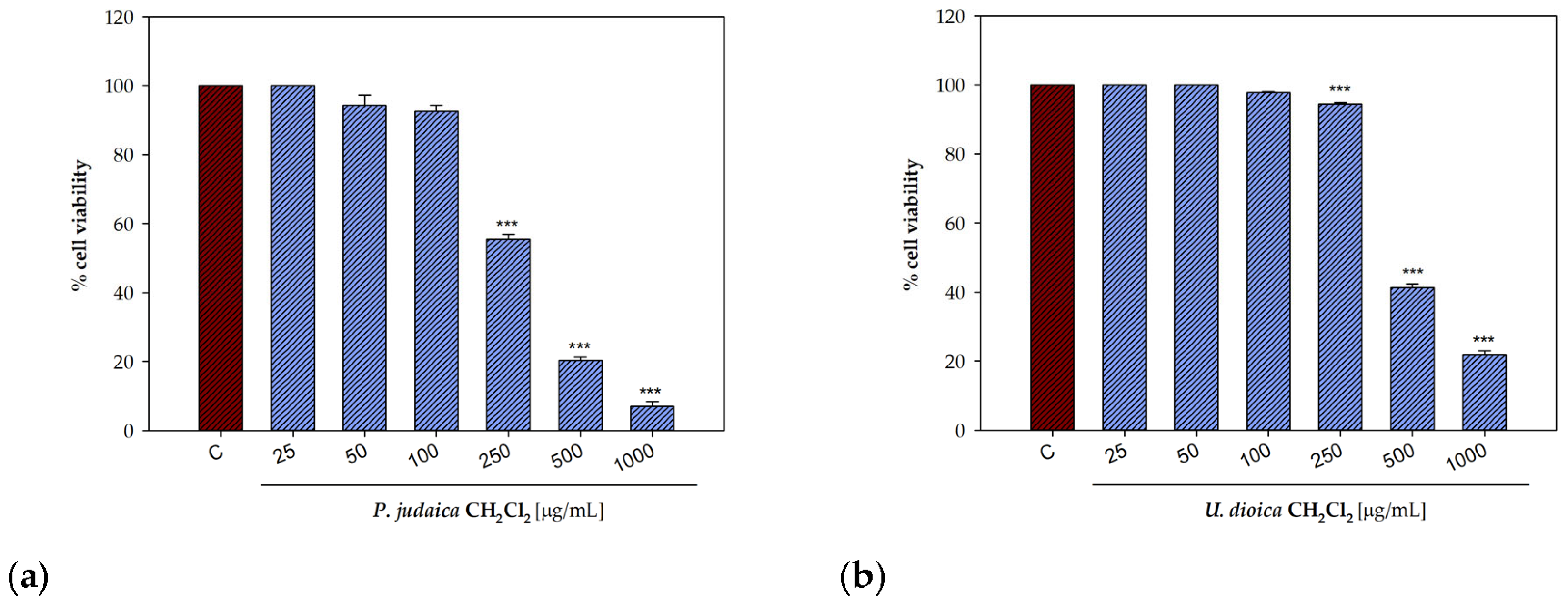
The raw extract from P. judaica exerted inhibitory activity, with an IC50 value equal to 690.3 ± 11.2 µg/mL and without showing any cytotoxicity on treated cells. The dichloromethane fraction was more effective in inhibiting the NO production, with an IC50 value equal to 104.2 ± 2.7 µg/mL. This sample showed a moderate cytotoxic effect on macrophages (IC50 = 277.4 ± 6.9), even if a significant difference was detected among the two IC50 values (p < 0.05, Bonferroni post hoc test, Table 2, Figure 2).
The dichloromethane fraction of U. dioica extract also exhibited promising biological activity with IC50 = 63.9 ± 1.8 µg/mL. While this fraction showed limited cytotoxic effects, a reduction in cell viability was noted at the highest concentrations tested (IC50 = 498.7 ± 10.9 µg/mL).

3.3. In Vitro Anti-Denaturation Effects on a Protein Model

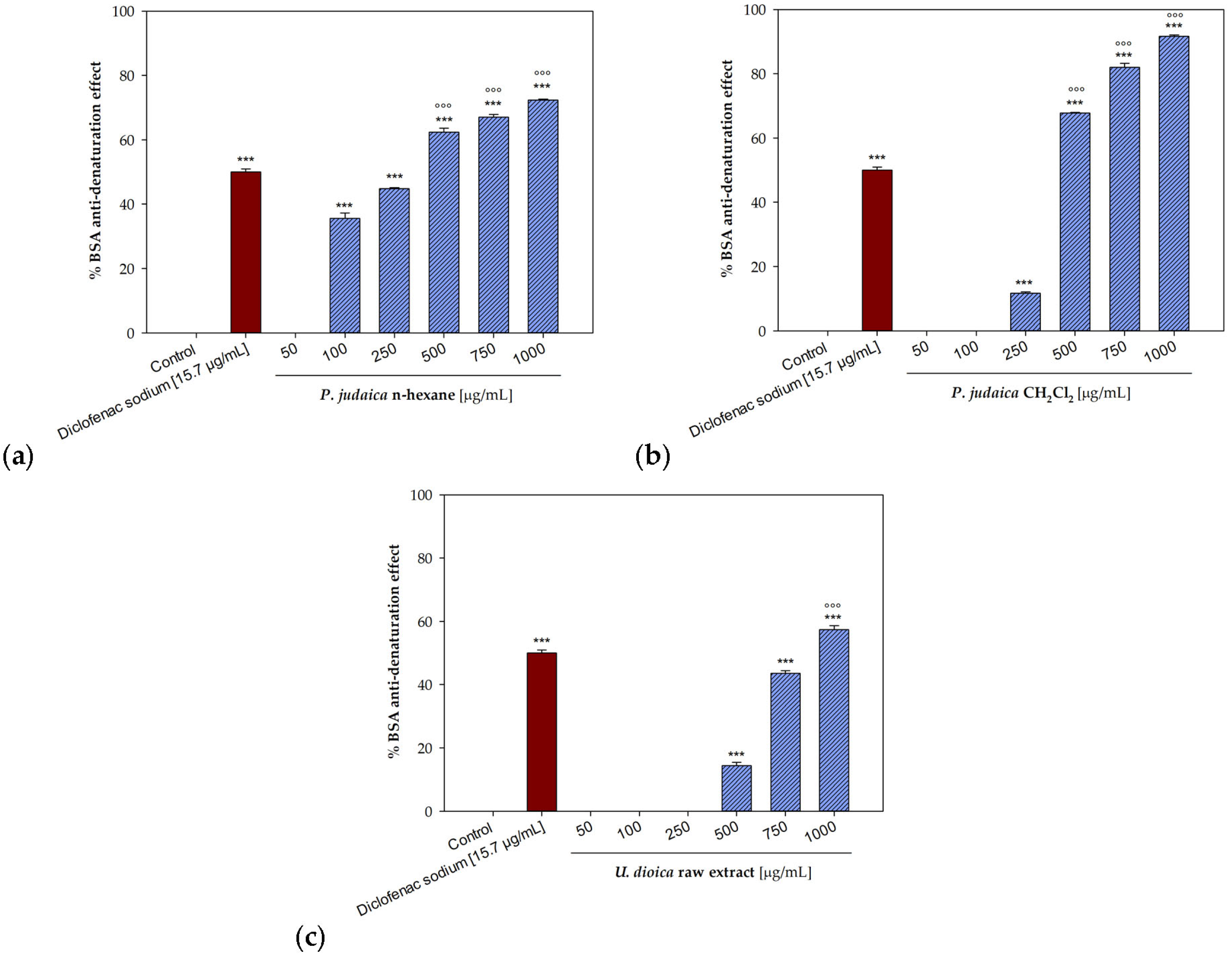
In this preliminary screening of the potential anti-arthritic activity of the investigated Urticaceae, the anti-denaturation properties were preliminary screened using a bovine serum albumin (BSA)-based model. While P. Judaica proved to be inactive, U. dioica raw extract showed a significant concentration-dependent ability to protect the protein from thermal denaturation. Specifically, at 1000 µg/mL, this last sample induced 57.36% inhibition of protein denaturation. Inhibition percentages equal to 43.59% and 14.45% were detected at 750 and 500 µg/mL (Figure 3c).
The most significant inhibitory effects were recorded for the two apolar fractions of P. Judaica. Specifically, at 1000 µg/mL, the n-hexane and dichloromethane fractions prevented BSA thermal denaturation by 72.35% and 91.71%, respectively (Figure 3a,b). The CH2Cl2 fraction was also particularly effective at the concentration of 750 and 500 µg/mL, with inhibition percentages equal to 82.01% and 67.85%, respectively, showing the lowest detectable effect (11.67%) at the concentration of 250 µg/mL. In contrast, the n-hexane fraction exhibited inhibition values around 67% and 62% at 750 and 500 µg/mL, respectively, while still providing a 35.54% protective effect at a concentration as low as 100 µg/mL.
The raw data were then fitted through nonlinear regression to deduce the IC50 parameters (Table 3).
U. dioica raw extract showed a concentration-dependent activity, with an IC50 value equal to 869.5 ± 8.2 µg/mL. However, the most interesting results were observed for the two fractions from P. judaica. The n-hexane sample was the most effective one, with IC50 = 321.6 ± 3.3 µg/mL (p < 0.05, Bonferroni post hoc test), followed by the dichloromethane fraction (IC50 = 424.4 ± 1.8 µg/mL).

3.4. In Vitro Inhibitory Potential on Pancreatic Lipase Enzyme

A screening of the potential nutraceutical value of the two investigated species was carried out thought the evaluation of the in vitro inhibitory properties on pancreatic lipase enzyme, with the aim to investigate the potential anti-obesity activity. To this goal, 4-nitrophenyl caprylate (NPC) was used as a substrate, and the release of the yellow chromogen p-nitrophenol due to the hydrolysis of NPC was spectrophotometrically monitored.
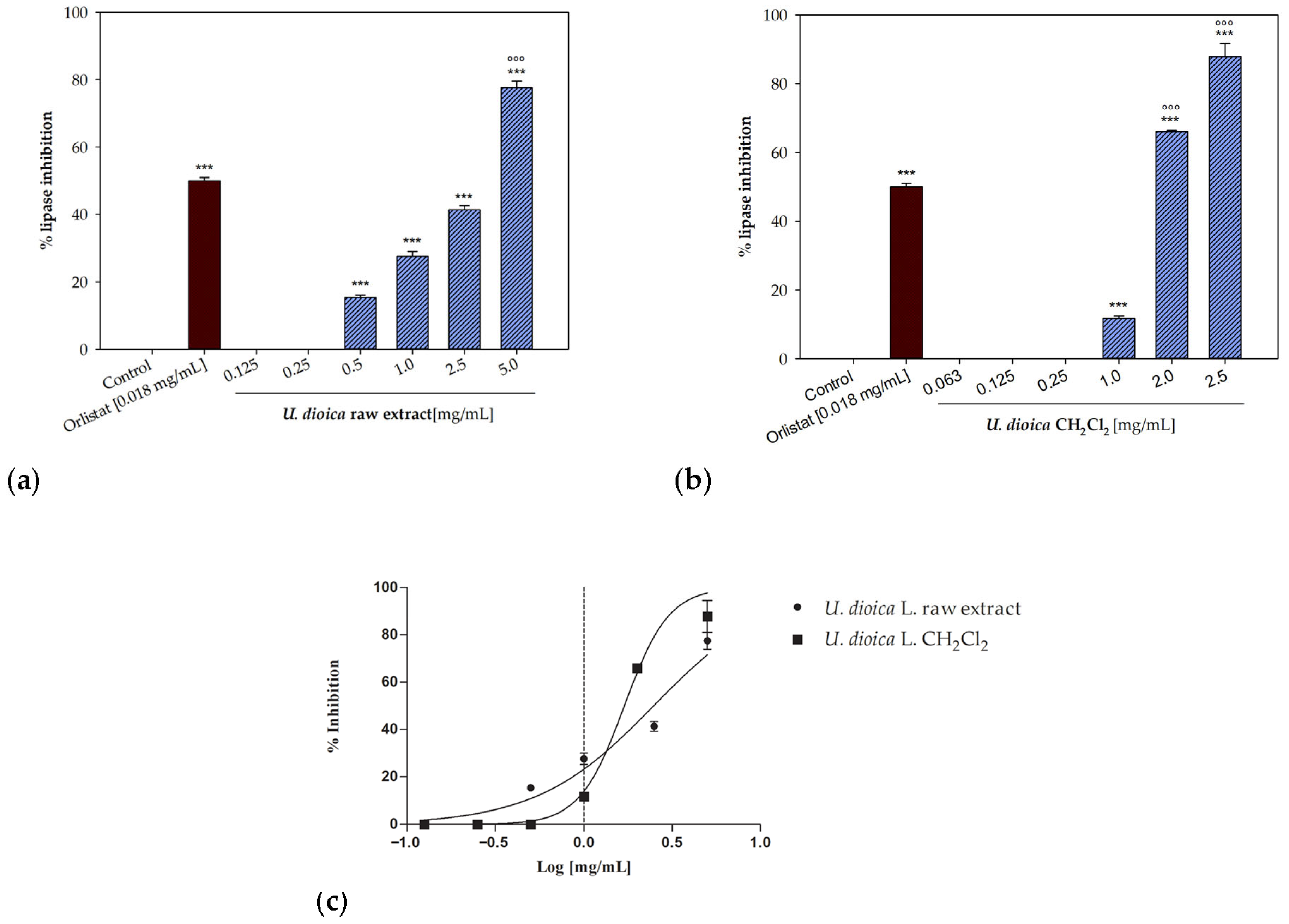
No activity was detected for the P. judaica crude extract and its fractions. On the contrary, a good inhibitory potential was observed for U. dioica species. Both the raw extract and the dichloromethane fraction were able to affect the enzyme, showing a concentration-dependent inhibitory activity (Figure 4).
The crude extract from U. dioica induced 77.52 ± 2.07% inhibition at a concentration of 5 mg/mL. The dichloromethane fraction induced a higher inhibition percentage, equal to 87.79 ± 3.88%, at even the lower concentration of 2.5 mg/mL (Figure 3b). This last sample was significantly more effective compared to the positive control also at a concentration of 2.0 mg/mL, with an inhibition percentage equal to 66.03 ± 0.49% (p < 0.001, Dunnett’s multiple comparison test).
Figure 3c illustrates the nonlinear fitting of the experimental data used to determine the IC50 parameters. While the U. dioica raw extract yielded an IC50 value of 2.49 ± 0.13 mg/mL, the dichloromethane fraction proved to be more effective, reaching an IC50 value equal to 1.67 ± 0.01 mg/mL.

3.5. Phytochemical Analysis

The concentration of polar constituents in both raw extracts was tentatively estimated using colorimetric assays to determine the total phenolic and total flavonoid content. U. dioica raw extract showed a significantly higher phenolic content compared to P. judaica, with values of 30.4 ± 0.6 and 8.0 ± 0.1 mg/g (expressed as chlorogenic acid equivalents per g of dry weight), respectively. A similar pattern was observed for the flavonoid content, with values equal to 0.57 ± 0.01 for U. dioica and 0.43 ± 0.02 mg/g for P. judaica.
The phytochemical investigation was mainly focused on the chemical characterization of nonpolar constituents, as the apolar h-hexane and dichloromethane fractions were investigated for their biological properties, showing interesting in vitro potential. The chemical constituents of the two isolated fractions (n-hexane and dichloromethane) were tentatively identified through GC–MS analyses (Figures S1 and S2). On the whole, 28 compounds were recognized, with the hexane fraction being particularly rich in fatty acids (Table S1). Fifteen compounds belonging to this class were identified, with palmitic acid being the main components of each species (about 8%), followed by linoleic and α-linolenic acids, particularly abundant in Parietaria judaica subsp. judaica (4.8% and 5.6%, respectively). Myristic, 14-methylpentadecanoic, stearic, arachidic, behenic, and lignoceric acid were also detected in both samples. Four terpenes, namely dihydroactinidiolide, neophytadiene, phytol, and phytone were also recognized in both Urticaceae, even if in different percentages. The two phytosterols, 22,23-dihydrobrassicasterol and β-sitosterol, were also detected. As regards the compounds identified in the two dichloromethane fraction, the most abundant component was vitamin E, identified in percentages equal to 3.9% in P. diffusa, followed by loliolide (1.8%), detected in the U. dioica sample (Table S2). Benzoic acid, methylethylmaleimide, 2,4-di-tert-butyilphenol, p-coumaric, and ferulic acids were also identified.
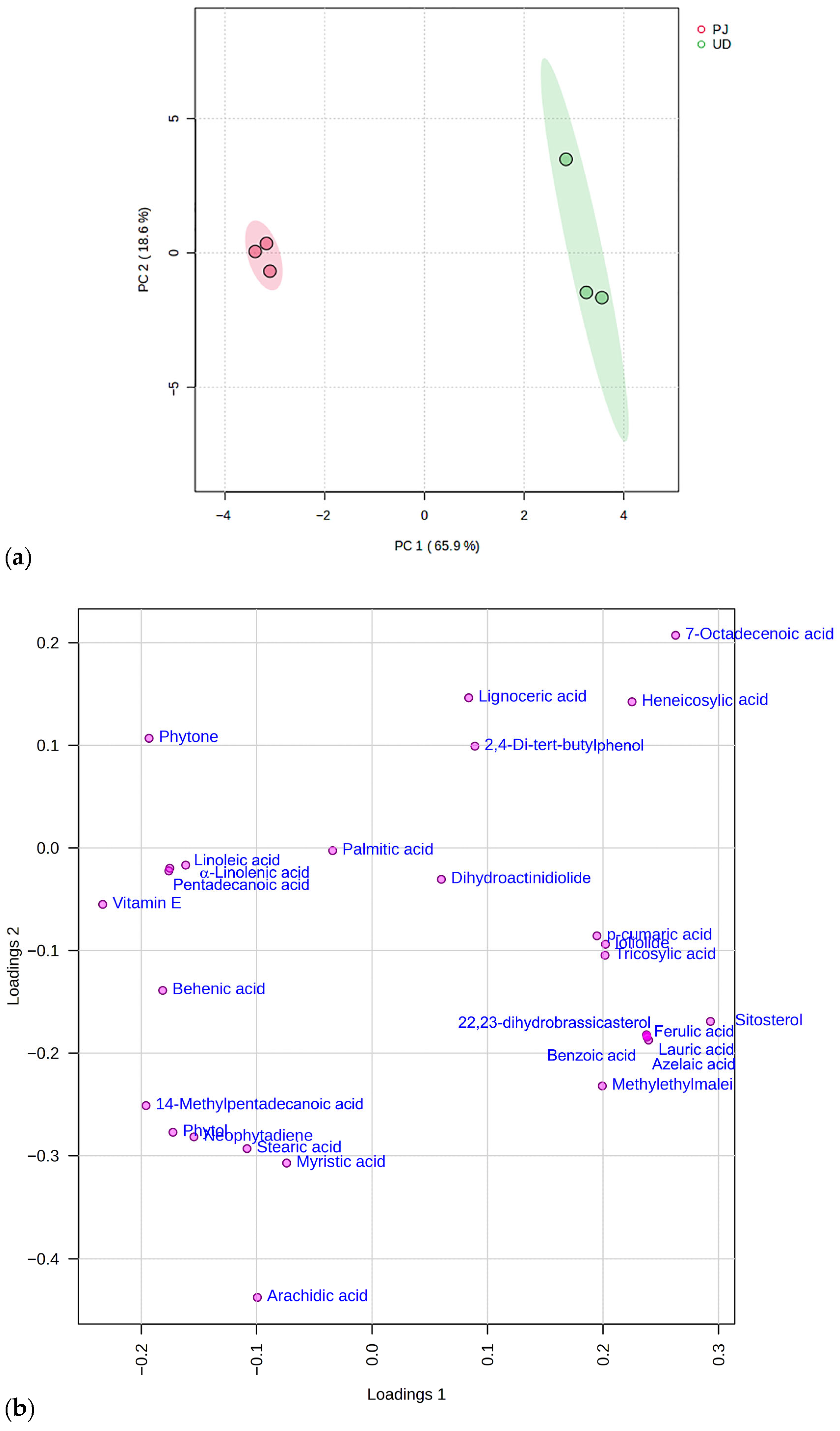
To visualize the distribution of identified metabolites across the two species, a principal component analysis (PCA) was performed. The analysis was based on a data matrix incorporating six samples (triplicates for species) and 28 descriptor variables. As illustrated in Figure 5, the scores plot and loading plots account for 84.5% of the total variance, with the first and the second principal components contributing 65.9% (PC-1) and 18.6% (PC-2), respectively.
The differences are explained by the loading plot of the components (Figure 5b). Positioned on the left half of the plot, with the lowest PC-1 score, the Parietaria judaica subsp. judaica (PJ) samples are characterized by high amounts of linoleic acid, α-linolenic acid, pentadecanoic acid, and vitamin E. Urtica dioica (UD) contains p-coumaric acid, loliolide, and tricolylic acid as the main discriminatory components separating these samples from the PJ ones. Lauric acid, azelaic acid, β-Sitosterol, 22,23 dihydrobrassicatserol, benzoic, and ferulic acids also characterize the UD samples.

3.6. Correlation Analysis

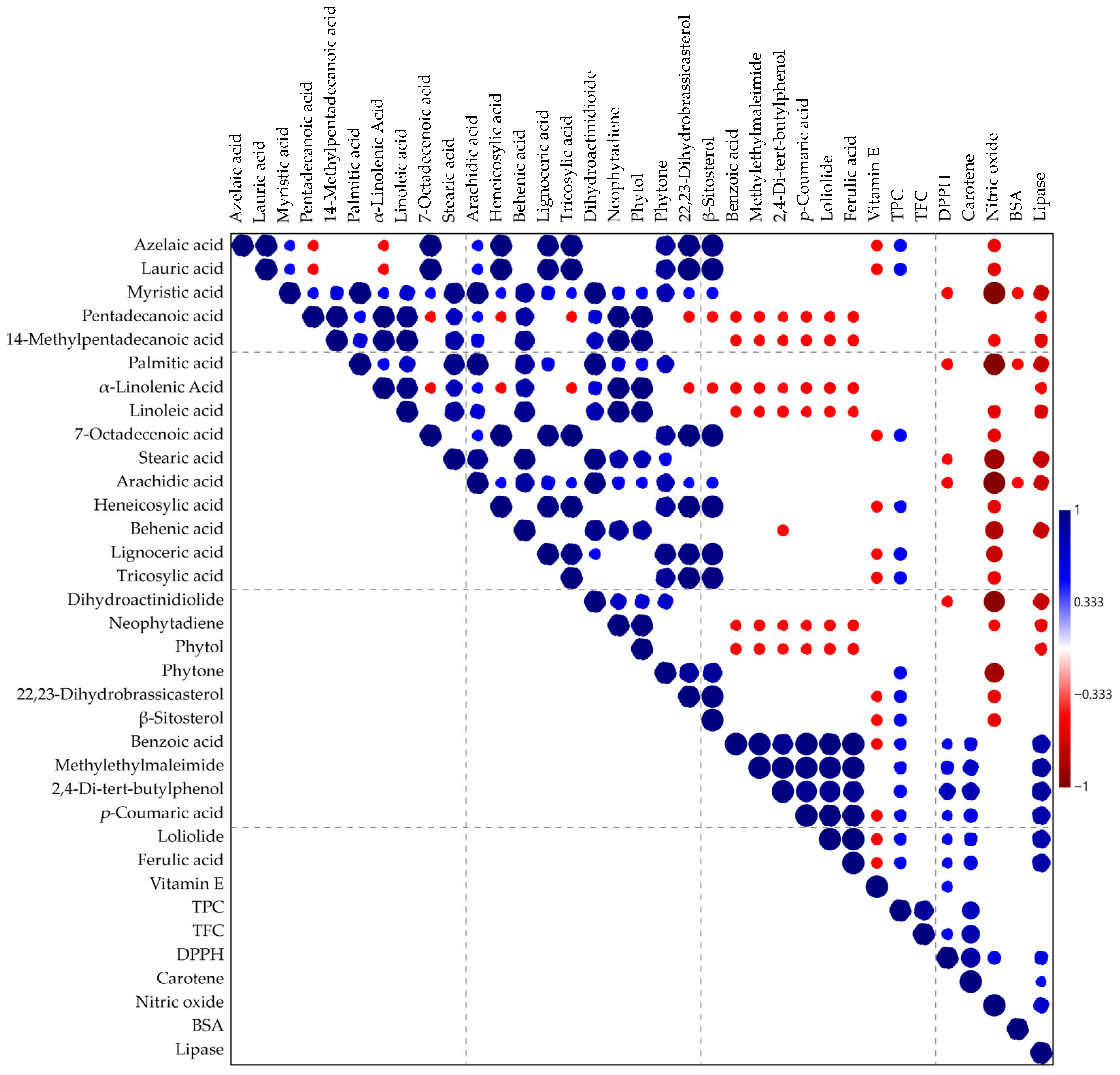
The relationship among the abundance of identified chemical compounds, the total phenolic and flavonoid content, and the biological properties of samples was assessed using the Pearson’s correlation coefficients, and it is illustrated in the correlation matrix of the entire data set presented in Figure 6.
Phenolic compounds such as 2,4-di-tert-butilphenol, p-coumaric acid, and ferulic acid exhibited significant positive correlation (p < 0.05) with total phenolic content and the antioxidant activity, namely the DPPH radical scavenging activity and the ability to protect linoleic acid from peroxidation (β-carotene bleaching assay). This correlation is well-known in the literature [20,21]. The same positive correlation was observed between these consistuents and the lipase inhibitory property (p < 0.001), with Pearson’s correlation coefficients of 0.88, 0.84, and 0.84, respectively. This result is also supported by the literature, as the influence of several phenolic acids on the activity of pancreatic lipase, particularly benzoic and ferulic acids, was reported by Karamać and Amarowicz [22]. Moreover, 2,4-di-tert-butylphenol has also been demonstrated to competitively and reversibly inhibit pancreatic lipase activity [23]. While the TPC is positively related to a better β-carotene bleaching inhibition, the flavonoid content shows a significant positive correlation with both DPPH and β-carotene bleaching test, suggesting that flavonoids are primarily responsible for the ability of investigated extracts to scavenge free radicals.
Meanwhile, the Pearson’s correlation coefficients between the percentage of lipase inhibition and the abundance of many fatty acids (namely myristic, palmitic, stearic, arachidic, and behenic acids) were comprised in the range between −0.70 and −0.73, implying a significant negative correlation (p ≤ 0.001).
The same trend was observed between most apolar compounds and the percentages of the nitric oxide inhibition: a significant negative correlation was observed between myristic, palmitic, stearic, arachidic, behenic, and lignoceric acids, as well as dihydroactinidiolide and phytone, and the inhibition of nitric oxide production (r ranging from 0.73 to 0.98, p < 0.001). Consistently, de Lima and colleagues reported that, at low concentrations, fatty acids increased NO production by the murine macrophage J774 cells, whereas at high concentrations, they cause cell death [24].

4. Discussion

Urtica dioica L. and Parietaria judaica L. subsp. judaica are two members of the Urticaceae family that, despite their sensitization and allergenic potential, were used in traditional medicine. Moreover, different pharmacological properties for both species have been described so far. Considering the traditional use of these plant species in the treatment of inflammatory conditions and joint pain and given the culinary use of nettle, the aim of this study was to perform a preliminary screening and a comparison of the in vitro biological properties of these species, with particular attention to their potential as regards the anti-arthritic and nutraceutical properties.
The antioxidant properties of P. judaica and U. dioica raw extracts were assessed in vitro using the two colorimetric assays, DPPH and β-carotene bleaching test. Both raw extracts showed concentration-dependent radical scavenging activity, with U. dioica sample (IC50 = 37.72 ± 0.51 μg/mL) being significantly more effective than P. judaica. As regards the obtained apolar fractions, the dichloromethane samples showed radical scavenging potency, with the CH2Cl2 fraction from U. dioica being more effective (IC50 = 20.32 ± 0.27 μg/mL) than the corresponding fraction from P. judaica (IC50 value equal to 103.00 ± 1.78 μg/mL). The two n-hexane fractions were not effective. On the contrary, the P. judaica raw extract was more effective than the U. dioica sample as regards the ability to inhibit lipid peroxidation, which was verified using the β-carotene bleaching test.
Our results agree with previous studies dealing with the antioxidant properties of U. dioica. Kukrić and colleagues tested the inhibitory activity of the 80% ethanol extract on the DPPH radical, reporting an IC50 value equal to 31.38 ± 0.10 (μg/g DW) [25], and Zouari Bouassida and colleagues described the lipid peroxidation inhibitory properties, reporting an IC50 value of 15 ± 0.001 μg/mL [26]. The radical scavenging potency of the investigated U. dioica sample was higher than those reported for the polyphenolic fractions isolated from the leaves and flower samples isolated from nettle from Lublin, analyzed by Wójcik-Borowska and coworkers, for which IC50 values of 78.56 ± 0.85 and 124.77 ± 1.38 μg/mL were detected [27]. As regards the raw extract from P. judaica L. subsp. judaica, only few studies were detected in the literature. The sample from Italy investigated here showed a higher radical scavenging potency compared to previous results reported for the hydroalcoholic extract from P. judaica from Iran [28].
Besides the evaluation of the antioxidant potential, the aim of this study was also to perform a screening dealing with the potential anti-arthritic effects of the investigated Urticaceae specie. These properties were tentatively assessed by studying the inhibitory effects of the samples on the well-known pro-inflammatory mediator nitric oxide (NO), which is involved in the progression of osteoarthritis and cartilage chondrocyte apoptosis, working as a catabolic element in the activation of TNFα and IL-1β [29], as well as through the evaluation of the anti-denaturation properties.
The Griess method is widely used for the detection of nitrites because of its simplicity and ease of use despite the limited sensitivity for NO2 (low detection limit of 1 µM), which makes it unsuitable for measuring micromolar levels of nitrite and nitrate in biological samples. However, overall, it must be noted that in vitro models cannot fully replicate the complexity of a living organism’s response; this remains a limitation of this kind of study [30].
The raw extract from P. judaica exerted inhibitory activity with an IC50 value = 690.3 ± 11.2 µg/mL and without showing any cytotoxicity on treated cells. The dichloromethane fraction was more effective in inhibiting the NO production, with an IC50 value equal to 104.2 ± 2.7 µg/mL, even if this sample showed cytotoxic effects on treated macrophages ah higher concentrations (IC50 = 277.4 ± 6.9). The dichloromethane fraction of U. dioica extract exhibited a significant biological activity, with an IC50 = 63.9 ± 1.8 µg/mL. These findings align with those of Carvalho and colleagues, who demonstrated that various Urtica extracts lack cytotoxic effects on macrophages [31]. Furthermore, Wójcik-Borowska and coworkers described the in vitro anti-inflammatory activity of an U. dioica polyphenolic fraction on human skin cells [27]. Such properties are consistent with existing literature reporting the in vivo anti-inflammatory efficacy of both U. dioica leaf extract [32,33] and seed fixed oil [34]. The anti-inflammatory potential of different extracts and subfractions from U. dioica leaf was also screened using the rat paw edema anti-inflammatory assay: the hexane extract (200 mg/kg bw) significantly inhibited the edema paw volume after 3 h of treatment, with the percent of inhibition (46.51%) being comparable to that of the standard indomethacin [35]. Moreover, Chira and colleagues evaluated the anti-inflammatory activity of the essential oil from U. dioica’s aerial parts using the same assay and describing a decrease in paw edema [36].
Arthritis represents a major chronic inflammatory disorder and a primary cause of global disability, resulting in substantial healthcare costs [37]. To evaluate the anti-arthritic potential of the two Urticaceae species under study, both crude extracts and their respective fractions were screened for their capacity to inhibit the heat-induced protein denaturation using BSA as a model. The U. dioica raw extract showed some inhibitory effects, with an IC50 = 869.5 ± 8.2 µg/mL, but the best results were observed for P. judaica n-hexane and dichloromethane fractions, with IC50 values equal to 321.6 ± 3.3 and 424.4 ± 1.8 µg/mL, respectively. Denaturation is the process in which proteins lose their secondary and tertiary structures as a result of different factors such as heat, organic solvents, concentrated inorganic salts, or strong acids or bases. Protein denaturation is a key feature of inflammation, as denatured proteins lose their biological functions. Also in this case, even if it is a well-known and accepted assay, it is necessary to point out that in vitro methods are not able to fully reproduce the complex interactions that happen in cells and tissues. Regarding this specific test, the way proteins are denatured might not accurately replicate the inflammatory processes that occur in vivo, as they measure a not clearly defined anti-denaturation effect rather than specifically inhibiting inflammatory mediators or pathways [30].
Our study agrees with the previous reports dealing with the anti-inflammatory and analgesic potential of U. dioica. Liao and colleagues described the anti-inflammatory and analgesic effects of a gel prepared using the methanol root extract from U. dioica on complete Freund’s adjuvant (CFA)-induced arthritic mice [38]. The therapeutic potential of these plants was further investigated by Abd-Nikfarjam and coworkers through a randomized, double-blind, placebo-controlled clinical trial. The obtained clinical findings demonstrated that both U. dioica and evening primrose lowered inflammatory markers and alleviated symptoms in patients suffering from rheumatoid arthritis [39].
Our results are also in accordance with previous in vitro studies: the hydroalcoholic extract of U. dioica from Brazil showed an in vitro anti-denaturation effect on egg albumin [40], and the ethanolic extracts of the aerial parts of U. dioica from Iran were also assessed for their inhibitory properties on BSA using the inhibition of albumin reaction denaturation technique [41].
The nutritional value of plants under study was also examined, reflecting the common use of U. dioica as a food ingredient. Since pancreatic lipase is a key enzyme for fat metabolism, its inhibition represents a primary target for assessing the plant’s nutraceutical efficacy. This test is a well-known and established procedure allowing a first screening of the potential activity of plant extracts and pure compounds. However, some limitations must be considered. For example, different experimental factors, including enzyme storage conditions, assay temperatures, buffers, and additives, may influence the hydrolysis rate in lipase assay [42]. No inhibitory effect was observed for P. judaica crude extract and its fractions. On the contrary, a good biological activity was showed by U. dioica species. Lipase inhibition occurred in a concentration-dependent manner for both the raw extract and its dichloromethane fraction, with IC50 values of 2.49 ± 0.13 and 1.67± 0.01 mg/mL, respectively. Differently from the Parietaria samples investigated here, the methanol extracts from P. judaica leaves from the Jenin area of Palestine showed anti-lipase activity, with an IC50 value of 38.9 ± 0.29 μg/mL [6]. Our results also diverge from the report by Zor et al., who found no inhibitory effect when testing the aqueous extracts from Turkish U. dioica aerial parts [43].
Finally, different phytochemicals were tentatively detected through GC–MS analyses, which allowed to verify the presence of different classes of more nonpolar compounds, namely fatty acids, terpenes, and phytosterols. The differences in the phytochemical composition of the two Urticaceae were highlighted by means of a PCA, and a correlation analysis allowed assessing the correlation between the detected phytochemicals and observed biological properties. A positive correlation was observed between the compounds present in the two dichloromethane fractions of both species and the inhibitory activity on the lipase enzyme. In particular, 2,4-di-tert-butylphenol was highlighted for its ability to inhibit pancreatic lipase, and p-coumaric acid and ferulic acid also showed strong positive correlations. Indeed, in our study, the best results were observed for the dichloromethane fraction of the species U. dioica, which was richer in these bioactive compounds compared to the same fraction from P. judaica. Pancreatic lipase is the primary catalyst for the hydrolysis of dietary triglycerides into absorbable free fatty acids and monoglycerides. Chemical constituents such as 2,4-di-tert-butylphenol act as competitive and reversible inhibitors. By binding to the lipase enzyme, they prevent it from breaking down fats in the digestive tract, thereby reducing lipid absorption and helping manage obesity.
On the other hand, a negative correlation was observed between the inhibitory properties on NO production and most fatty acids identified in the n-hexane fractions, namely myristic, palmitic, stearic, arachidic, behenic, and lignoceric acids, with the fractions from both species being ineffective, thus meaning they likely do not contribute to these specific medicinal effects. NO is a signaling molecule produced by the enzyme iNOS (inducible nitric oxide synthase) during inflammation. In the context of arthritis, an excess of NO leads to cartilage chondrocyte apoptosis and the progression of osteoarthritis. P. judaica fractions inhibited heat-induced protein denaturation. Since protein denaturation is a documented cause of inflammation and autoimmune responses, stabilizing these proteins prevents the trigger of downstream inflammatory cascades. Liquid–liquid extraction, which we utilized in our study design, is a well-established technique allowing for the partitioning of an analyte between two immiscible solvents, typically an aqueous solution and a water-immiscible organic solvent. By changing the second one, the targeted compounds can be preferably extracted into the organic phase. This chromatographic technique allows some advantages, such as the cost-effectiveness, simplicity, and speed of execution, as well as repeatability [44]. Liquid–liquid extraction separates chemical compounds based on their polarity and allows for isolating different chemical classes. For these reasons, the differences we observed among the plant extracts fractions, even if due to the use of liquid–liquid extraction, are attributable to and reflect the differences among the phytochemical profile of the two crude extracts. If a specific chemical class is responsible for a type of biological activity, the specific fraction can be more active than the whole crude extract, as fractionation allows to concentrate the active compounds and/or to remove interfering substances that reduce the overall effect [45,46].

5. Conclusions

In conclusion, this paper contributes to the scientific validation of the traditional use of the common Urticaceae Urtica dioica L. and Parietaria judaica subsp. judaica L. in the management of inflammatory conditions and joint pain. Our findings reveal that both species possess significant antioxidant activity, with U. dioica demonstrating a higher radical scavenging potency than P. Judaica, whose raw extract, conversely, was more effective in inhibiting lipid peroxidation in the β-carotene bleaching test. The anti-arthritic potential was successfully screened using the in vitro protein denaturation inhibition assay, suggesting the ability to stabilize proteins against inflammatory stressors. These results impact the broader field of ethnopharmacology by highlighting that, despite their sensitization and allergenic potential, these Urticaceae species also contain high-value bioactive compounds with specific therapeutic targets. Furthermore, our findings also point out a potential inhibitory effect of fat absorption, which should be interesting from a nutraceutical point of view. These results could be further exploited to guide the development of target-specific nutraceuticals and anti-inflammatory formulations. Future research should optimize the solvent partitioning method and focus on the bio-guided isolation of the specific metabolites responsible for these effects and the validation of their efficacy through advanced cell-based or in vivo models to confirm their safety and therapeutic potential.

Supplementary Materials

The following supporting information can be downloaded at: https://www.mdpi.com/article/10.3390/app16073155/s1, Table S1: Phytochemical profile of P. judaica L. subsp. judaica and U. dioica L. n-hexane fractions; Table S2: Phytochemical profile of P. judaica L. subsp. judaica and U. dioica L. dichloromethane fractions. Figure S1: GC-MS chromatograms of P. judaica L. subsp. judaica n-hexane (a) and dichloromethane fractions (b). Figure S2: GC-MS chromatograms of U. dioica L. n-hexane (a) and dichloromethane fractions (b).

Author Contributions

Conceptualization, M.M. and F.C.; methodology, M.M. and F.C.; formal analysis, M.M.; investigation, M.M., F.A., M.R.P., M.F., P.C., F.C. and G.S.; resources, F.C.; data curation, M.M.; writing—original draft preparation, M.M.; writing—review and editing, G.S. and F.C.; visualization, G.S.; supervision, F.C. All authors have read and agreed to the published version of the manuscript.

Funding

This research received no external funding.

Institutional Review Board Statement

Not applicable.

Informed Consent Statement

Not applicable.

Data Availability Statement

Data are contained within the article or Supplementary Materials.

Conflicts of Interest

The authors declare no conflicts of interest.

Abbreviations

The following abbreviations are used in this manuscript:
ANOVAanalysis of variance
4-NPC4-nitrophenyl caprylate
BSAbovine serum albumin
CFAcomplete Freund’s adjuvant
DMEMDulbecco’s modified Eagle’s medium
DMSOdimethyl sulfoxide
DPPH2,2-diphenyl-1-picrylhidrazyl radical
GC–MSgas chromatography–mass spectrometry
iNOSinducible nitric oxide synthase
L-NAMENG-nitro-L-arginine methyl ester
LPSlipopolysaccharide
MTT3-(4,5-dimethylthiazol-2-yl)-2,5-diphenyltetrazolium bromide
NOnitric oxide
PBS phosphate-buffered saline
PCAprincipal component analysis
TFCtotal flavonoid content
TPCtotal phenolic content

References

  1. WFO. 2025. Available online: http://www.worldfloraonline.org (accessed on 18 April 2025).
  2. Plants of the World Online. Available online: https://powo.science.kew.org/ (accessed on 18 April 2025).
  3. D’amato, G.; Ruffilli, A.; Sacerdoti, G.; Bonini, S. Parietaria pollinosis: A review. Allergy 1992, 47, 443–444. [Google Scholar] [CrossRef] [Scilit]
  4. Bielecka, I.; Natorska-Chomicka, D.; Dołomisiewicz, W.; Rodrigues Fortes, A.; Dos Santos Szewczyk, K. Phytochemical profile and selective anticancer activity of Parietaria judaica L. extracts. Molecules 2025, 30, 2739. [Google Scholar] [CrossRef] [Scilit]
  5. Manganelli, R.U.; Zaccaro, L.; Tomei, P.E. Antiviral activity in vitro of Urtica dioica L., Parietaria diffusa M. et K. and Sambucus nigra L. J. Ethnopharmacol. 2005, 98, 323–327. [Google Scholar] [CrossRef] [Scilit]
  6. Qadi, M.; Jaradat, N.; Al-Lahham, S.; Ali, I.; Abualhasan, M.N.; Shraim, N.; Hussein, F.; Issa, L.; Mousa, A.; Zarour, A.; et al. Antibacterial, anticandidal, phytochemical, and biological evaluations of pellitory plant. Biomed Res. Int. 2020, 2020, 6965306. [Google Scholar] [CrossRef] [Scilit] [PubMed]
  7. Bhusal, K.K.; Magar, S.K.; Thapa, R.; Lamsal, A.; Bhandari, S.; Maharjan, R.; Shrestha, S.; Shrestha, J. Nutritional and pharmacological importance of stinging nettle (Urtica dioica L.): A review. Heliyon 2022, 8, e09717. [Google Scholar] [CrossRef] [Scilit] [PubMed]
  8. Grauso, L.; De Falco, B.; Lanzotti, V.; Motti, R. Stinging nettle, Urtica dioica L.: Botanical, phytochemical and pharmacological overview. Phytochem. Rev. 2020, 19, 1341–1377. [Google Scholar] [CrossRef] [Scilit]
  9. Qasmi, M.; Fareed, M.M.; Ali, H.; Khan, Z.; Shityakov, S. Integrative multi-target analysis of Urtica dioica for gout arthritis treatment: A network pharmacology and clustering approach. Silico Pharmacol. 2024, 12, 88. [Google Scholar] [CrossRef] [Scilit]
  10. Chrubasik, J.E.; Roufogalis, B.D.; Wagner, H.; Chrubasik, S.A. A comprehensive review on nettle effect and efficacy profiles, Part I: Herba urticae. Phytomedicine 2007, 14, 423–435. [Google Scholar] [CrossRef] [Scilit]
  11. Dhouibi, R.; Affes, H.; Salem, M.B.; Hammami, S.; Sahnoun, Z.; Zeghal, K.M.; Ksouda, K. Screening of pharmacological uses of Urtica dioica and others benefits. Prog. Biophys. Mol. Biol. 2020, 150, 67–77. [Google Scholar] [CrossRef] [Scilit] [PubMed]
  12. Taheri, Y.; Quispe, C.; Herrera-Bravo, J.; Sharifi-Rad, J.; Ezzat, S.M.; Merghany, R.M.; Shaheen, S.; Azmi, L.; Mishra, A.P.; Sener, B.; et al. Urtica dioica-derived phytochemicals for pharmacological and therapeutic applications. Evid. Based Complement. Altern. Med. 2022, 2022, 4024331. [Google Scholar] [CrossRef] [Scilit]
  13. Musolino, V.; Marrelli, M.; Perri, M.R.; Palermo, M.; Gliozzi, M.; Mollace, V.; Conforti, F. Centranthus ruber (L.) DC. and Tropaeolum majus L.: Phytochemical profile, in vitro anti-denaturation effects and lipase inhibitory activity of two ornamental plants traditionally used as herbal remedies. Molecules 2022, 28, 32. [Google Scholar] [CrossRef] [Scilit]
  14. Singleton, V.L.; Rossi, J.A. Colorimetry of total phenolics with phosphomolybdic-Phosphotungstic acid reagents. Am. J. Enol. Vitic. 1965, 16, 144–158. [Google Scholar] [CrossRef] [Scilit]
  15. Rababah, T.M.; Hettiarachchy, N.S.; Horax, R. Total phenolics and antioxidant activities of fenugreek, green tea, black tea, grape seed, ginger, rosemary, gotu kola, and ginkgo extracts, vitamin E, and tert-butylhydroquinone. J. Agric. Food Chem. 2004, 52, 5183–5186. [Google Scholar] [CrossRef] [Scilit]
  16. Amorati, R.; Valgimigli, L. Advantages and limitations of common testing methods for antioxidants. Free Radic. Res. 2015, 49, 633–649. [Google Scholar] [CrossRef] [Scilit]
  17. Loizzo, M.R.; Marrelli, M.; Pugliese, A.; Conforti, F.; Nadjafi, F.; Menichini, F.; Tundis, R. Crocus cancellatus subsp. damascenus stigmas: Chemical profile, and inhibition of α-amylase, α-glucosidase and lipase, key enzymes related to type 2 diabetes and obesity. J. Enzym. Inhib. Med. Chem. 2016, 31, 212–218. [Google Scholar]
  18. Conforti, F.; Marrelli, M.; Statti, G.; Menichini, F.; Uzunov, D.; Solimene, U.; Menichini, F. Comparative chemical composition and antioxidant activity of Calamintha nepeta (L.) Savi subsp. glandulosa (Req.) Nyman and Calamintha grandiflora (L.) Moench (Labiatae). Nat. Prod. Res. 2012, 26, 91–97. [Google Scholar] [CrossRef] [Scilit] [PubMed]
  19. Marrelli, M.; Morrone, F.; Argentieri, M.P.; Gambacorta, L.; Conforti, F.; Avato, P. Phytochemical and biological profile of Moricandia arvensis (L.) DC.: An inhibitor of pancreatic lipase. Molecules 2018, 23, 2829. [Google Scholar] [CrossRef] [Scilit] [PubMed]
  20. Vuolo, M.M.; Lima, V.S.; Junior, M.R.M. Phenolic compounds: Structure, classification, and antioxidant power. In Bioactive Compounds; Segura Campos, M.R., Ed.; Woodhead Publishing: Sawston, UK, 2019; p. 33-502019. [Google Scholar]
  21. Rice-Evans, C.; Miller, N.; Paganga, G. Antioxidant properties of phenolic compounds. Trends Plant Sci. 1997, 2, 152–159. [Google Scholar] [CrossRef] [Scilit]
  22. Karamać, M.; Amarowicz, R. Inhibition of pancreatic lipase by phenolic acids-examination in vitro. Z. Naturforschung C 1996, 51, 903–906. [Google Scholar] [CrossRef] [Scilit] [PubMed]
  23. Guan, W.; Cheng, J.; McClements, D.J.; Tu, Z.; Chen, J.; Ma, D. Impact of 2, 4-di-tert-butylphenol on pancreatic lipase activity in emulsions: Multispectral, molecular docking, and in vitro digestion analysis. Food Chem. 2025, 470, 142730. [Google Scholar] [CrossRef] [Scilit]
  24. de Lima, T.M.; de Sa Lima, L.; Scavone, C.; Curi, R. Fatty acid control of nitric oxide production by macrophages. FEBS Lett. 2006, 580, 3287–3295. [Google Scholar] [CrossRef] [Scilit]
  25. Kukrić, Z.Z.; Topalić-Trivunović, L.N.; Kukavica, B.M.; Matoš, S.B.; Pavičić, S.S.; Boroja, M.M.; Savić, A.V. Characterization of antioxidant and antimicrobial activities of nettle leaves (Urtica dioica L.). Acta Period. Technol. 2012, 43, 257–272. [Google Scholar] [CrossRef] [Scilit]
  26. Zouari Bouassida, K.; Bardaa, S.; Khimiri, M.; Rebaii, T.; Tounsi, S.; Jlaiel, L.; Trigui, M. Exploring the Urtica dioica leaves hemostatic and wound-healing potential. BioMed Res. Int. 2017, 2017, 1047523. [Google Scholar] [CrossRef] [Scilit]
  27. Wójcik-Borowska, K.; Wójciak, W.; Żuk, M.; Luchowski, P.; Skalska-Kamińska, A.; Pacuła, W.; Sowa, I.; Wójciak, M. Oxidative stress protection and anti-inflammatory activity of polyphenolic fraction from Urtica dioica: In vitro study using human skin cells. Molecules 2025, 30, 2515. [Google Scholar] [CrossRef] [Scilit]
  28. Asadi-Samani, M.; Rafieian-Kopaei, M.; Lorigooini, Z.; Shirzad, H. A screening of anti-breast cancer effects and antioxidant activity of twenty medicinal plants gathered from Chaharmahal va Bakhtyari province. Iran. J. Pharm. Pharmacogn. Res. 2019, 7, 213–222. [Google Scholar] [CrossRef] [Scilit]
  29. Rahmati, M.; Mobasheri, A.; Mozafari, M. Inflammatory mediators in osteoarthritis: A critical review of the state-of-the-art, current prospects, and future challenges. Bone 2016, 85, 81–90. [Google Scholar] [CrossRef] [Scilit]
  30. Peiris, D.S.H.S.; Fernando, D.T.K.; Senadeera, S.P.N.N.; Ranaweera, C.B. Assessment of in vitro anti-inflammatory activity: A comprehensive review of methods, advantages, and limitations. Asian J. Res. Biochem. 2025, 15, 37–52. [Google Scholar] [CrossRef] [Scilit]
  31. Carvalho, A.R.; Costa, G.; Figueirinha, A.; Liberal, J.; Prior, J.A.; Lopes, M.C.; Cruz, M.T.; Batista, M.T. Urtica spp.: Phenolic composition, safety, antioxidant and anti-inflammatory activities. Food Res. Int. 2017, 99, 485–494. [Google Scholar] [CrossRef] [Scilit] [PubMed]
  32. Hajhashemi, V.; Klooshani, V. Antinociceptive and anti-inflammatory effects of Urtica dioica leaf extract in animal models. Avicenna J. Phytomed. 2013, 3, 193. [Google Scholar] [PubMed]
  33. Safari, V.Z.; Ngugi, M.P.; Orinda, G.; Njagi, E.M. Anti-pyretic, anti-inflammatory and analgesic activities of aqueous leaf extract of Urtica dioica L. in albino mice. Med. Aromat. Plants 2016, 5, 237. [Google Scholar]
  34. Tekin, M.; Özbek, H.; Him, A. Investigation of acute toxicity, anti-inflammatory and analgesic effect of Urtica dioica L. Pharmacologyonline 2009, 1, 1210–1215. [Google Scholar]
  35. Dar, S.A.; Ganai, F.A.; Yousuf, A.R.; Balkhi, M.U.H.; Bhat, T.M.; Sharma, P. Pharmacological and toxicological evaluation of Urtica dioica. Pharm. Biol. 2013, 51, 170–180. [Google Scholar] [CrossRef] [Scilit] [PubMed]
  36. Chira, A.; Rekik, I.; Rahmouni, F.; Ben Amor, I.; Gargouri, B.; Kallel, C.; Jamoussi, K.; Allouche, N.; El Feki, A.; Kadmi, Y.; et al. Phytochemical composition of Urtica dioica essential oil with antioxidant and anti-inflammatory properties: In vitro and in vivo studies. Curr. Pharm. Biotechnol. 2025, 26, 728–739. [Google Scholar] [CrossRef] [Scilit]
  37. Dunlop, D.D.; Manheim, L.M.; Yelin, E.H.; Song, J.; Chang, R.W. The costs of arthritis. Arthritis Care Res. 2003, 49, 101–113. [Google Scholar] [CrossRef] [Scilit]
  38. Liao, J.C.; Wei, Z.X.; Ma, Z.P.; Zhao, C.; Cai, D.Z. Evaluation of a root extract gel from Urtica dioica (Urticaceae) as analgesic and anti-inflammatory therapy in rheumatoid arthritis in mice. Trop. J. Pharm. Res. 2016, 15, 781–785. [Google Scholar] [CrossRef] [Scilit]
  39. Abd-Nikfarjam, B.; Abbasi, M.; Memarzadeh, M.; Farzam, S.A.; Jamshidian, A.; Dolati-Somarin, A. Therapeutic efficacy of Urtica dioica and evening primrose in patients with rheumatoid arthritis: A randomized double-blind, placebo-controlled clinical trial. J. Herb. Med. 2022, 32, 100556. [Google Scholar] [CrossRef] [Scilit]
  40. Cuinica, L.G.; Chissico, R.B. Anti-inflammatory properties of extract and quercetin from Urtica dioica L. Adv. Anal. Pharm. Chem. AAPC-104 2018, 2018, 1–5. [Google Scholar]
  41. Ali, M.R.H.C.; Khosroyar, S.; Mohammadi, A. Evaluation of in vitro anti-inflammatory activity of Harpagophytum procumbens and Urtica dioica against the denaturation of protein. Plant Arch. 2018, 18, 161–166. [Google Scholar]
  42. Vo, C.V.T.; Luu, N.V.; Nguyen, T.T.; Nguyen, T.T.; Ho, B.Q.; Nguyen, T.H.; Tran, T.-D.; Nguyen, Q.T. Screening for pancreatic lipase inhibitors: Evaluating assay conditions using p-nitrophenyl palmitate as substrate. All Life 2022, 15, 13–22. [Google Scholar] [CrossRef] [Scilit]
  43. Zor, M.; Özüpek, B.; Pekacar, S.; Deliorman Orhan, D. Antioxidants, enzyme inhibitory activities, and phytochemical profiles of seven medicinal plants grown with organic farming techniques. Turk. J. Agric. For. 2023, 47, 918–930. [Google Scholar] [CrossRef] [Scilit]
  44. Piestansky, J.; Olesova, D.; Matuskova, M.; Cizmarova, I.; Chalova, P.; Galba, J.; Majerova, P.; Mikus, P.; Kovac, A. Amino acids in inflammatory bowel diseases: Modern diagnostic tools and methodologies. Adv. Clin. Chem. 2022, 107, 139–213. [Google Scholar] [PubMed]
  45. Muhamad, I.I.; Hassan, N.D.; Mamat, S.N.; Nawi, N.M.; Rashid, W.A.; Tan, N.A. Extraction technologies and solvents of phytocompounds from plant materials: Physicochemical characterization and identification of ingredients and bioactive compounds from plant extract using various instrumentations. In Ingredients Extraction by Physicochemical Methods in Food; Academic Press: Cambridge, MA, USA, 2017; pp. 523–560. [Google Scholar]
  46. Jallali, I.; Waffo Teguo, P.; Smaoui, A.; Merillon, J.M.; Abdelly, C.; Ksouri, R. Bio-guided fractionation and characterization of powerful antioxidant compounds from the halophyte Inula crithmoïdes. Arab. J. Chem. 2020, 13, 2680–2688. [Google Scholar] [CrossRef] [Scilit]
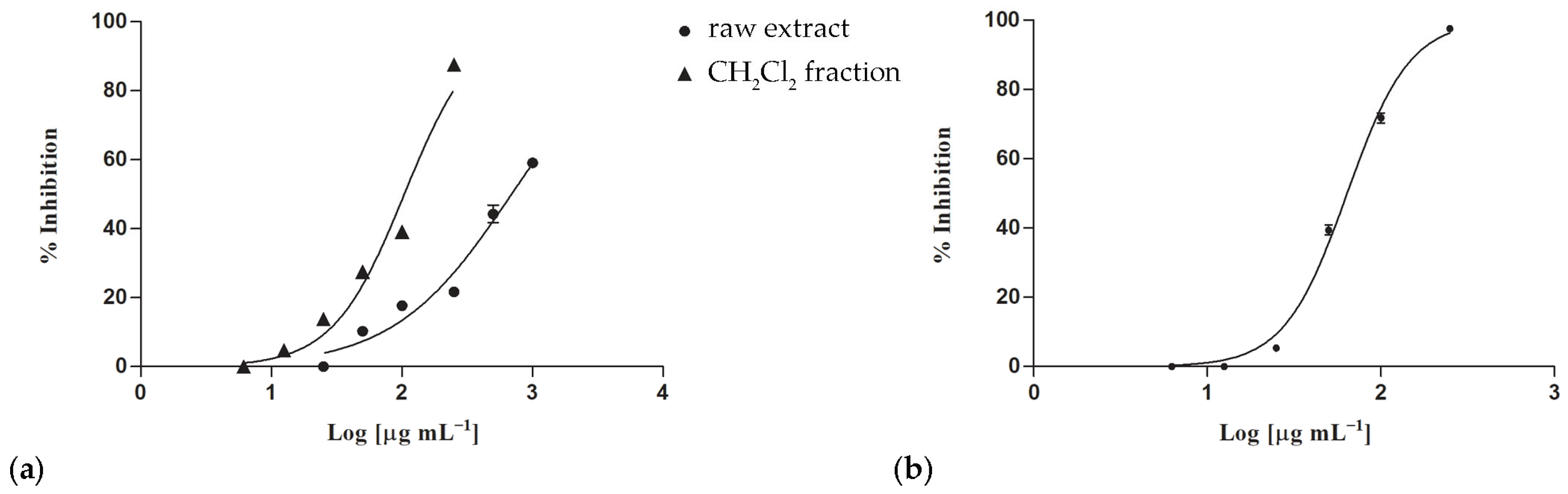
Figure 1. Inhibitory properties on nitric oxide production in LPS-stimulated RAW 264.7 macrophages induced by P. judaica L. subsp. judaica (a) and U. dioica L. (b) extracts and fractions. Data expressed as mean ± S.E.M (n = 3).
Applsci 16 03155 g001
Figure 2. Cell viability of LPS-stimulated RAW 264.7 cells treated with P. judaica L. subsp. judaica (a) and U. dioica L. (b) Dichloromethane fractions. C, control (untreated cells). Data expressed as mean ± S.E.M (n = 3). Significant difference versus control: *** p < 0.001 (Dunnett’s multiple comparison test).
Applsci 16 03155 g002
Figure 3. Inhibition of BSA denaturation induced by P. judaica L. subsp. judaica n-hexane (a) and dichloromethane fraction (b) and by U. dioica L. raw extract (c). Data expressed as mean ± S.E.M (n = 3). Diclofenac sodium was used as a positive control. Significant difference versus control: *** p < 0.001; significant difference versus positive control: °°° p < 0.001 (Dunnett’s multiple comparison test).
Applsci 16 03155 g003
Figure 4. Inhibitory effects on pancreatic lipase induced by U. dioica L. raw extract (a) and dichloromethane fraction (b). (c) Nonlinear regression curves. Data expressed as mean ± S.E.M (n = 3). Orlistat (0.018 mg/mL) was used as a positive control. Significant difference versus control: *** p < 0.001; significant difference versus positive control: °°° p < 0.001 (Dunnett’s multiple comparison test).
Applsci 16 03155 g004
Figure 5. PCA of chemical constituents identified with GC–MS analyses in the two Urticaceae extracts. (a) Scores plot. (b) Loading plot. PJ: Parietaria judaica L. subsp. judaica; UD, Urtica dioica L.
Applsci 16 03155 g005
Figure 6. Pearson correlation among detected chemical constituents, TPC (total phenolic content), TFC (total flavonoid content), antioxidant, nitric oxide production inhibition, and protein denaturation and lipase inhibitory properties.
Applsci 16 03155 g006
Table 1. Antioxidant activity of P. judaica L. subsp. judaica and U. dioica L. extracts and fractions.
SpeciesSampleIC50 (μg/mL)
DPPH Testβ-Carotene Bleaching Test
30 min60 min
Parietaria judaica L. subsp. judaicaRaw extract135.30 ± 3.07 e7.97 ± 0.52 b25.45 ± 1.00 d
n-hexane fractionn.a.n.a.n.a.
CH2Cl2 fraction103.00 ± 1.78 d 55.70 ± 2.08 fn.a.
Urtica dioica L.Raw extract37.72 ± 0.51 c17.84 ± 0.99 c30.12 ± 0.43 d
n-hexane fractionn.a.n.a.n.a.
CH2Cl2 fraction20.32 ± 0.27 b30.42 ± 0.92 d47.85 ± 0.62 e
Ascorbic acid 1 2.00 ± 0.01 a--
Propyl gallate 1 -1.00 ± 0.02 a 1.00 ± 0.02 a
Data are expressed as mean ± SEM (n = 3). n.a., not active. 1 Positive controls. Statistically significant differences along column (DPPH test) and between 30 and 60 min columns (β-carotene bleaching test) were assessed by one-way ANOVA and Bonferroni post hoc test.
Table 2. In vitro inhibitory properties of P. judaica L. subsp. judaica and U. dioica L. extracts and fractions on NO production.
SpeciesSampleIC50 (μg/mL)
NO InhibitionCytotoxicity
Parietaria judaica L. subsp. judaicaRaw extract690.3 ± 11.2 en.c.
n-hexane fractionn.a.n.c.
CH2Cl2 fraction104.2 ± 2.7 b277.4 ± 6.9 c
Urtica dioica L.Raw extractn.a.n.c.
n-hexane fractionn.a.n.c.
CH2Cl2 fraction63.9 ± 1.8 a,b498.7 ± 10.9 d
Indomethacin 1 58.0 ± 0.9 a-
L-NAME 1 45.9 ± 0.5 a-
Data are expressed as mean ± SEM (n = 3). n.a., not active. n.c., not cytotoxic. 1 Positive controls. Statistically significant differences were assessed by one-way ANOVA and Bonferroni post hoc test (p < 0.05).
Table 3. In vitro anti-denaturation potential of P. judaica L. subsp. judaica and U. dioica L. extracts and fractions.
SpeciesSampleBSA Denaturation Inhibition IC50 (μg/mL)
Parietaria judaica L. subsp. judaicaRaw extractn.a.
n-hexane fraction321.6 ± 3.3 b
CH2Cl2 fraction424.4 ± 1.8 c
Urtica dioica L.Raw extract869.5 ± 8.2 d
n-hexane fractionn.a.
CH2Cl2 fractionn.a.
Diclofenac 1 15.7 ± 0.2 a
Data are expressed as mean ± SEM (n = 3). n.a., not active. 1 Positive control. Statistically significant differences were assessed by one-way ANOVA and Bonferroni post hoc test (p < 0.05).
Disclaimer/Publisher’s Note: The statements, opinions and data contained in all publications are solely those of the individual author(s) and contributor(s) and not of MDPI and/or the editor(s). MDPI and/or the editor(s) disclaim responsibility for any injury to people or property resulting from any ideas, methods, instructions or products referred to in the content.