Review

Impact of Quercetin on Bone-Related Diseases

by
Paweł Polak
1,
Magdalena Dragan
2,
Antoni Wojciech Oniszczuk
2,
Emilia Skurko
2,
Kamila Kasprzak-Drozd
3,*,
Przemysław Niziński
4,
Anna Oniszczuk
3,* and
Karolina Wojtunik-Kulesza
3
1
Department of Orthopedics and Traumatology, Provincial Specialist Hospital in Biała Podlaska, Terebelska 57, 21-500 Biała Podlaska, Poland
2
Science Circle of the Department of Inorganic Chemistry, Medical University of Lublin, Dr. Witolda Chodźki 4a, 20-093 Lublin, Poland
3
Department of Inorganic Chemistry, Medical University of Lublin, Chodźki 4a, 20-093 Lublin, Poland
4
Department of Pharmacology, Medical University of Lublin, Radziwiłłowska 11, 20-080 Lublin, Poland
*
Authors to whom correspondence should be addressed.
Appl. Sci. 2026, 16(7), 3151; https://doi.org/10.3390/app16073151
Submission received: 5 March 2026 / Revised: 21 March 2026 / Accepted: 23 March 2026 / Published: 25 March 2026
(This article belongs to the Special Issue Innovations in Natural Products and Functional Foods)

Abstract

Quercetin (QE) is a widely distributed dietary flavonol with antioxidant and anti-inflammatory properties that has attracted interest as a modulator of bone remodeling and osteoporosis-related bone loss. In vitro data on osteoblasts, osteoclasts, and mesenchymal stem cells indicate that QE attenuates oxidative stress, suppresses pro-inflammatory signaling, and promotes osteogenic differentiation through modulation of pathways such as Nrf2/ARE, NF-κB, Wnt/β-catenin, and ER stress-related cascades. In vivo findings from animal models of estrogen deficiency, diabetes, and glucocorticoid-induced osteoporosis demonstrate that QE improves bone mineral density, trabecular microarchitecture, and biomechanical strength while reducing osteoclast number and activity, thereby attenuating osteoporotic bone deterioration. Collectively, preclinical evidence positions QE as a pleiotropic agent promoting osteoblastogenesis, inhibiting osteoclastogenesis, and balancing redox/inflammatory homeostasis in bone, despite bioavailability challenges. Future research should prioritize clinical trials with optimized formulations (e.g., nanoparticles) to validate efficacy, safety, and fracture outcomes in humans. The present review critically evaluates the chemical characteristics, pharmacokinetics, safety profile, and bone-targeted biological activity of QE, emphasizing effects on bone cells and skeletal metabolism.

1. Introduction

Quercetin (QE) is a polyphenolic compound identified chemically as 3,3′,4′,5,7-pentahydroxyflavone. It cannot be endogenously synthesized in the human body [1]. The name ‘quercetin’ comes from quercetum (it is a Latin word that refers to a specific type of oak wood or forest) and has been used since 1857 [2]. In nature, it occurs in multiple chemical forms, mostly as glycosides. This flavonoid is predominantly found in fruits, vegetables, nuts and seeds. Red onions are considered the most bioavailable food source of it. Identification and quantification of quercetin typically employ HPLC methods with acetic acid-based mobile phases, validated for precision in nanoparticles and biological samples. After consumption, QE undergoes digestion in the gastrointestinal tract and liver. Its metabolites can be detected in biological fluids such as urine or blood. Dietary supplements usually contain aglycone, the free state of quercetin. QE demonstrates antioxidant, anti-inflammatory, antiviral, anti-obesity, and antidepressant action. Moreover, it prevents cancer, diabetes, asthma, hypertension, and cardiovascular diseases [3].
The use of this substance is particularly noteworthy in the treatment of osteoporosis. Osteoporosis is a disorder of the skeletal system that significantly influences the quality of a patient’s life. This illness affects over 200 million women worldwide, with one-tenth of women aged 60 and one-fifth aged 70 impacted, contributing to a global health crisis where hip fractures are projected to double by 2050. The risk of this condition increases with age, and it is definitely more common among postmenopausal women. It leads to reductions in bone mass and consequently increases bone fragility. Osteoporosis typically affects the hip, spine and wrist regions [4,5].
The subject of QE is a popular and promising topic nowadays. The first studies about the positive impact of QE on bones are from 2008. Over the years, the topic gained more and more attention [6]. However, the majority of research in this field comprises experiments conducted in vitro, in silico, or utilizing animal models. This poses a significant challenge to the implementation of modern therapeutic interventions, as the findings derived from animal studies often do not accurately reflect the unique treatment requirements of human subjects. No human clinical trials specifically on quercetin’s bone-protective effects were identified in the literature; the evidence remains predominantly preclinical, including in vitro studies on osteoblasts/osteoclasts/MSCs and in vivo models like ovariectomized rats, diabetic rats, and glucocorticoid-induced osteoporosis. Therefore, the full validation of all methods used in treatments involving QE is impossible [7]. Nevertheless, the research on the subject under discussion is becoming more and more promising with each passing day.
This review analyzed the biological and pharmacological properties of QE. It is based on the in vitro and in vivo research conducted in recent years. The aim of this article is to understand the mechanisms of QE’s actions on the bones and skeletal system, including its influence on bone cells as well bone tissue metabolism pathways.

2. Chemical Properties, Pharmacokinetics, and Safety of Quercetin in Relation to Bone

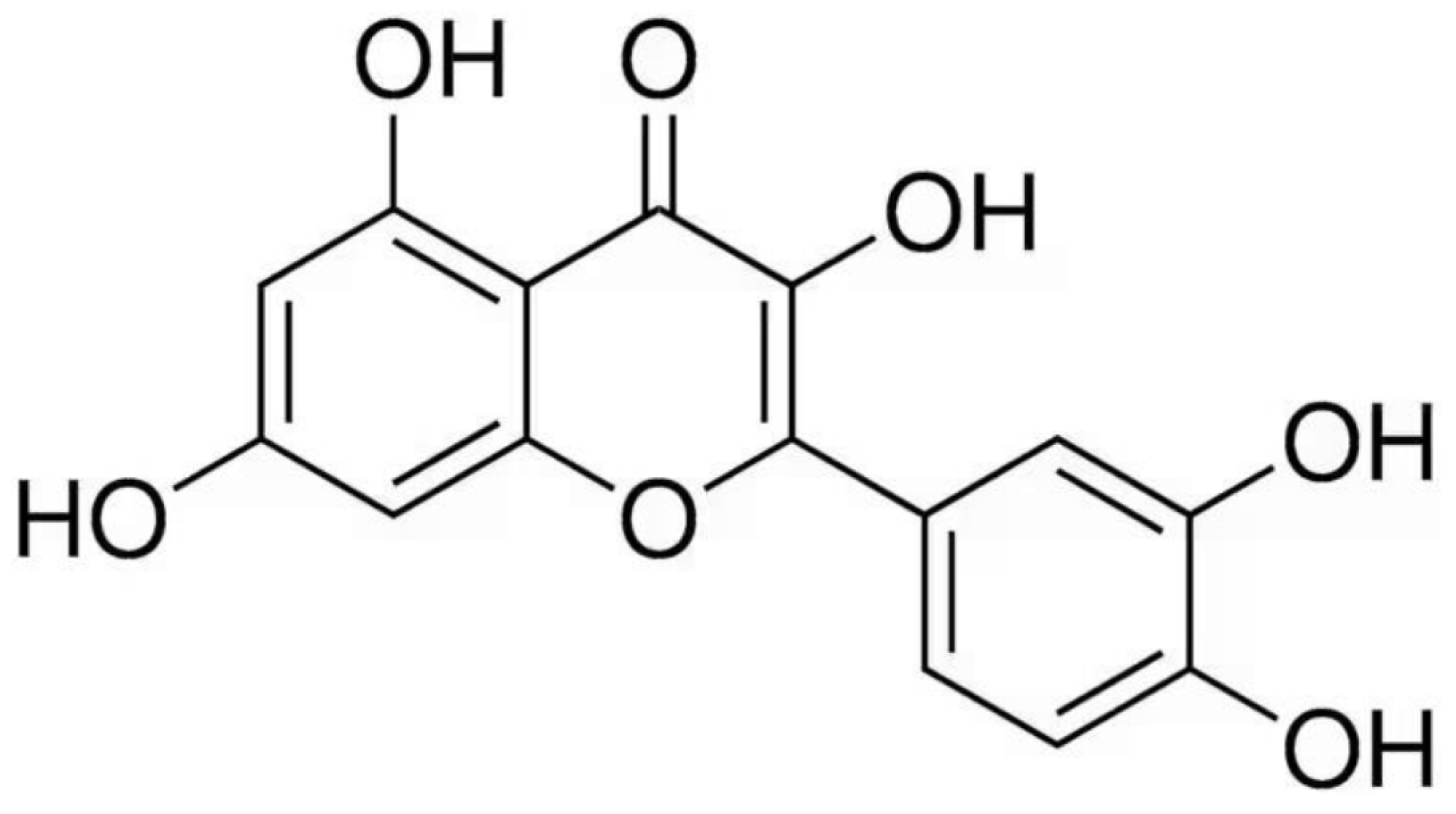
QE belongs to the group of secondary plant metabolites. It belongs to the flavonol class of flavonoids [8]. Biosynthesis of QE in plants takes place with the participation of the shikimic acid pathway and the phenylpropanoid pathway, in which anthranilate and phenylalanine, respectively, act as precursor compounds [9]. This compound is present in numerous fruits, vegetables, seeds and berries, including apples, onions, tea leaves, mangoes and other products of plant origin [10,11]. QE is one of the best-known flavonoids, with documented therapeutic properties. Due to its widespread occurrence in the human diet and its wide spectrum of biological activity, this compound is the subject of intensive research on its potential use in the prevention and treatment of many diseases. It is effective in the context of neurodegenerative diseases, cardiovascular diseases, cancer and diabetes. With respect to its chemical structure, it is a pentahydroxyflavone containing five hydroxyl groups located at positions 3, 3′, 4′, 5 and 7, which gives it a characteristic yellow color and provides it solubility in lipids and alcohol. The chemical formula of QE is presented in Figure 1. The QR molecule is composed of two aromatic rings connected by a three-carbon γ-pyran ring, to which hydroxyl groups that can undergo various substituent modifications are attached. Its strong antioxidant properties result from the polyphenolic nature of the structure, in particular from the presence of a catechol moiety in the aromatic ring B and hydroxyl groups located at positions 3 and 5 [12].
QE occurs naturally both as an aglycone and as glycosidic conjugates, with the glycoside forms being predominant in plant-derived foods. The glycosidic form of QE is formed by substituting a hydroxyl group—most often at the 3-molecule position—with a glycosyl residue such as glucose, rhamnose or rutinose. The introduction of a sugar fragment into the structure of QE results in an increase in its solubility in water compared to the aglycone form. The QE aglycone is practically insoluble in cold water and displays only limited solubility in hot water, whereas it is readily soluble in alcohols and lipid media [13].
Among dietary sources, the most prevalent forms include QE-3-O-glucoside (isoquercetin) as well as its glucuronide derivative, quercetin-3-O-glucuronide [14]. After oral administration, QE is mainly absorbed in the small intestine, especially in its upper sections, although its bioavailability is relatively low [15]. This is attributable to both its poor aqueous solubility and limited stability under gastrointestinal conditions, as well as to extensive first-pass metabolism [16].
In plant tissues, QE is primarily present in the form of hydrophilic glycosidic derivatives. These compounds can be hydrolyzed to a free aglycone form under the influence of β-glucosidases located in the small intestine, which promotes its absorption by passive diffusion through the intestinal epithelial barrier [17]. It is also assumed that intestinal sodium/glucose cotransporter-1 is involved in the absorption process, which allows direct transport of glycosidic forms of QE to the systemic circulation. After absorption, QE may undergo oxidation reactions, leading to the formation of reactive metabolites, such as quercetin–quinone and quercetin–quinone methides [18]. Upon passing through the intestinal wall, QE is rapidly biotransformed into enterocytes and hepatocytes. In these processes, phase II enzymes catalyze its coupling to various groups, forming glucuronides, sulfates, and methylated metabolites. Such conjugation significantly changes the chemical characteristics of the compound—metabolites are more polar and occur in systemic circulation in much higher concentrations than the free form (aglycone) [18,19]. As a result of metabolic transformations, conjugated QE derivatives are formed, which, compared to the parent form, are often characterized by reduced activity or a different profile of biological activity. Furthermore, QE is a substrate of efflux transporters, including P-glycoprotein, which actively remove it from enterocytes into the lumen of the intestine, leading to further limitation of its absorption [20]. The fraction of QE that has not been absorbed in the small intestine is broken down by the colon microbiota into simpler phenolic acids, which are then absorbed and transported to the liver, where further conjugation processes take place [21,22].
Early pharmacokinetic studies in humans indicated that after a single oral administration, QE has a very low bioavailability, estimated at approximately 2%. In the case of quercetin glucoside, which is its naturally occurring form, the degree of absorption in healthy volunteers at a dose of 100 mg was assessed at a level of 3% to 17% [23]. In a study by Goldberg et al. [24], oral administration of 10 mg QR (per 70 kg of body weight) in healthy men showed limited absorption, with maximum serum concentrations of total quercetin (free form + metabolites) reaching approximately 30 min after ingestion. Conjugated metabolites (glucuronides and sulfates) dominated the systemic circulation, and free quercetin accounted for only 17.2–26.9% of the maximum concentration. Total urinary excretion over 24 h was 2.9–7.0% of the ingested dose, with peak serum concentrations in the range of 10–40 nmol/L, indicating low systemic exposure [24]. The limited bioavailability of QE is most likely a consequence of its poor absorption in the gastrointestinal tract, intensive metabolic processes, and/or rapid elimination from the body [23]. The absorption efficiency also varies depending on the type of sugar attached to the QE molecule [25], the dietary matrix and other nutrients in the diet. Dietary fibers and antinutritional compounds bind quercetin, reducing its solubility and intestinal absorption; for example, high-fiber matrices slow hydrolysis of glycosides by β-glucosidases. Non-heme iron from plant foods and supplements chelates quercetin via its catechol moiety, inhibiting basolateral iron transport while mutually decreasing quercetin’s uptake. High-fat meals may enhance absorption via micelle formation, but excessive fiber or pH extremes (e.g., low gastric pH degrading to phenolic acids) further limit bioavailability to <10% [26]. In addition, it is suspected that endogenous factors may affect QE bioavailability [27].

3. Mechanisms of Action of Quercetin in the Context of Bones

It appears that QE affects bone health through a combination of pro-osteogenic, anti-resorptive, anti-inflammatory and antioxidant actions, which have been associated with the modulation of several signaling pathways in bone cells. However, as these pathways are highly interconnected and much of the available evidence comes from in vitro and animal studies, the precise contribution of each pathway to the bone-related effects of quercetin remains to be fully established. Below, selected mechanisms are briefly described and then expanded in subsequent sections.

3.1. Promotion of Osteogenesis

Osteogenesis is the process by which bone is formed. It begins with mesenchymal stem cells (MSCs) differentiating into osteoblasts, which synthesize the organic bone matrix (osteoid). This is then progressively mineralized into mature bone tissue [28]. QE can modulate osteogenesis by promoting the commitment and activity of osteoblast lineage cells, while protecting them from inflammatory and oxidative damage. The canonical Wnt/β-catenin pathway is essential for osteoblast differentiation and bone formation [29]. In the absence of Wnt ligands, β-catenin is phosphorylated by a destruction complex containing Axin, APC, CK1 and GSK-3β and subsequently degraded [30], which maintains low expression of osteogenic transcription factors such as Runx-2 and Osterix [31,32]. Although direct causality has not been established in all models, experimental data suggest that QE may influence this process by increasing Wnt3 and β-catenin levels and reducing GSK-3β activity [33]. This may allow β-catenin to accumulate and translocate to the nucleus, where it can activate the transcription of osteogenic genes via TCF/LEF [34].
Estrogen receptor (ER)-dependent signaling pathway is also of great importance. Bone morphogenetic protein 2 (BMP-2) and transforming growth factor beta (TGF-β) are central regulators of osteoblast differentiation and bone formation. They act through both canonical Smad-dependent pathways and non-canonical MAPK signaling [35]. In the canonical arm, BMP-2 or TGF-β binding to their type I and type II serine/threonine kinase receptors leads to the phosphorylation of receptor-regulated Smads (Smad1/5/8 for BMPs and Smad2/3 for TGF-β). These subsequently form complexes with Smad4 and translocate to the nucleus, which then work with osteogenic transcription factors, such as Runx2 and Osterix, to increase the expression of bone-specific genes [36]. These include alkaline phosphatase, type I collagen, osteopontin, and osteocalcin. This process promotes osteoblast maturation, extracellular matrix deposition, and mineralization [37]. Additionally, BMP/TGF-β receptors can activate ERK, p38 and JNK MAPKs [38,39,40], which modulate the phosphorylation and transcriptional activity of Runx2 and other factors, thereby further fine-tuning osteoblast differentiation and function [41]. QE appears to support this osteogenic axis at several levels; for instance, experimental data suggest that quercetin can increase the expression of BMP-2 and TGF-β and enhance Smad signaling downstream in MSCs and pre-osteoblasts. This is accompanied by the upregulation of RUNX2, osterix, osteopontin and osteocalcin, as well as increased ALP activity and matrix mineralization. This is consistent with a pro-osteoblastic effect. Notably, at least some of this response is mediated via the ER. Inhibition of ER signaling reduces quercetin-induced activation of the BMP-2/Smad pathways and decreases the expression of osteogenic markers in bone-marrow mesenchymal stem cells (BMSCs), therefore supporting the idea that QE acts as a phytestrogen, amplifying BMP/TGF-β-driven osteoblastogenesis under estrogen-deficient conditions [42,43].

3.2. Suppression of Osteoclastogenesis

The RANKL/RANK/OPG system is the primary regulatory axis of osteoclast differentiation and bone resorption [44]. RANKL, a protein produced by osteoblasts, osteocytes and immune cells, binds to RANK on osteoclast precursors, recruiting TRAF6 and activating NFκB, MAPKs and AP-1. These, in turn, induce NFATc1 and c-Fos, thereby driving the expression of osteoclast genes such as TRAP, cathepsin K and MMP9, which are necessary for cell fusion and matrix degradation [45,46]. Osteoprotegerin (OPG) functions as a soluble decoy receptor, sequestering RANKL. Consequently, the local RANKL/OPG ratio dictates the extent of osteoclastogenesis, and its imbalance is a hallmark of high turnover osteoporosis and a pivotal therapeutic target of anti-resorptive drugs [47]. QE has been demonstrated to modulate this axis by lowering RANKL and/or increasing OPG expression in osteoblast-lineage cells, thereby reducing the RANKL/OPG ratio [48]. In addition, it has been shown to directly inhibit RANKL-induced NF-κB/MAPK/AP-1 activation in osteoclast precursors, thus attenuating NFATc1 and osteoclast effector genes [5]. In the context of functional assays, these effects are manifested as a reduction in the number of TRAP-positive multinucleated osteoclasts, an impairment in F-actin ring formation, and the presence of smaller resorption pits [49]. In addition, the data demonstrate a decrease in osteoclast numbers and the preservation of trabecular microarchitecture in animal models, thus supporting the hypothesis that RANKL/RANK/OPG signaling is a significant anti-resorptive target of QE.

3.3. Integrated Control of Inflammation and Oxidative Stress via NF-κB and Nrf2

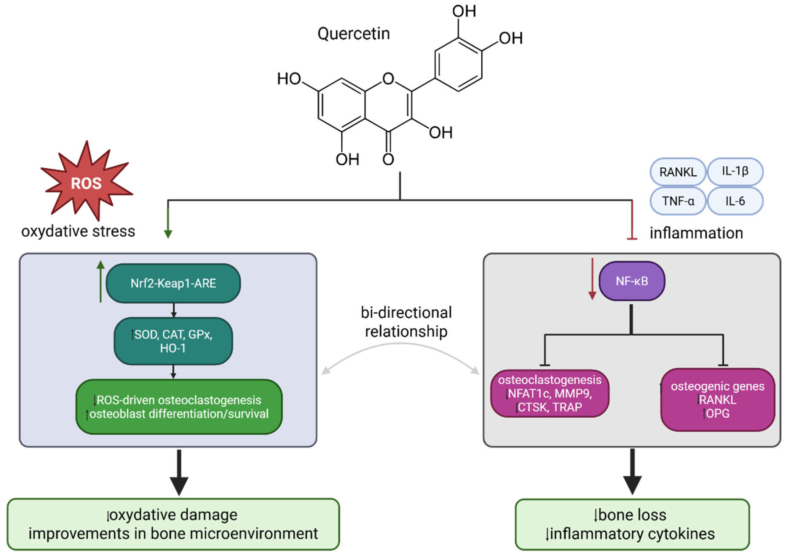
It is evident that chronic, low-grade inflammation and oxidative stress play a pivotal role in the process of bone loss [50]. The underlying mechanism involves the simultaneous promotion of osteoclastogenesis and impairment of osteoblast differentiation and survival by pro-inflammatory cytokines and reactive oxygen species (ROS), respectively [51,52]. NF-κB is a central transcription factor in this context: it is activated downstream of RANKL, TNF-α, IL-1β and IL-6 in osteoclast precursors and osteoblast-lineage cells, controlling genes essential for osteoclast formation and osteoblast function [5,52]. In osteoclast precursors, RANKL–RANK–TRAF6 signaling rapidly induces NF-κB, which cooperates with NFATc1 and c-Fos to drive the expression of osteoclast-specific genes and resorptive activity [5,51]. Conversely, in osteoblasts, NF-κB activation suppresses osteogenic programs and increases RANKL expression, thereby shifting bone remodeling towards resorption [53]. In turn, the Nrf2-Keap1-ARE pathway has been identified as a pivotal endogenous defense mechanism against oxidative stress in bone tissue [54]. In the presence of oxidative or electrophilic stress, Nrf2 dissociates from Keap1, translocates to the nucleus, and induces antioxidant and cytoprotective enzymes, including superoxide dismutase (SOD), catalase, glutathione peroxidase, and heme oxygenase-1 (HO-1). These enzymes function to limit ROS-induced damage in osteoblasts, osteocytes, and osteoclast precursors [55]. The results of experimental studies suggest that activation of Nrf2 provides protection to osteoblasts against apoptosis induced by ROS, whilst also promoting osteogenic differentiation and suppressing the process of osteoclastogenesis [56]. However, it has been observed that Nrf2 deficiency is associated with reduced bone mass and impaired trabecular structure [57]. What is more, Nrf2 and NF-κB are not independent—there is well-described, bidirectional crosstalk in which Nrf2 deficiency can enhance NF-κB-driven inflammatory output, whereas NF-κB activity can also modulate Nrf2 transcriptional function, linking redox balance with inflammatory tone [58]. The available evidence suggests that QE may modulate both NF-κB and Nrf2 signaling. However, given the close interaction between inflammatory and redox-sensitive pathways, these effects should be considered as part of an interconnected regulatory network, rather than as fully independent and directly established mechanisms [1]. It has been demonstrated to reduce IκB phosphorylation and NF-κB nuclear translocation in macrophages, osteoclast precursors and osteoblasts, which in turn lowers the production of TNF-α, IL-1β and IL-6, decreases RANKL expression and attenuates RANKL-induced osteoclastogenesis both in vitro and in vivo [59,60,61]. Concurrently, QE (in the form of glycoside isoquercitrin) activates Nrf2, elevates the expression of downstream antioxidant enzymes, and curtails biomarkers of oxidative damage (e.g., malondialdehyde) in osteoclasts, thereby promoting osteogenic differentiation and safeguarding these cells under conditions of oxidative or inflammatory stress [62]. Although available studies suggest that quercetin may modulate signaling pathways such as Nrf2/ARE, NF-κB, Wnt/β-catenin and ER stress-related cascades, these mechanisms should be interpreted with caution. These pathways are closely interconnected, and most of the current evidence originates from in vitro systems and animal models. An overview of the probable role of QE in inflammatory and oxidative stress reactions in bone tissue is shown in Figure 2.

4. In Vitro Model Data Supporting Quercetin’s Bone-Preserving Effects

In vitro determination of QE’s influence on bone cells is mainly linked with MSCs, murine or human osteoblast cells, which can be used for investigation of its effect on cell viability, proliferation, mineralization and expression of osteogenic genes [5]. MSCs are adult stem cells able to differentiate into osteoblasts, chondrocytes, myocytes, adipocytes, neurocytes and hepatocytes [5,63]. A significant aspect of bone metabolism is estrogen, a hormone with a pivotal impact on bone mineral density. Its level is associated with menopause, when a decrease in estrogen contributes to an increase in osteocyte apoptosis, leading to osteoporosis [64].
The influence of QE on osteocytes, osteoblasts and osteoclasts was presented by Khosla et al., who proposed that bone resorption and maintenance of bone formation are decreased by estrogen [65]. Significant in vitro studies were performed by Pang et al. [42], who decided to investigate the influence of QE on mouse bone-marrow mesenchymal stem cell (BMSC) proliferation and osteogenic differentiation. Scientists revealed that the flavonoid has a positive impact on BMSC proliferation, alkaline phosphatase (ALP) activity, extracellular matrix production and mineralization. The significant enhancement of BMSC proliferation was observed at concentrations ranging from 0.1 to 5 µM. Similar effects of quercetin have been detected previously by Zhou et al. [66], who revealed increased cell proliferation on days 1, 4 and 7 of their experiment, with 2 µM achieving the greatest effect. The evaluation was based on rBMSC isolated from 4-week-old male rats, which were used for future in vitro analyses. To evaluate the hypothesis that quercetin can induce osteogenic differentiation of BMSCs, the flavonoid was applied to rBMSCs at various concentrations. Its influence on proliferation, osteogenic differentiation and expression of angiogenic factors was evaluated. The most significant effect of the compound was observed at the concentration 2 µM. Additionally, extracellular signal-regulated protein kinases (ERK) were observed. An equally important activity of the quercetin hydrate (QH) was observed during in vitro studies towards osteoblast cell proliferation and differentiation using pre-osteoblastic cell lines MC3T3-E1 and MG-63 at two concentrations [67]. Significant increases in cell proliferation and viability at both concentrations, as well as more mineral matrices, were observed. These results indicate that QH can enhance osteoblast differentiation, improve matrix structuring, and development in vitro, as well as support bone health through anabolic effects on the skeleton. The positive influence of QE on osteoblast differentiation and expression of osteogenesis-related markers was observed at concentrations of the flavonoid ranging from 0.01 to 5 µM [68]. Positive effects of QE were also observed in studies based on MC3T3-E1 osteoblasts stimulating lipopolysaccharide (LPS) [69]. The effect suggests that the flavonoid can be an effective compound in case of abnormal human bone loss induced by LPS in chronic inflammatory diseases. Similarly important is evaluation of the compound as a potential agent in the prevention and treatment of osteoporosis. Detailed analyses of QE and rutin, individually and in combination, showed enhanced cell proliferation, osteoblastic differentiation and mineralization in UMR 106 cells, an epithelial-like cell that was isolated from the bone of a rat with osteosarcoma that is widely used as an osteoblast-like model [70]. The results, along with in vivo experiments, suggest that QUE is a promising candidate for treatment of glucocorticoid-induced osteoporosis.
Significant studies have been performed using the human fetal osteoblastic cell line (hFOB) that aimed to evaluate the effect of QE on increases in cell numbers [71]. Various tests, including MTT and trypan blue, confirmed the hypothesis that the flavonoid positively increases cell numbers and viability. Additionally, wound healing and cellular adhesion in osteoblasts were observed for 1 µM QE. It is interesting to note that QE at concentrations of 20–100 µM reduced cell number and viability in tumor osteoblasts (ROS 17/2.8 cells). Interesting results were also presented by Kim et al. [72], who focused on inhibition of cell proliferation and increasing osteogenic differentiation in human adipose stromal cells (hADSC). The findings indicate that the flavonoid is able to increase osteogenic differentiation as well as bone regeneration. The results suggest that quercetin’s effects are independent of estrogen receptor activation, providing insight into its mechanism of action. Interestingly, plants rich in QE have a positive impact on bone cells. An example is that the ethanolic fraction of Cissus quadrangularis can be enriched by rutin and quercetin (1.06 ± 0.12 mg/g). Human osteoblast-like MG-63 cells grown in this medium revealed higher ALP levels [73].
Flavonoids are characterized by high antioxidant and free radical scavenging activity. This feature can be used in bone therapy. It is known that QE provides protection against H2O2-, cigarette- and menadione-induced oxidative stress in MC3T3-E1 osteoblast cells and primary human osteoblasts [60,74].
A significant number of studies focus on osteoclastogenesis. Numerous results suggest that QE inhibits the formation of osteoclast-like cells, bone resorption pi and F-actin ring formation in RAW264.7 cells, human peripheral blood mononuclear cells (PBMCs) and bone-marrow macrophages treated with macrophage colony-stimulating factor (M-CSF) [75,76,77,78].
QE has been shown to reduce hydroxylysylpyridinoline levels in human peripheral blood mononuclear cells stimulated with M-CSF and RANKL, as well as in highly purified rabbit osteoclasts [61,79]. In addition, treatment with quercetin at concentrations of 15, 25 or 50 μM inhibited lipopolysaccharide-induced osteoclast differentiation and cell viability in RAW264.7 cells [80]. The effects of QE on bones, along with detailed findings, are presented in Table 1. Overall, most published studies indicate that quercetin promotes osteoblast differentiation, although a small number of reports show contrasting results. Such inconsistencies may stem from differences in the cell models employed and the concentrations of quercetin applied. Additionally, existing evidence indicates that quercetin consistently suppresses osteoclast differentiation, growth and maturation. Future investigation into the molecular mechanisms responsible for quercetin’s osteogenic effects is warranted.

5. In Vivo Evidence on the Impact of Quercetin on Bones

The potential of QE in promoting bone health has been the subject of extensive research, with numerous in vivo studies consistently demonstrating its protective properties in various models of osteoporosis and osteopenia. These experiments have examined the ability of quercetin to counteract reductions in bone mass in animals experiencing deficiencies in sex hormones, diabetes induced by streptozotocin, or glucocorticoid-induced decreases in bone density [4]. Ovariectomised (OVX) female rats have been the most commonly used model of postmenopausal osteoporosis [5]. As demonstrated by Yuan et al. [81], oral administration of QE at a dose of 50 mg/kg/day for eight consecutive weeks in OVX rats produced a notable improvement in bone quality parameters. Specifically, the supplementation of quercetin resulted in a significant enhancement of femoral bone mineral density, trabecular number (Tb.N), and trabecular thickness (Tb.Th), indicating a marked attenuation in estrogen deficiency-induced bone loss. Furthermore, biomechanical analysis of the femur demonstrated enhancements in both bone elasticity and maximal loading capacity, indicative of enhanced structural integrity and resistance to fracture. A subsequent study reported that the administration of continuous QE at an equivalent daily dose over a 30-day period resulted in a decline in the enzymatic activities of acid phosphatase and alkaline phosphatase, which are markers associated with bone resorption and formation, respectively. Concurrent with these biochemical changes, serum calcium and phosphorus concentrations increased significantly, indicating a positive modulation of mineral metabolism and an overall shift towards bone preservation [93]. In another experiment, the osteoprotective potential of quercetin was subjected to further evaluation in comparison with alendronate, the standard pharmacological therapy for postmenopausal osteoporosis [94]. This study employed a rat model of mandibular osteoporosis induced by ovariectomy to assess the relative efficacy of the two treatments in mitigating bone degeneration under estrogen-deficient conditions. The animals received either QE (50 mg/kg/d) or alendronate (6.25 mg/kg/w) by oral gavage for a period of eight weeks. The administration of the aforementioned substances to control groups occurred via intraperitoneal injection following bilateral ovariectomy, and the injection consisted of chloroquine (10 mg/kg, twice weekly) or the cytokine release inhibitor MCC950 (10 mg/kg, thrice weekly). It has been demonstrated that the ingestion of QE has a number of significant effects on the body. These include limiting body weight gain, improving bone microarchitecture, increasing bone mass and bone mineral density, reducing trabecular spacing and decreasing osteoclast numbers. The ability of QE to inhibit interleukins 1β and 18 was found to result from its capacity to downregulate the NLRP3 inflammasome pathway, thereby preventing osteoclast differentiation. Furthermore, QE was found to suppress autophagy and reduce the number of tartrate-resistant acid phosphatase-positive osteoclasts in OVX rats. These findings suggest that QE can preserve skeletal structure and mitigate bone loss in rats with osteoporosis. In a seminal study, Feng et al. [95] elucidated the molecular mechanisms underlying QE’s effects on the activation of the Wnt/β-catenin pathway and the suppression of NF-κB signaling. These observations counteract the inhibitory effect of tumor necrosis factor alpha (TNFα) on BMSC osteogenesis. In OVX-induced osteoporotic mice, QE significantly mitigated bone loss and structural damage and restored serum Metastasis Associated Lung Adenocarcinoma Transcript 1 (Malat1) levels. These findings indicate that QE has the capacity to reverse TNFα-induced impairment of BMSC differentiation and osteoporosis-related bone deterioration, a process that is dependent on Malat1. This suggests that QE has the potential to be a valuable therapeutic agent for the treatment of osteoporosis.
QE has also been reported to possess bone-preserving properties when administered via nanoparticles or hydroxyapatite bioceramic microspheres in models of osteoporosis. The utilization of delivery systems has been shown to enhance the bioavailability and bioactivity of therapeutic agents through several mechanisms, including the prolongation of half-life, the optimization of particle size, and the facilitation of controlled release [96]. In female albino rats subjected to bilateral osteoarthritis, the oral administration of 10 or 50 milligrams of QE-loaded phytosome nanoparticles per kilogram of body weight for 30 days had a marked effect on bone metabolism, as indicated by reduced bone turnover markers and increased serum mineral content [93]. In a separate investigation, QE was incorporated into hydroxyapatite bioceramic microspheres to create a composite delivery system designed for localized and sustained release within bone tissue. These QE-loaded hydroxyapatite microspheres were implanted into surgically induced bone defects located in the distal femoral diaphysis of OVX rats, a well-established model for postmenopausal osteoporosis. After an eight-week implantation period, microstructural analysis revealed a pronounced improvement in bone mineral density and trabecular thickness (Tb.Th) at the defect site, indicating enhanced new bone formation and mineralization. Moreover, the study demonstrated that QE exerted a dual biological effect by promoting the proliferation of BMSCs and stimulating their osteogenic differentiation. In parallel, QE upregulated the expression of angiogenic factors, thereby facilitating the coupling of osteogenesis and angiogenesis in the repaired tissue. Importantly, treatment with QE restored the receptor activator of nuclear factor kappa B ligand (RANKL)/osteoprotegerin (OPG) balance in a dose-dependent manner, suggesting a regulatory effect on bone remodeling. Mechanistically, several signaling cascades, including the extracellular signal-regulated kinase (ERK), p38 mitogen-activated protein kinase (MAPK), and protein kinase B, were implicated in mediating these effects. Available evidence also points to potential crosstalk among these pathways, contributing to the integrated regulation of osteoblastic activity and vascularization. Furthermore, the hydroxyapatite microspheres featuring a micro-nano hybrid surface provided a controlled and sustained release of QE, which amplified its osteoinductive capacity and angiogenic potential in vivo. It can be concluded that hydroxyapatite-based microspheres have the potential to function as effective carriers for the delivery of QE. Additionally, they can be considered as prospective bone graft substitutes for the purpose of promoting osteoporotic bone regeneration [48]. In contrast, the dietary supplementation of phytochemical blends in ovariectomised aged Fischer 344 rats failed to show any anti-osteoporotic effects [5]. In one study, the experimental design consisted of two distinct treatment groups, each receiving a diet supplemented with a specific combination of bioactive compounds. The first group was fed a high-dose formulation containing 1000 mg/kg of QE, 2400 IU/kg of vitamin D3, 500 mg/kg of genistein, and 200 mg/kg of resveratrol. The second group received a low-dose diet, which included 2000 mg/kg of QE, 2400 IU/kg of vitamin D3, 1000 mg/kg of genistein, and 400 mg/kg of resveratrol. Despite four weeks of dietary intervention, neither formulation produced a statistically significant change in femoral bone mineral density or bone microarchitectural parameters. The lack of noticeable bone-protective effects, in contrast to findings from other studies, may be attributed to potential antagonistic or competitive interactions among the constituent polyphenols and micronutrients in the mixture, which could have diminished QE’s osteoprotective efficacy [97]. Further insight into the structure–activity relationship of QE derivatives was provided by research on quercitrin, a naturally occurring QE glycoside formed through conjugation with the deoxy sugar rhamnose. In an experiment conducted by Xing et al. [98], ovariectomized female Sprague-Dawley rats were orally administered quercitrin at doses of 50, 100, or 200 mg/kg/day for a period of 60 days. The treatment elicited marked improvements in bone mechanical and biochemical parameters: quercitrin significantly elevated bone mineral density at the distal femur, increased maximum energy absorption, and enhanced fracture load and stiffness at the femoral neck.
Fayed et al. [99] provided further valuable insights into the anti-osteoporotic properties of isoquercitrin by investigating its effects in OVX rat model. In their study, isoquercitrin was administered orally at varying doses, with the highest dose (60 mg/kg/day) producing the most pronounced improvements in bone strength. Specifically, this treatment significantly enhanced lumbar vertebral compression strength, suggesting that isoquercitrin contributes to increased bone density and improved biomechanical resilience of osteoporotic bone tissue. Moreover, biochemical analyses revealed that treatment with isoquercitrin led to a reduction in bone turnover markers, indicating a beneficial modulation of bone remodeling dynamics. This implies that isoquercitrin can suppress excessive bone resorption while promoting bone formation, thereby restoring the balance between these two crucial processes in osteoporotic conditions. The choice of isoquercitrin in this study was supported by earlier findings highlighting its superior pharmacokinetic and pharmacodynamic characteristics compared to its parent flavonol, quercetin, and its glycoside analog, quercitrin. Previous research has shown that isoquercitrin possesses greater bioavailability, allowing for more efficient absorption and systemic distribution, as well as higher antioxidative capacity, which enhances its ability to counteract oxidative stress—a critical factor contributing to osteoblast dysfunction and bone loss. These properties make isoquercitrin a particularly promising flavonoid derivative for the prevention and management of osteoporosis [100].
There is a robust body of evidence indicating a strong correlation between diabetes mellitus and osteopenia, a heightened propensity for bone fractures, and a protracted healing process following bone fractures. The potential of QE to mitigate diabetes-associated bone loss has been investigated using streptozotocin (STZ)-induced diabetic rat models in two independent studies. In the first investigation, experimental diabetes was induced via a single intraperitoneal (i.p.) injection of STZ at a dose of 50 mg/kg. Following eight weeks of hyperglycemia, the diabetic rats received QR treatment (15 mg/kg/day, i.p.) for a subsequent period of four weeks. Evaluation of bone structural and mechanical parameters revealed that QE administration significantly enhanced bone volume fraction, trabecular number, and trabecular thickness. Moreover, biomechanical testing indicated improved maximum load-bearing capacity at both the femoral mid-diaphysis and femoral neck. Concurrently, treated animals exhibited elevated calcium and magnesium concentrations in bone tissue compared with untreated diabetic controls. In addition to these skeletal effects, QE effectively lowered blood glucose levels while restoring plasma insulin content, underscoring its dual role in glycemic regulation and bone protection under diabetic conditions [101]. In a subsequent study, a modified diabetic model was established in male Sprague-Dawley rats through intraperitoneal administration of STZ at 100 mg/kg on two consecutive days. After four weeks of diabetes development, the animals received QE at doses of either 30 mg/kg or 50 mg/kg daily for an eight-week treatment period. Untreated diabetic rats demonstrated markedly elevated serum glucose levels, disrupted bone remodeling, and deterioration of bone microarchitecture. Histomorphometric analyses revealed thinning and disorganization of trabecular bone, accompanied by reductions in mechanical strength and alterations in serum bone turnover markers. Remarkably, QR supplementation reversed many of these degenerative changes, improving structural integrity and restoring bone formation–resorption balance toward values observed in healthy controls [102].
Elevated production of ROS, in conjunction with disrupted glucose metabolism, severely impairs the bioenergetic efficiency and adenosine triphosphate (ATP) synthesis capacity of MSCs derived from osteoporotic bone. These alterations compromise osteogenic potential and cellular regeneration within the osteoporotic microenvironment. To address these metabolic and oxidative challenges, Chen et al. [103] engineered a multifunctional implant system by fabricating calcium carbonate–quercetin–chromium (CaCO3-QR-Cr) nanoparticles through ion coordination chemistry and embedding them into a ROS-responsive gelatin/chitosan coating applied to a titanium substrate (designated Ti/Gel/CaCO3-QR-Cr). The conceptual design aimed to synergistically enhance ATP production and mineralization in osteoporotic MSCs by integrating the antioxidative capacity of QE with the glucose metabolism-modulating properties of the QR-Cr complex. The Ti/Gel/CaCO3-QR-Cr interface was engineered to undergo controlled degradation in response to elevated ROS levels characteristic of osteoporotic bone tissue. This degradation behavior facilitated a sustained, localized release of CaCO3-QE-Cr nanoparticles into the surrounding microenvironment for over 28 days. Functionally, this controlled release system effectively scavenged excess intracellular and extracellular ROS, thereby alleviating oxidative stress, while concurrently enhancing glucose uptake and metabolic flux in osteoporotic MSCs. As a result, cellular bioenergetics were restored, and osteogenic differentiation was markedly improved, leading to enhanced formation of mineralized extracellular matrix. In in vivo assays, titanium implants modified with the Ti/Gel/CaCO3-QE-Cr composite coating significantly accelerated new bone formation and improved bone-implant contact area compared with unmodified controls. These findings demonstrate not only increased osteogenic activity around the implant site but also superior mechanical stability and integration between the titanium surface and host bone under osteoporotic conditions. This study introduces an innovative biomaterial platform that couples antioxidative and metabolic regulation for the targeted restoration of osteoporotic bone function. It also establishes a promising foundation for the rational design of next-generation titanium-based implants capable of actively promoting osseointegration in compromised metabolic bone environments.
Glucocorticoid therapy is among the most frequent causes of secondary osteoporosis [104]. The experimental studies were conducted in which glucocorticoids were administered subcutaneously (s.c.) on repeated occasions to induce an osteoporotic phenotype in rats. In the first test, female Sprague-Dawley rats were given a drug that made them lose bone. Then they were given a different drug called QE. QE treatment had a positive effect on a number of bone-related factors, including increasing Tb, Th, the number of osteoblasts and the moment of inertia. It also strengthened the bone and increased osteocalcin levels, as this study pointed out [105]. A promising strategy for treating osteoporosis is represented by topical delivery of quercetin via deformable transfersomes [106]. These transfersomes exhibit favorable physicochemical properties, including high entrapment efficiency, small particle size, an appropriate zeta potential and a low polydispersity index. The demonstration of excellent skin permeation in rat models was made by the transfersomes when incorporated into chitosan films. In a glucocorticoid-induced osteoporosis rat model, bone loss was evident by day 30. Compared to the positive control group, substantial improvements were seen in the treatment initiated on day 45, which involved the use of chitosan films loaded with quercetin transfersomes. It is interesting to note that the thickness, length, and density of the thigh bone (the femur) increased a lot. This increase was also accompanied by higher levels of calcium, phosphorus, an enzyme called alkaline phosphatase, and another enzyme called tartrate-resistant acid phosphatase in the blood. The tensile strength of osteoporotic femurs was restored to levels comparable with those of healthy controls, and histomicrographic analysis revealed reduced bone disruption and lytic lesions. These outcomes reflect quercetin’s ability to suppress osteoclastogenesis and osteoblast apoptosis, thereby increasing osteoblast numbers and enhancing bone mineralization. Overall, QE-loaded transfersome–chitosan films represent a superior alternative to oral QE administration, offering convenient ‘anytime, anywhere’ application—such as via a wristband—even during work, thereby improving patient adherence. Subsequent research employed network pharmacology analyses alongside in vivo models to elucidate the mechanistic basis of QE anti-osteoporotic effects [107]. This approach identified 55 putative molecular targets linked to QE’s therapeutic action, with the TNF signaling pathway emerging as a central mediator of its pharmacological benefits. Molecular docking simulations confirmed particularly robust binding affinities for IL1 β, RelA, and NFKB1 (ranking as the top three), providing a molecular rationale for QR’s capacity to modulate inflammatory and bone remodeling cascades in osteoporosis. Complementary validation through zebrafish models and quantitative PCR assays yielded results consistent with these predictions, reinforcing the pathway’s involvement. Collectively, these findings offer a comprehensive framework for QE’s anti-osteoporotic mechanism, laying a robust foundation for targeted mechanistic investigations and potential clinical translation. Beyond the common forms of osteoporosis associated with estrogen deficiency, diabetes, or aging, quercetin has also demonstrated therapeutic potential in bone loss induced by other pathological conditions. Osteoporosis and an increased susceptibility to fractures frequently occur as secondary complications in patients suffering from chronic liver diseases such as primary biliary cholangitis and hepatic cirrhosis. These conditions disrupt mineral metabolism and bone remodeling due to systemic inflammation, oxidative stress, and altered bile acid homeostasis. To explore quercetin’s protective efficacy under such hepatic stress, a rat model of cirrhosis-induced osteoporosis was established through complete bile duct ligation, which reproducibly induces cholestasis and subsequent bone deterioration [108]. In this model, administration of QE at a dose of 150 µmol/kg/day for four weeks markedly mitigated bone loss associated with cirrhosis. Morphological and biomechanical assessments revealed that quercetin treatment significantly improved both trabecular and cortical bone parameters, including increased bone volume, enhanced trabecular connectivity, and restoration of cortical thickness. Furthermore, mechanical testing indicated improved bone strength and resistance to fracture in quercetin-treated rats relative to untreated bile duct ligation controls. These findings highlight quercetin’s capacity to counteract cirrhosis-related bone deterioration, likely through its antioxidative, anti-inflammatory, and metabolic regulatory effects. They underscore the broader therapeutic potential of quercetin in managing secondary osteoporosis arising from systemic pathological stress beyond metabolic or endocrine origins [108].
Bone defects can be defined as fractures or malformations of the bone. The etiology of these conditions may be attributed to factors such as injury, tumors or infections. Recent studies have demonstrated the efficacy of QE in promoting bone healing in rats and rabbits. Song and his team [109] made a hole in the heads of female rats that was 4 mm wide. They put a special material called a scaffold into the hole. This special material was made from QE, silk, and hydroxyapatite. They also put BMSCs into the scaffold. A special type of X-ray called a ‘microcomputed tomography scan’ was used to analyze the bone. This scan was done six weeks after the implant. The scan showed that the bone was much stronger and thicker than normal. Histological assessments further confirmed that the defect sites treated with MSC-seeded QE/silk fibroin/hydroxyapatite scaffolds exhibited increased bone matrix thickness, boosted collagenous tissue formation, and enhanced tissue ingrowth. Another study looked at what happens when a special sponge made from duck’s foot collagen and hydroxyapatite is used to treat problems in the head and neck area. This sponge had a really good effect on the problems it was used to treat. Eight weeks after surgery, the treated defects showed increased bone mineral density, bone volume and new bone formation [110]. Córdoba et al. [111] conducted a study investigating the osteoregulatory effects of QE-functionalized implants using a rabbit tibial defect model. In this experiment, bilateral cylindrical drill-hole defects were created in the proximal tibiae of rabbits, and four titanium implants coated with a nanostructured layer of QE were inserted into each tibia. The quercetin nanocoating was designed to provide a localized and sustained release of the flavonoid at the bone–implant interface, thereby modulating local bone remodeling processes. Molecular analyses revealed that the expression of key osteoclast-related genes—including cathepsin K, vacuolar-type H+-ATPase, and matrix metalloproteinase-9—was markedly downregulated in bone tissue surrounding the QE-coated implants compared with uncoated controls. In parallel, serum analysis demonstrated a significant reduction in RANKL concentrations, indicating suppression of osteoclastogenesis and bone resorption activity. Complementary findings were reported by Babosová et al. [112], who evaluated the osteogenic influence of systemically administered QE in healthy, five-month-old female rabbits. QE was delivered intramuscularly at doses of 10 µg/kg or 100 µg/kg, three times per week over a defined treatment period. Quantitative histomorphometric analyses revealed a dose-dependent increase in cortical bone thickness in treated animals relative to controls, indicating that quercetin enhances cortical bone apposition even under physiological (non-pathological) conditions. These observations further support QE’s dual modulatory action—suppressing bone resorption while promoting bone formation—across both systemic and localized modes of administration.
The majority of studies conducted on this subject have demonstrated that the application of QE technology results in enhanced bone strength and health in individuals with low bone density, holes in the bone, and broken bones by increasing bone thickness and strength. The delivery of QE is typically accomplished through either oral administration, parenteral injection, or direct muscular injection. However, two studies reported unexpected negative or null results, which may be attributable to interference from co-administered phytochemicals attenuating quercetin’s bone-protective effects (this requires further confirmation) or the limited impact of QE on bone parameters in healthy, non-osteoporotic animals. The outcomes of the present study are extremely encouraging, and thus, researchers are currently conducting further investigations into the mechanisms by which QE operates directly on bone cells and the processes that regulate bone metabolism [43]. Other cases of quercetin’s effects on bone health, not described in the text, are included in Table 2.
In general, QE exhibits pronounced bone-protective effects in estrogen-deficient states, particularly in ovariectomized rodent models, where it improves BMD and trabecular microarchitecture, and reduces osteoclast number and bone loss markers [94,119]. In one recent OVX study, these effects were linked to modulation of the intestinal flora–SCFA–inflammatory axis [119]. These actions may be partly related to ER-dependent osteogenic signaling and inhibition of RANKL-driven osteoclastogenesis [42,61]. Although most studies have been conducted in female hyperestrogenic models, limited data from male diabetic and orchiectomized animals also suggest beneficial skeletal effects [101,102,116,117]. In diabetic osteoporosis models, quercetin improves bone remodeling, microarchitecture, and selected metabolic parameters related to glucose homeostasis [101,102,117]. Protective effects have also been reported in glucocorticoid-induced osteoporosis [105] and in retinoic acid-induced bone loss models [120]. However, the available evidence remains predominantly preclinical, and human data are still lacking [5].

6. Conclusions

QE and its glycosides consistently enhance bone quantity and quality across diverse osteopenic models, including estrogen deficiency, diabetes, and glucocorticoid excess. In ovariectomized rodents, QE increases bone mineral density, trabecular number/thickness, cortical thickness, and mechanical strength of long bones/vertebrae, while normalizing histomorphometric indices. Analogous benefits occur in diabetic/glucocorticoid-induced osteoporosis, restoring microarchitecture, load-bearing capacity, and turnover markers like ALP/acid phosphatase. In vitro, QE promotes osteoblastogenesis from bone-marrow/adipose MSCs, boosting proliferation, ALP activity, mineralization, and markers (Runx2, osterix, osteocalcin). It exerts anti-resorptive effects by elevating OPG, suppressing RANKL, and downregulating osteoclast genes (NFATc1, TRAP, cathepsin K). Local QE delivery via scaffolds, microspheres, or nanocoatings accelerates critical-size defect repair and osseointegration by increasing bone volume/implant contact and curbing peri-implant osteoclasts. Preclinical safety data reveal a wide therapeutic window, with glycosides (quercitrin, isoquercitrin) often outperforming aglycone due to better bioavailability. QE acts pleiotropically on osteoblasts/osteoclasts, redox (Nrf2), inflammation (NF-κB), and angiogenesis, positioning it as an adjunct for osteoporosis prevention/treatment.

7. Future Perspectives

QE’s low solubility, rapid metabolism, and ~2–17% oral bioavailability necessitate advanced formulations like nanoparticles, transfersomes, hydroxyapatite composites, and graphene systems for sustained bone-targeted release. QE–vitamin D interactions warrant exploration for synergistic osteoblast/osteoclast regulation, given the limited current evidence [97,121,122]. Critical needs include human RCTs assessing optimal dosing/regimens, long-term safety, and head-to-head comparisons with bisphosphonates/denosumab on BMD, microarchitecture, fractures, and patient outcomes. Nutraceutical applications in at-risk groups (postmenopausal and diabetic) and bioactive implants for osteoporotic defects represent near-term priorities. Mechanistic studies should clarify metabolite contributions, age/sex/pathology modulators, and interactions with standard therapies.

Author Contributions

Conceptualization, P.P., K.K.-D., P.N. and A.O.; methodology, P.P., K.W.-K. and P.N.; data collection, P.P., E.S. and K.K.-D.; writing—original draft preparation, P.P., M.D., A.W.O., K.K.-D., P.N., A.O. and K.W.-K.; supervision, A.O. and K.W.-K. All authors have read and agreed to the published version of the manuscript.

Funding

This research was funded by Medical University of Lublin, internal grant number DS 12.

Institutional Review Board Statement

Not applicable.

Informed Consent Statement

Not applicable.

Data Availability Statement

No new data were created.

Acknowledgments

Figure 2 was Created in BioRender. Niziński, P. (2026) https://BioRender.com/y3ldcmi.

Conflicts of Interest

The authors declare no conflicts of interest.

Abbreviations

The following abbreviations are used in this manuscript:
AKTprotein kinase B
ALPalkaline phosphatase
AMPKAMP-activated protein kinase
ANG-1angiopoietin-1
AP-1activator protein 1
APCadenomatous polyposis coli
AREantioxidant response element
ATPadenosine triphosphate
BclB-cell lymphoma protein family
Bcl-2B-cell lymphoma 2
β-cateninbeta-catenin
bFGFbasic fibroblast growth factor
BMDbone mineral density
BMP-2bone morphogenetic protein 2
BMSCbone-marrow mesenchymal stem cell
BV/TVbone volume/total volume
CaCO3calcium carbonate
Cbfα1core-binding factor subunit alpha 1
CHOPC/EBP homologous protein
CK1casein kinase 1
COL1collagen type I
Crchromium
CRPC-reactive protein
Ct.Thcortical thickness
CTXC-terminal telopeptide of type I collagen
ERestrogen receptor
ERKextracellular signal-regulated kinase
ERSendoplasmic reticulum stress
FasFas cell surface death receptor
GPRC6AG protein-coupled receptor family C group 6 member A
GRP78glucose-regulated protein 78
GSK-3βglycogen synthase kinase 3 beta
H2O2hydrogen peroxide
HO-1heme oxygenase-1
hADSChuman adipose-derived stromal cells
hFOBhuman fetal osteoblastic cells
IL-1βinterleukin-1 beta
IL-6interleukin-6
i.p.intraperitoneal
IRE1inositol-requiring enzyme 1
JNKc-Jun N-terminal kinase
LPSlipopolysaccharide
M-CSFmacrophage colony-stimulating factor
MCC950NLRP3 inflammasome inhibitor
MC3T3-E1murine pre-osteoblastic cell line
MDAmalondialdehyde
MG-63human osteoblast-like cell line
MMP9matrix metalloproteinase 9
MSCsmesenchymal stem cells
mTORmammalian target of rapamycin
MTT3-(4,5-dimethylthiazol-2-yl)-2,5-diphenyltetrazolium bromide
NF-κBnuclear factor kappa B
NFATc1nuclear factor of activated T-cells, cytoplasmic 1
NFKB1NF-κB subunit 1 (p50)
NLRP3NLR family pyrin domain containing 3
NO *nitric oxide radical
Nrf2nuclear factor erythroid 2–related factor 2
OPGosteoprotegerin
OVXovariectomized
p16cyclin-dependent kinase inhibitor 2A
PARPpoly(ADP-ribose) polymerase
PBMCsperipheral blood mononuclear cells
PCRpolymerase chain reaction
PERKprotein kinase RNA-like endoplasmic reticulum kinase
PTHparathyroid hormone
QEquercetin
QHquercetin hydrate
RANKreceptor activator of nuclear factor κB
RANKLreceptor activator of nuclear factor κB ligand
RelANF-κB subunit p65 (RelA)
ROSreactive oxygen species
Runx-2runt-related transcription factor 2
s.c.subcutaneous
SMIstructure model index
SnCsenescent cells
SODsuperoxide dismutase
STZstreptozotocin
STZ-NAstreptozotocin nicotinamide
Tb.Bv/Tb.Tvtrabecular bone volume fraction
Tb.Ntrabecular number
Tb.Sptrabecular separation/spacing
Tb.Thtrabecular thickness
TCF/LEFT-cell factor/lymphoid enhancer factor
TGF-βtransforming growth factor beta
TGF-β1transforming growth factor beta 1
Tititanium
TNF-αtumor necrosis factor alpha
TRAF6TNF receptor-associated factor 6
TRAPtartrate-resistant acid phosphatase
UMR 106rat osteoblast-like osteosarcoma cell line
VEGFvascular endothelial growth factor

References

  1. Mirza, M.A.; Mahmood, S.; Hilles, A.R.; Ali, A.; Khan, M.Z.; Zaidi, S.A.A.; Iqbal, Z.; Ge, Y. Quercetin as a Therapeutic Product: Evaluation of Its Pharmacological Action and Clinical Applications—A Review. Pharmaceuticals 2023, 16, 1631. [Google Scholar] [CrossRef]
  2. Kim, J.K.; Park, S.U. Quercetin and Its Role in Biological Functions: An Updated Review. EXCLI J. 2018, 17, 856–863. [Google Scholar] [CrossRef]
  3. Inchingolo, A.D.; Inchingolo, A.M.; Malcangi, G.; Avantario, P.; Azzollini, D.; Buongiorno, S.; Viapiano, F.; Campanelli, M.; Ciocia, A.M.; De Leonardis, N.; et al. Effects of Resveratrol, Curcumin and Quercetin Supplementation on Bone Metabolism-A Systematic Review. Nutrients 2022, 14, 3519. [Google Scholar] [CrossRef] [PubMed]
  4. Feng, Y.; Dang, X.; Zheng, P.; Liu, Y.; Liu, D.; Che, Z.; Yao, J.; Lin, Z.; Liao, Z.; Nie, X.; et al. Quercetin in Osteoporosis Treatment: A Comprehensive Review of Its Mechanisms and Therapeutic Potential. Curr. Osteoporos. Rep. 2024, 22, 353–365. [Google Scholar] [CrossRef]
  5. Wong, S.K.; Chin, K.-Y.; Ima-Nirwana, S. Quercetin as an Agent for Protecting the Bone: A Review of the Current Evidence. Int. J. Mol. Sci. 2020, 21, 6448. [Google Scholar] [CrossRef] [PubMed]
  6. Yang, A.; Wang, J.; Yang, W.; Yang, Z.; Zhang, L.; Li, H. Bibliometric Mapping of Quercetin Research: Analysis of the Most-Cited Articles (2000–2023). Food Sci. Nutr. 2025, 13, e70500. [Google Scholar] [CrossRef]
  7. Wong, R.W.K.; Rabie, A.B.M. Effect of Quercetin on Preosteoblasts and Bone Defects. Open Orthop. J. 2008, 2, 27–32. [Google Scholar] [CrossRef] [PubMed]
  8. Gupta, M.; Ahmad, J.; Sahoo, M.R.; Waiwut, P.; Mishra, A. Quercetin as Therapeutic Modality for Inflammatory Diseases: Where We Are and Where to Go? Food Biosci. 2026, 76, 108149. [Google Scholar] [CrossRef]
  9. Dhanaraj, S. A Critical Review on Quercetin Bioflavonoid and Its Derivatives: Scope, Synthesis, and Biological Applications with Future Prospects. Arab. J. Chem. 2023, 16, 104881. [Google Scholar] [CrossRef]
  10. Vinayak, M.; Maurya, A.K. Quercetin Loaded Nanoparticles in Targeting Cancer: Recent Development. Anticancer. Agents Med. Chem. 2019, 19, 1560–1576. [Google Scholar] [CrossRef]
  11. Markowska, J.; Kasprzak-Drozd, K.; Niziński, P.; Dragan, M.; Kondracka, A.; Gondek, E.; Oniszczuk, T.; Oniszczuk, A. Quercetin: A Promising Candidate for the Management of Metabolic Dysfunction-Associated Steatotic Liver Disease (MASLD). Molecules 2024, 29, 5245. [Google Scholar] [CrossRef] [PubMed]
  12. Majid, I.; Majid, D.; Makroo, H.A.; Dar, B.N. Enhancing the Bioavailability and Gut Health Benefits of Quercetin from Sprouted Onions: A Comprehensive Review in the Context of Food-Derived Bioactives. Food Chem. Adv. 2024, 4, 100725. [Google Scholar] [CrossRef]
  13. Ulusoy, H.G.; Sanlier, N. A Minireview of Quercetin: From Its Metabolism to Possible Mechanisms of Its Biological Activities. Crit. Rev. Food Sci. Nutr. 2020, 60, 3290–3303. [Google Scholar] [CrossRef] [PubMed]
  14. Li, Z.; Li, Y.; Jiang, T.; Wang, Y.; Li, C.; He, Z. Quercetin and Its Metabolites: Mechanistic Insights as the Basis of Their Therapeutic Potential in NAFLD and HCC. Molecules 2025, 30, 4441. [Google Scholar] [CrossRef] [PubMed]
  15. Solnier, J.; Zhang, Y.; Roh, K.; Kuo, Y.C.; Du, M.; Wood, S.; Hardy, M.; Gahler, R.J.; Chang, C. A Pharmacokinetic Study of Different Quercetin Formulations in Healthy Participants: A Diet-Controlled, Crossover, Single- and Multiple-Dose Pilot Study. Evid. Based Complement. Altern. Med. 2023, 2023, 9727539. [Google Scholar] [CrossRef]
  16. Joseph, A.; Shanmughan, P.; Balakrishnan, A.; Maliakel, B.; M, K.I. Enhanced Bioavailability and Pharmacokinetics of a Natural Self-Emulsifying Reversible Hybrid-Hydrogel System of Quercetin: A Randomized Double-Blinded Comparative Crossover Study. ACS Omega 2022, 7, 46825–46832. [Google Scholar] [CrossRef]
  17. Ader, P.; Wessmann, A.; Wolffram, S. Bioavailability and Metabolism of the Flavonol Quercetin in the Pig. Free Radic. Biol. Med. 2000, 28, 1056–1067. [Google Scholar] [CrossRef]
  18. Xiong, F.; Zhang, Y.; Li, T.; Tang, Y.; Song, S.-Y.; Zhou, Q.; Wang, Y. A Detailed Overview of Quercetin: Implications for Cell Death and Liver Fibrosis Mechanisms. Front. Pharmacol. 2024, 15, 1389179. [Google Scholar] [CrossRef]
  19. Guo, X.-D.; Zhang, D.-Y.; Gao, X.-J.; Parry, J.; Liu, K.; Liu, B.-L.; Wang, M. Quercetin and Quercetin-3-O-Glucuronide Are Equally Effective in Ameliorating Endothelial Insulin Resistance Through Inhibition of Reactive Oxygen Species-Associated Inflammation. Mol. Nutr. Food Res. 2013, 57, 1037–1045. [Google Scholar] [CrossRef]
  20. Saran, V.; Jeeva, A.; Syamala, G. Nanotechnology-Enabled Delivery Systems to Improve the Bioavailability and Efficacy of Polyphenolic Compounds. Pharmacol. Res.-Nat. Prod. 2026, 10, 100532. [Google Scholar] [CrossRef]
  21. Thilakarathna, S.H.; Rupasinghe, H.P.V. Flavonoid Bioavailability and Attempts for Bioavailability Enhancement. Nutrients 2013, 5, 3367–3387. [Google Scholar] [CrossRef]
  22. Manach, C.; Williamson, G.; Morand, C.; Scalbert, A.; Rémésy, C. Bioavailability and Bioefficacy of Polyphenols in Humans. I. Review of 97 Bioavailability Studies. Am. J. Clin. Nutr. 2005, 81, 230S–242S. [Google Scholar] [CrossRef] [PubMed]
  23. Li, Y.; Yao, J.; Han, C.; Yang, J.; Chaudhry, M.T.; Wang, S.; Liu, H.; Yin, Y. Quercetin, Inflammation and Immunity. Nutrients 2016, 8, 167. [Google Scholar] [CrossRef] [PubMed]
  24. Goldberg, D.M.; Yan, J.; Soleas, G.J. Absorption of Three Wine-Related Polyphenols in Three Different Matrices by Healthy Subjects. Clin. Biochem. 2003, 36, 79–87. [Google Scholar] [CrossRef] [PubMed]
  25. Graefe, E.U.; Wittig, J.; Mueller, S.; Riethling, A.K.; Uehleke, B.; Drewelow, B.; Pforte, H.; Jacobasch, G.; Derendorf, H.; Veit, M. Pharmacokinetics and Bioavailability of Quercetin Glycosides in Humans. J. Clin. Pharmacol. 2001, 41, 492–499. [Google Scholar] [CrossRef]
  26. Liu, L.; Barber, E.; Kellow, N.J.; Williamson, G. Improving Quercetin Bioavailability: A Systematic Review and Meta-Analysis of Human Intervention Studies. Food Chem. 2025, 477, 143630. [Google Scholar] [CrossRef]
  27. Guo, Y.; Bruno, R.S. Endogenous and Exogenous Mediators of Quercetin Bioavailability. J. Nutr. Biochem. 2015, 26, 201–210. [Google Scholar] [CrossRef]
  28. Gilbert, S.F. Osteogenesis: The Development of Bones. In Developmental Biology, 6th ed.; Sinauer Associates: Sunderland, MA, USA, 2000. [Google Scholar]
  29. Hu, L.; Chen, W.; Qian, A.; Li, Y.-P. Wnt/β-Catenin Signaling Components and Mechanisms in Bone Formation, Homeostasis, and Disease. Bone Res. 2024, 12, 39. [Google Scholar] [CrossRef]
  30. Stamos, J.L.; Weis, W.I. The β-Catenin Destruction Complex. Cold Spring Harb. Perspect. Biol. 2013, 5, a007898. [Google Scholar] [CrossRef]
  31. Xu, J.; Li, Z.; Hou, Y.; Fang, W. Potential Mechanisms Underlying the Runx2 Induced Osteogenesis of Bone Marrow Mesenchymal Stem Cells. Am. J. Transl. Res. 2015, 7, 2527–2535. [Google Scholar]
  32. Liu, Q.; Li, M.; Wang, S.; Xiao, Z.; Xiong, Y.; Wang, G. Recent Advances of Osterix Transcription Factor in Osteoblast Differentiation and Bone Formation. Front. Cell Dev. Biol. 2020, 8, 601224. [Google Scholar] [CrossRef]
  33. Predes, D.; Maia, L.A.; Matias, I.; Araujo, H.P.M.; Soares, C.; Barros-Aragão, F.G.Q.; Oliveira, L.F.S.; Reis, R.R.; Amado, N.G.; Simas, A.B.C.; et al. The Flavonol Quercitrin Hinders GSK3 Activity and Potentiates the Wnt/β-Catenin Signaling Pathway. Int. J. Mol. Sci. 2022, 23, 12078. [Google Scholar] [CrossRef]
  34. Cao, L.; Wang, J. Roles of Wnt/β-Catenin Signaling in Osteoporosis, Disease Pathogenesis, and Natural Compound Intervention. Biomol. Ther. 2026, 34, 65–79. [Google Scholar] [CrossRef] [PubMed]
  35. Chen, G.; Deng, C.; Li, Y.-P. TGF-β and BMP Signaling in Osteoblast Differentiation and Bone Formation. Int. J. Biol. Sci. 2012, 8, 272–288. [Google Scholar] [CrossRef]
  36. Liao, J.; Wu, T.; Zhang, Q.; Shen, P.; Huang, Z.; Wang, J.; Zhang, P.; Lin, S.; Pi, J.; Zhang, N.; et al. TGF-β/BMP Signaling in Skeletal Biology: Molecular Mechanisms, Regulatory Networks, and Therapeutic Implications in Development, Regeneration, and Disease. Bone Res. 2026, 14, 6. [Google Scholar] [CrossRef] [PubMed]
  37. Huang, W.; Yang, S.; Shao, J.; Li, Y.-P. Signaling and Transcriptional Regulation in Osteoblast Commitment and Differentiation. Front. Biosci. 2007, 12, 3068–3092. [Google Scholar] [CrossRef] [PubMed]
  38. Rodríguez-Carballo, E.; Gámez, B.; Ventura, F. P38 MAPK Signaling in Osteoblast Differentiation. Front. Cell Dev. Biol. 2016, 4, 40. [Google Scholar] [CrossRef]
  39. Rahman, M.S.; Akhtar, N.; Jamil, H.M.; Banik, R.S.; Asaduzzaman, S.M. TGF-β/BMP Signaling and Other Molecular Events: Regulation of Osteoblastogenesis and Bone Formation. Bone Res. 2015, 3, 15005. [Google Scholar] [CrossRef]
  40. Matsuguchi, T.; Chiba, N.; Bandow, K.; Kakimoto, K.; Masuda, A.; Ohnishi, T. JNK Activity Is Essential for Atf4 Expression and Late-Stage Osteoblast Differentiation. J. Bone Miner. Res. 2009, 24, 398–410. [Google Scholar] [CrossRef]
  41. Wu, M.; Wu, S.; Chen, W.; Li, Y.-P. The Roles and Regulatory Mechanisms of TGF-β and BMP Signaling in Bone and Cartilage Development, Homeostasis and Disease. Cell Res. 2024, 34, 101–123. [Google Scholar] [CrossRef]
  42. Pang, X.-G.; Cong, Y.; Bao, N.-R.; Li, Y.-G.; Zhao, J.-N. Quercetin Stimulates Bone Marrow Mesenchymal Stem Cell Differentiation through an Estrogen Receptor-Mediated Pathway. BioMed. Res. Int. 2018, 2018, 4178021. [Google Scholar] [CrossRef]
  43. Deng, T.-T.; Ding, W.-Y.; Lu, X.-X.; Zhang, Q.-H.; Du, J.-X.; Wang, L.-J.; Yang, M.-N.; Yin, Y.; Liu, F.-J. Pharmacological and Mechanistic Aspects of Quercetin in Osteoporosis. Front. Pharmacol. 2024, 15, 1338951. [Google Scholar] [CrossRef]
  44. De Leon-Oliva, D.; Barrena-Blázquez, S.; Jiménez-Álvarez, L.; Fraile-Martinez, O.; García-Montero, C.; López-González, L.; Torres-Carranza, D.; García-Puente, L.M.; Carranza, S.T.; Álvarez-Mon, M.Á.; et al. The RANK–RANKL–OPG System: A Multifaceted Regulator of Homeostasis, Immunity, and Cancer. Medicina 2023, 59, 1752. [Google Scholar] [CrossRef]
  45. Kobayashi, Y.; Udagawa, N.; Takahashi, N. Action of RANKL and OPG for Osteoclastogenesis. Crit. Rev. Eukaryot. Gene Expr. 2009, 19, 61–72. [Google Scholar] [CrossRef]
  46. Kim, J.H.; Kim, N. Regulation of NFATc1 in Osteoclast Differentiation. J. Bone Metab. 2014, 21, 233–241. [Google Scholar] [CrossRef]
  47. Tobeiha, M.; Moghadasian, M.H.; Amin, N.; Jafarnejad, S. RANKL/RANK/OPG Pathway: A Mechanism Involved in Exercise-Induced Bone Remodeling. BioMed. Res. Int. 2020, 2020, 6910312. [Google Scholar] [CrossRef]
  48. Zhou, Y.; Wu, Y.; Ma, W.; Jiang, X.; Takemra, A.; Uemura, M.; Xia, L.; Lin, K.; Xu, Y. The Effect of Quercetin Delivery System on Osteogenesis and Angiogenesis under Osteoporotic Conditions. J. Mater. Chem. B 2017, 5, 612–625. [Google Scholar] [CrossRef]
  49. Kodama, J.; Kaito, T. Osteoclast Multinucleation: Review of Current Literature. Int. J. Mol. Sci. 2020, 21, 5685. [Google Scholar] [CrossRef]
  50. Luo, J.; Li, L.; Shi, W.; Xu, K.; Shen, Y.; Dai, B. Oxidative Stress and Inflammation: Roles in Osteoporosis. Front. Immunol. 2025, 16, 1611932. [Google Scholar] [CrossRef]
  51. Han, J.; Yang, K.; An, J.; Jiang, N.; Fu, S.; Tang, X. The Role of NRF2 in Bone Metabolism—Friend or Foe? Front. Endocrinol. 2022, 13, 813057. [Google Scholar] [CrossRef]
  52. Boyce, B.F.; Li, J.; Yao, Z.; Xing, L. Nuclear Factor-Kappa B Regulation of Osteoclastogenesis and Osteoblastogenesis. Endocrinol. Metab. 2023, 38, 504–521. [Google Scholar] [CrossRef]
  53. Wu, Z.; Li, W.; Jiang, K.; Lin, Z.; Qian, C.; Wu, M.; Xia, Y.; Li, N.; Zhang, H.; Xiao, H.; et al. Regulation of Bone Homeostasis: Signaling Pathways and Therapeutic Targets. MedComm 2024, 5, e657. [Google Scholar] [CrossRef]
  54. Che, J.; Yang, X.; Jin, Z.; Xu, C. Nrf2: A Promising Therapeutic Target in Bone-Related Diseases. Biomed. Pharmacother. 2023, 168, 115748. [Google Scholar] [CrossRef]
  55. Chen, F.; Xiao, M.; Hu, S.; Wang, M. Keap1-Nrf2 Pathway: A Key Mechanism in the Occurrence and Development of Cancer. Front. Oncol. 2024, 14, 1381467. [Google Scholar] [CrossRef]
  56. Zhang, F.; Peng, W.; Zhang, J.; Dong, W.; Yuan, D.; Zheng, Y.; Wang, Z. New Strategy of Bone Marrow Mesenchymal Stem Cells against Oxidative Stress Injury via Nrf2 Pathway: Oxidative Stress Preconditioning. J. Cell. Biochem. 2019, 120, 19902–19914. [Google Scholar] [CrossRef]
  57. Ibáñez, L.; Ferrándiz, M.L.; Brines, R.; Guede, D.; Cuadrado, A.; Alcaraz, M.J. Effects of Nrf2 Deficiency on Bone Microarchitecture in an Experimental Model of Osteoporosis. Oxidative Med. Cell. Longev. 2014, 2014, 726590. [Google Scholar] [CrossRef]
  58. Gao, W.; Guo, L.; Yang, Y.; Wang, Y.; Xia, S.; Gong, H.; Zhang, B.-K.; Yan, M. Dissecting the Crosstalk Between Nrf2 and NF-κB Response Pathways in Drug-Induced Toxicity. Front. Cell Dev. Biol. 2022, 9, 809952. [Google Scholar] [CrossRef]
  59. Xiao, J.; Zhang, G.; Chen, B.; He, Q.; Mai, J.; Chen, W.; Pan, Z.; Yang, J.; Li, J.; Ma, Y.; et al. Quercetin Protects against Iron Overload-Induced Osteoporosis through Activating the Nrf2/HO-1 Pathway. Life Sci. 2023, 322, 121326. [Google Scholar] [CrossRef]
  60. Braun, K.F.; Ehnert, S.; Freude, T.; Egaña, J.T.; Schenck, T.L.; Buchholz, A.; Schmitt, A.; Siebenlist, S.; Schyschka, L.; Neumaier, M.; et al. Quercetin Protects Primary Human Osteoblasts Exposed to Cigarette Smoke through Activation of the Antioxidative Enzymes HO-1 and SOD-1. Sci. World J. 2011, 11, 2348–2357. [Google Scholar] [CrossRef]
  61. Wattel, A.; Kamel, S.; Prouillet, C.; Petit, J.-P.; Lorget, F.; Offord, E.; Brazier, M. Flavonoid Quercetin Decreases Osteoclastic Differentiation Induced by RANKL via a Mechanism Involving NF Kappa B and AP-1. J. Cell Biochem. 2004, 92, 285–295. [Google Scholar] [CrossRef]
  62. Liu, Y.; Li, T.; Bai, J.; Liu, W.; Wang, Z.; Feng, C.; Pu, L.; Wang, X.; Liu, H. Isoquercitrin Attenuates the Osteoclast-Mediated Bone Loss in Rheumatoid Arthritis via the Nrf2/ROS/NF-κB Pathway. Biochim. Biophys. Acta (BBA)-Mol. Basis Dis. 2024, 1870, 166977. [Google Scholar] [CrossRef]
  63. Ullah, I.; Subbarao, R.B.; Rho, G.J. Human Mesenchymal Stem Cells—Current Trends and Future Prospective. Biosci. Rep. 2015, 35, e00191. [Google Scholar] [CrossRef]
  64. Thapa, S.; Nandy, A.; Rendina-Ruedy, E. Endocrinal Metabolic Regulation on the Skeletal System in Post-Menopausal Women. Front. Physiol. 2022, 13, 1052429. [Google Scholar] [CrossRef]
  65. Khosla, S.; Oursler, M.J.; Monroe, D.G. Estrogen and the Skeleton. Trends Endocrinol. Metab. 2012, 23, 576–581. [Google Scholar] [CrossRef]
  66. Zhou, Y.; Wu, Y.; Jiang, X.; Zhang, X.; Xia, L.; Lin, K.; Xu, Y. The Effect of Quercetin on the Osteogenesic Differentiation and Angiogenic Factor Expression of Bone Marrow-Derived Mesenchymal Stem Cells. PLoS ONE 2015, 10, e0129605. [Google Scholar] [CrossRef]
  67. Wyche, D.; Walewska, K.; Belcher, J. Quercetin Hydrate Promotes Osteoblast Differentiation and Mineralization in Vitro. FASEB J. 2019, 33, 452.17. [Google Scholar] [CrossRef]
  68. Meng, X.; Chen, X.; Meng, W.; Han, C.; Qian, X.; Zhang, Z. Low Concentration of Quercetin Promotes BDNF Expression and Osteoblast Differentiation during Fracture Healing via TrkB-ERK1/2 Signaling Pathway. Exp. Cell Res. 2025, 449, 114569. [Google Scholar] [CrossRef]
  69. Wang, X.-C.; Zhao, N.-J.; Guo, C.; Chen, J.-T.; Song, J.-L.; Gao, L. Quercetin Reversed Lipopolysaccharide-Induced Inhibition of Osteoblast Differentiation through the Mitogen-activated Protein Kinase Pathway in MC3T3-E1 Cells. Mol. Med. Rep. 2014, 10, 3320–3326. [Google Scholar] [CrossRef] [PubMed]
  70. Priya, P.S.; Nayak, S.P.R.R.; Margesan, T.; Chitra, V.; Almutairi, M.H.; Almutairi, B.O.; Kumaradoss, K.M.; Namasivayam, S.K.R.; Arockiaraj, J. Investigating the Osteoprotective Effects of Quercetin and Rutin from Terminalia chebula in Glucocorticoid-Induced Osteoporosis In Vitro Cell Line and In Vivo Zebrafish Model. S. Afr. J. Bot. 2024, 175, 712–722. [Google Scholar] [CrossRef]
  71. Lezcano, V.; Morelli, S.; González-Pardo, V. Molecular and Cellular Outcomes of Quercetin Actions on Healthy and Tumor Osteoblasts. Biochimie 2022, 199, 46–59. [Google Scholar] [CrossRef] [PubMed]
  72. Kim, Y.J.; Bae, Y.C.; Suh, K.T.; Jung, J.S. Quercetin, a Flavonoid, Inhibits Proliferation and Increases Osteogenic Differentiation in Human Adipose Stromal Cells. Biochem. Pharmacol. 2006, 72, 1268–1278. [Google Scholar] [CrossRef]
  73. Ruangsuriya, J.; Charumanee, S.; Jiranusornkul, S.; Sirisa-Ard, P.; Sirithunyalug, B.; Sirithunyalug, J.; Pattananandecha, T.; Saenjum, C. Depletion of β-Sitosterol and Enrichment of Quercetin and Rutin in Cissus Quadrangularis Linn Fraction Enhanced Osteogenic but Reduced Osteoclastogenic Marker Expression. BMC Complement. Med. Ther. 2020, 20, 105. [Google Scholar] [CrossRef]
  74. Fatokun, A.A.; Tome, M.; Smith, R.A.; Darlington, L.G.; Stone, T.W. Protection by the Flavonoids Quercetin and Luteolin against Peroxide- or Menadione-Induced Oxidative Stress in MC3T3-E1 Osteoblast Cells. Nat. Prod. Res. 2015, 29, 1127–1132. [Google Scholar] [CrossRef]
  75. Siddiqui, J.A.; Sharan, K.; Swarnkar, G.; Rawat, P.; Kumar, M.; Manickavasagam, L.; Maurya, R.; Pierroz, D.; Chattopadhyay, N. Quercetin-6-C-β-D-Glucopyranoside Isolated from Ulmus Wallichiana Planchon Is More Potent than Quercetin in Inhibiting Osteoclastogenesis and Mitigating Ovariectomy-Induced Bone Loss in Rats. Menopause 2011, 18, 198–207. [Google Scholar] [CrossRef]
  76. Oršolić, N.; Goluža, E.; Dikić, D.; Lisičić, D.; Sašilo, K.; Rođak, E.; Jeleč, Z.; Lazarus, M.V.; Orct, T. Role of Flavonoids on Oxidative Stress and Mineral Contents in the Retinoic Acid-Induced Bone Loss Model of Rat. Eur. J. Nutr. 2014, 53, 1217–1227. [Google Scholar] [CrossRef]
  77. Abdelkarem, H.M.; Fadda, L.M.; Kaml, O.R. Alleviation of Bone Markers in Rats Induced Nano-Zinc Oxide by Qurecetin and α-Lipolic Acid. Toxicol. Mech. Methods 2016, 26, 692–699. [Google Scholar] [CrossRef] [PubMed]
  78. Zhang, L.; Tian, Z.; Li, W.; Wang, X.; Man, Z.; Sun, S. Inhibitory Effect of Quercetin on Titanium Particle-Induced Endoplasmic Reticulum Stress (ERS)-Related Apoptosis and in Vivoosteolysis. Biosci. Rep. 2017, 37, BSR20170961. [Google Scholar] [CrossRef]
  79. Wattel, A.; Kamel, S.; Mentaverri, R.; Lorget, F.; Prouillet, C.; Petit, J.-P.; Fardelonne, P.; Brazier, M. Potent Inhibitory Effect of Naturally Occurring Flavonoids Quercetin and Kaempferol on In Vitro Osteoclastic Bone Resorption. Biochem. Pharmacol. 2003, 65, 35–42. [Google Scholar] [CrossRef]
  80. Guo, C.; Hou, G.; Li, X.; Xia, X.; Liu, D.; Huang, D.; Du, S. Quercetin Triggers Apoptosis of Lipopolysaccharide (LPS)-Induced Osteoclasts and Inhibits Bone Resorption in RAW264.7 Cells. Cell Physiol. Biochem. 2012, 30, 123–136. [Google Scholar] [CrossRef]
  81. Yuan, Z.; Min, J.; Zhao, Y.; Cheng, Q.; Wang, K.; Lin, S.; Luo, J.; Liu, H. Quercetin Rescued TNF-Alpha-Induced Impairments in Bone Marrow-Derived Mesenchymal Stem Cell Osteogenesis and Improved Osteoporosis in Rats. Am. J. Transl. Res. 2018, 10, 4313–4321. [Google Scholar]
  82. Lee, H.; Nguyen, T.T.; Kim, M.; Jeong, J.-H.; Park, J.-B. The Effects of Biodegradable Poly(Lactic-Co-Glycolic Acid)-Based Microspheres Loaded with Quercetin on Stemness, Viability and Osteogenic Differentiation Potential of Stem Cell Spheroids. J. Periodontal Res. 2018, 53, 801–815. [Google Scholar] [CrossRef] [PubMed]
  83. Yamaguchi, M.; Hamamoto, R.; Uchiyama, S.; Ishiyama, K. Effects of Flavonoid on Calcium Content in Femoral Tissue Culture and Parathyroid Hormone-Stimulated Osteoclastogenesis in Bone Marrow Culture in Vitro. Mol. Cell Biochem. 2007, 303, 83–88. [Google Scholar] [CrossRef]
  84. Srivastava, S.; Bankar, R.; Roy, P. Assessment of the Role of Flavonoids for Inducing Osteoblast Differentiation in Isolated Mouse Bone Marrow Derived Mesenchymal Stem Cells. Phytomedicine 2013, 20, 683–690. [Google Scholar] [CrossRef]
  85. Li, Y.; Wang, J.; Chen, G.; Feng, S.; Wang, P.; Zhu, X.; Zhang, R. Quercetin Promotes the Osteogenic Differentiation of Rat Mesenchymal Stem Cells via Mitogen-Activated Protein Kinase Signaling. Exp. Ther. Med. 2015, 9, 2072–2080. [Google Scholar] [CrossRef][Green Version]
  86. Choi, E.M. Protective Effect of Quercitrin against Hydrogen Peroxide-Induced Dysfunction in Osteoblastic MC3T3-E1 Cells. Exp. Toxicol. Pathol. 2012, 64, 211–216. [Google Scholar] [CrossRef]
  87. Son, Y.-O.; Kook, S.-H.; Choi, K.-C.; Jang, Y.-S.; Choi, Y.-S.; Jeon, Y.-M.; Kim, J.-G.; Hwang, H.-S.; Lee, J.-C. Quercetin Accelerates TNF-Alpha-Induced Apoptosis of MC3T3-E1 Osteoblastic Cells Through Caspase-Dependent and JNK-Mediated Pathways. Eur. J. Pharmacol. 2008, 579, 26–33. [Google Scholar] [CrossRef]
  88. Son, Y.-O.; Kook, S.-H.; Choi, K.-C.; Jang, Y.-S.; Jeon, Y.-M.; Kim, J.-G.; Lee, K.-Y.; Kim, J.; Chung, M.-S.; Chung, G.-H.; et al. Quercetin, a Bioflavonoid, Accelerates TNF-Alpha-Induced Growth Inhibition and Apoptosis in MC3T3-E1 Osteoblastic Cells. Eur. J. Pharmacol. 2006, 529, 24–32. [Google Scholar] [CrossRef]
  89. Notoya, M.; Tsukamoto, Y.; Nishimura, H.; Woo, J.-T.; Nagai, K.; Lee, I.-S.; Hagiwara, H. Quercetin, a Flavonoid, Inhibits the Proliferation, Differentiation, and Mineralization of Osteoblasts in Vitro. Eur. J. Pharmacol. 2004, 485, 89–96. [Google Scholar] [CrossRef] [PubMed]
  90. Casado-Díaz, A.; Anter, J.; Dorado, G.; Quesada-Gómez, J.M. Effects of Quercetin, a Natural Phenolic Compound, in the Differentiation of Human Mesenchymal Stem Cells (MSC) into Adipocytes and Osteoblasts. J. Nutr. Biochem. 2016, 32, 151–162. [Google Scholar] [CrossRef] [PubMed]
  91. Woo, J.-T.; Nakagawa, H.; Notoya, M.; Yonezawa, T.; Udagawa, N.; Lee, I.-S.; Ohnishi, M.; Hagiwara, H.; Nagai, K. Quercetin Suppresses Bone Resorption by Inhibiting the Differentiation and Activation of Osteoclasts. Biol. Pharm. Bull. 2004, 27, 504–509. [Google Scholar] [CrossRef]
  92. Prouillet, C.; Mazière, J.-C.; Mazière, C.; Wattel, A.; Brazier, M.; Kamel, S. Stimulatory Effect of Naturally Occurring Flavonols Quercetin and Kaempferol on Alkaline Phosphatase Activity in MG-63 Human Osteoblasts Through ERK and Estrogen Receptor Pathway. Biochem. Pharmacol. 2004, 67, 1307–1313. [Google Scholar] [CrossRef] [PubMed]
  93. Abd El-Fattah, A.I.; Fathy, M.M.; Ali, Z.Y.; El-Garawany, A.E.-R.A.; Mohamed, E.K. Enhanced Therapeutic Benefit of Quercetin-Loaded Phytosome Nanoparticles in Ovariectomized Rats. Chem.-Biol. Interact. 2017, 271, 30–38. [Google Scholar] [CrossRef]
  94. Xiong, Y.; Huang, C.-W.; Shi, C.; Peng, L.; Cheng, Y.-T.; Hong, W.; Liao, J. Quercetin Suppresses Ovariectomy-Induced Osteoporosis in Rat Mandibles by Regulating Autophagy and the NLRP3 Pathway. Exp. Biol. Med. 2023, 248, 2363–2380. [Google Scholar] [CrossRef]
  95. Feng, L.; Yang, Z.; Hou, N.; Wang, M.; Lu, X.; Li, Y.; Wang, H.; Wang, Y.; Bai, S.; Zhang, X.; et al. Long Non-Coding RNA Malat1 Increases the Rescuing Effect of Quercetin on TNFα-Impaired Bone Marrow Stem Cell Osteogenesis and Ovariectomy-Induced Osteoporosis. Int. J. Mol. Sci. 2023, 24, 5965. [Google Scholar] [CrossRef]
  96. Zhang, Y.; Liu, H. Development of Bioadhesive Microspheres for Oral Bioavailability Enhancement of Berberine Hydrochloride. Int. J. Polym. Sci. 2016, 2016, 4235832. [Google Scholar] [CrossRef]
  97. Ambati, S.; Miller, C.N.; Bass, E.F.; Hohos, N.M.; Hartzell, D.L.; Kelso, E.W.; Trunnell, E.R.; Yang, J.-Y.; Della-Fera, M.A.; Baile, C.A.; et al. Synergistic Phytochemicals Fail to Protect Against Ovariectomy Induced Bone Loss in Rats. J. Med. Food 2018, 21, 1044–1052. [Google Scholar] [CrossRef] [PubMed]
  98. Xing, L.; Ni, H.; Wang, Y. Quercitrin Attenuates Osteoporosis in Ovariectomized Rats by Regulating Mitogen-Activated Protein Kinase (MAPK) Signaling Pathways. Biomed. Pharmacother. 2017, 89, 1136–1141. [Google Scholar] [CrossRef]
  99. Fayed, H.A.; Barakat, B.M.; Elshaer, S.S.; Abdel-Naim, A.B.; Menze, E.T. Antiosteoporotic Activities of Isoquercitrin in Ovariectomized Rats: Role of Inhibiting Hypoxia Inducible Factor-1 Alpha. Eur. J. Pharmacol. 2019, 865, 172785. [Google Scholar] [CrossRef]
  100. Li, X.; Jiang, Q.; Wang, T.; Liu, J.; Chen, D. Comparison of the Antioxidant Effects of Quercitrin and Isoquercitrin: Understanding the Role of the 6″-OH Group. Molecules 2016, 21, 1246. [Google Scholar] [CrossRef]
  101. Kanter, M.; Altan, M.F.; Donmez, S.; Ocakci, A.; Kartal, M.E. The Effects of Quercetin on Bone Minerals, Biomechanical Behavior, and Structure in Streptozotocin-Induced Diabetic Rats. Cell Biochem. Funct. 2007, 25, 747–752. [Google Scholar] [CrossRef] [PubMed]
  102. Liang, W.; Luo, Z.; Ge, S.; Li, M.; Du, J.; Yang, M.; Yan, M.; Ye, Z.; Luo, Z. Oral Administration of Quercetin Inhibits Bone Loss in Rat Model of Diabetic Osteopenia. Eur. J. Pharmacol. 2011, 670, 317–324. [Google Scholar] [CrossRef]
  103. Chen, M.; Li, M.; Wei, Y.; Xue, C.; Chen, M.; Fei, Y.; Tan, L.; Luo, Z.; Cai, K.; Hu, Y. ROS-Activatable Biomimetic Interface Mediates in-Situ Bioenergetic Remodeling of Osteogenic Cells for Osteoporotic Bone Repair. Biomaterials 2022, 291, 121878. [Google Scholar] [CrossRef] [PubMed]
  104. Briot, K.; Roux, C. Glucocorticoid-Induced Osteoporosis. RMD Open 2015, 1, 2547–2556. [Google Scholar] [CrossRef] [PubMed]
  105. Derakhshanian, H.; Djalali, M.; Djazayery, A.; Nourijelyani, K.; Ghadbeigi, S.; Pishva, H.; Saedisomeolia, A.; Bahremand, A.; Dehpour, A.R. Quercetin Prevents Experimental Glucocorticoid-Induced Osteoporosis: A Comparative Study with Alendronate. Can. J. Physiol. Pharmacol. 2013, 91, 380–385. [Google Scholar] [CrossRef]
  106. Pandit, A.P.; Omase, S.B.; Mute, V.M. A Chitosan Film Containing Quercetin-Loaded Transfersomes for Treatment of Secondary Osteoporosis. Drug Deliv. Transl. Res. 2020, 10, 1495–1506. [Google Scholar] [CrossRef]
  107. Hu, Y.; Yuan, W.; Cai, N.; Jia, K.; Meng, Y.; Wang, F.; Ge, Y.; Lu, H. Exploring Quercetin Anti-Osteoporosis Pharmacological Mechanisms with In Silico and In Vivo Models. Life 2022, 12, 980. [Google Scholar] [CrossRef]
  108. Derakhshanian, H.; Ghadbeigi, S.; Rezaian, M.; Bahremand, A.; Javanbakht, M.H.; Golpaie, A.; Hosseinzadeh, P.; Tajik, N.; Dehpour, A.R. Quercetin Improves Bone Strength in Experimental Biliary Cirrhosis. Hepatol. Res. 2013, 43, 394–400. [Google Scholar] [CrossRef] [PubMed]
  109. Song, J.E.; Tripathy, N.; Lee, D.H.; Park, J.H.; Khang, G. Quercetin Inlaid Silk Fibroin/Hydroxyapatite Scaffold Promotes Enhanced Osteogenesis. ACS Appl. Mater. Interfaces 2018, 10, 32955–32964. [Google Scholar] [CrossRef]
  110. Song, J.E.; Tian, J.; Kook, Y.J.; Thangavelu, M.; Choi, J.H.; Khang, G. A BMSCs-Laden Quercetin/Duck’s Feet Collagen/Hydroxyapatite Sponge for Enhanced Bone Regeneration. J. Biomed. Mater. Res. Part A 2020, 108, 784–794. [Google Scholar] [CrossRef]
  111. Córdoba, A.; Manzanaro-Moreno, N.; Colom, C.; Rønold, H.J.; Lyngstadaas, S.P.; Monjo, M.; Ramis, J.M. Quercitrin Nanocoated Implant Surfaces Reduce Osteoclast Activity In Vitro and In Vivo. Int. J. Mol. Sci. 2018, 19, 3319. [Google Scholar] [CrossRef]
  112. Babosova, R.; Duranova, H.; Omelka, R.; Kovacova, V.; Adamkovicova, M.; Grosskopf, B.; Capcarova, M.; Martiniakova, M. Structural Changes in Femoral Bone Microstructure of Female Rabbits after Intramuscular Administration of Quercetin. Acta Vet. Scand. 2016, 58, 43. [Google Scholar] [CrossRef][Green Version]
  113. Vakili, S.; Zal, F.; Mostafavi-pour, Z.; Savardashtaki, A.; Koohpeyma, F. Quercetin and Vitamin E Alleviate Ovariectomy-Induced Osteoporosis by Modulating Autophagy and Apoptosis in Rat Bone Cells. J. Cell. Physiol. 2021, 236, 3495–3509. [Google Scholar] [CrossRef]
  114. Wang, Y.; Che, L.; Chen, X.; He, Z.; Song, D.; Yuan, Y.; Liu, C. Repurpose Dasatinib and Quercetin: Targeting Senescent Cells Ameliorates Postmenopausal Osteoporosis and Rejuvenates Bone Regeneration. Bioact. Mater. 2023, 25, 13–28. [Google Scholar] [CrossRef]
  115. Mousavi, S.; Vakili, S.; Zal, F.; Savardashtaki, A.; Jafarinia, M.; Sabetian, S.; Razmjoue, D.; Veisi, A.; Azadbakht, O.; Sabaghan, M.; et al. Quercetin Potentiates the Anti-Osteoporotic Effects of Alendronate through Modulation of Autophagy and Apoptosis Mechanisms in Ovariectomy-Induced Bone Loss Rat Model. Mol. Biol. Rep. 2023, 50, 3693–3703. [Google Scholar] [CrossRef]
  116. Baş, A.; Albeniz, I. Investigation of the Effects of Eugenol and Quercetin on Bone Loss in STZ-NA Induced Diabetic Rats Utilizing Micro CT. J. Diabetes Metab. Disord. 2022, 21, 637–646. [Google Scholar] [CrossRef]
  117. Sun, J.; Pan, Y.; Li, X.; Wang, L.; Liu, M.; Tu, P.; Wu, C.; Xiao, J.; Han, Q.; Da, W.; et al. Quercetin Attenuates Osteoporosis in Orchiectomy Mice by Regulating Glucose and Lipid Metabolism via the GPRC6A/AMPK/mTOR Signaling Pathway. Front. Endocrinol. 2022, 13, 849544. [Google Scholar] [CrossRef]
  118. Ge, Y.-W.; Feng, K.; Liu, X.-L.; Zhu, Z.-A.; Chen, H.-F.; Chang, Y.-Y.; Sun, Z.-Y.; Wang, H.-W.; Zhang, J.-W.; Yu, D.-G.; et al. Quercetin Inhibits Macrophage Polarization through the P-38α/β Signalling Pathway and Regulates OPG/RANKL Balance in a Mouse Skull Model. J. Cell. Mol. Med. 2020, 24, 3203–3216. [Google Scholar] [CrossRef] [PubMed]
  119. Feng, R.; Wang, Q.; Yu, T.; Hu, H.; Wu, G.; Duan, X.; Jiang, R.; Xu, Y.; Huang, Y. Quercetin Ameliorates Bone Loss in OVX Rats by Modulating the Intestinal Flora-SCFAs-Inflammatory Signaling Axis. Int. Immunopharmacol. 2024, 136, 112341. [Google Scholar] [CrossRef] [PubMed]
  120. Oršolić, N.; Jeleč, Ž.; Nemrava, J.; Balta, V.; Gregorović, G.; Jeleč, D. Effect of Quercetin on Bone Mineral Status and Markers of Bone Turnover in Retinoic Acid-Induced Osteoporosis. Pol. J. Food Nutr. Sci. 2018, 68, 149–162. [Google Scholar] [CrossRef]
  121. Lau, A.J.; Politi, R.; Yang, G.; Chang, T.K.H. Cell-Based and in Silico Evidence against Quercetin and Structurally-Related Flavonols as Activators of Vitamin D Receptor. J. Steroid Biochem. Mol. Biol. 2016, 163, 59–67. [Google Scholar] [CrossRef] [PubMed]
  122. Bose, S.; Chaudhari, V.S.; Kushram, P. 3D Printed Scaffolds with Quercetin and Vitamin D3 Nanocarriers: In Vitro Cellular Evaluation. J. Biomed. Mater. Res. A 2024, 112, 2110–2123. [Google Scholar] [CrossRef] [PubMed]
Figure 1. Chemical formula of quercetin.
Applsci 16 03151 g001
Figure 2. Integrated regulation of oxidative stress and inflammatory signaling in bone tissue by quercetin. The left panel illustrates the activation of the Nrf2–Keap1–ARE pathway, which increases the expression of antioxidant enzymes, including SOD, CAT, GPx, and HO-1. This, in turn, leads to a reduction in ROS-driven osteoclastogenesis, supports osteoblast differentiation and survival, and limits oxidative damage in the bone microenvironment. The right panel illustrates the inhibition of NF-κB signaling triggered by RANKL and pro-inflammatory cytokines such as TNF-α, IL-1β, and IL-6, resulting in suppression of osteoclastogenic mediators and regulation of the RANKL/OPG axis. The collective impact of these factors culminates in the attenuation of inflammatory cytokine activity, the restoration of redox balance, and the facilitation of the prevention of bone loss. ↑ increase/upregulation, ↓ decrease/downregulation. Created in BioRender. Niziński, P. (2026) https://BioRender.com/y3ldcmi.
Applsci 16 03151 g002
Table 1. Effects of quercetin on bone based on in vitro studies.
Type of CellQE ConcentrationStudy ResultsRef.
Murine osteoblastic MC3T3-E1 cells treated with H2O2 or menadione1–10 μMIncrease: cell viability[74]
Rat bone-marrow-derived MSCs treated with TNF-α1 μMIncrease: cel viability, calcium nodule formation, osterix, Runx-2, β-catenin;
Decrease: pNF-κB
[81]
Stem-cell spheroids cultured in osteogenic medium1 μg/mLIncrease: ALP, Runx-2[82]
Rat femoral-diaphyseal and metaphyseal tissues1 or 10 μMIncrease: calcium content[83]
Mouse bone-marrow-derived MSCs25–50 μMIncrease: mineralization, ALP, cell proliferation, osteopontin, Runsx-2, osteocalcin, osterix, osteoprotegerin[84]
Rat bone-marrow-derived MSCs0.1, 1 or 10 μMIncrease: ALP, COL1, cell differentiation, Cbfα1, TGF-β1, BMP-2, cell differentiation, p-p38, p-JNK[85]
Rat bone-marrow-derived MSCs1 μMIncrease: ALP, COL1, Runx-2, osteocalcin, osteopontin, BMP-2, ANG-1, VEGF, osteoprotegerin, bFGF, p-ERK, p-p38, p-AKT;
Decrease: RANKL
[48]
Primary human osteoblasts exposed to cigarette-smoke medium25, 50, 100 μMIncrease: SOD, cell viability, HO-1, p-Nrf2, p-ERK1/2;
Decrease: ROS
[60]
Murine osteoblastic MC3T3-E1 cells treated with H2O21 μg/mLIncrease: cell growth, collagen, mineralization, ALP;
Decrease: RANKL, MDA, protein carbonyl, nitrotyrosine
[86]
Murine osteoblastic MC3T3-E1 cells treated with TNF-α1–10 μMIncrease: apoptosis, Fas activation, PARP cleavage, degradation of procaspase-8, caspase-8, caspase-3, AP-1 activity, p-JNK
Decrease: cell viability, Bcl-2, cytochrome c
[87,88]
Rat calvarial osteoblast-like cells0.1–10 μMDecrease: cell proliferation, ALP, osteocalcin, deposition of calcium, mineralised nodules[89]
MSCs induced to differentiate into osteoblasts10 μMDecrease: cell proliferation, mineralization, ALP, COL1, osteocalcin[90]
RAW264.7 cells treated with RANKL40–160 μmol/LIncrease: Bcl
Decrease: cell apoptosis, osteoclast number, PERK, caspase-12, caspase-3, IRE1, TNF-α, IL-1β, IL-6, GRP78, CHOP, TRAP, RANK
[78]
Mouse bone-marrow cells treated with PTH0.01–1 μMDecrease: osteoclast number[83]
Highly purified rabbit osteoclasts50 μMIncrease: apoptotic osteoclast
Decrease: resorption pit area, ROS, hydroxylysylpyridinoline
[79]
RAW264.7 cells treated with M-CSF and RANKL2–5 μMIncrease: disruption of actin ring
Decrease: osteoclast formation, pit formation, TRAP activity
[91]
Human osteoblast-like MG-63 cells1–50 μMIncrease: ALP, p-ERK, estrogen receptor signaling[92]
Human adipose-tissue-derived stromal cells5 μMIncrease: Runx-2, osteogenic differentiation, ALP, osteopontin, p-ERK, Runx-2, BMP-2[72]
Human PBMCs treated with M-CSF and RANKL1–10 μMDecrease: resorbed area, osteoclast number, hydroxylysylpryridinoline[75]
Abbreviations: AKT, protein kinase B; ALP, alkaline phosphatase; ANG-1, angiopoietin-1; AP-1, activator protein 1; Bcl, B-cell lymphoma protein family; Bcl-2, B-cell lymphoma 2; BMP-2, bone morphogenetic protein 2; Cbfα1, core-binding factor subunit alpha 1; CHOP, C/EBP homologous protein; COL1, collagen type I; ERK, extracellular signal-regulated kinase; Fas, Fas cell surface death receptor; GRP78, glucose-regulated protein 78; HO-1, heme oxygenase-1; IL-1β, interleukin-1 beta; IL-6, interleukin-6; IRE1, inositol-requiring enzyme 1; JNK, c-Jun N-terminal kinase; M-CSF, macrophage colony-stimulating factor; MDA, malondialdehyde; MSCs, mesenchymal stem cells; NF-κB, nuclear factor kappa B; Nrf2, nuclear factor erythroid 2–related factor 2; PARP, poly(ADP-ribose) polymerase; PBMCs, peripheral blood mononuclear cells; PERK, protein kinase RNA-like endoplasmic reticulum kinase; PTH, parathyroid hormone; QE, quercetin; RANK, receptor activator of nuclear factor κB; RANKL, receptor activator of nuclear factor κB ligand; ROS, reactive oxygen species; Runx-2, runt-related transcription factor 2; SOD, superoxide dismutase; TGF-β1, transforming growth factor beta 1; TNF-α, tumor necrosis factor alpha; TRAP, tartrate-resistant acid phosphatase; VEGF, vascular endothelial growth factor; bFGF, basic fibroblast growth factor.
Table 2. The effect of quercetin on bone conditions.
Type of AnimalInterventionFindingsRef.
OVX rats;
8-week-old female Sprague-Dawley
15 mg/kg/day↑ serum calcium, bone weight, bone volume,
trabeculae volume, the total number of osteocytes
and osteoblasts, LC3, beclin1, caspase 3
↓ total number of osteoclasts, serum osteocalcin,
Bcl-2
[113]
OVX rats;
10-week-old female Wistar
QE 50 mg/kg/day + Dasatinib mg/kg/day↑ femur trabecular bone microarchitecture, BV/
TV, trabecular number
↓ Tb.Sp, SMI, SnC, p16
[114]
OVX rats;
adult 3-month-old female Sprague-Dawley rats
QE 15 mg/kg/day  +  Alendronate 5 µg/kg/day↑ BMD, BV/TV, Tb.N, osteocyte, osteoblast
↓ Tb.Sp, Beclin-1, Caspase-3
[115]
Iron overload-induced osteoporosis mouse model;
6–8-week-old female C57BL/6
QE 50 or 100 mg/kg/day↑ BV/TV, Tb.Th, Tb.N;
↓ SMI
[59]
STZ-NA-induced diabetic rats;
200–250 g male Wistar
QE 50 mg/kg/day↑ BMD, Tb.Bv/Tb.Tv, Tb.N, Tb.Th, Ct.Th, Tb.Sp;
↓ SMI
[116]
Osteoporosis in orchiectomy mice;
8-week-old male C57BL/6
QE 75, 150 mg/kg/day↑ bone mass, bone strength, bone microstructure,
stride length and frequency, insulin-like growth
factor-1, high-density lipoprotein, GPRC6A,
phospho-AMPK/AMPK;
↓ phospho-mTOR/mTOR
[117]
Ti particle-induced osteolysis in female C57BL/6 adult miceQE 2 or 5 mg/kg/day↑ BV/TV
↓ total porosity, erosion area, osteoclast number
[118]
Ti particle-induced osteolysis in 6-week-old male BALB/C mice (weighing 18 ± 5 g) QE 50 or 100 mg/kg/day↑ bone area
↓osteolysis, osteoclast number, PERK, IRE1, GRP78, CHOP, cleaved caspase-12,
cleaved caspase-3, Bcl-2
[78]
Zinc oxide nanoparticles (600
mg/kg/day, 5 days); male Wistar albino rats weighing 170–200 g
QE 200 mg/kg/day↑ Bone ALP, TNF-α
↓ CTX, NO *; DNA damage, IL-6, CRP
[77]
↑ increase/upregulation, ↓ decrease/downregulation. Abbreviations: AMPK, AMP-activated protein kinase; mTOR: mammalian target of rapamycin; ALP, alkaline phosphatase; BMD, bone mineral density; BV/TV, bone volume/total volume; Bcl-2, B-cell lymphoma 2; CHOP, CCAAT/enhancer-binding protein homologous protein; CRP, C-reactive protein; Ct.Th, cortical bone thickness; CTX, C-terminal telopeptide of type 1 collagen; DNA, deoxyribonucleic acid; GPRC6A, G protein-coupled receptor family C, Group 6, Subtype A; GRP78, glucose-regulated protein; IL-6, interleukin-6; IRE1, inositol-requiring enzyme 1; NO *, nitric oxide radical; OVX, ovariectomized; p16, multiple tumor suppressor 1; PERK, protein kinase RNA-like endoplasmic reticulum kinase; SMI, skeletal muscle mass index; SnC, senescent cell; STZ-NA, streptozotocin nicotinamide; Tb.Bv/Tb.Tv, trabecular volume fraction; Tb.N, trabecular number; Tb.Sp, trabecular separation/spacing; Tb.Th, trabecular thickness; TNF-α, tumor necrosis factor-α.
Disclaimer/Publisher’s Note: The statements, opinions and data contained in all publications are solely those of the individual author(s) and contributor(s) and not of MDPI and/or the editor(s). MDPI and/or the editor(s) disclaim responsibility for any injury to people or property resulting from any ideas, methods, instructions or products referred to in the content.