Review

UV Radiation: Applications on Surfaces in the Food Industry

1
Centre for the Research and Technology of Agroenvironmental and Biological Sciences (CITAB, LA Inov4Agro), Universidade de Trás-os-Montes e Alto Douro (UTAD), Quinta de Prados, 5000-801 Vila Real, Portugal
2
Institute of Innovation, Training and Sustainability of Agro-Food Production (INOV4AGRO), Universidade de Trás-os-Montes e Alto Douro (UTAD), Quinta dos Prados, 5000-801 Vila Real, Portugal
3
Research Centre-Vila Real (CQ-VR), Department of Biology and Environment, School of Life Sciences and Environment, Universidade de Trás-os-Montes e Alto Douro (UTAD), Quinta de Prados, 5000-801 Vila Real, Portugal
4
Castros S. A., São Félix da Marinha, 4410-160 Vila Nova de Gaia, Portugal
5
CIMO, LA SusTEC, Instituto Politécnico de Bragança, Campus de Santa Apolónia, 5300-253 Bragança, Portugal
*
Author to whom correspondence should be addressed.
Appl. Sci. 2026, 16(4), 1877; https://doi.org/10.3390/app16041877
Submission received: 13 January 2026 / Revised: 4 February 2026 / Accepted: 9 February 2026 / Published: 13 February 2026
(This article belongs to the Section Chemical and Molecular Sciences)

Abstract

Ultraviolet radiation, particularly in the UVC sub-band 200–280 nm, is a non-thermal disinfection technology capable of inactivating a broad spectrum of microorganisms primarily through nucleic acid damage and protein oxidation. Its effectiveness depends on wavelength, irradiance, exposure time, environmental conditions, and microbial characteristics, such as species and repair capacity. In food processing environments, where equipment surfaces and packaging materials are critical control points for microbial contamination, UVC offers several advantages, including the absence of chemical residues, and compatibility with sustainable sanitization strategies. However, efficacy is strongly influenced by surface properties. Smooth, non-porous, reflective materials (stainless steel, glass), and photocatalytic metal coatings, enhance UVC performance, whereas rough, porous, or fibrous surfaces reduce penetration and create shadowing effects that limit microbial inactivation. This review synthesizes current evidence on UV-based decontamination in the food industry, highlighting both its potential and limitations. The findings emphasize that, although UVC radiation is effective in microbial control, its implementation must consider the complex interactions between surface properties, microorganisms and irradiation parameters, requiring optimization for each environment and application. Further research is therefore needed into: (i) wavelength-tuned systems, (ii) hybrid technologies (UV–plasma or UV-photocatalysis), (iii) material integrity and durability of materials under repeated exposure, and (iv) emerging alternative light sources.

1. Introduction

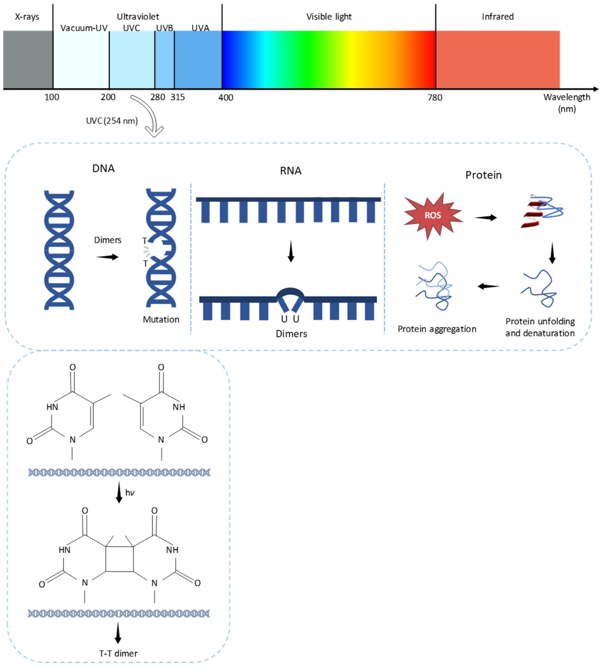
The ultraviolet (UV) region is part of the light spectrum (100–400 nm) that lies between visible and X-ray regions and is divided into three subregions: UVA (315–400 nm) that has the longest wavelengths and lowest energy and causes skin tanning, UVB (280–315 nm) with intermediate wavelengths responsible for skin tanning, sunburns, and skin cancer, and UVC (200–280 nm), also known as the UV-germicidal irradiation region, which has high-energy radiation suitable for microbial inactivation (Figure 1) [1]. UVC radiation, with the shortest wavelengths, and most UVB, do not reach the Earth’s atmosphere as they are absorbed by the stratospheric ozone layer, whereas UVA and a small part of UVB reaches the Earth’s surface and can be absorbed by living beings [2,3,4]. The UVC irradiation dose (J/m2) is defined as the irradiance (W/m2) multiplied by the exposure time (in seconds, s). Higher UVC irradiance and longer exposure times generally result in higher disinfection efficacy, since more UVC photons can interact with and damage the genetic material of the microorganisms [5,6,7].
UVC irradiation deserves special attention, particularly in the 250–260 nm wavelength range, since it is highly lethal to a wide range of microorganisms, including bacteria, mold, and yeast [8,9]. When UVC light penetrates the cell wall of a microorganism, it is absorbed by nucleic acids (DNA and RNA) and most proteins [5,10,11]. The damage promoted by UVC light is mainly due the formation of intrastrand dimers, more specifically cyclobutyl–pyrimidine dimers (CPDs) and pyrimidine–pyrimidine photoproducts, resulting in photodimerization, where two adjacent bases in the DNA/RNA sequence bind together. Pyrimidines and purines bases are strong UVC photon absorbers; however, pyrimidines (cytosine, thymine and uracil) absorb about 10-fold more than purines, so photoproducts derived from pyrimidines are the most important ones [5,12]. These dimers can occur in the same strand or between adjacent strands, and their presence inhibits the progress of DNA polymerase and RNA polymerase II, the enzymes responsible for DNA replication and RNA transcription, respectively. Consequently, dimers will prevent normal function of DNA assembly and impair essential cellular processes, leading to mutagenesis and microbial death [5,13,14,15]. Therefore, pyrimidine-derived photoproducts are the major contributors to UV-induced microbial inactivation. DNA is more sensitive to UV light, while RNA is more resistant due to the presence of the reactive hydroxyl group on its ribose sugar. However, since RNA is a temporary molecule used for protein synthesis with a high turnover, the long-term effects differ from DNA damage [12]. Prolonged exposure of RNA to UV leads to inhibition of protein synthesis and disturbs RNA–protein cross-links [16]. UVC radiation can also lead to the formation of other types of photoproducts, which can contribute to cell death. Photohydration reactions can occur under UV light in which the pyrimidines cytosine and uracil bond with elements from water molecules [5]. The formation of reactive free radicals, like reactive oxygen species (ROS) and oxidative damage, can cause structural changes in proteins such as denaturation, unfolding, and aggregation (Figure 1) [17].
UVC efficacy varies depending on the microbial state (vegetative cells are more susceptible than endospores and fungal spores) and on the type of microorganisms involved, with viruses and bacteria being less resistant than fungi [5,18]. In bacteria, endospores are very resistant to UV radiation, followed by the vegetative forms of Gram-positive and Gram-negative bacteria [19]. In fungal spores, the effect of UV radiation differs among species, with some having thin-walled hyaline conidia while others have pigmented conidia, some dark ones containing melanin. Melanin, being photoprotective, due to the high UV absorbance [20], increases the survival of fungal spores, while non-melanin compounds are less defensive against ultraviolet radiation [8,21,22].
Microorganisms exposed to UVC have developed sophisticated mechanisms to repair DNA damage that can result in a reduced final inactivation. These repair mechanisms can be divided into two classes, namely photoreactivation, a light-dependent repair that operates through the enzyme photolyase, and dark repair, a light-independent repair that requires multiple enzymes and nutrients for energy [5,23]. Photoreactivation is a natural process in which visible and UV wavelengths promote a partial recovery. This process unfolds in two stages: initially, an enzyme–substrate complex forms at the site of the DNA damage and, subsequently, a photolytic reaction takes place, where light energy is absorbed to facilitate repair of the damage. Dark repair, on the other hand, requires multiple enzymes and nutrients for energy [5,24]. In both cases, pyrimidine dimers are the main repair targets, and the DNA repair mechanism, nucleotide excision repair (NER), leads to the deletion of the damaged strand by endonucleases and the complementary strand of DNA is used as template [5]. Photoreactivation can be controlled by adjusting parameters such as temperature, UVC dosage, and wavelength spectrum [25,26,27].
Disinfection in the food industry poses a challenge for ensuring the microbiological safety of food, as it is directly related to preventing food contamination and food poisoning. Despite technological advances and constant vigilance by health authorities, foodborne illness outbreaks still occur, and contaminations can appear throughout the entire production chain, from the field to final consumption [28,29]. Surfaces that come in contact with food, especially equipment during processing, are critical points for microbial risk. The materials used and the effectiveness of sanitization or disinfection directly influence the presence and persistence of pathogenic microorganisms. Thus, failures in sanitizing or disinfection processes can compromise product quality and safety [28,29]. Surface decontamination strategies are generally classified into thermal and non-thermal methods [30]. Traditional thermal treatments such as hot water or steam require heat-resistant materials and often involve high energy consumption and extended treatment times [31]. Non-thermal approaches can be chemical or physical. Chemical methods employ approved disinfectants such as peroxide, ozone, activated water, and organic acids, usually after thorough cleaning and rinsing [30,32], while physical methods include ultrasound, UV radiation, and cold plasma (CP) [30]. Non-thermal alternatives, although sometimes more expensive, provide effective sanitization or disinfection with low surface stress [30,32]. Dry sanitization methods such as hot air, UVC radiation, pulsed light (PL), gaseous ozone (O3), and CP can enhance traditional approaches, offering improved sanitization efficiency and reduced environmental impact due to the absence of waste generation [33,34,35]. Alternating sanitization methods with different mechanisms of action can help reduce microbial resistance and increase the range of antimicrobial activity [36].
UVC disinfection is a non-thermal, non-toxic, and non-ionizing technique that requires reduced energy consumption and mild processing conditions, and leaves no chemical residue, making it useful as an alternative, environmentally friendly disinfection method in food and healthcare industries [30,32,37,38]. The effectiveness of UVC germicidal action depends on several key factors like the intensity and duration of exposure (higher intensity and exposure times increase microbial inactivation), distance from UVC source (increasing distances reduces effectiveness) [5,15], surface characteristics (reflective surfaces enhance UVC exposure) [39], type and load of microorganism and environmental conditions (relative humidity, ambient lighting and shadowing) [40]. A major limitation of UVC treatment is its small penetration depth into organic matter, which limits its effectiveness against microorganisms under the surface layer in both solids and liquids [40,41,42]. Excessive or improper UVC application can induce mutations, potentially increasing microbial pathogenicity or resistance [5,23].
This review aims to gather and critically analyze the available scientific evidence on the application of UV radiation to control microorganisms on surfaces in the food industry. The objective is to evaluate the effectiveness of UV technology as a sanitization or disinfection method, highlighting its impact on different types of microorganisms and on various materials commonly used in food production environments. An extensive comparison of materials investigated in published studies will be conducted to identify behavioral patterns related to surface characteristics, such as roughness, porosity, and reflectivity, as well as, where possible, the responses of different microorganisms to UV treatment. By consolidating current knowledge and identifying knowledge gaps, this review intends to provide a clearer scientific basis to the food industry for decision-making regarding the use of UV radiation as a complementary tool to conventional cleaning and disinfection methods.

2. Methodology

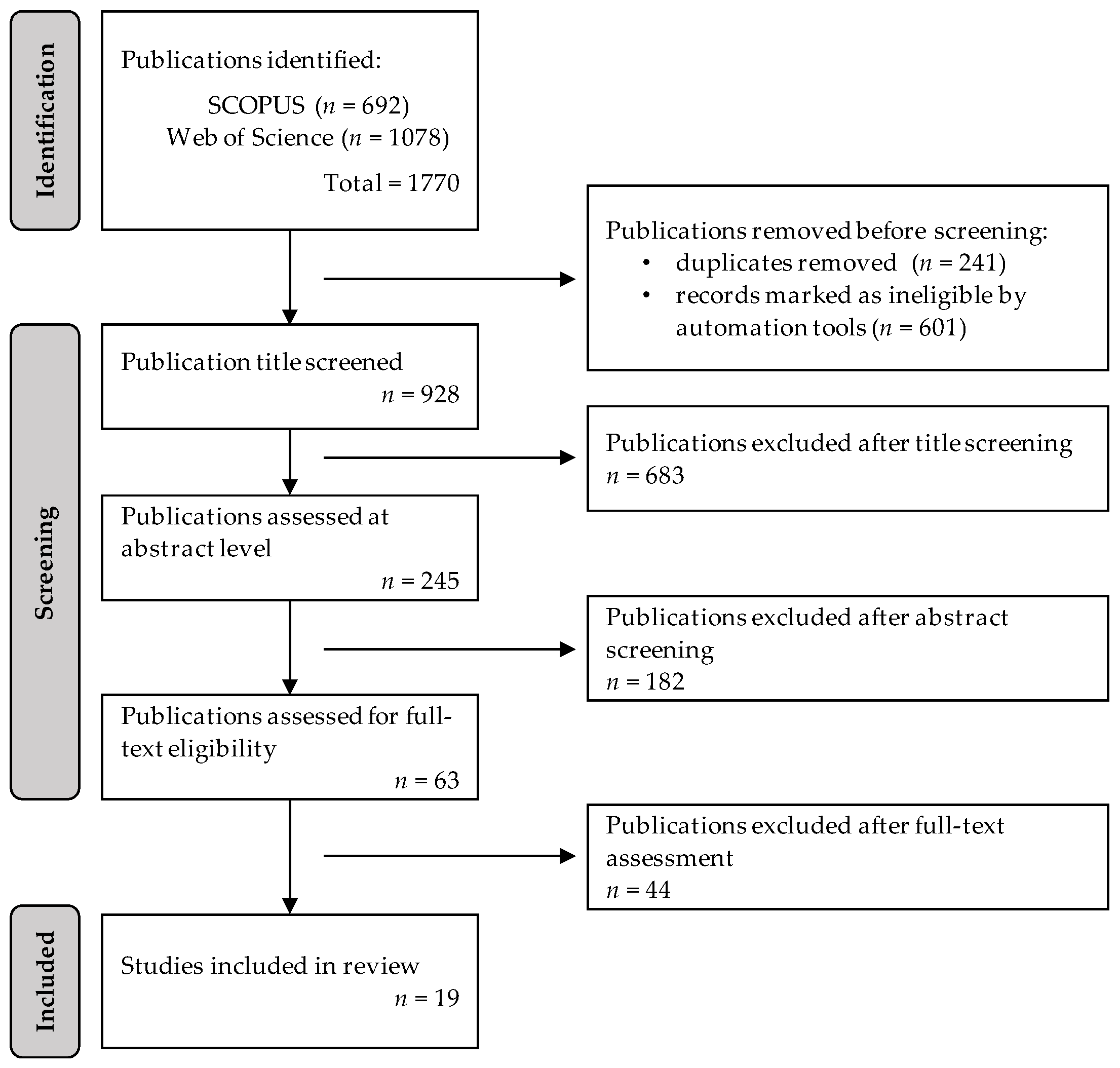
Relevant articles were gathered from Scopus and Web of Science databases. The literature review was performed using the keywords “food” AND “industry” AND “UV”. The final search was conducted on 18 June 2025. After a thorough review and elimination process, only original research articles written in English and specifically addressing the use of UV for disinfection on industrial surfaces were included in this study (Figure 2). To ensure reliability and minimize potential bias, two authors independently screened and evaluated the articles. The selection process was carried out in two stages: first, only full-text research articles were taken into consideration. On the contrary, review papers, conference proceedings, books, book chapters, and non-English publications were excluded. Then, after removing duplicates, the remaining records were screened for relevance at the level of the title, followed by the abstract and, finally, the full-texts of the remaining studies were carefully assessed for eligibility. After full-text evaluation, only 19 original research articles met the inclusion criteria and were therefore considered suitable for analysis in this study. The initial 1770 articles identified spanned the period from 2005 to 2026. Following the screening and selection process, 19 articles were ultimately included for analysis, all published from 2010 onwards.
In addition to the 19 articles selected for analysis, many others that contributed to deepening the understanding of the topics covered were consulted, resulting in a large bibliographic reference. Many of these additional references were essential to support and contextualize the effects of UV radiation, and to rigorously characterize the materials studied, allowing for a more precise description of their physical, chemical, and functional properties. During the literature review, several relevant studies related to different types of food matrices were also identified, which enriches the discussion. Thus, the reference list resulted not only from direct citations present in the body of the text, but also from the need to ensure consistency, scientific rigor, and the theoretical foundation of all the conclusions presented.
Table 1 presents a set of relevant information for understanding and comparing the different studies, including the surface type where tests were performed, the microorganisms evaluated, the species or the strains used, the type of UV radiation applied, and the respective wavelength. In addition, the irradiance of the lamps used and the radiation doses reported by the authors are presented, either as a single value or a range of values. The organization of the data in Table 1 follows a chronological order, which makes it possible to observe the evolution of the methodological approaches over time.

3. Types of Industrial Surfaces

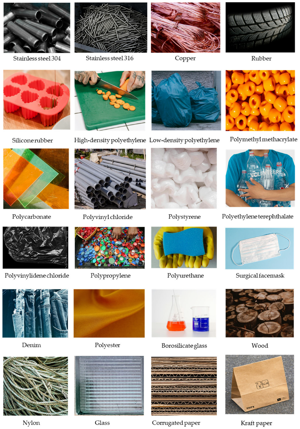
Industrial surfaces are found in various environments such as food processing plants, pharmaceutical manufacturing, hospitals, and cleanrooms. The type of surface determines the appropriate disinfection method based on the material, risk of contamination, and regulatory requirements. The most common types of industrial surfaces are stainless steel (SS), plastic polymers, glass surfaces, rubber and elastomeric surfaces, concrete floors and walls, wooden surfaces, and coated metallic surfaces (Figure 3).
Surfaces of SS are very common in food processing, pharmaceutical, and medical industries. There are many types of these surfaces, differing in chemical composition, properties and applications (Table 2). Stainless steel is primarily an alloy of Fe (50–72%), C (0.03–0.08%), containing at least 10.5% Cr, which gives it toughness, rust resistance, and versatility. Also, these surfaces are non-porous and easy to clean. There are several categories of SS: (i) SS 304 (or AISI 304 SS, American Iron and Steel Institute classification), a category of Austenitic SS, the most widely used type, is known for its high nickel and chromium content, which improves corrosion resistance. It is very easy to clean, which makes it perfect for food-related use, medical devices, and pipes. (ii) The SS 304L, the low C version of SS 304, offers resistance to wear and high temperatures, making it suitable for harsh environments. It is also used in off-shore construction, cisterns and tubes for chemical tankers, warehousing and the overland transport of chemicals, food and beverages. (iii) SS 316 (or AISI 316 SS) stands out for its high Mo content, which gives high protection against media containing chlorides and non-oxidizing acids. It is used in harsh environments and in offshore technology, production, storage and land transport of chemicals, food and beverages. (iv) The SS 316L is similar to 316 SS, but has a very low nickel release, making it hypoallergenic. This property is appreciated in medical devices, jewelry, body piercings, kitchenware and food storage.
Elastomers are common surfaces with industrial applications (Table 2), such as rubbers and silicone rubber. Both natural (rubber) and the synthetic rubbers (Ethylene Propylene Diene Monomer—EPDM, nitrile, neoprene) are known for their elasticity, versatility, and durability, and their wide range of applications [60]. Silicone rubber is a polymer of silicon, oxygen and other elements, characterized by high elasticity, resistance and stability to temperature, radiation (UV and ionizing), and chemicals, as well as water-proofing and biocompatibility [61].
Plastic polymers are derived from petroleum and have porous surfaces (Table 2). They are common in equipment housings, cleanroom walls, packaging areas, and food containers. Both the porosity and the sensitivity to harsh chemicals varies with the type of polymer. (i) High-density polyethylene (HDPE) is safe and very resistant to chemicals and corrosion, long-lasting, and used in piping, cutting boards, and containers. It is also used in plastic surgery and in skeletal and facial reconstruction [62]. (ii) Low-density polyethylene (LDPE), is a flexible polymer, known for its transparency and its good chemical and impact resistance. It is also known for its low resistance to high temperature and corrosion. It is commonly used in food packaging, irrigation, and coatings. Furthermore, it is applied in medical devices [63]. (iii) Polymethyl methacrylate (PMMA) is known for its transparency, high durability, thermal stability, and excellent chemical resistance, making it ideal for laboratory and industrial use (optics, aerospace and electronics) [64]. (iv) Polycarbonate (PC) is a transparent thermoplastic, resistant to impact and fracture, lightweight (an excellent alternative to glass) and thermal-resistant (−20 °C to 140 °C). With similar characteristics to PMMA, it is used in protection and as a glass substitute [65]. (v) Polyvinyl chloride (PVC) is a high-strength thermoplastic material and the third-most widely produced plastic polymer [66]. It is used in food packages, pipes, medical devices, and in construction. (vi) Polystyrene (PS) is a polymer of aromatic hydrocarbon styrene. It is stiff, lightweight, and transparent. PS is most commonly used in foam packaging [67]. (vii) Polyethylene terephthalate (PET) is a polyester and is transparent, resistant to impact, moisture, and chemicals, and is commonly used in storing food and beverages and in houseware [68]. (viii) Polyvinylidene Chloride (PVdC) is a homopolymer of vinylidene chloride that forms transparent films, and, because it has low permeability to gases, moisture, fat and aromas, it is used in food wrapping [69]. (ix) Polypropylene (PP) [70] and (x) Polyurethane (PU or PUR) result from a reaction between an isocyanate and a polyol, and are very light, insulated and flexible. Also, they are known for their high mechanical strength and good temperature resistance. These materials are available as rigid (surfaces) and flexible (foams, paints) types, and are applied in many areas [71].
Glass is a mixture of silica with sodium carbonate, and calcium carbonate. It is transparent amorphous, hard, non-porous, chemically inert, and has low thermal and electrical conductivity. Stainless steel, plastic polymers and glass are the most commonly used materials. Among the various characteristics of these materials, roughness is a factor to consider. Irregular or rough surfaces can lead to non-uniform UV dose distribution due to the geometric shielding effect, particularly when microorganisms are located in surface depressions or protected microstructures [72]. Furthermore, increased surface roughness has been found to intensify microbial adhesion and facilitate biofilm formation, which in turn reduces the effectiveness of UV irradiation [73]. Therefore, surface roughness represents an important parameter that should be considered along with material composition when evaluating UV disinfection performance on industrial surfaces.
Although less common, disinfecting fabrics or textiles by UV radiation may be of interest, as cleaning these surfaces can be difficult and time-consuming. These types of materials include synthetic fibers (nylon, polyester) and natural fibers (cotton). Cotton is a natural fiber known for being soft, breathable and comfortable (e.g., denim, dyed cotton, which are durable and thick). Synthetic fibers as polyester (based on petroleum chemicals) and nylon (polyamide fiber) are lightweight, highly resistant to abrasion and water, and are elastic and durable [74].
Table 2. Types of materials used on surfaces in industry or for industrial use and their composition and properties of interest. AISI—American Iron and Steel Institute. Sources: stainless steel [75,76]; rubber [77]; silicon [78]; plastic polymers [62].
Surface MaterialCompositionPropertiesApplications
Stainless Steel (SS)
SS 304 (same as AISI 304 SS)Fe (balance), C (0.07%), Cr (18–20%), Ni (8–12%)Excellent corrosion resistance in a wide variety of environments and mediaKitchen and food-related use, medical devices, and pipes
SS 304L (low alloy)Fe (balance), Cr (17.50–19.50%), Ni (8–10.50%), Mn (0–2%), C (0.03%)Superior corrosion resistance than SS 304. With Cr. Resistance to wear and high temperatures makes it suitable for harsh environments, ranging from industrial plants to marine settingsHandling aggressive chemicals or high-temperature processes. Chemical reactors, storage tanks, and pipelines. Kitchenware, brewery, food, dairy and pharmaceutical equipment
SS 316 (same as AISI 316 SS)Fe (balance), C (0.07–0.08%), Cr (16.5–18.5%), Ni (10–13%), Mo (2–2.5%)High resistance to corrosion. For harsh environments, e.g., chloride exposureMedical applications, food and beverages, marine transport and off-shore construction
SS 316L
(ow alloy)
Fe (balance), C (0.03%), Cr (16.5–18.5%), Ni (10–13%), Mo (2–2.5%)Untreated, and treated with DURALTI® (special anodizing with TiO2, approved for food contact). HypoallergenicJewelry, implants, body piercings, surgical instruments, cooking and food storage
Elastomers
RubberNatural: polyisoprene, sulfur. Synthetic: e.g., EPDM, nitrile, butyl, neopreneElasticity, versatility, durability. High elongation before breaking point and/or strong resistance to tearing, resistance to chemicals (O3, acids and alkalis)Surgical gloves, footwear, hoses, industrial flooring, molds, kitchenware, bottle caps
Silicone rubberPolymer of silicon, O, C, and HElasticity, resistant and stable [−50 °C to 250 °C]. Resistant to radiation (UV, alpha-, beta- and gamma-rays), water-proofing, and biocompatible. Silicone surface is non-porousCookware, food packing medical applications (skin contact, medical devices, long term implants)
Plastic polymers
High-density polyethylene (HDPE)Linear polymer of ethyleneHigh tensile strength, high chemical resistance, low water absorption and durabilityPipes, cutting boards, food and beverages containers, detergent and bleach containers. Also used in plastic surgery
Low-density polyethylene (LDPE)Branched polymer of ethyleneA flexible polymer, transparent, ease of processing, good chemical and impact resistanceFood storage (flexible films, bags, packaging), irrigation tubing, and coatings. Pediatric orthotics and prosthetics
Polymethyl methacrylate (PMMA)Polymer of methyl methacrylateTransparent plastic, high durability and chemical and impact resistance, and thermal stableCasting resin, in coatings, cutting-edge industries, and medical applications
Polycarbonate (PC)Polymer of polycarbonateSimilar characteristics to PMMASafety helmets, bullet-proof glass, glass substitute (car headlamp lenses, baby-feeding bottles, roofing, glazing)
Polyvinyl chloride (PVC)Chlorinated ethyleneLightweight, durable, low-cost, and easy processabilityFood packages (the rigid form), pipes, medical devices, construction (good flame retardant)
Polystyrene (PS)Polymer of styreneStiff, lightweight, transparent, with low resistance to high temperatures. Can be solid or foamedPackaging (Styrofoam containers), construction, and medical equipment
Polyethylene terephthalate (PET)Polymer of terephthalic acid and ethylene glycol, ethylene glycol and dimethyl terephthalateTransparent, and resistant to impact, moisture, alcohols, and diluted acids, transparent to microwave radiationFood and beverages, household containers, houseware
Polyvinylidene chloride (PVdC)Polymer of vinylidene chloride with vinyl chlorideTransparent films with low gas permeability, moisture, and fat permeabilityCling film for food wrapping, blister packaging
Polypropylene (PP)Polymer of propyleneChemical-resistant, strong, lightweight, good fatigue resistance and ability to withstand high temperaturesFood packaging, automotive components, medical industry (trays, simple handles and body contact plates, surgical face masks), textiles, fibers
Polyurethane (PU or PUR)From a reaction between an isocyanate and a polyolVery light, insulated and flexible. Also, high mechanical strength and good temperature resistanceUsed on furniture, water tanks, paints, automotive components, mops
Glass
Glass sheet, borosilicate glassMixture of silica with sodium carbonate, and calcium carbonateAmorphous, transparent, hard, chemical resistant, durable, non-porous, and low conductivity to heat and electricityUse in the electronics, construction, energy, transport, automotive, medical and laboratory equipment industries

4. Disinfection of Industrial Surfaces by UV

The most tested type of surface in the revised studies was SS, untreated or treated, followed by plastics, rubber and elastomers, other metallic surfaces (aluminum, copper), textiles, wood and paper (Table 3). These surfaces are the most used in industry, and SS is one of the most common contact surface materials in food processing [79]. Weng et al. reported that, on SS, a dose of 720 mJ/cm2 inhibited S. Typhimurium, but not E. coli and L. monocytogenes, which were only inhibited at 2160 mJ/cm2 [44]. On SS, at 254 nm, and under the droplet and smear contamination models, the bacteria E. coli and S. aureus and the yeast C. albicans were fully inhibited (100%), with an approximately 4-log reduction, at 14,004 mJ/cm2. By contrast, the inhibition of Aspergillus fumigatus was negligible when the drop method was applied (6.34%), and increased in the smear method (89.96%). This result suggests that microenvironments within droplets can shield organisms from UVC exposure, a factor that should be considered in practical decontamination scenarios [49].
The bacteria E. coli and L. innocua exhibited different inactivation patterns on different surfaces and under UVC (254 nm): SS 304, medical grade 99.999% copper, and copper-deposited polymer plastic sheets (CuPoly). The final inactivation effectiveness ranked SS > Cu > CuPoly, for both bacteria. The reduction of E. coli on SS and Cu showed an almost linear inactivation trend, and reached a >6-log reduction at 990 mJ/cm2, but, on CuPoly and a dose of 438 mJ/cm2, it reached a maximum 3.1-log reduction. The maximum inactivation levels for L. innocua on SS, Cu, and CuPoly were 6.1 (219 mJ/cm2), 5.3, and 4.5 log at 438 mJ/cm2, respectively [50].
On SS 316L, E. coli was more resistant than S. Enteritidis and Pseudomonas fragi, showing only 1.7- to 2.63-log reductions across doses of 1–6 mJ/cm2, while S. Enteritidis and P. fragi displayed reductions between 2.1 to 3.74 log under the same conditions [59]. As pointed out by Sharma et al. sated that the surface roughness of SS is strongly influenced by its grade and can present differences in values greater than 10 times [59]. On SS surfaces and using 265 nm LEDs, a L. monocytogenes reduction of approximately 2 log was achieved after exposure to 11, 33 and 55 mJ/cm2 [57]. Interestingly, the biofilms formed at 4 °C were less susceptible to UVC than the ones formed at room temperature. At 4 °C, and after 2.5 min (27 mJ/cm2) of exposure to UVC, there was a reduction of 1.1 log that increased to 1.2 log (5 min, 55 mJ/cm2). At room temperature, a dose of 27 mJ/cm2 provoked a reduction of 1.5 log, and a log reduction of 2.5 was reached at a dose of 55 mJ/cm2 [57].
The bacteria of the genus Alicyclobacillus, a spore-forming and Gram-variable bacteria, poses serious quality problems for the food industry. On SS 304, and at a dose of 2620 mJ/cm2, the biofilm of A. acidocaldarius was susceptible (3-log reduction), followed by the biofilms of A. acidoterrestris and A. cycloheptanicus, with reductions around 2 log, and A. herbarius was less susceptible, not exceeding a 2-log reduction [41]. These findings emphasize that disinfection protocols must be tested against the most resistant organisms expected in real-world conditions. On SS and under UVC radiation for 15, 30 and 60 min, the spores of B. cereus, a frequent Gram-positive foodborne pathogen, were reduced by 1.06 log, 1.18 log and 1.68 log, respectively [48]. The authors do not mention either the wavelength or the irradiance of the lamp used.
The UVC disinfection response on different solid substrates seems to be correlated with physical properties, such as roughness values (Table 3, highest CuPoly > SS > Cu) and the water contact angles (CuPoly (108.57°) > Cu (88.50°) > SS (31.30°)) [50]. Differences in hydrophobicity influence bacterial distribution on hydrophobic surfaces (>65.0°), causing bacterial clustering and shading, which decrease inactivation [80]. Also, the high reflectivity of the surface can affect the disinfection performance. The log reduction of E. coli and L. innocua increased by 113–271% after 3 min UVC treatment in the presence of the reflective Al lining [50].
The impact of UV radiation on microorganisms, in addition to depending on the type of surface, also varies with its finishing. Using SS surfaces without or with different finishes, on the bare SS surface, an UV irradiance at 1200 µW/cm2 for 5 min (720 mJ/cm2) inhibited S. Typhimurium, but not E. coli and L. monocytogenes, which required a higher dose (2160 mJ/cm2). On PU-coated SS, a dose of 360 mJ/cm2 was sufficient to inhibit L. monocytogenes, but not E. coli and S. Typhimurium, which were only inhibited at 2160 mJ/cm2 and 720 mJ/cm2, respectively. On the contrary, on a SS surface coated with PU and TiO2, it took a dose of 360 mJ/cm2 to inhibit both S. Typhimurium and E. coli, and a dose of 720 mJ/cm2 to inhibit L. monocytogenes [44]. Also, Chen et al. [50] reported that UVC disinfection on bead-blasted SS 304 was very effective, reducing E. coli ATCC 25922 by more than 6 log when applied a dose of 437 mJ/cm2. Although reductions in L. innocua were comparable, a rapid initial reduction followed by a plateau was observed, unlike E. coli, which showed a linear trend reduction [50].
After 1 h under UVA (365 nm), the number of L. monocytogenes-adherent cells in bare SS was reduced by 2 log, while, in SS covered with 4 layers of TiO2, there was a 4-log reduction [58]. Another study, which tested the effect of combining acid/enzymatic detergents with UVA light on L. monocytogenes biofilms formed on SS 304 and PU surfaces, demonstrated that the treatment efficacy varied between the surfaces and the treatment combination. The highest reduction level (5 log) was obtained on the SS 304 surface after 10 min of treatment with acid detergent, followed by 10 min of exposure to UVA. On the PU surface, a reduction of 2.60 log was achieved after 30 min of treatment with enzymatic solution, followed by 10 min of exposure to UVA [56].
Perhaps the most outstanding findings across the several SS studies related to the surface finish and its grade was done by Gabriel et al., who demonstrated the impact of different finishes on SS 304 and 316 on S. enterica inactivation. For instance, in the SS 304 mirror finish, a dose of 62.46 mJ/cm2 was required for the complete inactivation of S. enterica. In the 2B finish, complete inactivation required a dose of 81.99 mJ/cm2, while, in the hairline finish, 101.53 mJ/cm2 was required, indicating that bacteria on this surface were more resistant to inactivation. On the other hand, on SS 316 with a hairline finish, bacterial inactivation was easier (46.81 mJ/cm2), followed by 2B (66.34 mJ/cm2) and mirror (93.78 mJ/cm2) finishes. These data indicate that reflectivity interacts with microbial adhesion and exposure to UVC in complex ways [45]. However, it is important to note that the surface roughness of SS can vary substantially depending on its grade. In addition to SS, other surfaces are used in industry. Copper surfaces, particularly medical-grade copper, demonstrate strong antimicrobial activity when exposed to UV (254 nm). Chen et al. reported results of an over 6-log reduction of E. coli and a 5.3-log reduction of L. innocua within just 3 minutes of treatment, or a dose of 90 mJ/cm2 [50]. Epelle et al. obtained a 100% reduction of E. coli and S. aureus, and a 99.83% reduction of the yeast C. albicans on copper surfaces after 15 min of irradiation at 15.56 mW/cm2 (fluence of 14,004 mJ/cm2), regardless the inoculation method. However, the same study showed that the susceptibility of A. fumigatus to UV depends not only on the surface used, but also on the contamination method. When spread in droplet form, A. fumigatus was resistant to treatment (5.5% of reduction), being more susceptible in the smear form, where it showed a 72.04% reduction [49]. On the other hand, maximum inactivation levels of 3.1 log units were reported for E. coli and 4.5 log for L. innocua on copper-deposited polymer sheets [50].
Another surface tested in the reviewed publications was aluminum, where effective inactivation by UVC was achieved. Weng et al. found reductions comparable to those on SS, with the three tested species E. coli, S. Typhimurium and L. monocytogenes, inhibited at a dose of 2160 mJ/cm2. The research also tested the impact of the aluminum finish on bacteria UVC inactivation. On a surface of aluminum coated with PU, a dose of 360 mJ/cm2 was sufficient to inhibit S. Typhimurium, but not E. coli, and L. monocytogenes, which were only inhibited at 2160 mJ/cm2. However, on a surface of aluminum coated with PU and TiO2, a dose of 360 mJ/cm2 was sufficient to inhibit S. Typhimurium and E. coli, while L. monocytogenes was inhibited at 720 mJ/cm2 [44].
The effect of the finish on microbial susceptibility to UV radiation is related to its roughness and reflectivity. Research performed by Cerbo et al., on the effect of three roughness levels (R0.25, R0.5 and R1, for 0.25, 0.5 and 1.0 µm, respectively) of Aluminum ANTICORODAL alloy 6082 T6 on the susceptibility of seven bacterial species to UVC, found that the efficacy of the UVC depended on the species, and, in some species, on the roughness of the aluminum surface. For untreated aluminum, the results showed that, after 12 h of exposure to UVC, the Gram-negative bacteria (P. aeruginosa, S. Typhimurum, and Yersinia enterocolitica) were more susceptible than the Gram-positive bacteria (L. monocytogenes, S. aureus, E. faecalis, and B. cereus) to the UVC treatment. Also, the susceptibility to UVC varied with roughness: in all the Gram-negative species, and after 12 h of UVC treatment, no growth was detected on roughness 1 µm (R1), contrary to the Gram-positive bacteria. In general, the susceptibility of the bacteria varied with roughness: R0.25 > R0.5 > R1 (Table 3). The susceptibility to UVC of the same bacterial species increased when the surface was treated with a layer of aluminum oxide combined with titanium oxide—DURALTI®. On all tested roughness levels, the complete bacterial inactivation by UVC was achieved [47]. The DURALTI® surface itself has a different impact on the survival of bacteria. Gram-negative bacteria are affected to an increasing extent with surface roughness (R0.25 > R0.5 > R1). The susceptibility to UVC on DURALTI® varies greatly among Gram-positive bacteria: S. aureus exhibited identical behavior to Gram-negative bacteria (R0.25 > R0.5 > R1), L. monocytogenes, a reverse behavior to Gram-negative bacteria (R1 > R0.5 > R0.25), B. cereus was more susceptible at R0.5 with DURALTI®, and the susceptibility of E. faecalis was not affected by the DURALTI® roughness [47]. The TiO2 finishing may have a bacteriostatic effect due to photocatalytic reactions between the aluminum surface and titanium [81], which under UVC becomes bactericidal, by enhancing the oxidative stress on bacteria [47].
On glass sheet, a decrease of 3.8 log CFU/cm2 was reported for S. aureus after treatment with UVC (222 nm) at 9.1 mW/cm2 for 60 s (546 mJ/cm2). When the UVC was combined with CP, a 4.5 log CFU/cm2 was observed for S. aureus after 60 s of CP + UVC [53]. Borosilicate glass was identified as one of the best-performing surfaces among those tested (SS 316L and silicone rubber), being the most favorable for UVC disinfection. Results showed high log reductions in the three tested bacteria at a dose of 6 mJ/cm2: E. coli (3.39 log), S. Enteritidis (4.40 log), and P. fragi (4.16 log), compared to SS 316L (reductions of 2.63, 3.63 and 3.74, respectively) and silicon rubber (reductions of 2.5, 3.51 and 3.67, respectively to E. coli, S. Enteritidis, and P. fragi) [59].
Disinfection with UV in polymers varies widely in performance depending on their composition and structure. Morey et al. tested several belts made of thermoplastic and elastomers and found that UV light (254 nm) can rapidly reduce the load of a four-strain cocktail of L. monocytogenes on conveyor belts (CB), but the degree of reduction is dependent on the type of belting material (Table 3). Under the lowest tested dose, 5.53 mJ/cm2, the reduction range for L. monocytogenes was [log 3.4–log 4.2]. At 5.95 mJ/cm2, on belt 1, L. monocytogenes was not detected (ND). At a dose of 16.59 mJ/cm2, the survival population was ND < 0.74 < 1.31 < 1.73, respectively, depending on the type of belt (Table 3). After exposure to 5.95 mW/cm2 for 3 s (17.85 mJ/cm2), L. monocytogenes reduced to below the detection limit, 3.2 log CFU/cm2 (ND) on belts 1, 2, and 3, contrary to belt 4, which had a survival population of 1.4 log CFU/cm2. The survival of L. monocytogenes varied significantly between similar materials such as CB1 (Ropanyl DM 8/2 A2 + 04 Light Blue thermoplastic polyurethane) and CB4 (Ropanyl DM thermoplastic polyurethane 04 + 04 White food-grade Amerol). On smoother surfaces, inactivation was more rapid and complete, compared with the rougher surface (belt 4) that allowed bacterial survival, which demonstrates the importance of surface texture on protecting cells from the UV radiation [82]. The results obtained by Morey et al. showed that low doses are effective in reducing populations of L. monocytogenes, suggesting a potential application for the sanitization of moving CBs in a processing plant [43].
Along with the thermoplastics, silicon rubber is a type of surface frequently found in the food industry. Sharma et al. studied the efficacy of low UV doses (1–6 mJ/cm2) in three bacterial species inoculated on silicone rubber, and found that P. fragi and S. Enteritidis were easier to reduce than E. coli. At 6 mJ/cm2, the mean log reduction of P. fragi was 3.67 log, followed by S. Enteritidis (3.51 log), while E. coli only reduced 2.50 log. Also, it was demonstrated that, in the tested conditions, the reduction values obtained for each species on silicon rubber were similar to those obtained on SS 316L (Table 3). Once more, these results highlight that different microbial species respond differently to UVC even on the same surfaces [59]. Ashrafudoulla et al. also reported a significant reduction [0.80–4.74 log CFU/cm2] of S. Thompson cells on silicon rubber, but using higher doses (60–300 mJ/cm2) [54] compared to S. Enteriditis [59]. Even when endospore bacteria are used, UVC can reduce bacterial load. Four Alicyclobacillus species (A. acidoterrestris, A. cycloheptanicus, A. herbarius, and A. acidocaldarius) in biofilm, forming on rubber under a strong UVC dose (2.52 J/cm2 or 2520 mJ/cm2), were reduced by approximately 2–3 log. The species A. acidocaldarius was the most susceptible (~3 log CFU/cm2 under 2.52 J/cm2), contrary to A. herbarius, which was particularly resistant, showing a reduction of less than 2 log. The results show that the inactivation of spore-forming bacteria on rubbery materials can be difficult [41].
A study of UVA disinfection on food packaging polymers like PVC, PS, PET and PVDC cling film, uncoated and coated with TiO2, found that, in the uncoated substrates, both E. coli and S. Typhimurium exhibited a steady growth over the incubation time, confirming that UVA irradiation is not bactericidal. Escherichia coli and S. Typhimurium were more easily inactivated on PET > PVDC > PS > PVC, but S. Typhimurium was more resilient to inactivation. On PET and after 60 min of exposure (9 kJ/cm2), E. coli was completely inactivated, while S. Typhimurium was 97.8% reduced. At 120 min (18 kJ/cm2) E. coli was fully inactivated in all surfaces, but S. Typhimurium continued to grow in PVC [52]. The biofilm of L. monocytogenes formed in PU showed greater tolerance to UVA than the one formed on SS: L. monocytogenes was reduced from 6.05 to <1.0 log CFU/cm2 on SS treated with an acid detergent and UVA (10 min), and reduced by 3.6 log on PU with the same treatment and time of exposure [56]. On the other hand, for thin polypropylene films, a 4.1 log reduction was observed for S. aureus after 60 s of a combined treatment with CP and UV, which compares to results obtained for paper, but in this case the context of the combined treatment must be considered [53].
Disinfection studies conducted on HDPE caps, a type of plastic very resistant to chemicals and corrosion, contaminated with conidia or ascospores from various fungal species, reported that the first decimal reduction time (1D) values depended on the species and whether it was in a single layer or multiple layers. Some species were rapidly reduced when in a single layer, such as Aspergillus hiratsukae SSICA 3913, Aspergillus montevidense SSICA 28,219, and Talaromyces bacillisporus SSICA 10915, with 1D of 13.7 s, 12.2 s and 9.7 s, respectively. On the contrary, Aspergillus brasiliensis showed greater resistance (1D = 24.9 s) than the other three species, but still significantly less than Chaetomium globosum (1D = 99.9 s). As multi-layer on HPDE caps, A. hiratsukae (1D = 30.3 s) and A. montevidense (1D = 51.6 s) were the most susceptible species, while T. bacillisporus (1D = 147.1 s), C. globosum (1D = 153.2 s), and A. brasiliensis (1D = 188.2 s) were very resistant. In single-layer, the ascospores of C. globosum were the less susceptible, followed by the conidia of A. brasiliensis, swapping places when in multi-layer. These results highlight the importance of the applied inoculum when testing microbial susceptibility to UV, since the number of layers interfere with the energy of irradiation, resulting in different inactivation rates [22]. Also, intrinsic microbial characteristics such as pigmentation, type of spores, and cell wall thickness must be considered. For instance, A. brasiliensis belongs to the Aspergillus section Nigri, known for their strong pigmentation due to melanin. When the synthesis of melanin was inhibited in A. brasiliensis, the hypopigmented phenotype significantly increased its sensitivity to biocides [83].
The disinfection by UVC of two of the surfaces most used by fruit producers, such as nylon and polycarbonate, was studied after contamination with three strains of L. monocytogenes, in equal ratio and an initial population of 1 × 10⁸ CFU/mL. The biofilm treated under 850 µW/cm2 irradiation for 2 (102 mJ/cm2) and 5 (255 mW/cm2) min, were little affected: a reduction of 0.15–0.75 log CFU/cm2 in nylon and 0.93–1.33 log CFU/cm2 in polycarbonate, concluding that the texture of these materials could explain the low success of UVC light in disinfecting them, as they require higher doses for a meaningful reduction [51]. Finally, when comparing PMMA and SS, both non-porous surfaces, Epelle et al. reported a 100% inactivation in E. coli, S. aureus, and C. albicans on PMMA, but the A. fumigatus susceptibility was dependent on the contamination mode (9% in droplet versus 92.16% in smear) [49]. This result corroborates the ones obtained by Racchi et al. on the importance of the number of layers of the inoculum in some species [22]. Also, the results confirm the general rule that smooth, non-porous surfaces like PMMA and SS allow a more effective and predictable inactivation.
Along with glass sheet, Sheng et al. tested the efficacy of a combined treatment of CP and UVC (222 nm) for 60 s on the disinfection of corrugated and kraft paper. In corrugated paper, the population of S. aureus was reduced 1.5 log CFU/cm2, whereas, in Kraft paper, the reduction was 2.4 log CFU/cm2. This difference likely results from the porosity/absorption of the material (Figure 3) [53].
The research articles that studied the use of UV disinfection on SS surfaces used UV-lamps emitting different wavelengths, including UVA and UVC (Table 3). Among these, UVC was the most widely used [41,44,45,48,49,50,54,57,59], followed by UVA applications that appeared in only two studies [56,58].
Within the polymer category, plastics, thermoplastics and rubber, the cited research employed lamps emitting UVA and UVC. Notably, even within the UVC range, differences were observed. Sheng et al. and Sharma et al. used, respectively, a UV-lamp of 222 nm and 279 nm wavelengths [53,59], in contrast to other authors, who applied UVC radiation at 254 nm [22,41,43,49,51]. Ashrafudoulla et al. reported the use of UVC but did not specify the wavelength [54]. Others employed UVA [52]. Many of the research articles considered in this review mention that porous and fibrous materials pose the greatest obstacles to UV disinfection. A strong bacterial reduction was reported on surgical facemasks (Table 3), but, when A. fumigatus was inoculated as droplets, it was poorly inactivated (12.75% reduction), showing how porous fibers shield embedded mold spores. In textiles, denim was identified as one of the most challenging textiles, showing limited inactivation even under prolonged exposures, due to its thickness and dense texture. Textile blends of cotton/polyester performed better, allowing complete bacterial and fungal reductions under the same conditions. In all three types of fabric, C. albicans was 100% inactivated. Escherichia coli was completely inactivated (100%) in cotton/polyester, but not in denim. The species S. aureus was completely inactivated in cotton/polyester and surgical masks (100%), but not in denim, where a reduction of 99.90%, or 3 log was achieved [49].
In recent years, UV treatments have been increasingly recognized as sustainable decontamination technologies that ensure microbial safety in industry. Two studies that evaluated UVC disinfection on equipment used in industry showed that UVC treatment alone or combined with calcined calcium reduces or eliminates bacterial contaminants. Kayaardı et al. performed a study on the equipment surfaces used in a catering facility, such as a meat grinder knife MT, cutting knife CT, cut-proof glove CG, and knife sharper KS. These surfaces were analyzed before and after UV treatment, showing that the population of aerobic mesophilic bacteria was reduced in all tested materials: KS 2.61 log CFU/cm2, MT 1.99 log CFU/cm2, CT 0.48 log CFU/cm2, and CG 0.43 log CFU/cm2. The population of yeast, molds, E. coli, and coliforms after UVC treatment was reduced to bellow the detection limit (<1 log CFU/cm2) [55]. Moreover, in a bulk food bag manufacturing facility, it was found that gloves, fabrics, utensils, and machine surfaces were contaminated with coliforms, fecal coliforms, and Staphylococcus spp. (≤3.68 log CFU/unit). They demonstrated that washing gloves with calcined calcium, followed by UV drying, and UV treatment of finished product storage rooms, combined with sanitizing floors using 0.02% calcined calcium, significantly reduced or eliminated bacterial contaminants (1.0–3.68 log CFU/unit) from surfaces [46]. These studies highlight the importance of using UV treatment, alone or in combination with chemical sanitization, when dealing with high-risk production environments.

5. Disinfection of Food Matrices by UV

Along with packaging materials and surfaces in contact with food, UV disinfection has been studied in various food matrices such as fresh and processed fruits [41,84,85,86,87,88,89,90,91], leafy green vegetables [92], salad components [57], meat and meat products [93,94], seafood and fish [95,96], dairy products and cheese [97], shelled eggs [54], grains and seeds [98], wheat flour [99], powders and seasonings [100,101], and roasted coffee beans [102]. Table 4 presents the methodological characteristics of the selected publications, detailing the food matrices evaluated and the UV irradiation conditions applied. Information includes UV wavelength, irradiation intensity, applied dose, and irradiation time, allowing for comparison of the experimental setups used across the studies.
In the microbiological control of winemaking, UVC radiation has shown potential in controlling fermentative microorganisms, albeit with the use of high doses (1 kJ/L), once Saccharomyces cerevisiae is more resistant to UVC than Hanseniaspora uvarum. In the same study, the authors obtained promising results in the disinfection of wine by UVC, which could replace the use of SO2. Additionally, when compared to the thermal disinfection of wine, UVC radiation showed a better performance on the preservation of aromatic compounds [103]. More recently, it was reported that that UVC disinfection of wine must is highly dependent on the initial microbial load and must turbidity, which are highly variable. In wine must-treated by UVC, the oxidation of polyphenols and ortho-diphenols increased and must color increased [104]. These authors demonstrated that, in wine from UVC-treated must, the aroma-relevant compounds, such as alcohols, terpenes, and C13-norisoprenoids, decreased [104].

6. Pros and Cons of UV Treatment

UV radiation disinfection, particularly UVC at 254 nm, presents clear advantages due to its high antimicrobial efficacy (Table 3). The UV technology also demonstrates great versatility, being effective on a wide variety of surfaces relevant to the food industry. The application of coatings, especially those based on TiO2, emerges as a decisive factor in enhancing the effectiveness of UV radiation, allowing for high levels of microbial reduction with lower doses. This is particularly important when UVA is used. The combination of UVA with photocatalytic surfaces shows significant potential, demonstrating that integrated approaches can broaden the application spectrum of UV disinfection [52].
The use of UV radiation in industry has many other benefits that include rapid and environmentally friendly disinfection without chemical residues, low operating costs after proper setup, and a synergistic effect with disinfectants [105,106], and seems a valid procedure in the management of biofilm [107].
However, the use of UV treatments has several important constrains. One of the most important is the irregular effect of the dose. The effectiveness of UV radiation strongly depends on the microbial species, and, within the species, the strain, and the vegetative state, with the surface material, surface characteristics, and the applied dose, which leads to inconsistent results among the studies analyzed (Table 3). In addition, on certain surfaces, such as textiles and surgical masks, the variability associated with the contamination method compromises reproducibility and comparison of results [49]. Other drawbacks include its limited penetration [82], its ineffectiveness on soiled surfaces [108] and its impact on materials, especially wood and plastics, over time [109,110]. In addition, during UVC application, ozone generation may occur [111], which is a risk for children, the elderly and people with asthma [112].
An undesirable effect of UVC radiation that may arise in less susceptible cells/species is mutations, which may increase microbial resistance to subsequent UV exposures [113], or increase their resistance to other environmental stressors such as desiccation and starvation [114]. Also, the exposure to UV radiation may increase the resistance to antibiotics, a potential risk that needs to be assessed [115]. Additionally, the use of UV radiation raises safety concerns as it is harmful to human skin and eyes, especially the UVC and UVB wavelengths, being regulated by legal standards that establish clear guidelines for safe use. The standards include requirements for UV devices, workplace safety, and the quality and labelling of UV-irradiated products, ensuring effective protection for people and optimal use of UV technology [116].
The industrial use of UV-lamps may be limited by cost or by efficiency. Thus, the type of UV-lamp is a factor that needs to be considered. Among the articles analyzed, only three mention the type of lamp used, which includes low-pressure mercury lamps [48,50] and UVC-LEDs [57]. Low-pressure mercury lamps, emitting strongly at 254 nm, are efficient (up to 35%) and provide high UV output. However, they contain Hg, are fragile, require a warm-up time, and have a lifespan of about 8000–12,000 h. These lamps have been used over the last 90 years for UV-based disinfection, and continue to lead the germicidal UV market, namely in the healthcare industry. But, in the sequence of the Minamata Convention on Mercury in 2017, due to the hazards it poses to health and the environment, UVC light-emitting diodes (UVC-LEDs) were developed as a mercury-free light source, emitting narrow-band wavelengths (255–280 nm). These UV-lamps are among the most promising alternatives, offering compact and chip-scale solutions, instant activation, higher lifespan (approximately two times that of mercury lamps) and low-cost benefits. Nevertheless, the wall plug efficiency of UVC LEDs remains relatively low (up to 8%), with most of the energy applied to the device converting into heat, which limits their suitability for large-scale or high-demand applications [117,118]. Other potential alternatives for specific applications include excited dimer lamps (excimer) and pulsed xenon lamps, which produce different UV spectra and are used in industrial and scientific contexts. Despite the fact that excimer lamps, which emit radiation at 222 nm or 172 nm, are based on older and non-chip technology, they recently attracted attention because they are safer for humans compared to other more conventional UV sources. Excimer lamps have instant start and operate at cool temperatures, but are less efficient (up to 15%), and their production is limited. Xenon UV-lamps have been replaced, despite their potential use in sterilization and disinfection, especially in the medical field [119,120,121,122,123]. However, further research is required to comprehensively assess their effectiveness against a broad range of microorganisms. Each technology presents distinct advantages in energy efficiency, spectral purity, lifetime, and system integration potential, guiding source-selection for research and commercial UVC applications, depending on dose and safety priorities.
Also, the cost of the lamps varies with the lamp type. Low-pressure lamps and UVC-LEDs have similar costs (0.9–87 € for small units to several hundred euros for industrial lamps), but UVC-LEDs have a high cost per watt and require thermal management. The prices of the excimer lamps vary from 260 to 2600 €, depending on voltage [124,125,126,127]. These prices have been converted to euro (1 dollar = 0.86 euro on 18 November 2025).
Overall, the need to adapt specific protocols to each type of surface and environment constitutes an additional challenge to the widespread and standardized implementation of UV sanitization or disinfection. Table 5 summarizes the main strengths and limitations of UV disinfection technology.

7. Future Research and Conclusions

In food processing, UV irradiation has been used to disinfect or sanitize packaging materials, surfaces in contact with food, and liquid foods, fruits, vegetables, shelled eggs, meat, and ready-to-eat meat products, since wavelength, fluence, dose, and exposure time can ensure consistent microbial control. Nonetheless, surface material variability must be considered, as different materials (plastics, metals, glass, and food surfaces) interact differently with UV, influencing penetration depth and overall efficacy. Additionally, energy efficiency and sustainability are important factors. Optimizing UV systems for minimal energy use while maintaining effective microbial inactivation is essential for environmentally responsible implementation. Future research should further explore the long-term effects of repeated UV exposure on surface integrity, as well as the integration of UV-photocatalytic or UV–plasma systems for sustainable, high-efficiency sterilization. Furthermore, other wavelengths, such as 405 nm (blue LEDs), safer than UV radiation, should be considered in future studies, as positive results were reported regarding the effects of this type of light [128]. The use of these technologies with precise wavelength and of combined approaches offers promising avenues for enhancing antimicrobial performance while maintaining safety and efficiency.
Among available technologies, UVC irradiation is one of the most efficient and reliable methods for microbial inactivation. As demonstrated, numerous studies have shown that UVC light, particularly at 254 nm, rapidly and effectively inactivates a broad range of microorganisms on diverse surfaces. When applied alone or combined with surface treatments, UVC can significantly reduce or even eliminate microbial populations. Metallic surfaces, particularly copper, stainless steel, and TiO2-coated aluminum, consistently show high levels of microbial elimination at relatively low UVC doses due to their reflective and photocatalytic properties, which enhance UV exposure. In contrast, porous, fibrous, and multilayered materials such as denim, wood, paper, and composite plastics, present significant challenges and often require extended exposure times. Smooth, non-porous, and transparent materials such as polished stainless steel, borosilicate glass, and certain plastics demonstrate superior performance by maximizing UV penetration and minimizing shadowing effects.
The compiled results demonstrate that UV irradiation is an effective disinfection method capable of significantly reducing microorganisms on a wide variety of surfaces. However, its performance is highly dependent on numerous interacting factors. Microbial characteristics (such as species, strain, cell state, and cell wall structure), surface properties (including smoothness, porosity, opacity, and reflectivity), and irradiation parameters (UV wavelength and fluence) all strongly influence efficacy. Smooth, reflective, or photocatalytic surfaces generally enhance UV efficiency, whereas rough, porous, or opaque materials hinder UV penetration. This high sensitivity to multiple variables limits the reliability and scalability of UV disinfection, as it is not a universal solution and requires careful, application-specific optimization of both surface conditions and irradiation parameters to achieve consistent results.
Beyond these technical considerations, UVC industrial application must also account for practical factors including operational costs, energy efficiency, system design, and maintenance requirements. When properly integrated into sanitization programs, UVC systems represent a cost-effective and environmentally sustainable solution. Their non-chemical nature reduces residue formation and minimizes the risk of secondary contamination, making them particularly attractive for sectors such as food processing and pharmaceuticals, which require microbial control.
In conclusion, this review reaffirms that UVC irradiation is a powerful tool for surface disinfection in industrial environments. Its performance depends on material properties, environmental conditions, and operational parameters. Continued research into UVC mechanisms, wavelength optimization, and combined disinfection strategies will further expand its applicability. The integration of UVC technology represents a significant step forward in the quest for safer, cleaner and more efficient industrial processes, ensuring higher standards of microbial safety and overall product quality.

Author Contributions

Conceptualization, A.S., R.M. and S.S.; methodology, A.S., R.M. and S.S.; validation, A.S., P.R. and A.A.D.; formal analysis, R.M. and S.S.; investigation, R.M., S.S. and N.B.S.; data curation, A.S., P.R., A.I. and A.A.D.; writing—original draft preparation, R.M. and S.S.; writing—review and editing, A.S., R.M., S.S., C.A. and N.B.S.; visualization, A.S.; supervision, A.S., P.R. and A.A.D.; project administration, A.S.; funding acquisition, A.S. and P.M. All authors have read and agreed to the published version of the manuscript.

Funding

This research was funded by Vine&Wine Portugal Project, co-financed by “PRR—Plano de Recuperação e Resiliência, Agendas Mobilizadoras para Inovação Empresarial” and the European Next Generation EU Funds within the scope of the Mobilizing Agendas for Reindustrialization, C644866286-00000011. R.M. and C.A. are grateful to Vine&Wine for their PhD grants (BI/UTAD/80/2022 and BI/UTAD/81/2022). We are also grateful to the FCT-Portuguese Foundation for Science and Technology, under the projects CITAB (UID/040033/2025, https://doi.org/10.54499/UID/04033/2025), and the Associated Laboratory Inov4Agro (LA/P/0126/2020; https://doi.org/10.54499/LA/P/0126/2020), CQ-Vila Real (UIDB/00616/2020), CIMO (UIDB/00690/2025, https://doi.org/10.54499/UIDB/00690/2020; UIDP/00690/2025, https://doi.org/10.54499/UIDP/00690/2020), and to the Associated Laboratory SusTEC (LA/P/0007/2020; https://doi.org/10.54499/LA/P/0007/2020).

Institutional Review Board Statement

Not applicable.

Informed Consent Statement

Not applicable.

Data Availability Statement

No new data were created or analyzed in this study.

Conflicts of Interest

Authors Nabiha Ben Sedrine and Paulo Mendes were employed by the company Castros S. A. The remaining authors declare that the research was conducted in the absence of any commercial or financial relationships that could be construed as a potential conflict of interest.

Abbreviations

The following abbreviations are used in this manuscript:
1DFirst decimal reduction time
AlGaNAluminum gallium nitride
AISEAmerican Iron and Steel Institute
CBConveyor belts
CGCut-proof glove
CPCold plasma
CPDsCyclobutyl–pyrimidine dimers
CTCutting knife
CuPolyCopper-deposited polymer plastic sheets
EPDMEthylene Propylene Diene Monomer
HDPEHigh-density polyethylene
KSKnife sharper
LDPELow-density polyethylene
MTMeat grinder knife
NDNot detected
NPNot provided
O3Gaseous ozone
PCPolycarbonate
PETPolyethylene terephthalate
PLPulsed light
PMMAPolymethyl methacrylate
PPPolypropylene
PSPolystyrene
PU or PURPolyurethane
PVCPolyvinyl chloride
PVdCPolyvinylidene chloride
ROSReactive oxygen species
SSStainless steel
UVC-LEDsUVC light-emitting diodes
UVUltraviolet

References

  1. Song, K.; Mohseni, M.; Taghipour, F. Application of ultraviolet light-emitting diodes (UV-LEDs) for water disinfection: A review. Water Res. 2016, 94, 341–349. [Google Scholar] [CrossRef] [PubMed]
  2. Bernhard, G.H.; Bais, A.F.; Aucamp, P.J.; Klekociuk, A.R.; Liley, J.B.; McKenzie, R.L. Stratospheric ozone, UV radiation, and climate interactions. Photochem. Photobiol. Sci. 2023, 22, 937–989. [Google Scholar] [CrossRef]
  3. Schmalwieser, A.W.; Klotz, B.; Schwarzmann, M.; Baumgartner, D.J.; Schreder, J.; Schauberger, G.; Blumthaler, M. The Austrian UVA-Network. Photochem. Photobiol. 2019, 95, 1258–1266. [Google Scholar] [CrossRef] [PubMed]
  4. Zhou, Y.; Meng, X.; Belle, J.H.; Zhang, H.; Kennedy, C.; Al-Hamdan, M.Z.; Wang, J.; Liu, Y. Compilation and spatio-temporal analysis of publicly available total solar and UV irradiance data in the contiguous United States. Environ. Pollut. 2019, 253, 130–140. [Google Scholar] [CrossRef]
  5. Kowalski, W. (Ed.) Ultraviolet Germicidal Irradiation Handbook: UVGI for Air and Surface Disinfection, 1st ed.; Springer: New York, NY, USA, 2009; pp. 1–501. [Google Scholar]
  6. Sinha, R.P.; Häder, D.P. UV-induced DNA damage and repair: A review. Photochem. Photobiol. Sci. 2002, 1, 225–236. [Google Scholar] [CrossRef] [PubMed]
  7. Templeton, M.R.; Antonakaki, M.; Rogers, M. UV Dose–Response of Acinetobacter baumannii in Water. Environ. Eng. Sci. 2009, 26, 697–701. [Google Scholar] [CrossRef]
  8. Begum, M.; Hocking, A.; Miskelly, D. Inactivation of food spoilage fungi by ultraviolet (UVC) irradiation. Int. J. Food Microbiol. 2009, 129, 74–77. [Google Scholar] [CrossRef]
  9. López-Malo, A.; Palou, E. Ultraviolet Light and Food Preservation. In Novel Food Processing Technologies, 1st ed.; Barbosa-Cánovas, G.V., Tapia, M.S., Cano, M.P., Eds.; CRC Press: New York, NY, USA, 2004; pp. 405–422. [Google Scholar]
  10. Daryany, M.K.; Hosseini, S.M.; Raie, M.; Fakharie, J.; Zareh, A. Study on continuous (254 nm) and pulsed UV (266 and 355 nm) lights on BVD virus inactivation and its effects on biological properties of fetal bovine serum. J. Photochem. Photobiol. B. 2009, 94, 120–124. [Google Scholar] [CrossRef]
  11. Wang, C.; Lu, S.; Zhang, Z. Inactivation of airborne bacteria using different UV sources: Performance modeling, energy utilization, and endotoxin degradation. Sci. Total Environ. 2019, 655, 787–795. [Google Scholar] [CrossRef]
  12. Kundu, L.M.; Linne, U.; Marahiel, M.; Carell, T. RNA Is More UV Resistant than DNA: The Formation of UV-Induced DNA Lesions is Strongly Sequence and Conformation Dependent. Chem. Eur. J. 2004, 10, 5697–5705. [Google Scholar] [CrossRef]
  13. Britt, A.B. Repair of DNA Damage Induced by Ultraviolet Radiation. Plant. Physiol. 1995, 108, 891–896. [Google Scholar] [CrossRef]
  14. Gayán, E.; Condón, S.; Álvarez, I. Biological Aspects in Food Preservation by Ultraviolet Light: A Review. Food Bioprocess Technol. 2014, 7, 1–20. [Google Scholar] [CrossRef]
  15. Singh, H.; Bhardwaj, S.K.; Khatri, M.; Kim, K.H.; Bhardwaj, N. UVC radiation for food safety: An emerging technology for the microbial disinfection of food products. Chem. Eng. J. 2021, 417, 128084. [Google Scholar] [CrossRef]
  16. Esteban-Serna, S.; McCaughan, H.; Granneman, S. Advantages and limitations of UV cross-linking analysis of protein–RNA interactomes in microbes. Mol. Microbiol. 2023, 120, 477–489. [Google Scholar] [CrossRef]
  17. Pattison, D.I.; Davies, M.J. Actions of ultraviolet light on cellular structures. In Cancer: Cell Structures, Carcinogens and Genomic Instability, 1st ed.; Bignold, L.P., Ed.; Birkhäuse: Basel, Switzerland, 2006; Volume 96, pp. 131–157. [Google Scholar]
  18. Tomás, A.L.; Reichel, A.; Silva, P.M.; Silva, P.G.; Pinto, J.; Calado, I.; Campos, J.; Silva, I.; Machado, V.; Laranjeira, R.; et al. UV-C irradiation-based inactivation of SARS-CoV-2 in contaminated porous and non-porous surfaces. J. Photochem. Photobiol. B Biol. 2022, 234, 112531. [Google Scholar] [CrossRef]
  19. Gladka, G.; Hovorukha, V.; Romanovskaya, V.; Tashyrev, O. Correlation Between Resistance to UV Irradiation and the Taxonomic Position of Microorganisms. Environ. Res. Eng. Manag. 2021, 77, 67–75. [Google Scholar] [CrossRef]
  20. Anderson, J.G.; Rowan, N.J.; MacGregor, S.J.; Fouracre, R.A.; Farish, O. Inactivation of Food-Borne Enteropathogenic Bacteria and Spoilage Fungi Using Pulsed-Light. IEEE Trans. Plasma Sci. 2000, 28, 83–88. [Google Scholar] [CrossRef]
  21. Durrell, L.W.; Shields, L.M. Fungi Isolated in Culture from Soils of the Nevada Test Site. Mycologia 1960, 52, 636–641. [Google Scholar] [CrossRef]
  22. Racchi, I.; Scaramuzza, N.; Hidalgo, A.; Cigarini, M.; Berni, E. Sterilization of food packaging by UV-C irradiation: Is Aspergillus brasiliensis ATCC 16404 the best target microorganism for industrial bio-validations? Int. J. Food Microbiol. 2021, 357, 109383. [Google Scholar] [CrossRef] [PubMed]
  23. Demeersseman, N.; Saegeman, V.; Cossey, V.; Devriese, H.; Schuermans, A. Shedding a light on ultraviolet-C technologies in the hospital environment. J. Hosp. Infect. 2023, 132, 85–92. [Google Scholar] [CrossRef]
  24. Sanz, E.N.; Dávila, I.S.; Balao, J.A.A.; Alonso, J.M.Q. Modelling of reactivation after UV disinfection: Effect of UV-C dose on subsequent photoreactivation and dark repair. Water Res. 2007, 41, 3141–3151. [Google Scholar] [CrossRef]
  25. Oguma, K.; Katayama, H.; Ohgaki, S. Photoreactivation of Escherichia coli after Low- or Medium-Pressure UV Disinfection Determined by an Endonuclease Sensitive Site Assay. Appl. Environ. Microbiol. 2002, 68, 6029–6035. [Google Scholar] [CrossRef]
  26. Quek, P.H.; Hu, J.Y.; Chu, X.N.; Feng, Y.Y.; Tan, X.L. Photoreactivation of Escherichia coli following medium-pressure ultraviolet disinfection and its control using chloramination. Water Sci. Technol. 2006, 53, 123–129. [Google Scholar] [CrossRef] [PubMed]
  27. Wen, G.; Wan, Q.; Deng, X.; Cao, R.; Xu, X.; Chen, Z.; Wang, J.; Huang, T. Reactivation of fungal spores in water following UV disinfection: Effect of temperature, dark delay, and real water matrices. Chemosphere 2019, 237, 124490. [Google Scholar] [CrossRef]
  28. Faille, C.; Cunault, C.; Dubois, T.; Bénézech, T. Hygienic design of food processing lines to mitigate the risk of bacterial food contamination with respect to environmental concerns. Innov. Food Sci. Emerg. Technol. 2018, 46, 65–73. [Google Scholar] [CrossRef]
  29. Nerín, C.; Aznar, M.; Carrizo, D. Food contamination during food process. Trends Food Sci. Technol. 2016, 48, 63–68. [Google Scholar] [CrossRef]
  30. Skåra, T.; Rosnes, J.T. Emerging Methods and Principles in Food Contact Surface Decontamination/Prevention. In Innovation and Future Trends in Food Manufacturing and Supply Chain Technologies, 1st ed.; Leadley, C., Ed.; Woodhead Publishing: Stavanger, Norway, 2016; pp. 151–172. [Google Scholar]
  31. Otto, C.; Zahn, S.; Rost, F.; Zahn, P.; Jaros, D.; Rohm, H. Physical Methods for Cleaning and Disinfection of Surfaces. Food Eng. Rev. 2011, 3, 171–188. [Google Scholar] [CrossRef]
  32. Gabrić, D.; Galić, K.; Timmerman, H. Cleaning of Surfaces. In Handbook of Hygiene Control in the Food Industry, 2nd ed.; Lelieveld, H., Holah, J., Gabrić, D., Eds.; Woodhead Publishing: Cambridge, UK, 2016; pp. 447–463. [Google Scholar]
  33. Bangar, S.P.; Suri, S.; Nayi, P.; Phimolsiripol, Y. Cold plasma for microbial safety: Principle, mechanism, and factors responsible. J. Food Process. Preserv. 2022, 46, e16850. [Google Scholar] [CrossRef]
  34. Chacha, J.S.; Zhang, L.; Ofoedu, C.E.; Suleiman, R.A.; Dotto, J.M.; Roobab, U.; Agunbiade, A.O.; Duguma, H.T.; Mkojera, B.T.; Hossaini, S.M.; et al. Revisiting Non-Thermal Food Processing and Preservation Methods—Action Mechanisms, Pros and Cons: A Technological Update (2016–2021). Foods 2021, 10, 1430. [Google Scholar] [CrossRef]
  35. Rao, Y.; Shang, W.; Yang, Y.; Zhou, R.; Rao, X. Fighting Mixed-Species Microbial Biofilms with Cold Atmospheric Plasma. Front. Microbiol. 2020, 11, 1000. [Google Scholar] [CrossRef]
  36. Alonso, V.P.P.; Furtado, M.M.; Iwase, C.H.T.; Brondi-Mendes, J.Z.; da Silva Nascimento, M. Microbial resistance to sanitizers in the food industry: Review. Crit. Rev. Food Sci. Nutr. 2024, 64, 654–669. [Google Scholar] [CrossRef]
  37. Koca, N.; Urgu, M.; Saatli, T.E. Ultraviolet Light Applications in Dairy Processing. In Technological Approaches for Novel Applications in Dairy Processing, 1st ed.; Koca, N., Ed.; InTech: Garching, Germany, 2018; pp. 3–22. [Google Scholar]
  38. Tchonkouang, R.D.; Lima, A.R.; Quintino, A.C.; Cristofoli, N.L.; Vieira, M.C. UV-C Light: A Promising Preservation Technology for Vegetable-Based Nonsolid Food Products. Foods 2023, 12, 3227. [Google Scholar] [CrossRef] [PubMed]
  39. Cheng, Y.; Chen, H.; Sánchez Basurto, L.A.; Protasenko, V.V.; Bharadwaj, S.; Islam, M.; Moraru, C.I. Inactivation of Listeria and E. coli by Deep-UV LED: Effect of substrate conditions on inactivation kinetics. Sci. Rep. 2020, 10, 3411. [Google Scholar] [CrossRef]
  40. Bintsis, T.; Litopoulou-Tzanetaki, E.; Robinson, R.K. Existing and potential applications of ultraviolet light in the food industry —A critical review. J. Sci. Food. Agric. 2000, 80, 637–645. [Google Scholar] [CrossRef]
  41. do Prado, D.B.; dos Anjos Szczerepa, M.M.; Capeloto, O.A.; Astrath, N.G.C.; dos Santos, N.C.A.; Previdelli, I.T.S.; Nakamura, C.V.; Mikcha, J.M.G.; de Abreu Filho, B.A. Effect of ultraviolet (UV-C) radiation on spores and biofilms of Alicyclobacillus spp. in industrialized orange juice. Int. J. Food Microbiol. 2019, 305, 108238. [Google Scholar] [CrossRef]
  42. Sastry, S.K.; Datta, A.K.; Worobo, R.W. Ultraviolet Light. J. Food Sci. 2000, 65, 90–92. [Google Scholar] [CrossRef]
  43. Morey, A.; Mckee, S.R.; Dickson, J.S.; Singh, M. Efficacy of Ultraviolet Light Exposure Against Survival of Listeria monocytogenes on Conveyor Belts. Foodborne Path. Dis. 2010, 7, 737–740. [Google Scholar] [CrossRef] [PubMed]
  44. Weng, X.; Van Niekerk, J.; Neethirajan, S.; Warriner, K. Characterization of antimicrobial efficacy of photocatalytic polymers against food-borne biofilms. LWT Food Sci. Technol. 2016, 68, 1–7. [Google Scholar] [CrossRef]
  45. Gabriel, A.A.; Ballesteros, M.L.P.; Rosario, L.M.D.; Tumlos, R.B.; Ramos, H.J. Elimination of Salmonella enterica on common stainless steel food contact surfaces using UV-C and atmospheric pressure plasma jet. Food Control 2018, 86, 90–100. [Google Scholar] [CrossRef]
  46. Zaman, S.; Ahmed, S.; Kabir, M.R.; Bari, M.L. Microbiological risk assessment and simple cost-effective ways to reduce the risk in bulk food bags manufacturing company. J. Food Saf. 2018, 38, e12497. [Google Scholar] [CrossRef]
  47. Di Cerbo, A.; Mescola, A.; Iseppi, R.; Canton, R.; Rossi, G.; Stocchi, R.; Loschi, A.R.; Alessandrini, A.; Rea, S.; Sabia, C. Antibacterial Effect of Aluminum Surfaces Untreated and Treated with a Special Anodizing Based on Titanium Oxide Approved for Food Contact. Biology 2020, 9, 456. [Google Scholar] [CrossRef]
  48. Kim, S.S.; Kim, S.H.; Park, S.H.; Kang, D.H. Inactivation of Bacillus cereus Spores on Stainless Steel by Combined Superheated Steam and UV-C Irradiation Treatment. J. Food Prot. 2020, 83, 13–16. [Google Scholar] [CrossRef] [PubMed]
  49. Epelle, E.I.; Macfarlane, A.; Cusack, M.; Burns, A.; Mackay, W.G.; Rateb, M.E.; Yaseen, M. Application of Ultraviolet-C Radiation and Gaseous Ozone for Microbial Inactivation on Different Materials. ACS Omega 2022, 7, 43006–43021. [Google Scholar] [CrossRef] [PubMed]
  50. Chen, H.; Moraru, C.I.; Protasenko, V.V. Maximizing the disinfection effectiveness of 254 nm UV-C light with a special design unit: Simulation and experimental approaches. Front. Food. Sci. Technol. 2023, 3, 1223829. [Google Scholar] [CrossRef]
  51. Manville, E.; Nwadike, L.; Trinetta, V. Combined Effects of Sanitizers and UV-C Light on Listeria monocytogenes Biofilm Growth and Survivability on Produce-Harvesting Materials Used in the Tree Fruit Production Industry. Food Prot. Trends 2023, 43, 376–382. [Google Scholar] [CrossRef]
  52. Phuinthiang, P.; Channei, D.; Ratananikom, K.; Nakaruk, A.; Khanitchaidecha, W. Antibacterial properties of TiO2 nano coating on food packaging surfaces against Escherichia coli and Salmonella typhimurium. Sur. Eng. 2023, 39, 433–444. [Google Scholar] [CrossRef]
  53. Sheng, X.; Yan, W.; Zhao, L.; Qian, J.; Li, S.; Ye, Z.; Zhang, J.; Wang, J. Cold plasma-222 nm UV: A new cold sterilizing method for food contact surfaces. Food Control 2023, 152, 109870. [Google Scholar] [CrossRef]
  54. Ashrafudoulla, M.; Park, J.; Toushik, S.H.; Shaila, S.; Ha, A.J.-w.; Rahman, M.A.; Park, S.H.; Ha, S.-D. Synergistic mechanism of UV-C and postbiotic of Leuconostoc mesenteroides (J.27) combination to eradicate Salmonella Thompson biofilm in the poultry industry. Food Control 2024, 164, 110607. [Google Scholar] [CrossRef]
  55. Kayaardı, S.; Uyarcan, M.; Atmaca, I.; Yıldız, D.; Benzer Gürel, D. Effect of non-thermal ultraviolet and ultrasound technologies on disinfection of meat preparation equipment in catering industry. Food Sci. Technol. Int. 2024, 30, 282–289. [Google Scholar] [CrossRef]
  56. Lima, L.S.; Müller, T.N.; Ansiliero, R.; Schuster, M.B.; Silva, B.L.; Jaskulski, I.B.; da Silva, W.P.; Moroni, L.S. Biofilm formation by Listeria monocytogenes from the meat processing industry environment and the use of different combinations of detergents, sanitizers, and UV-A radiation to control this microorganism in planktonic and sessile forms. Braz. J. Microbiol. 2024, 55, 2483–2499. [Google Scholar] [CrossRef]
  57. Marques, A.P.; Santos, C.; Sério, J.; Crespo, M.T.B.; Pereira, V.J. Enhancing food safety: Employing ultraviolet-C light emitting diodes for water, leaf, and surface disinfection. Innov. Food Sci. Emerg. Technol. 2024, 98, 103848. [Google Scholar] [CrossRef]
  58. Aboudalle, A.; Barthomeuf, M.; Castel, X.; Le Gendre, L.; Pissavin, C. Antibacterial activity of photocatalytic titanium dioxide (TiO2) thin films for Listeria monocytogenes biofilms disinfection. Photochem. Photobiol. 2026, 102, 93–104. [Google Scholar] [CrossRef] [PubMed]
  59. Sharma, A.; Singh, A.; Pendyala, B.; Balamurugan, S.; Patras, A. Inactivation of deposited bioaerosols on food contact surfaces with UV-C light emitting diode devices. Appl. Environ. Microbiol. 2025, 91, e0109324. [Google Scholar] [CrossRef]
  60. Walker-Rubber. Available online: https://www.walker-rubber.co.uk/knowledge-hub/rubber-material-properties-367 (accessed on 10 October 2025).
  61. Allen. Available online: https://allen.in/jee/chemistry/silicones (accessed on 6 October 2025).
  62. Acmeplastics. Available online: https://www.acmeplastics.com/what-is-hdpe?srsltid=AfmBOooX2yoLOQNH138YbdFeoR-pYxz_AxNIirF6utde_3Vm4BIxsD2J (accessed on 6 October 2025).
  63. Curbell Plastics. Available online: https://www.curbellplastics.com/materials/plastics/ldpe/?srsltid=AfmBOoo0ZGEN2nE7ZukbdjbHO0EucyQWbPwwiFtKR0uJm1ANxeiI71ja (accessed on 6 October 2025).
  64. Goodfellow. Available online: www.goodfellow.com/eu/material/polymers/pmma-polymethyl-methacrylate (accessed on 6 October 2025).
  65. SpecialChem. Available online: https://www.specialchem.com/plastics/guide/polycarbonate-pc-plastic (accessed on 6 October 2025).
  66. SpecialChem. Available online: https://www.specialchem.com/plastics/guide/polyvinyl-chloride-pvc-plastic (accessed on 6 October 2025).
  67. Singh, A.; Chauhan, A.; Gaur, R. A comprehensive review on the synthesis, properties, environmental impacts, and chemiluminescence applications of polystyrene (PS). Discov. Chem. 2025, 2, 47. [Google Scholar] [CrossRef]
  68. SpecialChem. Available online: https://www.specialchem.com/plastics/guide/polyethylene-terephthalate-pet-plastic (accessed on 6 October 2025).
  69. Brody, A.L. Packaging of Foods. In Encyclopedia of Food Microbiology, 2nd ed.; Batt, C.A., Tortorello, M.-L., Eds.; Elsevier: Amsterdam, The Netherlands, 2014; pp. 1017–1027. [Google Scholar]
  70. Ensinger Plastics. Available online: https://www.ensingerplastics.com/en/thermoplastic-materials/pp-plastic (accessed on 6 October 2025).
  71. Purplan. Available online: https://www.purplan.com/en/glossary/term/polyurethanes-pur (accessed on 6 October 2025).
  72. Guerrero-Beltrán, J.A.; Barbosa-Cánovas, G.V. Review: Advantages and Limitations on Processing Foods by UV Light. Food Sci. Technol. Int. 2004, 10, 137–147. [Google Scholar] [CrossRef]
  73. Donlan, R.M. Biofilms: Microbial life on surfaces. Emerg. Infect. Dis. 2002, 8, 881–890. [Google Scholar] [CrossRef]
  74. Italian Artisan. Available online: https://italianartisan.com/what-is-nylon-fabric-origin-composition-and-properties/ (accessed on 8 October 2025).
  75. Jindal Stainless. Available online: https://www.jindalstainless.com/ (accessed on 8 October 2025).
  76. Thyssenkrupp Materials. Available online: https://www.thyssenkrupp-materials.co.uk/stainless-steel-304-14301.html (accessed on 10 October 2025).
  77. National Plastics. Available online: https://nationalplastics.net.au/what-is-rubber/ (accessed on 10 October 2025).
  78. Magazine Elkem. Available online: https://magazine.elkem.com/material-science-insights/what-is-silicone-rubber-made-of/ (accessed on 10 October 2025).
  79. Zaffora, A.; Di Franco, F.; Santamaria, M. Corrosion of stainless steel in food and pharmaceutical industry. Curr. Opin. Electrochem. 2021, 29, 100760. [Google Scholar] [CrossRef]
  80. Woodling, S.E.; Moraru, C.I. Influence of Surface Topography on the Effectiveness of Pulsed Light Treatment for the Inactivation of Listeria innocua on Stainless-steel Surfaces. J. Food Sci. 2005, 70, M345–M351. [Google Scholar] [CrossRef]
  81. Di Cerbo, A.; Pezzuto, F.; Scarano, A. Cytotoxic and Bacteriostatic Activity of Nanostructured TiO2 Coatings. Pol. J. Microbiol. 2016, 65, 225–229. [Google Scholar] [CrossRef] [PubMed]
  82. de Sá Silva, C.A.; de Andrade, N.J.; Soares, N.d.F.F.; Ferreira, S.O. Evaluation of ultraviolet radiation to control microorganisms adhering to low-density polyethylene films. Braz. J. Microbiol. 2003, 34, 175–178. [Google Scholar] [CrossRef]
  83. Pascoe, M.J.; Maillard, J.Y. The role of melanin in Aspergillus tolerance to biocides and photosensitizers. Lett. Appl. Microbiol. 2021, 72, 375–381. [Google Scholar] [CrossRef]
  84. Hosseini, F.; Akhavan, H.; Maghsoudi, H.; Hajimohammadi-Farimani, R.; Balvardi, M. Effects of a rotational UV-C irradiation system and packaging on the shelf life of fresh pistachio. J. Sci. Food Agric. 2019, 99, 5229–5238. [Google Scholar] [CrossRef] [PubMed]
  85. Nicolau-Lapeña, I.; Colás-Medà, P.; Viñas, I.; Alegre, I. Inactivation of Escherichia coli, Salmonella enterica and Listeria monocytogenes on apple peel and apple juice by ultraviolet C light treatments with two irradiation devices. Int. J. Food Microbiol. 2022, 364, 109535. [Google Scholar] [CrossRef]
  86. Andreone, A.; Kozono, M.L.; Schenk, M.; Guerrero, S. A multidimensional evaluation of the effects of sweetener selection and UV-C treatment on orange juice and pectin-based confectionery gels. J. Sci. Food Agric. 2024, 104, 3013–3026. [Google Scholar] [CrossRef]
  87. Colás-Medà, P.; Nicolau-Lapeña, I.; Viñas, I.; Neggazi, I.; Alegre, I. Bacterial Spore Inactivation in Orange Juice and Orange Peel by Ultraviolet-C Light. Foods 2021, 10, 855. [Google Scholar] [CrossRef]
  88. Feliciano, R.J.; Estilo, E.E.C.; Nakano, H.; Gabriel, A.A. Ultraviolet-C resistance of selected spoilage yeasts in orange juice. Food Microbiol. 2019, 78, 73–81. [Google Scholar] [CrossRef] [PubMed]
  89. Kutlu, B.; Taştan, Ö.; Baysal, T. Decontamination of frozen cherries by innovative light-based technologies: Assessment of microbial inactivation and quality changes. Food Control 2022, 141, 109149. [Google Scholar] [CrossRef]
  90. Abadias, M.; Colás-Medà, P.; Viñas, I.; Bobo, G.; Aguiló-Aguayo, I. Application of an innovative water-assisted ultraviolet C light technology for the inactivation of microorganisms in tomato processing industries. Food Microbiol. 2021, 94, 103631. [Google Scholar] [CrossRef] [PubMed]
  91. Salazar, F.; Pizarro-Oteíza, S.; Molinett, S.; Labbé, M. Effect of Optimized UV-LED Technology on Modeling, Inactivation Kinetics and Microbiological Safety in Tomato Juice. Foods 2024, 13, 430. [Google Scholar] [CrossRef]
  92. Collazo, C.; Charles, F.; Aguiló-Aguayo, I.; Marín-Sáez, J.; Lafarga, T.; Abadias, M.; Viñas, I. Decontamination of Listeria innocua from fresh-cut broccoli using UV-C applied in water or peroxyacetic acid, and dry-pulsed light. Innov. Food Sci. Emerg. Technol. 2019, 52, 438–449. [Google Scholar] [CrossRef]
  93. Bryant, M.T.; Degala, H.L.; Mahapatra, A.K.; Gosukonda, R.M.; Kannan, G. Inactivation of Escherichia coli K12 by pulsed UV light on goat meat and beef: Microbial responses and modelling. Int. J. Food Sci. Technol. 2021, 56, 563–572. [Google Scholar] [CrossRef]
  94. Byun, K.-H.; Na, K.W.; Ashrafudoulla, M.; Choi, M.W.; Han, S.H.; Kang, I.; Park, S.H.; Ha, S.-D. Combination treatment of peroxyacetic acid or lactic acid with UV-C to control Salmonella Enteritidis biofilms on food contact surface and chicken skin. Food Microbiol. 2022, 102, 103906. [Google Scholar] [CrossRef] [PubMed]
  95. Colejo, S.; Alvarez-Ordóñez, A.; Prieto, M.; González-Raurich, M.; López, M. Evaluation of ultraviolet light (UV), non-thermal atmospheric plasma (NTAP) and their combination for the control of foodborne pathogens in smoked salmon and their effect on quality attributes. Innov. Food Sci. Emerg. Technol. 2018, 50, 84–93. [Google Scholar] [CrossRef]
  96. Fan, L.; Liu, X.; Dong, X.; Dong, S.; Xiang, Q.; Bai, Y. Effects of UVC light-emitting diodes on microbial safety and quality attributes of raw tuna fillets. LWT Food Sci. Technol. 2021, 139, 110553. [Google Scholar] [CrossRef]
  97. Keklik, M.N.; Elik, A.; Demirci, A.; Koçer, G. Surface decontamination of white cheese by pulsed UV light treatment. J. Food Saf. Food Qual. 2020, 71, 83–106. [Google Scholar] [CrossRef]
  98. Popović, V.; Fairbanks, N.; Pierscianowski, J.; Biancaniello, M.; Zhou, T.; Koutchma, T. Feasibility of 3D UV-C treatment to reduce fungal growth and mycotoxin loads on maize and wheat kernels. Mycotoxin Res. 2018, 34, 211–221. [Google Scholar] [CrossRef]
  99. Du, L.; Jaya Prasad, A.; Gänzle, M.; Roopesh, M.S. Inactivation of Salmonella spp. in wheat flour by 395 nm pulsed light emitting diode (LED) treatment and the related functional and structural changes of gluten. Food Res. Int. 2020, 127, 108716. [Google Scholar] [CrossRef]
  100. Dogu-Baykut, E.; Gunes, G. Ultraviolet (UV-C) radiation as a practical alternative to decontaminate thyme (Thymus vulgaris L.). J. Food Process. Preserv. 2019, 43. [Google Scholar] [CrossRef]
  101. Nyhan, L.; Przyjalgowski, M.; Lewis, L.; Begley, M.; Callanan, M. Investigating the Use of Ultraviolet Light Emitting Diodes (UV-LEDs) for the Inactivation of Bacteria in Powdered Food Ingredients. Foods 2021, 10, 797. [Google Scholar] [CrossRef] [PubMed]
  102. Byun, K.-H.; Park, S.Y.; Lee, D.U.; Chun, H.S.; Ha, S.-D. Effect of UV-C irradiation on inactivation of Aspergillus flavus and Aspergillus parasiticus and quality parameters of roasted coffee bean (Coffea arabica L.). Food Addit. Contam. Part A. 2020, 37, 507–518. [Google Scholar] [CrossRef]
  103. Durner, D.; Diesler, K.; Golombek, P.; Kromm, L.; Stahl, M.; Briviba, K.; Scharfenberger, M.; Fischer, U. Inactivation of microorganisms by UV-treatment of must and wine. BIO Web Conf. 2017, 9, 02001. [Google Scholar] [CrossRef]
  104. Diesler, K.; Golombek, P.; Kromm, L.; Scharfenberger-Schmeera, M.; Durner, D.; Schmarr, H.; Stahl, M.R.; Briviba, K.; Fischer, K. UV-C treatment of grape must: Microbial inactivation, toxicological considerations and influence on chemical and sensory properties of white wine. Innov. Food Sci. Emerg. Technol. 2019, 52, 291–304. [Google Scholar] [CrossRef]
  105. Zhao, J.; Zhu, H.; Tao, C.; Wang, Z.; Deng, N.; Huang, X. A Combination of UV and Disinfectant for Inactivating Viable but Nonculturable State Pseudomonas aeruginosa: Efficiency and Mechanisms. Water 2024, 16, 1302. [Google Scholar] [CrossRef]
  106. Dykstra, T.S.; O’Leary, K.C.; Chauret, C.; Andrews, R.C.; Gagnon, G.A. Impact of UV and secondary disinfection on microbial control in a model distribution system. Environ. Eng. Sci. 2007, 6, 147–155. [Google Scholar] [CrossRef]
  107. Palma, F.; Díaz-Navarro, M.; Visedo, A.; Sanz-Ruíz, P.; Brandi, G.; Schiavano, G.F.; Guembe, M. Assessment of the anti-biofilm effect of UV-C irradiation (254 nm) against healthcare associated infections related microorganisms. Front. Microbiol. 2025, 16, 1570334. [Google Scholar] [CrossRef]
  108. Browne, K.L.; Crowley, J.D.; Tan, C.J.; O’Sullivan, C.B.; Walsh, W.R. Effect of ultraviolet-C light on the environmental bacterial bioburden in various veterinary facilities. Am. J. Vet. Res. 2021, 82, 582–588. [Google Scholar] [CrossRef]
  109. Andrady, A.L.; Heikkilä, A.M.; Pandey, K.K.; Bruckman, L.S.; White, C.C.; Zhu, M.; Zhu, L. Effects of UV radiation on natural and synthetic materials. Photochem. Photobiol. Sci. 2023, 22, 1177–1202. [Google Scholar] [CrossRef]
  110. Kang, Y.; Jo, H.H.; Kim, S. Effects of UV degradation on building materials with emphasis on microplastic generation potential. J. Hazard. Mater. 2025, 483, 136521. [Google Scholar] [CrossRef]
  111. Tsolak, S.; Bud, E.; Bucur, S.M.; Păcurar, M.; Man, A.; Manuc, D. Efficacy of UVC radiation in reducing bacterial load on dental office surfaces. Dent. J. 2025, 13, 596. [Google Scholar] [CrossRef] [PubMed]
  112. United States Environmental Protection Agency. Available online: https://www.epa.gov/ground-level-ozone-pollution/health-effects-ozone-pollution (accessed on 5 January 2026).
  113. Sommer, R.; Lhotsky, M.; Haider, T.; Cabaj, A. UV Inactivation, Liquid-Holding Recovery, and Photoreactivation of Escherichia coli O157 and Other Pathogenic Escherichia coli Strains in Water. J. Food Prot. 2000, 63, 1015–1020. [Google Scholar] [CrossRef]
  114. Goldman, R.P.; Travisano, M. Experimental evolution of ultraviolet radiation resistance in Escherichia coli. Evolution 2011, 65, 3486–3498. [Google Scholar] [CrossRef]
  115. Guo, M.T.; Yuan, Q.-B.; Yang, J. Microbial selectivity of UV treatment on antibiotic-resistant heterotrophic bacteria in secondary effluents of a municipal wastewater treatment plant. Water Res. 2013, 47, 6388–6394. [Google Scholar] [CrossRef]
  116. Guidelines, Laws, and Standards in the Field of UV Radiation. Available online: https://www.advanced-uv.de/en/uv-normen-und-regelwerke/ (accessed on 25 November 2025).
  117. Onushkin, G.; Ruschel, J.; Piva, F.; Buffolo, M.; Rass, J.; Davis, J.L.L.; Trivellin, N.; De Santi, C.; Driel, V.W.; Meneghini, M. Efficiency- and Lifetime-Limiting Effects of Commercially Available UVC LEDs: A Review. J. Phys. Photonics 2025, 7, 032002. [Google Scholar] [CrossRef]
  118. The Role of Wall Plug Efficiency in UVC LED Technology. Available online: https://www.cisuvc.com/the-role-of-wall-plug-efficiency-in-uvc-led-technology (accessed on 5 January 2026).
  119. Kneissl, M.; Seong, T.; Huan, J.; Amano, H. The emergence and prospects of deep-ultraviolet light-emitting diode technologies. Nat. Photonics 2019, 13, 233–244. [Google Scholar] [CrossRef]
  120. Amano, H.; Collazo, R.; De Santi, C.; Einfeldt, S.; Funato, M.; Glaab, J.; Hagedorn, S.; Hirano, A.; Hirayama, H.; Ishii, R.; et al. The 2020 UV emitter roadmap. J. Phys. D Appl. Phys. 2020, 53, 503001. [Google Scholar] [CrossRef]
  121. UV Curing Sources. Available online: https://www.gewuv.com/uv-curing-sources/ (accessed on 19 October 2025).
  122. Płonka, I.; Pieczykolan, B. Thermal methods, ultraviolet radiation, and ultrasonic waves for the treatment of waterborne pathogens. In Waterborne Pathogens, 2nd ed.; Bridle, H., Ed.; Elsevier: Amsterdam, The Netherlands, 2020; pp. 143–167. [Google Scholar]
  123. Rajkhowa, S. Heat, solar pasteurization, and ultraviolet radiation treatment for removal of waterborne pathogens. In Waterborne Pathogens, 2nd ed.; Bridle, H., Ed.; Elsevier: Amsterdam, The Netherlands, 2020; pp. 169–187. [Google Scholar]
  124. Far UVC Excimer Lamps. Available online: https://www.prolampsales.com/collections/far-uvc-excimer-lamps (accessed on 19 October 2025).
  125. IN-M39CTGU1. Available online: https://www.digikey.pt/pt/products/detail/inolux/IN-M39CTGU1/12155151?gclsrc=aw.ds&gad_source=1&gad_campaignid=20195109022&gclid=CjwKCAiAlrXJBhBAEiwA-5pgwh4utTSDuxBjvEsBUjF2geRP-w1K7fHYTkLTmhgNvlpS-G_Qs5n0CRoCbjwQAvD_BwE (accessed on 19 October 2025).
  126. ProLampSales. Available online: https://www.prolampsales.com (accessed on 19 October 2025).
  127. UV Medium Pressure Lamps. Available online: https://www.prolampsales.com/collections/uv-medium-pressure-lamps (accessed on 19 October 2025).
  128. Chen, H.; Cheng, Y.; Moraru, C.I. Blue 405 nm LED light effectively inactivates bacterial pathogens on substrates and packaging materials used in food processing. Sci. Rep. 2023, 13, 15472. [Google Scholar] [CrossRef] [PubMed]
Figure 1. Light Spectrum showing the ultraviolet region, particularly UVC radiation which, when penetrating the cell, leads to DNA, RNA and protein damage. T—thymine; U—uracil; T-T dimer.
Applsci 16 01877 g001
Figure 2. Flow chart of the literature review process.
Applsci 16 01877 g002
Figure 3. Examples of materials commonly used in industry.
Applsci 16 01877 g003
Table 1. Methodological characteristics from the selected publications that studied the in situ application of UV radiation. NP: not provided; SS: stainless steel; HDPE: high-density polyethylene; LDPE: low-density polyethylene; PVC: polyvinyl chloride; PS: polystyrene; PET: polyethylene terephthalate; PVDC: polyvinylidene Chloride; PP: polypropylene. 1 Currently named by thermotolerant; 2 The tests were conducted at various lamp-source heights, which prevented the calculation of irradiance.
Surface TypeTarget MicroorganismUV Radiation
(λ nm)
Irradiation
(mW/cm2)
Dose
(mJ/cm2)
Reference
Thermoplastics (4 types)Listeria monocytogenes serotypes 3A, 4A, 4B and 4CUVC (254)5.53 and 5.95[5.53–17.85][43]
SS, aluminumEscherichia coli ATCC 25992, L. monocytogenes, Pseudomonas aeruginosa BK-76, Salmonella enterica subsp. enterica ser. TyphimuriumUVC (254)
UVA (365)
1.2[360–2160][44]
SS 304, SS 316S. Typhimurium ATCC 14028, Salmonella enterica subsp. diarizonae ATCC 12325, Salmonella enterica subsp. enterica ser. Abortusequi ATCC 9842, Salmonella enterica subsp. enterica ser. Enteritidis, Salmonella enterica subsp. enterica ser. Montevideo, and Salmonella enterica subsp. enterica ser. InfantisUVC (254)5.21[46.86–101.52][45]
Food bags, fabrics, hand gloves and swabs (hands, surfaces)Aerobic mesophilic bacteria, total coliforms, total fecal 1 coliforms, Staphylococcus spp., Streptococcus spp.Vacuum-UV (185)
UVC (253.7)
UVA (365)
NPNP[46]
SS 304, rubberAlicyclobacillus spp.:
(A. acidoterrestris 0244T, A. herbarius 0246T, A. cycloheptanicus 0297T, A. acidocaldarius 0299T)
UVC (254)1.4[420–2520][41]
Two types of Aluminum alloy 6082 T6 surfaces: Untreated, and treated with DURALTI® (with TiO2). With three surface roughnesses: 0.25, 0.5, and 1 µmE. coli ATCC 25922, S. Typhimurium ATCC 1402, Yersinia enterocolitica ATCC 9610, P. aeruginosa ATCC 27588, Staphylococcus aureus ATCC 6538, Enterococcus faecalis ATCC 29212, Bacillus cereus ATCC 14579, L. monocytogenes NCTT 10888UVC (253)NPNP[47]
SS 304B. cereus ATCC 10876, ATCC 13061, and ATCC 14579UVC (254)NPNP[48]
HDPE screw capsAspergillus brasiliensis ATCC 16404, A. hiratsukae SSICA 3913, A. montevidensis SSICA 28219, Chaetomium globosum ATCC 6205, Talaromyces bacillisporus SSICA 10915UVC (253.7)0.127[2.54–101.6][22]
SS, polymethyl methacrylate, copper, surgical facemask, fabrics (denim, cotton-polyester)E. coli, S. aureus, Candida albicans, A. fumigatusUVC (254)[0.077–15.56][23.1–14,004][49]
SS 304 glass finish, medical-grade 99.999% copper metal sheets, copper deposited polymer sheetsListeria innocua, E. coli ATCC 25922UVC (254)2[20–990][50]
Wood (unfinished basswood Tilia Americana), nylon, polycarbonateL. monocytogenes: L2624 (serotype 1/2b), L2626 (serotype 1/2a), and J2230 (serotype 4b)UVC (254)0.85102 and 255[51]
PVC food packages, PS containers, PET food containers, PVDC film for foodE. coli ATCC 25922, S. Typhimurium TISTR 1469UVA (ND)2500[4,500,000–27,000,000][52]
Glass sheet, PP film, corrugated paper, and kraft paperS. aureus ATCC 6538UVC (222)9.1546[53]
SS, siliconBiofilm of Salmonella enterica subsp. enterica ser. ThompsonUVC (253.7)1[60–300][54]
Meat grinder knife, cutting knife, cut-proof glove, knife sharpenerAerobic mesophilic bacteria, yeasts and molds, E. coli and coliforms, Salmonella spp.UVC (253.7)NP[1070–3060][55]
SS 304, polyurethaneListeria spp.UVA (365)NPNP[56]
SSL. monocytogenesUVC (265)0.2039[11–55][57]
SS 316L. monocytogenes ScottAUVA (365)NPNP[58]
SS 316, silicone rubber, borosilicate glassE. coli C3040 (kanamycin resistant), Salmonella Enteritidis ATCC 4931, Pseudomonas fragi ATCC 4973UVC (279)0.07[1–6][59]
Table 3. Types of industrial surfaces submitted to UV disinfection and the associated experimental outcomes.
Surface TypesResultsReference
Stainless steel
Stainless steelUVC (254–365 nm)—Irradiation at 1200 µW/cm2 for 5 min (360 mJ/cm2) was sufficient to inhibit S. Typhimurium, but not E. coli and L. monocytogenes, which were inhibited at a dose of 720 mJ/cm2[44]
Stainless steel coated with PUUVC (254–365 nm)—Irradiation at 1200 µW/cm2 for 5 min (360 mJ/cm2) inhibited L. monocytogenes, but not E. coli and S. Typhimurium, which were inhibited at 2160 mJ/cm2 and 720 mJ/cm2, respectively[44]
Stainless steel coated with PU + TiO2UVC (254–365 nm)—Irradiation at 1200 µW/cm2 for 5 min (360 mJ/cm2) inhibited both S. Typhimurium and E. coli, but not L. monocytogenes, which was inhibited at 720 mJ/cm2[44]
Stainless steelUVC (254 nm)—Complete inactivation of E. coli, S. aureus and C. albicans was achieved under both contamination models (droplet and smear), corresponding to a 100% reduction (4.12-log reduction). Aspergillus fumigatus showed a reduction of 6.34% in droplet and 89.86% in smear methods. All results were obtained under a fluence of 14,004 mJ/cm2[49]
Stainless steelUVC (253.7 nm)—Treatment at 60–300 mJ/cm2 reduced the population of S. Thompson biofilm on stainless steel by 1.28–3.23 log CFU/cm2[54]
Stainless steel 304 with a glass bead-blasted finishUVC (254 nm)—After 3 min of exposure (437 mJ/cm2), E. coli ATCC 25922 was reduced by more than 6 log, while L. innocua FSL C2-008 reached a maximum inactivation level of 6.1 log under a 219 mJ/cm2 dose[50]
Stainless steel 304UVA (365 nm)—The reduction of L. monocytogenes load after 10 min of exposure was 5.0 log CFU/cm2[56]
Stainless steel 304UVC (254 nm)—On 2B finish, a dose of 81.99 mJ/cm2 achieved the maximum log reduction of S. enterica. Among all 304 SS samples, the hairline finish required the highest dose (101.53 mJ/cm2). The mirror finish required the lowest dose (62.46 mJ/cm2)[45]
Stainless steel 304UVC (254 nm)—Under a 2520 mJ/cm2 dose, the biofilms of A. acidoterrestris showed a reduction of approximately 2.0 log CFU/cm2. Alicyclobacillus herbarius reduced less than 2.0 log, indicating higher resistance. The A. cycloheptanicus reduction was around 2.0 log, and A. acidocaldarius was the most sensitive on SS, with a reduction of approximately 3.03 log[41]
Stainless steel 304UVC irradiation for 15, 30, and 60 min reduced B. cereus by 1.06 log, 1.18 log and 1.68 log, respectively[48]
Stainless steel 316UVC (254 nm)—The 2B finish required 66.34 mJ/cm2 for a maximum log reduction (S. enterica serovars), and hairline finish required the lowest dose (46.81 mJ/cm2). Mirror finish required the highest dose (93.78 mJ/cm2) [45]
Stainless steelUVC (265 nm)—Exposure of L. monocytogenes to UVC resulted in an approximate 2-log reduction at fluences of 11, 33 and 55 mJ/cm2[57]
Stainless steel AISI 316 bare and covered with TiO2UVA (365 nm)—Listeria monocytogenes-adherent cells reduced 2 log after 1 h of exposure in bare SS, and reduced 4 log in SS covered with 4 layers of TiO2[58]
Stainless steel 316LUVC (279 nm)—Bacteria reduction was dose-dependent. E. coli reduction was dose-dependent: 1.7 log reduction at 1 mJ/cm2 and 2.63 log reduction at 6 mJ/cm2. S. Enteritidis was more sensitive: 2.1 log reduction at 1 mJ/cm2 and 3.63 log at 6 mJ/cm2. Pseudomonas fragi was the most sensitive: 2.13 log reduction at 1 mJ/cm2 and 3.74 log reduction at 6 mJ/cm2. Overall, SS surfaces supported effective inactivation, although E. coli was more resistant than Salmonella spp. and P. fragi[59]
Copper
CopperUVC (254 nm)—Complete inactivation of E. coli and S. aureus was achieved under both contamination models (droplet and smear), corresponding to a 100% reduction (4.12 log). Candida albicans was reduced by 99.83 and 100% in droplet and smear models, respectively. By contrast, A. fumigatus was more resistant, with a 5.05% reduction in the droplet model and 72.04% in the smear model. All results were obtained under an irradiance of 15.56 mW/cm2 for 15 min (14,004 mJ/cm2)[49]
Medical-grade 99.999% copper metal sheetsUVC (254 nm)—After 3 min of exposure to 0.5 mW/cm2 (90 mJ/cm2), E. coli was reduced by more than 6 log, while L. innocua was reduced by 5.3 log[50]
Copper deposited polymer sheetsUVC (254 nm)—After 3 min of exposure to 0.5 mW/cm2 (90 mJ/cm2), E. coli was reduced by 3.1 log, while L. innocua was reduced by 4.5 log[50]
Aluminum
AluminumUVC (254–365 nm)—UV irradiation at 1200 µW/cm2 for 30 min (2160 mJ/cm2) inhibited all the three tested species: E. coli, S. Typhimurium and L. monocytogenes[44]
Aluminum coated with PUUVC (254–365 nm)—Irradiation at 1200 µW/cm2 for 5 min (360 mJ/cm2) inhibited S. Typhimurium, but not E. coli and L. monocytogenes, which were only inhibited at a dose of 2160 mJ/cm2[44]
Aluminum coated with PU + TiO2UVC (254–365 nm)—Irradiation at 1200 µW/cm2 for 5 min (360 mJ/cm2) inhibited both S. Typhimurium and E. coli, but L. monocytogenes was only inhibited at a dose of 720 mJ/cm2[44]
ANTICORODAL alloy 6082 T6, untreated aluminumUVC (253 nm)—After 12 h exposure to UV and an initial inoculum of 106 CFU/mL, E. coli counts were 21.67 CFU/mL on roughness R0.25, 58.33 CFU/mL on R0.5. Pseudomonas aeruginosa counts were 13.33 CFU/mL on R0.25 and R0.5. S. Typhimurium counts were 9.66 CFU/mL on R0.25 and 19.67 CFU/mL on R0.5. Yersinia enterocolitica counts were 13.33 CFU/mL on R0.25 and 11.00 CFU/mL on R0.5. On R1 and for E. coli, P. aeruginosa, S. Typhimurium and Y. enterocolitica, the counts were lower than the limit of detection. On R1, L. monocytogenes counts were 38.33 CFU/mL, S. aureus 16.67 CFU/mL, E. faecalis 11.67 CFU/mL, and B. cereus 38.33 CFU/mL[47]
ANTICORODAL alloy 6082 T6, Al treated with DURALTI®UVC (253 nm)—For all bacterial species, no detectable counts were reported at all roughness levels. The surface itself differentially affected bacterial survival[47]
Glass
Glass sheetUVC (222 nm)—Staphylococcus aureus was reduced from 6.3 log CFU/cm2 to 2.8 log CFU/cm2 after 60 s at 9.1mW/cm2 (fluence of 546 mJ/cm2). The combination of UV and cold plasma (CP) reduced the population by 5.4 log CFU/cm2[53]
Borosilicate glassUVC (279 nm)—After applying a dose of 6 mJ/cm2, E. coli reduced 3.39 log, S. Enteritidis 4.40 log, and P. fragi 4.16 log[59]
Thermoplastics
Food-grade conveyor beltsUVC (254 nm)—Listeria monocytogenes reduced to below the detection limit, 3.2 CFU/cm2 (ND) on belts 1, 2, and 3 after exposure to 5.95 mW/cm2 for 3 s (17.85 mJ/cm2), contrary to belt 4, which had a survival of 1.4 log CFU/cm2. At 5.95 mJ/cm2, on belt 1, L. monocytogenes was ND. At a dose of 16.59 mJ/cm2, the survival population was ND < 0.74 < 1.31 < 1.73, respectively, for belts 3, 1, 2 and 4[43]
Rubber
Silicone rubberUVC (279 nm)—The mean reduction of E. coli ranged from 1.66 log (1 mJ/cm2) to 2.50 log (6 mJ/cm2), S. Enteritidis from 2.25 log (1 mJ/cm2) to 3.51 log (6 mJ/cm2), and P. fragi from 2.41 log (1 mJ/cm2) up to 3.67 log (6 mJ/cm2)[59]
Silicon rubberUVC (253.7 nm)—Treatment reduced the population of wild strains of S. Thompson by 0.80–4.74 log CFU/cm2 (60–300 mJ/cm2)[54]
RubberUVC (254 nm)—After 30 min of treatment at 25.2 kJ/m2 (2.52 J/cm2), A. acidoterrestris and A. cycloheptanicus reduced approximately 2.0 log CFU/cm2. For A. herbarius, the reduction was less than 2.0 log, indicating higher resistance. Alicyclobacillus acidocaldarius was the most susceptible species, and was reduced by 3.26 log[41]
Plastics
Plastic food packages (PVC)UVA—After 60 min of irradiation, E. coli reduced 85.91%, and S. Typhimurium reduced 68.94%. For 100% of elimination, E. coli and S. Typhimurium required more time, 120 (18 kJ/cm2) and 180 min (27 kJ/cm2), respectively[52]
Styrofoam
containers (PS)
UVA—At 9 kJ/cm2, E. coli reduced 96.5% and S. Typhimurium 74.79%. For 100% of elimination E. coli and S. Typhimurium required higher doses, or high exposure time, 120 (18 kJ/cm2) and 180 min (27 kJ/cm2), respectively[52]
Transparent PET food containersUVA—At 9 kJ/cm2, E. coli reduced 99.85% and S. Typhimurium 97.8%. For 100% of elimination, E. coli and S. Typhimurium required higher doses, or high exposure time, 120 (18 kJ/cm2) and 180 min (27 kJ/cm2), respectively[52]
PVDC cling film for food wrappingUVA—After 60 min of irradiation, the reduction of E. coli was 97.14%, and, for S. Typhimurium, 83.71%. For 100% of inactivation, E. coli and S. Typhimurium required a time of 120 (18 kJ/cm2) and 180 min (27 kJ/cm2), respectively[52]
Polypropylene (PP) filmUVC (222 nm) + CP—After 60 s of treatment, S. aureus was reduced from 6.5 log CFU/cm2 to 4.1 log CFU/cm2[53]
Polyurethane (PU)UVA (365 nm)—The reduction of L. monocytogenes after 10 and 30 min of exposure was 3.6 log CFU/cm2 and 3.77 log CFU/cm2, respectively[56]
High-density polyethylene (HDPE) screw capsUVC (253.7 nm) –The first decimal reduction (1D-value) varied with the species and the number of layers. A. hiratsukae, 1D-value = 13.7 ± 4.1 s (single-layer) and 30.3 ± 4.7 s (multi-layer). A. montevidensis had similar behavior: 12.2 ± 2.2 s (single-layer) and 51.6 ± 4.4 s (multi-layer). T. bacillisporus, the most sensitive in single-layer (9.7 ± 0.8 s), became highly resistant in multi-layer (147.1 ± 49.8 s). C. globosum was resistant in single- and multi-layer, with 99.9 ± 16.4 s, and 153.2 ± 55.1 s, respectively. A. brasiliensis was moderately resistant in single-layer (24.9 ± 4.0 s), but the most resistant in multi-layer (188.2 ± 26.5 s)[22]
PolycarbonateUVC (254 nm)—With irradiation of 850 µW/cm2, the number of recovered L. monocytogenes cells was 6.75 log CFU/cm2 after 2 min (102 mJ/cm2) and 6.35 log CFU/cm2 after 5 min (255 mJ/cm2) of exposure. The initial cell number was 7.68 ± 0.06 log CFU/cm2[51]
NylonUVC (254 nm)—With irradiation of 850 µW/cm2, the number of recovered L. monocytogenes cells was 9.45 log CFU/cm2 (2 min exposure) and 8.85 log CFU/cm2 (5 min exposure). The initial cell number was 9.60 ± 0.32 CFU/cm2[51]
Polymethyl methacrylate (PMMA)UVC (254 nm)—Complete inactivation of E. coli, S. aureus and C. albicans achieved in both contamination models (droplet and smear); A. fumigatus was resistant in the droplet model (only 9% reduction), versus a 92.16% reduction in the smear model. All results were obtained under an irradiance of 15.56 mW/cm2 for 15 min (14.0 J/cm2)[49]
Paper
Corrugated paperUVC (222 nm) + CP—After treatment (60 s), S. aureus reduced 1.5 log CFU/cm2[53]
Kraft paperUVC (222 nm) + CP—After treatment (60 s), S. aureus reduced 2.4 log CFU/cm2[53]
Natural materials
WoodUVC (254 nm)—The number of recovered cells was 8.70 log CFU/cm2 after 2 min of exposure and 8.52 log CFU/cm2 after 5 min of exposure[51]
Textiles
DenimUVC (254 nm)—Under droplet model, E. coli was reduced 98.90% (2.05 log) and S. aureus 99.90% (3.0 log). Candida albicans and A. fumigatus in the droplet model achieved 100% reduction. All results were obtained under an irradiance of 15.56 mW/cm2 for 15 min (14 J/cm2)[49]
Cotton and polyesterUVC (254 nm)—Complete inactivation of E. coli and S. aureus was achieved under both contamination models (droplet and smear), corresponding to a 100% reduction (4.12 log). Candida albicans and A. fumigatus showed a reduction of 100% in droplet models. All results were obtained under a dose of 14 J/cm2[49]
Composites
Surgical facemaskUVC (254 nm)—In the droplet contamination model, E. coli showed a 99.87% reduction (3.36 log), achieving 100% (4.12 log) of reduction in the smear model. For S. aureus, a complete reduction (100%, 4.12 log) was achieved for both models, a result also achieved in C. albicans. In contrast, A. fumigatus exhibited only a 12.75% reduction (droplet model), but showed a higher reduction (93.24%) in the smear model[49]
Equipment
Meat grinder knifeUVC (253.7 nm)—Aerobic mesophilic bacteria decreased from 4.88 log CFU/cm2 to 2.89 log CFU/cm2. Yeasts, molds and E. coli were reduced to below the detection limits (<1 log CFU/cm2). For coliforms, the initial population of 2.71 log CFU/cm2 was reduced to <1 log CFU/cm2[55]
Cutting knifeUVC (253.7 nm)—The initial bacterial population of total aerobic mesophilic decreased from 5.37 log CFU/cm2 to 4.89 log CFU/cm2. Yeasts, molds, E. coli and coliforms were reduced to below the detection limit (<1 log CFU/cm2)[55]
Cut-proof gloveUVC (253.7 nm)—The initial bacterial population of total aerobic mesophilic decreased from 5.44 log CFU/cm2 to 5.01 log CFU/cm2. Yeasts, molds, E. coli and coliforms were reduced to below the detection limit (<1 log CFU/cm2)[55]
Knife sharpenerUVC (253.7 nm)—Aerobic mesophilic bacteria decreased from 5.45 log CFU/cm2 to 2.84 log CFU/cm2. Yeasts, molds, E. coli and coliforms were reduced to below the detection limit (< 1 log CFU/cm2)[55]
Bulk food bags, woven fabrics, workers’ hand gloves and swabs (workers’ hands, table surfaces, floor, utensils, and printing and feed-machine surface)UVC (253.7 nm)—The bulk bag manufacturing process was grossly contaminated with multiple types of bacteria (≤3.68 log CFU/unit), coliforms (≤3.63 log CFU/unit), fecal coliform (1.0–1.25 log CFU/unit) and Staphylococcus spp. (≤3.6 log CFU/unit) on workers’ gloves and different sections. The combinations of calcinated calcium (CCa; 0.02%), followed by UV light, were able to reduce (1.0–3.68 log CFU/unit) or eliminate the bacterial contaminants from hand gloves, finished products and floor surfaces[46]
Table 4. Methodological characteristics of selected publications on food matrices. NP: not provided.
Food MatricesUV Radiation
(λ nm)
Irradiation
(mW/cm2)
Dose
(mJ/cm2)
Irradiation Time (s)Reference
Apple juice254NP[95.2–3644.1]NP[85]
Apple peel254NP[602.4–10,665.9]NP[85]
Orange juice2544.454.451[88]
Orange juice2541.4[4.20–2520][300–1200][41]
Orange juice254[10.09–10.79][3140–37,060][300–3600][87]
Orange peel254[9.78–10.01][30–5990][3–600][87]
Tomato juice273–2750.19922511260[91]
Plum tomatoes254[1.1–1.14][70–340][60–420][90]
Fresh pistachio2545 × 10−7210 and 450420 and 900[84]
Frozen cherries254Distance from the lamp:
10 cm—7.1
20 cm—5.6
[3000–12,000]420, 840, 1260, 1680 at 10 cm and 534, 1062, 1596,
2130 at 20 cm
[89]
Fresh-cut broccoli254Two lamps—0.246
Four lamps—0.398
[30–50]120[92]
Salad (lettuce and arugula leaves)2650.2039110600[57]
Goat meat and beefNPNPNPNP[93]
Chicken skin253.71.0300 or 600300 or 600[94]
Smoked salmon254NP[1–1000][1–900][95]
Raw tuna fillets2752.0[500–4000][250–2000][96]
White cheese200–110Distance from the lamp:
5 cm—2.06
8 cm—1.52
13 cm—0.98
Distance from the lamp:
5 cm—[7600–91,220]
8 cm—[4890–123,690]
13 cm—[4890–58,620]
[5–60][97]
Shelled eggs253.71.0[60–300][60–300][54]
Maize and wheat kernels253.73.15[10–100]NP[98]
Wheat flour395450[270–1620][600–3600][99]
Thyme (Thymus vulgaris L.)25426.7[25,700–205,600][960–7680][100]
Powdered food ingredients 2544.0[20–160][5–40][101]
Powdered food ingredients 2703.2[16–128][5–40][101]
Powdered food ingredients 365340[1700–12,600][5–40][101]
Roasted coffee bean253.71.0[1800–7200][1800–7200][102]
Table 5. Summary of strong points and limitations of UV disinfection technology.
Strong PointsLimitations
High antimicrobial efficacy: UVC at 254 nm effective across diverse microorganismsVariable effectiveness: Depends on species, strain, surface type, and dose
Surface versatility: Works on multiple surfaces; enhanced by coatings like TiO2Limited penetration: Ineffective on soiled surfaces or within materials
Environmentally friendly: Chemical-free, low operational costs, synergistic with disinfectantsMaterial damage: Can degrade plastics, wood, and other materials over time
Lamp technology options: Low-pressure mercury lamps (efficient), UVC-LEDs (mercury-free, long lifespan), excimer lamps (safe for humans)Safety risks: Harmful to skin and eyes; potential microbial resistance and mutation
Regulatory framework: Standards for safe use, device quality, and UV-irradiated productsCost and implementation challenges: Expensive lamps (LEDs, excimer), thermal management, surface-specific protocols required
Disclaimer/Publisher’s Note: The statements, opinions and data contained in all publications are solely those of the individual author(s) and contributor(s) and not of MDPI and/or the editor(s). MDPI and/or the editor(s) disclaim responsibility for any injury to people or property resulting from any ideas, methods, instructions or products referred to in the content.