Article

Study on Waveform Superposition and Ultrasonic Gain During Nonlinear Propagation of Ultrasound in Fibrin Clots

College of Aerospace Engineering, Chongqing University, Chongqing 400044, China
*
Author to whom correspondence should be addressed.
Appl. Sci. 2026, 16(2), 1137; https://doi.org/10.3390/app16021137
Submission received: 17 November 2025 / Revised: 10 January 2026 / Accepted: 14 January 2026 / Published: 22 January 2026

Abstract

Fibrin clots with strain-hardening characteristics exhibit pronounced material nonlinearity and acoustic dispersion under ultrasound, leading to waveform distortion and shock formation during finite-amplitude wave propagation. However, peak-shock stress is limited by viscoelastic dissipation and dispersion, constraining the efficiency of ultrasound in applications such as thrombus ablation. To overcome this limitation, a shock wave amplification method using designed multi-wave-packet sequences is proposed. Based on a power-law model from quasi-static compression tests, shock generation under a single sinusoidal pulse was first simulated. The dual-wave-packet chasing strategy was then developed, in which the amplitude, frequency, and time delay of the second packet were tuned to achieve effective superposition with the precursor. The waveform superposition factor (WSF) was introduced for quantitative evaluation. Numerical results demonstrate that this strategy can significantly increase the peak-shock-wave stress, with a maximum gain of 22.7%. Parametric analysis further identified amplitude as the dominant factor influencing wavefront steepness and amplification effectiveness. This study provides a novel method and theoretical support for developing efficient and controllable ultrasonic sequences for thrombolysis.

1. Introduction

Thromboembolic diseases are ranked as the third leading cause of vascular mortality worldwide. Currently, vascular interventional procedures remain the mainstream treatment for acute thrombotic diseases. Among these, pharmacological thrombolysis is associated with a high risk of systemic bleeding [1], while mechanical thrombectomy may cause permanent damage to the vascular endothelium [2]. In contrast, ultrasound thrombolysis technology, recognized for its non-invasive nature, precise energy focusing, and favorable safety profile, has demonstrated significant therapeutic potential in clinical trials [3,4]. Consequently, its physical mechanisms have become an important research focus in the interdisciplinary field of biomedical engineering and clinical medicine.
A widely recognized classification by the World Federation for Ultrasound in Medicine and Biology (WFUMB) divides the mechanisms of ultrasonic thrombolysis into three types: mechanical, cavitation, and thermal effects [5,6,7]. Unlike cavitation effects and thermal effects, the mechanical effect of ultrasound causes damage to the thrombus structure through various mechanical mechanisms such as high-frequency vibration [8], acoustic radiation force [5], inducing blood flow to generate shear stress [9], and enhancing enzymatic action [10]. So far, relevant studies on the mechanical effects of ultrasound waves in online elastic media have achieved quite rich results [11,12,13]. However, for biological materials such as thrombi, which have the characteristic of gradual hardening, the related research is still insufficient. These studies are crucial for analyzing the role of mechanical effects in ultrasound thrombolysis. At present, the research in this field mainly focuses on two aspects: the dynamic response of the medium and the nonlinear propagation of sound waves.

1.1. Principles and Current Status of Major Technical Pathways for Ultrasound Thrombolysis

As a therapy combining physical and mechanical effects and targeted enhancement capabilities, ultrasound thrombolysis technology has evolved over several decades from an adjuvant method into a promising new treatment paradigm. Currently, its development is characterized by a distinct diversification of technical pathways.
The first is the relatively traditional sonothrombolysis. Ultrasound, through cavitation effects (generating and oscillating microbubbles near the thrombus) and mechanical stress, alters thrombus structure, promotes drug penetration, and simultaneously activates endothelial function, providing biochemical-level assistance for thrombolysis [14]. Clinical studies have shown that compared to drug thrombolysis alone, its combination with ultrasound can significantly improve the rate of vascular recanalization. However, it is essentially an auxiliary enhancement method and cannot completely replace drugs. For large-volume or chronic thrombi, treatment relying solely on this technology may require a longer duration. Furthermore, there is the interventional ultrasound-enhanced catheter-directed thrombolysis (UTCD), which delivers low-frequency ultrasound energy directly to the interior or surface of the thrombus through an interventional catheter. The ultrasonic cavitation effect directly loosens the thrombus while increasing the permeability of locally perfused thrombolytic drugs into the thrombus [15]. This treatment modality is invasive, requiring vascular interventional surgery and carrying associated risks related to puncture and catheterization.
Building upon classic treatment methods, histotripsy represents the forefront of high-intensity therapeutic ultrasound as a non-thermal, mechanical ablation technology. It involves emitting extremely short (microsecond-scale), high-intensity pulsed ultrasound waves towards the target thrombus, generating a dense cloud of microbubbles at the focal point via inertial cavitation effects. The violent expansion and collapse of these bubble clouds produce powerful mechanical forces that physically disintegrate thrombus cells and fibrin networks [16]. It operates through a purely physical mechanism, avoiding drug-related bleeding risks. Moreover, the cavitation effect has a clear pressure threshold, causing minimal damage to normal tissues below this threshold. However, the core action of histotripsy lies in the dynamics of exogenous bubble clouds. Its destructive efficacy depends on the generation and distribution of cavitation nuclei, and the energy is released primarily through the random, discrete collapse of bubbles.
Therefore, the future development of ultrasound thrombolysis lies in leveraging the inherent mechanical properties of the thrombus itself. By optimizing energy delivery strategies at the mechanical level to achieve more focused and controllable pure mechanical energy (non-thermal effect) action, the ultimate goal is to attain more predictable therapeutic outcomes.

1.2. The Biomechanical Characteristics of Thrombi and Ultrasonic Response

The biomechanical properties of thrombi play a decisive role in their ultrasonic response. Microscopically, beyond network fiber breakage caused by acoustic radiation force [5] or fluid shear stress [9], the destruction of the fibrin network under ultrasonic mechanical action also includes multi-modal and multi-level characteristics. Ariëns et al. [17] confirmed through in vitro experiments that high-frequency ultrasound can reduce fibrin bundle diameter and increase clot porosity; at the same time, Tang et al. [18] captured the dynamic expansion process of the internal pores of the blood clot under ultrasonic action using high-speed microscopic imaging.
At the molecular mechanism level, Liu et al. [19] revealed fatigue fracture of fibrin under cyclic loading, manifested as an irreversible transformation from α-helices to β-sheets, leading to stress localization and crack propagation. Sakharov et al. [10] compared the thrombolytic efficiency of various plasminogen activators (PAs) under ultrasound assistance, finding that ultrasound (3 MHz) increased the dissolution rate approximately 1.5-fold, due to ultrasound-enhanced transport of plasminogen to the thrombus surface. Adzerikho et al. [20] reported that low-frequency ultrasound (20 kHz–1 MHz) combined with thrombolytics can enhance enzymatic fibrin degradation and decrease the risk of distal embolism; further research showed that within this frequency range, the thrombus destruction rate exhibits a linear relationship with acoustic intensity [21].
The aforementioned studies have shown that the current ultrasonic mechanical effects achieve thrombolysis enhancement through two distinct pathways: directly destroying the spatial topological structure of fibrin and regulating the enzymatic hydrolysis kinetics. Its mechanism is independent of the widely accepted mechanism of ultrasonic thrombolysis, which is dominated by the impact of micro-jets produced by the cavitation effect. Currently, in the research on thrombolysis mechanisms, there is still a lack of in-depth understanding of the changes in the spatial topological structure of fibrin under low-frequency ultrasonic action (such as increased porosity and the dynamic expansion of internal cracks in the blood clot), especially the lack of quantitative description of its association with the regulation of enzymatic hydrolysis kinetics.

1.3. Mechanical Modeling of Thrombi

From the above research, it can also be observed that current studies on the enhancement effect of ultrasound thrombolysis are mostly based on empirical descriptions of experimental phenomena. To further understand the damage mechanism and optimize the gain pathway, primary emphasis should focus on the mechanical properties of the clot. Currently, based on tensile experiments using ex vivo [22,23] and in vitro [24] thrombus samples, as well as compressive [25,26] and shear vibration experiments [27], researchers have developed various models within the linear viscoelastic framework, such as the Kelvin–Voigt [28], Maxwell [29], and generalized Maxwell models [30]. However, these models are largely based on small-strain and isotropic assumptions.
For biological materials, fibrin clots often exhibit nonlinear mechanical behavior under finite deformation. A number of mechanical models within a nonlinear elastic framework have been further developed. Tashiro et al. [31] established a nonlinear visco-hyperelastic model for finite element analysis; Fereidoonnezhad et al. [32] proposed a hyperelastic model capable of accurately capturing volumetric changes and isochoric deformations of clots; Kumacheva et al. [33] experimentally and theoretically revealed the responses such as strain-hardening in filamentous gels (e.g., fibrin) under biaxial constraints; Piechocka et al. [34] developed a multi-scale structural model (fibril–fiber–network) that successfully captures the bending–stretching coupling behavior under large deformations. These studies highlight the importance of strain-hardening behavior in fibrin clots for mechanical modeling and ultrasound propagation simulations

1.4. Research on Nonlinear Acoustic Propagation and Beamforming

In the field of ultrasound propagation, the theoretical modeling of nonlinear acoustic fields has been emphasized by researchers. A dual-cross transducer HIFU acoustic field calculation method was proposed by Roohi et al. [35], in which full coupling simulation of the acoustic pressure–temperature field was achieved for the first time by coupled solution of the dual-phase bio-heat transfer equation and the Westervelt equation. A finite difference algorithm based on the time-domain propagation factor was developed by Purrington et al. [36], through which nonlinear acoustic propagation in biological tissue was successfully simulated based on the Westervelt equation, and the applicability of the thermoviscous fluid model in liver tissue was verified. A systematic comparison of the Westervelt, KZK, and SEB spherical wave equations for nonlinear acoustic field prediction was conducted by Sheng et al. [37], and it was found that the KZK equation is suitable for small-aperture focused spherical transducers, while the SEB equation is applicable to large-aperture ones. Traveling wave solutions of wavefront, kink, or shock wave type for the one-dimensional viscoelastic Burgers equation with the objective Gordon–Schowalter time derivative were investigated by Ramos et al. [38], and it was observed that the thickness of the shock wave decreases with an increase in the dimensionless relaxation parameter.
It has been confirmed by previous studies that the mechanical effects of ultrasound can enhance thrombolytic efficacy by disrupting the spatial structure of fibrin [18,19,20], and the progressively hardening nature of thrombi is recognized as key to ultrasound distortion and the formation of internal shock waves [39]. However, the above conclusions were drawn based on studies of single waves. In the context of ultrasound enhancement and multi-wave superposition, the most widely used ultrasound amplification method at present is High-Intensity Focused Ultrasound (HIFU). Through ultrasonic transducers, multiple low-intensity ultrasound beams emitted externally are focused on a specific target within the human body, where significant thermal effects are produced. As a result, the targeted tumor tissue is mainly caused to undergo coagulative necrosis via heating, making this approach unsuitable for thrombolysis [40,41]. Furthermore, other superposition methods aimed at improving ultrasound image quality and suppressing acoustic noise are implemented, including nonlinear compounding with plane-wave steering angles [42], coherent plane-wave compounding [43], and coherence-factor-based angular compounding [44]. From the above research, it can be seen that studies on ultrasound superposition and enhancement in the field of thrombus treatment remain extremely scarce. Moreover, existing studies have focused on extrinsic superposition of multiple traveling waves, while the phenomenon of neighboring waves in a traveling wave sequence chasing and superposing with one another may provide new insights for enhancing ultrasound-mediated thrombolysis. How to actively control and enhance shock wave effects through the design of ultrasound sequences (such as multi-wave packets) still lacks in-depth investigation.
In summary, the aforementioned studies have captured two phenomena: the disruption of the spatial topology of fibrin networks and the modulation of enzymatic degradation kinetics by the mechanical effects of ultrasound. Furthermore, acoustic field modeling has been predominantly conducted under the assumption that the medium is linear or nonlinear elastic. However, the specific influence of the mechanical properties of fibrin clots on the nonlinear propagation of ultrasound has not been thoroughly investigated. Moreover, few studies have explored the gain mechanism of ultrasonic sequences in terms of enhancing the acoustic field effects aimed at material disruption.
This study proposes an innovative “wave-packet-chasing” strategy that effectively amplifies shock waves generated within the thrombus by precisely controlling the parameters of two sequential wave packets. By leveraging the inherent strain-hardening characteristics of the thrombus, this approach provides a novel pathway for achieving highly efficient and localized targeted thrombolysis. Firstly, quasi-static compression tests were performed on fibrin clots with varying concentrations, through which a concentration-dependent power-law constitutive model was obtained. Combined with a nonlinear wave equation, numerical simulations were conducted to model the shock wave formation process induced by a single sinusoidal pulse. Based on these simulations, the dual-wave-packet chasing strategy was proposed, and ultrasonic sequences were designed to achieve trailing-wave superposition within the propagating waveform. This approach resulted in a significant stress gain in the shock wave generated by the preceding wave packet. Additionally, a method for rapidly localizing the shock wave formation position was introduced. Finally, a parametric analysis was carried out to evaluate the influence of ultrasonic parameters on dual-wave-packet chasing and shock wave gain. The schematic of the dual-wave-packet chasing strategy for shock wave amplification in a strain-hardening fibrin clot is shown in Figure 1.

2. Governing Equations

2.1. Constitutive Equation of Fibrin Clot

The mechanical properties of fibrin clots are dominated by their microscopic network structure. An increase in fibrin concentration has been shown to significantly enhance the progressively hardening effect of the clot through elevated crosslinking density and fiber bundle diameter, resulting in a concentration-dependent nonlinear mechanical response [34]. In this study, the mechanical behavior of clots with different concentrations was analyzed through quasi-static compression tests, through which the underlying mechanism is quantitatively revealed.
  • Uniaxial Compression Test
Fibrin clots were prepared using bovine fibrinogen, industrial-grade bovine thrombin (5000 U), and Tris-HCl buffer (containing 5 mM CaCl2, pH = 8.0), all sourced from Shanghai Aladdin Biochemical Technology. The required mass of fibrinogen powder was weighed using an analytical balance (accuracy: 0.1 mg), dissolved in the buffer solution, and stirred thoroughly for 5 min. Polymerization was initiated by adding 3 U/mL thrombin. The mixture was then poured into cylindrical molds (diameter: 20 mm; height: 5 mm) and incubated at a constant temperature of 37 °C for 4 h to form gel-state clots. Quasi-static compression tests were conducted using an Anton Paar MCR 702e rheometer, at a constant displacement rate of 0.5 mm/min
2.
Concentration-Dependent Constitutive Equation for fibrin Clots with progressive hardening
Experimental results indicated that the force–displacement curves of the clots exhibited significant progressive hardening characteristics. This hardening behavior is primarily governed by the microstructure of the fibrin network and is described by the following viscoelastic constitutive relation:
σ = 0 ε G ε d ε + η ε ( t ) d t
where η is the viscous dissipation coefficient. G(ε)is the strain-dependent tangent modulus, whose hardening behavior satisfies
G ε = G 0 μ ε m
In this equation, G0 is the initial elastic modulus; μ is a dimensionless coefficient; m is the power-law exponent that characterizes the hardening behavior.
It is assumed that the influence of fibrin concentration Df on the initial elastic modulus G0 follows a power-law relationship:
G 0 = φ D f m
where φ is a proportionality constant related to fiber stiffness and crosslinking efficiency, and m is the power-law exponent reflecting microstructural nonlinearity. Curve fitting of the experimental data indicated that the power-law exponent m and the fibrin concentration Df satisfy
m = q ln D f + p ,   D f 10 , 35     mg / mL
where q and p are regression coefficients.
Assuming the clot deformation satisfies the small-strain assumption and the contribution of the viscous term is negligible, its effect can be disregarded. The constitutive equation can thus be rewritten as
σ = 0 ε G 0 μ ε m d ε
By integrating, the power-law constitutive model is obtained as
σ ( ε , D f ) = φ ( D f ) ε m ( D f )
The engineering stress and engineering strain are
σ e = F π d 0 / 2 2 ,     ε e = Δ l l 0
where d0 = 1.57 × 10⁻⁵ s is the initial diameter of the sample, l0 is the initial thickness of the sample, and F and Δl represent the measured force and displacement, respectively.
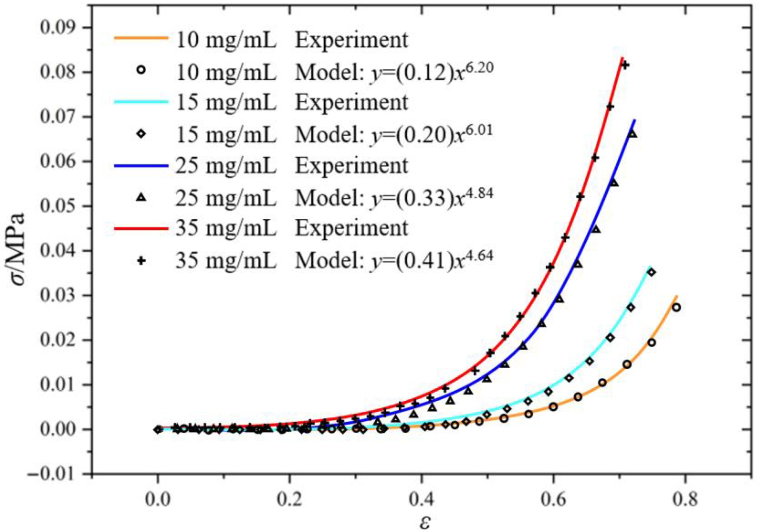
The engineering stress–strain relationships were obtained according to the experimental data of force–displacement, and then fitted using the aforementioned power-law constitutive model, with the results shown in Figure 2.

2.2. Nonlinear Wave Equation

The nonlinear stress–strain relationship of fibrin clots directly influences the mathematical form of the governing equation for acoustic wave propagation. Therefore, the simulations were based on the Westervelt equation, which incorporates the material’s nonlinearity.
Within the Lagrangian framework, the equation of motion accounting for finite deformation conditions is given by
ρ 0 2 u t 2 = x P
where ρ0 is the initial mass density in the reference configuration, u is the displacement vector, x is the coordinate, ∇ denotes the divergence operator with respect to the material coordinate, and P is the first Piola–Kirchhoff stress tensor. The deformation gradient F tensor is defined as
F i j = δ i j + u i x j
where I = δij is the second-order identity tensor. Introducing the Green strain tensor E to characterize finite deformation,
E i j = 1 2 k = 1 3 F k i F k j δ i j
Assuming the strain energy of the fibrin clot depends on both local stretching and volume change, and is entirely determined by the Green strain tensor E, the strain energy density function can be expressed as W = W(E). The first Piola–Kirchhoff stress tensor P can be derived from this strain energy density function:
P i j = F j k W E i k
We assume the fibrin clot satisfies the isotropy hypothesis. For nonlinear acoustic wave propagation problems under small-strain assumptions, W is expanded to the third order:
W = 1 2 ! C i j k l E i j E k l + 1 3 ! C i j k l m n E i j E k l E m n + o E 4
where Cijkl and Cijklmn are the second-order and third-order elastic constant tensors, respectively.
Combining Equation (12) and Equation (11) yields
P i j = C i j k l u k x l + L i j k l m n 2 u k x l u m x n + L i j k l m n p q 6 u k x l u m x n u p x q + o E 3
where Lijklmn = Cijklmn + Cijlnδkm + Cjklnδim + Cjlmnδik, and the expressions for the higher-order coefficients Lijklmnpq are provided in [38]. Note that LijklmnLjiklmn, indicating that the non-symmetry of P is a second-order effect. Combining Equation (8) and Equation (13), the nonlinear equation of motion is obtained:
ρ 0 2 u i t 2 = 2 u k x j x l C i j k l + L i j k l m n u m x n + L i j k l m n p q u m x n u p x q
From Equation (14), it follows that
2 u 1 t 2 c l 2 2 u 1 x 2 = 3 c l 2 + c 111 ρ 0 u 1 x 2 u 1 x 2 + c l 2 + c 166 ρ 0 u 2 x 2 u 2 x 2 + u 3 x 2 u 3 x 2                                                                           + 3 + 3 c 111 + c 1111 ρ 0 c l 2 1 2 ! u 1 x 2
2 u 2 t 2 c t 2 2 u 2 x 2 = c l 2 + c 166 ρ 0 u 1 x 2 u 2 x 2 + u 2 x 2 u 1 x 2
2 u 3 t 2 c t 2 2 u 3 x 2 = c l 2 + c 166 ρ 0 u 1 x 2 u 3 x 2 + u 3 x 2 u 1 x 2
where cl and ct are the propagation velocities of the longitudinal and transverse waves, respectively, and the nonlinear terms cijk and cijkl are given by
c i j k = 3 W E i E j E k ,   c i j k l = 4 W E i E j E k E l
For purely longitudinal motion (v = w = 0), Equations (15)–(17) simplify to
2 u t 2 = c l 2 2 u x 2 g u x
The power-law constitutive relation (Equation (6)) satisfied the theory of rate-independent materials where stress is solely a function of strain [39], and given that it is a continuously differentiable function with a positive first derivative, the wave speed cl is
c l = 1 ρ 0 d σ d ε = m ( D f ) k ( N f ) ρ 0 ε m ( D f ) 1
The nonlinear term in the wave equation is expressed as
g u x = 1 + 3 + c 111 ρ 0 c l 2 u x + 3 + 3 c 111 + c 1111 ρ 0 c l 2 1 2 ! u x 2 +
The nonlinear parameter can be represented as
β = 3 + c 111 ρ 0 c l 2 , γ = 3 + 3 c 111 + c 1111 ρ 0 c l 2
In this study, β and γ are treated as constants, with their value constrained to the range [5] to satisfy the concentration dependence of the clot. The primary focus is on investigating the influence of higher-order hardening effects under uniaxial compression on nonlinear wave propagation.

3. Numerical Simulation of the Shock Wave Formation Process

The propagation characteristics of stress waves in strain-hardening materials can be summarized as follows: As strain gradually increases due to external loading, the material enters the hardening stage, and the velocity of stress waves is governed by the instantaneous tangent modulus. Given the monotonically increasing hardening rate of such materials (i.e., the tangent modulus of the stress–strain curve continuously rises with increasing strain), subsequently generated stress waves will propagate at higher velocities. This wave velocity gradient manifests in spacetime such that later-generated, higher-stress wavefronts travel faster than earlier, lower-stress wavefronts. As a result, the former gradually overtake the latter during propagation. This velocity-gradient-induced wavefront convergence leads to a continuous increase in the spatial gradients of physical quantities such as stress and strain near the wavefront.
Based on the COMSOL Multiphysics (Version 6.3) finite element analysis platform, the computational domain of the one-dimensional fibrin clot model was spatially discretized using edge elements, with a total of 3000 mesh elements. The characteristic element length was set to Δx = 1.0 × 10⁻⁶ m.
The transient analysis was performed using the coefficient-form partial differential equation (PDE) module, employing the MUMPS direct solver and the automatic Newton iteration method for numerical simulation. The time-stepping for the transient solver was selected as the implicit generalized-α method, with the time step parameter set to Δt = 1 × 10⁻⁸ s, and data output at 1 × 10⁻⁷ s intervals.
The primary simulation parameters were defined as follows: frequency f = 20 kHz, hardening exponent n = 2, hardening coefficient k = 10 MPa, nonlinear parameter ratio β/γ = 5, γ = −1, with the remaining parameters as shown in Table 1.
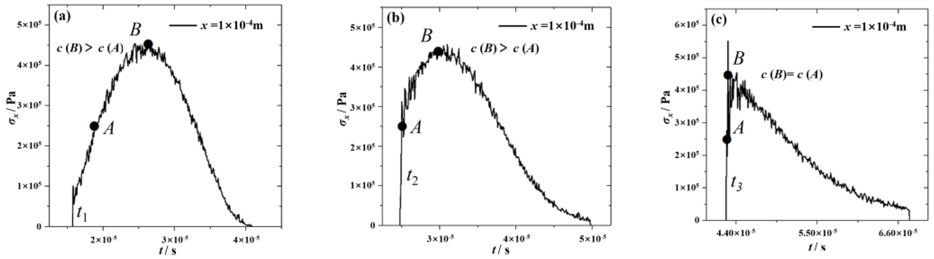
The one-dimensional numerical simulation results are illustrated in Figure 3. At t1 = 1.57 × 10⁻⁵ s, the ultrasonic wave exhibited a continuous and smooth distribution at x = 1.0 × 10⁻⁴ m, consistent with the sinusoidal periodic displacement boundary condition. The wave velocity c(B) at point B was significantly higher than that at point A. When the system evolved to t2 = 2.47 × 10⁻⁵ s, at x = 1.0 × 10⁻³ m, the trailing point B (which experiences a higher stress state, resulting in a larger material tangent modulus and corresponding wave velocity) gradually closed the distance to the leading point A. This caused a significant increase in the slope of the wavefront and even led to the formation of partially vertical stress profiles. At the critical moment t3 = 4.26 × 10⁻⁵ s, when the trajectories of the two wavefronts intersect at point x = 1.5 × 10⁻³ m, the originally continuous wave profile develops into a shock wavefront. At this stage, the wave velocities at points A and B become equal, and both are located on the shock wavefront. This indicated that the trailing wave B had caught up with the leading wave A. These observations aligned well with the shock wave evolution process described in existing studies for progressively hardening materials.
Based on the numerical results, two shock wave localization methods were proposed:
The Average Steepness Factor (ASF) was introduced to quantify the distortion degree of nonlinear ultrasonic waves within the material:
ASF = E p ˙ + E p ˙
where E p ˙ and E p ˙ + represent the average negative slope and average positive slope, respectively, defined as follows:
E p ˙ = P T n e g ,       E p ˙ + = P T p o s
where P = pmaxpmin is the difference between the maximum and minimum amplitudes within the selected time period, and Tneg, Tpos are the durations of the negative and positive slopes within the same time period.
The ASF value increased monotonically as the ultrasonic wave propagated into the clot interior, with the theoretical shock wave formation corresponding to ASF→∞. However, capturing shock waves in ASF theory encountered two numerical bottlenecks: First, the artificial viscosity introduced by traditional discrete schemes smoothed stress gradients, preventing ASF from reaching theoretical infinity. Although high-order schemes can suppress dissipation, numerical oscillations at the shock still affect the accuracy of shock wave capture. Second, there was the difficulty of mesh division. When the ASF number approached infinity, the grid size had to satisfy the shock characteristic width, and the computational cost increased exponentially with the increase in ASF. This paper set ASF = 100 as the shock wave formation threshold to locate the shock wave position.
From the perspective of energy transfer mechanisms, continuous waveform distortion led to energy transfer from the fundamental frequency to higher harmonics. The accumulation of harmonic energy led to the formation of shock waves. To characterize this, the Total Harmonic Distortion (THD) was introduced:
THD ( x ) = i = 2 N A i ( x ) A 1 ( x ) 2 × 100 %
where A1 is the fundamental frequency amplitude at the left point x, and Aᵢ represents the amplitudes of the other higher harmonics.
The shock formation position xTHD is identified as the extremum point of the THD function:
x THD = x d ( THD ( x ) ) d x = 0
In this section, numerical simulations under a single sinusoidal pulse successfully reproduced wavefront overtaking, convergence, and shock formation phenomena attributable to the strain-hardening behavior of the clot. The simulation results were consistent with reference [39] and classical theories. This established a foundation for subsequent investigations involving dual-wave-packet sequences. Both the Average Steepness Factor (ASF) and Total Harmonic Distortion (THD) methods were applied to calibrate the shock formation position, ensuring accuracy in subsequent analyses of gain effects.

4. Shock Wave Amplification Induced by Dual-Wave-Packet Sequences

Based on the previous analysis, when an external load is applied to a strain-hardening material, continuous strain variation is induced. Due to differences in wave velocity, wave overtaking occurs within the waveform occurs, leading to the formation of shock waves. Such shock waves arise from the distortion of ultrasonic waveforms and do not inherently produce shock wave amplification. Therefore, this section extended the concept of velocity-difference-induced wave overtaking to interactions between wave packets. Through designing wave packet sequences, overtaking and superposition of different wave packets were achieved to realize shock wave amplification, and the influence of ultrasonic parameters on the amplification mechanism was discussed.

4.1. Chasing and Superposition of Wave Packets

To characterize the superposition degree of wave packet sequences with different amplitudes at a specific moment, the following criterion was defined:
R % = k n WSF WSF × 100 %             n = 1 , 2 , , 5
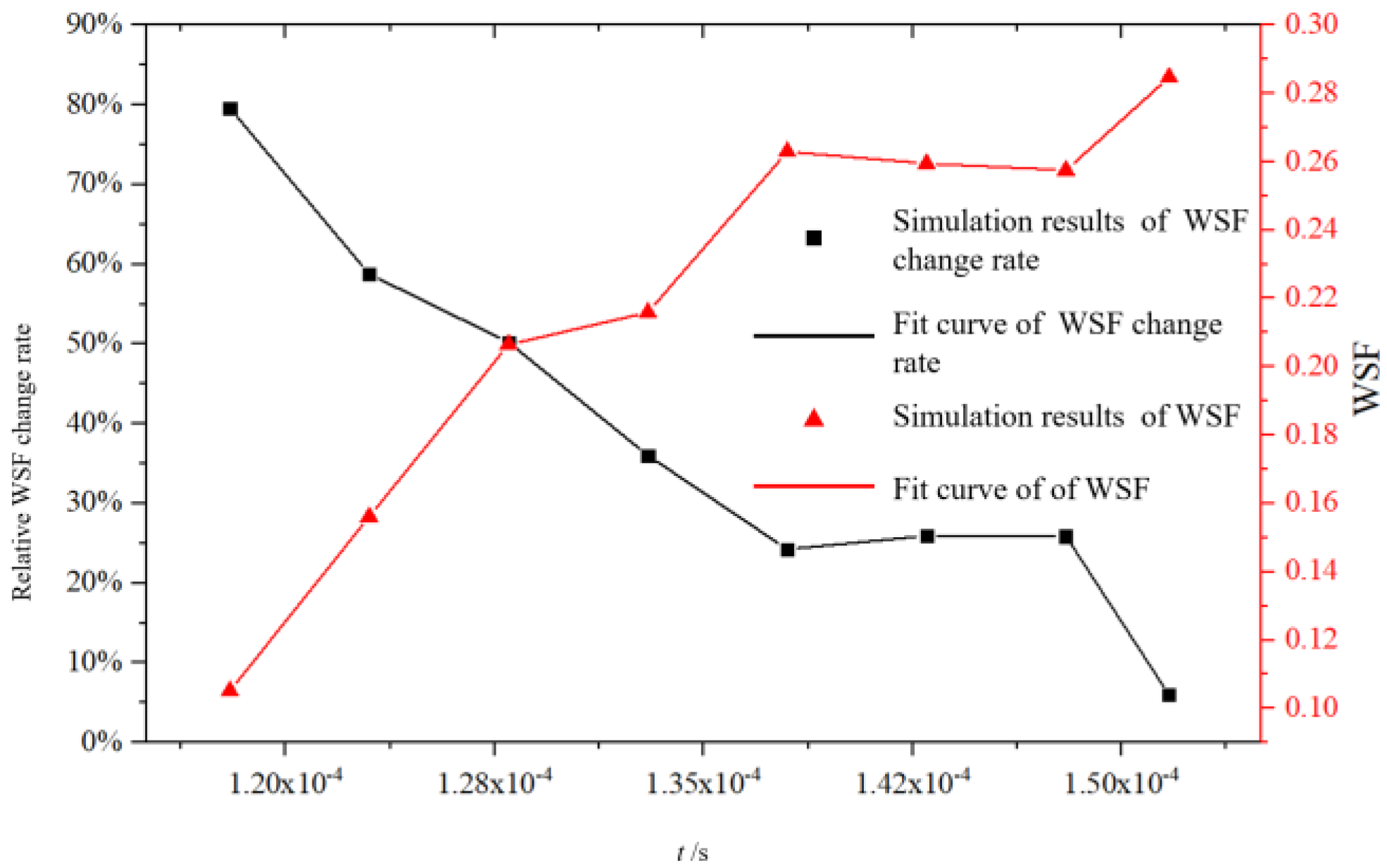
where WSF represents the waveform superposition factor, which is used to characterize the wavefront slope at a given moment, and R is the relative WSF rate of change. A smaller change in R rate indicates more stable variations in the wavefront slope within the considered interval.
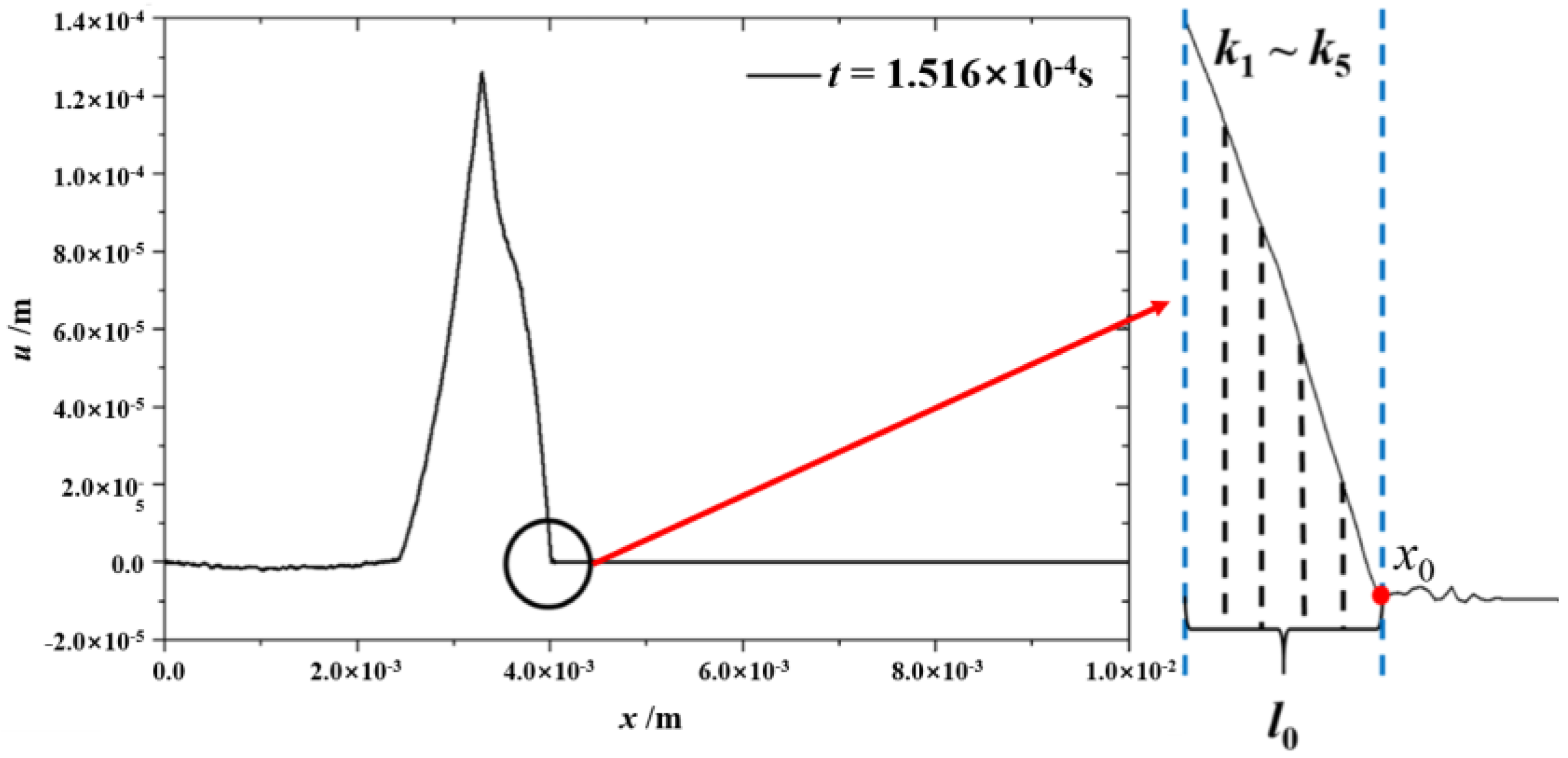
The calculation diagram of WSF is shown in Figure 4: first, the point where the wavefront portion of the displacement curve initially intersects the Y-axis was taken as the reference point; then, a waveform superposition characteristic distance l0, corresponding to 2r times the grid length, was taken leftward from the reference point to obtain a continuous segment of the wavefront displacement curve; this segment was evenly divided into r sections, and the slopes k1 ~ kᵣ of each section were calculated. The absolute value of the mean of their sum was taken as the WSF:
WSF = n = 1 r k n r    
In this study, the threshold for the relative WSF change rate was set as 10%, meaning that when this value is ≤10%, the superposition of wave packet sequences with different amplitudes can be considered sufficiently adequate.
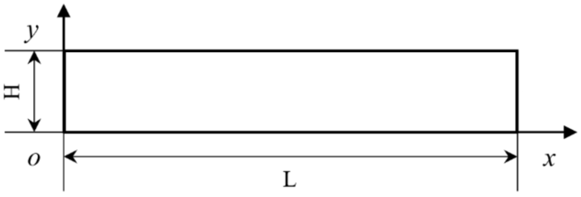
Now, consider the chasing interaction between two sinusoidal wave packets. To capture the process related to the chasing and superposition of wave packet sequences with different amplitudes, the computational model considered planar longitudinal wave propagation in a two-dimensional homogeneous medium as shown in Figure 5.
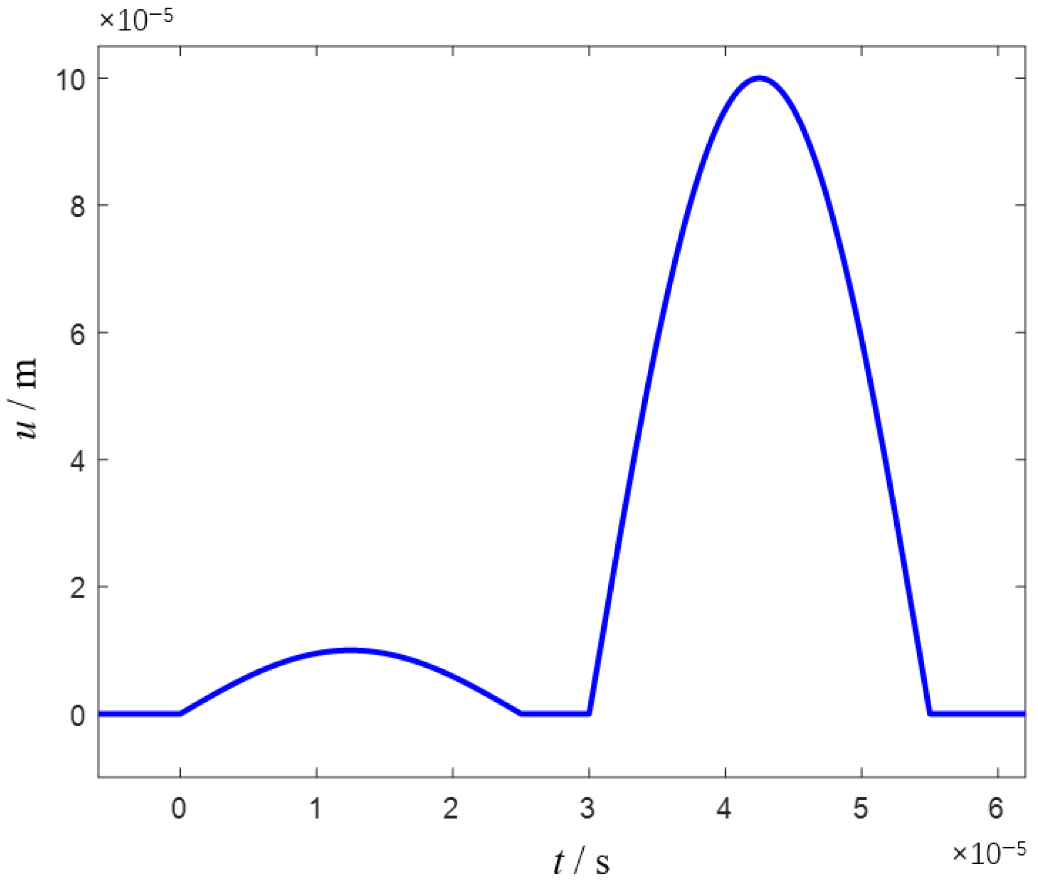
The model had a length L of 10 mm and a height H of 0.2 mm, with a total computation time of 6T. The computational domain was discretized using a uniform grid of 20,000 points. Time integration was performed using an explicit scheme with a fixed time step of 10−8 s. This time step satisfies the CFL stability condition, ensuring the stability of the numerical solution. The top and bottom boundaries of the model are free boundaries. The left boundary was a fixed boundary where the excitation was applied. A displacement boundary condition, as specified by the formula in the manuscript, was imposed at the left end, set as the dual-wave-packet form of Equation (29), as illustrated in Figure 6. The right end was set as a non-reflecting boundary to simulate a semi-infinite medium, thereby preventing wave reflections from interfering with the results. The relevant parameters were set as follows: frequency f = 20 kHz, hardening exponent n = 2, hardening coefficient k = 1.0 MPa, nonlinear parameter ratio β = −5, γ = −1, number of grid elements 20,000, duty cycle 90.9%.
f t = 0.1 × 10 4 sin 2 π f t                                       t 0 , 0.5 / f 0                                                                                                         t 0.5 / f , 0.6 / f 1.0 × 10 4 sin 2 π f t 1.2 π             t 0.6 / f , 1.1 / f
First, the degree of dual-wave superposition was examined. The relative WSF rate of change and WSF variation over time are shown in Figure 7. The starting point of the time axis was determined based on the criterion that the wavefront displacement curve within the length interval l0 could capture wave packets of two different amplitudes. The results indicate that as the wave packet sequences were superposed with different amplitudes, the relative WSF rate of change gradually decreased from a peak of approximately 80% to 6%, demonstrating that the wave packet sequences had sufficiently superposed. Meanwhile, the wavefront slope WSF gradually increased, ultimately rising from 0.10513 to 0.28469 after full superposition—an increase of approximately 171%.
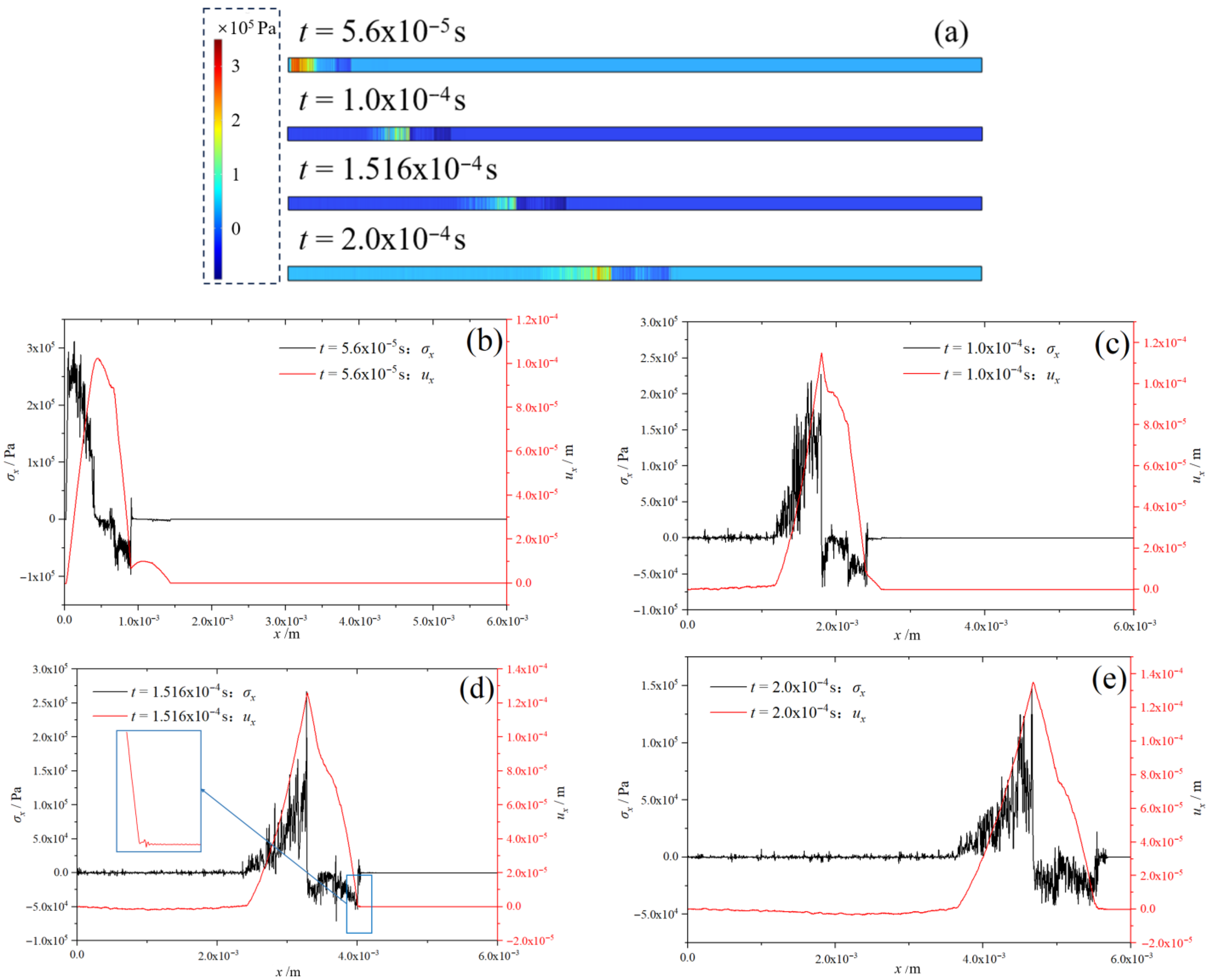
Subsequently, an analysis was conducted by extracting the variation in stress and displacement with coordinates at different time steps. The stress nephograms and corresponding line plots at different moments during wave propagation are shown in Figure 8. At t = 5.6 × 10⁻⁵ s, the boundary condition of trailing wave A (with a displacement amplitude U0 = 1.0 × 10⁻⁴ m) had completed its application and partially overtaken leading wave B (with an amplitude U0 = 0.1 × 10⁻⁴ m). Simultaneously, due to the progressively hardening characteristics of the clot, wave A was undergoing internal overtaking, causing distortion in the displacement curve. A strong stress discontinuity appeared at x = 0.4 × 10⁻³ m. However, due to its smaller amplitude, the strain induced by wave B corresponded to the low-strain segment of the constitutive curve of the progressively hardening material. The minimal difference in wave velocity resulted in an insignificant internal overtaking effect within wave B, so its displacement curve remained nearly sinusoidal, as shown in Figure 8b.
At t = 1.0 × 10⁻⁴ s, a discontinuity in displacement had already emerged at x = 1.8 × 10⁻³ m, forming a distinct shock wavefront. This indicates that the internal overtaking within wave A had concluded, with a corresponding stress peak of 0.22 MPa. However, the wavefront profile of wave B remained near x = 2.5 × 10⁻³ m, indicating that the superposition of the wave packet sequences with different amplitudes was not yet complete, as shown in Figure 8c. At t = 1.516 × 10⁻⁴ s, the shock wavefront continued to propagate longitudinally to x = 4.0 × 10⁻³ m. The magnified view within the blue frame in Figure 8d reveals that the displacement curve at the wavefront was smooth and showed no significant characteristics of the preceding wave. The corresponding stress peak here was 0.27 MPa, which was 0.05 MPa greater than the stress peak observed in Figure 8c—an increase of approximately 22.7%. According to the conclusions in Section 3, the stress peak should continuously decay after the shock wavefront forms. However, the superposition of wave packet sequences with different amplitudes had induced this increase in the stress peak. At t = 2.0 × 10⁻⁴ s, as the shock wavefront propagates to x = 4.7 × 10⁻³ m, the corresponding stress peak decays to 0.15 MPa.
The 22.7% gain was the maximum value obtained from a systematic parameter sweep over both time delays and amplitude ratios. Specifically, the frequency remained 20 KHz; the duty cycle was varied within the range of [100%, 85%], and the displacement amplitude range was [9 × 10⁻⁵ m, 1.3 × 10⁻⁴ m].
To ensure the reliability of the numerical results, a rigorous mesh independence study was conducted. Simulations for the same case were performed using three different grid densities: 20,000, 40,000, and 80,000 points. A comparison of the results showed that the relative error in the calculated shock formation position and peak-shock stress between the different grids was less than 0.5%. This conclusively demonstrates that the selected grid resolution (20,000 points) yielded converged results that are consistent with those from finer meshes.
The above analysis demonstrated that in a fibrin clot exhibiting progressive hardening, both the leading and trailing waves in the dual-wave-packet sequence undergo waveform distortion. The trailing wave gradually overtook the leading wave until their superposition was fully achieved, resulting in a gain effect on the formed shock wave. This process led to a 22.7% increase in the peak stress of the shock wave.
Existing studies have confirmed a clear positive correlation between the mechanical force of ultrasonic shock waves and the thrombolysis rate: at a fixed frequency, higher acoustic intensity leads to stronger mechanical force and a higher thrombolysis rate [45]. Higher ultrasound intensity indeed poses two main potential risks: mechanical damage to the vessel wall and the generation of harmful embolic fragments due to rapid, uncontrolled thrombus fragmentation. Notably, research on ultrasound intensity thresholds indicates the existence of a critical intensity (approximately 21.6 W/cm2); below this value, the extent of thrombus damage is minimal (approximately 11–15%); once this threshold is exceeded, the maximum damage degree increases rapidly with intensity [21]. Furthermore, when the intensity (which determines the peak stress) surpasses a specific critical point, the destructive effect is significantly enhanced, exhibiting a strong nonlinear characteristic. Therefore, solely relying on increasing ultrasound intensity is not a viable strategy. Our proposed ‘wave-packet chasing and superposition’ strategy does not aim to infinitely amplify shock wave energy. Instead, it seeks to more efficiently concentrate energy at specific locations within the thrombus through precise waveform modulation, promoting controllable disruption of its internal structure while maintaining the existing energy input. Compared to traditional methods that simply increase single-pulse amplitude, our strategy offers intrinsic safety advantages.
However, the current continuum model did not consider inertial cavitation effects. According to nonlinear acoustic theory, the propagation of finite-amplitude compression waves (positive pressure) in media such as fibrin clots leads to waveform distortion, generating higher harmonics that include rarefaction waves (negative pressure). This provides a theoretical possibility for inertial cavitation. Meanwhile, our numerical simulation results (Figure 8e) indicated that the peak negative pressure induced by material nonlinearity was approximately 0.075 MPa, theoretically, since both wave packets provide positive pressure. Existing research clearly shows that the cavitation threshold for blood containing ultrasound contrast agents is on the order of megapascals, while the threshold for pure blood or tissue is even higher (typically >9 MPa) [46]. Evidently, the negative pressure level generated in our simulations is significantly below these cavitation thresholds, suggesting a low probability of significant inertial cavitation within the current theoretical framework.
This characteristic represents a potential safety advantage of our strategy: it primarily amplifies mechanical shock by leveraging the material’s strain-hardening properties through precise waveform design, rather than relying on uncontrolled cavitation bubbles as the main disruption mechanism. Nonetheless, we fully recognize that in practical high-intensity pulse sequence treatments, improper parameter design (e.g., timing, duty cycle) could substantially increase cavitation risk. Therefore, future engineering explorations could draw on existing studies by optimizing pulse forms (e.g., employing short-pulse, low-duty-cycle sequences) [47]. This approach holds promise for synergistically combining controlled cavitation mechanical forces with shock wave amplification effects while effectively managing thermal effects, thereby achieving efficient and synergistic thrombus ablation.

4.2. Parametric Analysis

During the dual-wave-packet interaction process, several parameters involved in the ultrasonic boundary conditions, such as frequency, amplitude (unless otherwise specified, changes in amplitude in parametric analyses refer to the second wave packet with larger amplitude), and duty cycle, exert a significant impact on the interaction and superposition of wave packets with different amplitudes. Therefore, a controlled variable approach was employed in this section to analyze and discuss the effects of the aforementioned parameters, while all other relevant parameters were held constant.

4.2.1. Influence of Frequencies on the Location and Magnitude of the Stress Peak

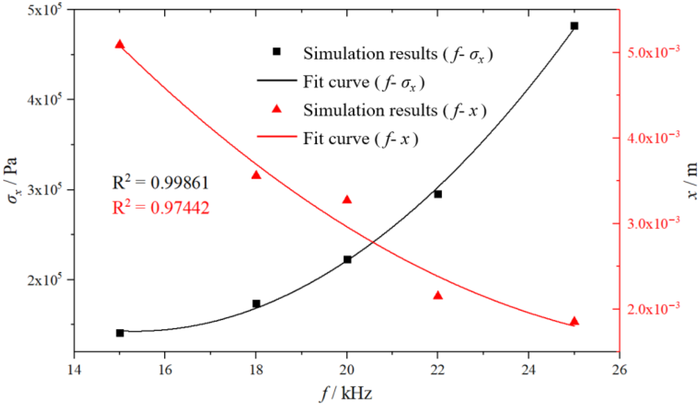
The influence of five different frequencies (15 kHz, 18 kHz, 20 kHz, 22 kHz, and 25 kHz) on the location and magnitude of the stress peak at full superposition of wave packet sequences with different amplitudes was discussed. The numerical simulation results were fitted using a quadratic polynomial, as shown in Figure 9.
The simulation results indicate that as the frequency increased from 15 kHz to 25 kHz, the location of the stress peak shifted forward from x = 5.09 × 10⁻³ m to x = 1.85 × 10⁻³ m, corresponding to a 63.7% advancement. Meanwhile, the magnitude of the stress peak increased from 0.14 MPa to 0.48 MPa, representing an increase of 243%. It was demonstrated that an increase in amplitude resulted in a forward shift of the stress peak position and a significant enhancement in its magnitude. The amplitude was directly correlated with the intensity of ultrasonic energy, where larger amplitudes correspond to higher input power. This led to more pronounced strain variations, thereby inducing a more significant strain-hardening effect. At a constant frequency, variations in amplitude directly altered the tangent slope of the waveform profile, consequently modifying the wave velocity at different waveform positions. These modifications further accelerated the wave overtaking processes occurring both within and between wave packets.

4.2.2. Influence of Amplitude on the Location and Magnitude of the Stress Peak

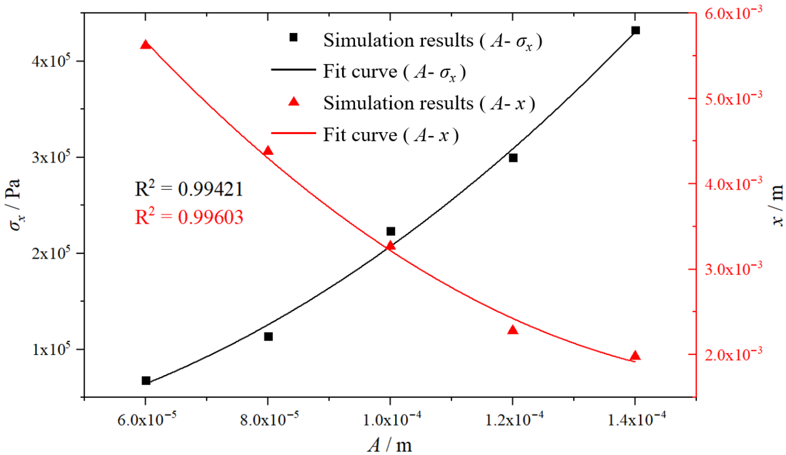
The influence of five different amplitudes (0.6 × 10⁻⁴ m, 0.8 × 10⁻⁴ m, 1.0 × 10⁻⁴ m, 1.2 × 10⁻⁴ m, and 1.4 × 10⁻⁴ m) on the location and magnitude of the stress peak at full superposition of wave packet sequences with different amplitudes was discussed. The numerical simulation results were fitted using a quadratic polynomial, as shown in Figure 10.
The simulation results indicated that as the amplitude increased, the location of the stress peak shifted forward from x = 5.62 × 10⁻³ m to x = 1.98 × 10⁻³ m, corresponding to a 64.8% advancement. Meanwhile, the magnitude of the stress peak increased from 0.07 MPa to 0.43 MPa, representing an increase of 514%. This demonstrated that an increase in amplitude led to a forward shift in the location of the stress peak and a significant increase in its magnitude. The amplitude directly affected the intensity of ultrasonic energy, with larger amplitudes corresponding to stronger input power. This resulted in more pronounced strain variations, thereby inducing a more significant strain-hardening effect. At a constant frequency, a change in amplitude directly altered the tangent slope of the waveform curve, leading to variations in wave velocity at different positions of the waveform. This further accelerated the overtaking process both within and between wave packets.

4.2.3. Influence of Duty Cycle on the Location and Magnitude of the Stress Peak

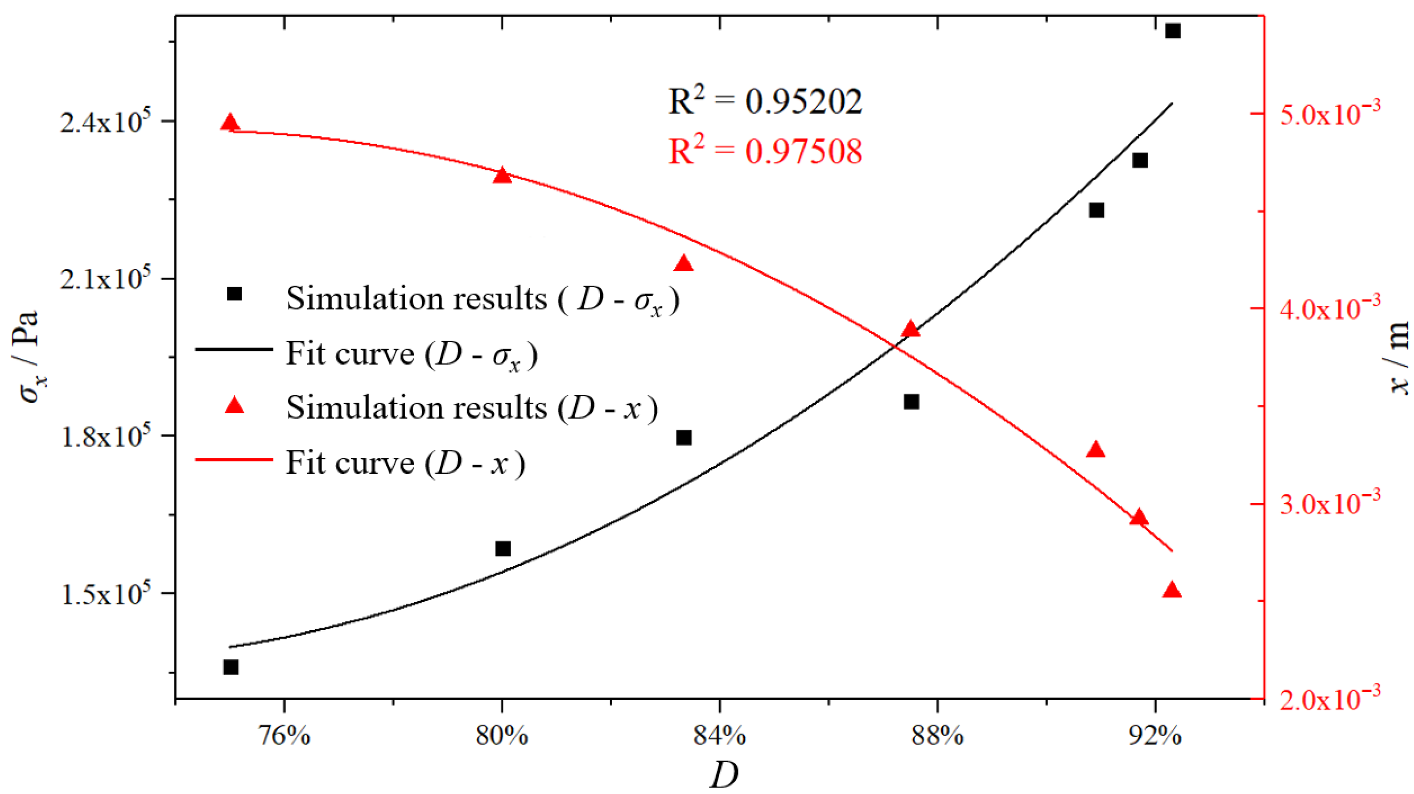
The influence of seven different duty cycles D (75%, 80%, 83.3%, 87.5%, 90.9%, 91.7%, and 92.3%) on the location and magnitude of the stress peak at full superposition of wave packet sequences with different amplitudes was discussed. The numerical simulation results were fitted using a quadratic polynomial, as shown in Figure 11.
The simulation results indicated that as the duty cycle increased from 75% to 92.3%, the location of the stress peak shifted forward from x = 4.95 × 10⁻³ m to x = 2.55 × 10⁻³ m, corresponding to a 48.5% advancement. Meanwhile, the magnitude of the stress peak increased from 0.14 MPa to 0.26 MPa, representing an 85.7% increase. Although an increase in the duty cycle also led to a forward shift in the stress peak location and an increase in its magnitude, the mere reduction in zero-value intervals did not significantly affect stress distribution or waveform changes. The duty cycle most directly influences ultrasonic wave propagation: a decrease in the duty cycle corresponds to an increase in the initial zero-value intervals between wave packets, thereby delaying the pursuit and superposition of wave packet sequences with different amplitudes. Parametric analysis determined the shock wave formation position range as xASF ∈ (1.85 × 10⁻³ m, 5.62 × 10⁻³ m). Although all three ultrasonic parameters affected the location and magnitude of the stress peak as well as the WSF value, their respective influence weights differed.
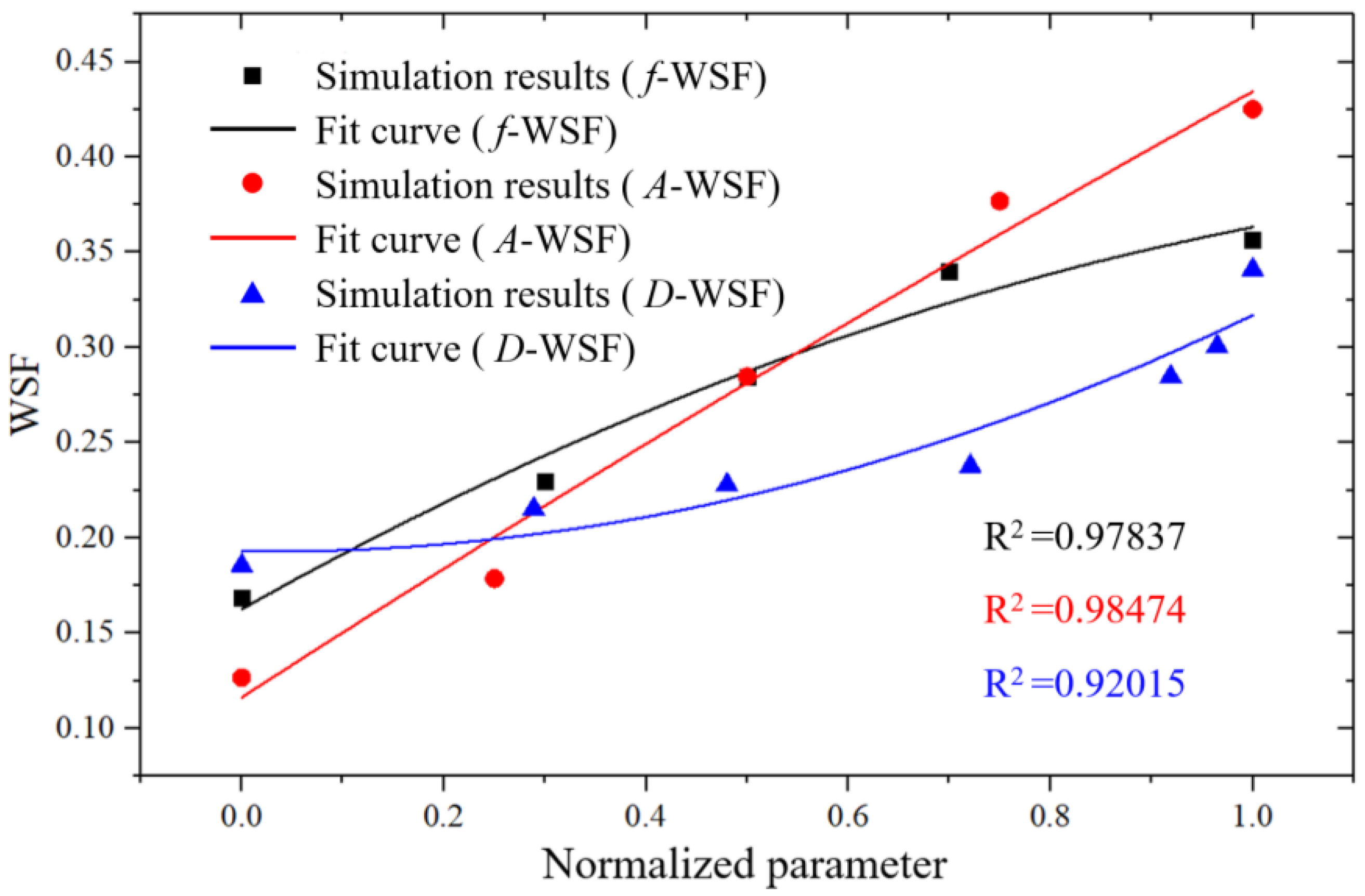
To facilitate efficient determination of treatment protocols in clinical practice, it is necessary to focus on targeted adjustments of specific parameters. To quantify the relative influence of each parameter on the WSF, normalization was performed on frequency, amplitude, and duty cycle, with the value range of each parameter proportionally mapped to the [0, 1] interval. Taking frequency as an example, the five frequency values (15 kHz, 18 kHz, 20 kHz, 22 kHz, 25 kHz) correspond to normalized parameter values of 0, 0.3, 0.5, 0.7, and 1, respectively. The final results are presented in Figure 12.
The above results indicated that amplitude had the most significant impact on the WSF. An increase in amplitude implied an expansion of the strain variation range, which in turn led to a greater range of variation in the slope of the progressively hardening constitutive curve—i.e., a larger range of wave velocity variation. This intensified the overtaking effects both within and between wave packets (waveform distortion), making the shock wave effect more pronounced, the wavefront displacement curve steeper, and ultimately corresponding to a higher shock wave stress peak.
The results of this study indicate that by leveraging the nonlinear properties of the material and designing the delay time, amplitude, and frequency of dual-wave packets, it is possible to achieve shock wave gain at specific locations—essentially, a form of directional aggregation of wave energy. The underlying physical principles and the observed patterns are highly consistent with experimental trends in the existing literature. For example, Waters et al. designed a linear frequency modulation (chirp) excitation waveform to compensate for the dispersion of flexural waves, enabling different frequency components to arrive simultaneously at the target location. This experiment achieved a significant amplification of the local shock response, directly demonstrating the experimental validation of “achieving wavefront superposition and shock amplification by adjusting the emission delays of different frequency groups (i.e., allowing subsequent waves to ‘chase’ and catch up with the leading wave)” [48]. Wang et al. generated acoustic wave packets through resonant excitation in a ring-shaped Bose–Einstein condensate and observed that the propagation speed of waves (i.e., density perturbations) is related to the local density of the fluid. This is analogous to the principles of classical nonlinear acoustics in this study. Under strong excitation, the denser parts of the wave packet propagate faster, causing the waveform to steepen at the front and broaden at the rear during propagation. Under intense excitation, the generation of supersonic shock waves was observed [49]. This aligns with the physical principles in this study, which utilize the nonlinear properties of the material and design key parameters such as delay time, frequency, and amplitude of dual ultrasonic wave packets to achieve dual-wave chasing, superposition, and shock wave amplification.
In the field of ultrasonic therapy, Edsall et al. precisely controlled the “time delay” of dual-frequency pulses, causing coherent superposition of acoustic waves during nonlinear propagation in tissues. They demonstrated that variations in time delay on the order of microseconds or even nanoseconds can fundamentally alter the instantaneous waveform of the superimposed waves, thereby achieving more efficient and precise tissue ablation effects compared to single-frequency pulses. In red blood cell phantom experiments, the lesions produced by dual-frequency pulses were more precise and exhibited higher ablation efficiency [50]. The above discussion demonstrates that these experimental results are consistent with the trends observed in this study’s numerical simulations, where shock wave gain is achieved by adjusting the delay time, frequency, and amplitude of the second wave packet.
The implementation of the dual-wave packet chasing strategy places clear demands on the damping and bandwidth of the transducer. This outlines a clear engineering pathway: achieving ideal waveform transmission through subsequent material optimization and transducer design. Existing theoretical models and experimental trends fully support the feasibility of this pathway in principle.
In terms of acoustic implementation, the strategy can be realized on two levels: a single-frequency dual-wave-packet sequence and a dual-frequency dual-wave-packet sequence. (1) For single-frequency dual-wave-packet sequences, damping can be increased and bandwidth broadened by optimizing transducer structure (e.g., enhancing acoustic backing) [51] and employing broadband materials (e.g., novel composite piezoelectric materials, PMN-PT single crystals) [52], laying the groundwork for short-pulse emission. Based on this, waveform generation and precisely controlled silent intervals between two waveforms, with nanosecond-level accuracy, can be realized via counters within a Field-Programmable Gate Array (FPGA). (2) For dual-frequency dual-wave-packet sequences, two independent transducer elements, each optimized for frequencies f1 and f2, respectively, can be used. These elements are controlled by the same FPGA but operate through separate transmission channels and are driven sequentially according to the designed order.
It must be emphasized that despite the therapeutic advantages of this technical approach, its potential safety risks require careful consideration. If unintended beam superposition occurs due to timing errors, it could damage healthy vascular walls or other adjacent tissues. Therefore, substantial future in vitro and preclinical experiments are necessary to determine the precise “therapeutic window” and safety parameter thresholds for different treatment scenarios. This is a prerequisite for achieving safe clinical applications.

5. Conclusions

This study reveals the physical mechanism of shock wave amplification induced by dual-wave-packet ultrasonic sequences in fibrin clots through numerical simulations. The main conclusions are summarized as follows:
(1)
A power-law constitutive model for fibrin clots with concentration dependence was established through microforce uniaxial compression experiments. Combined with the third-order nonlinear wave equation, the numerical model successfully reproduced the single-wave shock wave formation process, validating the reliability of the model.
(2)
An innovative dual-wave-packet chasing strategy was proposed, extending the waveform distortion induced by the nonlinear elasticity of fibrin clots from single-wave scenarios to multi-wave pursuit in progressively hardening materials. This approach successfully achieved inter-wave superposition, resulting in a significant amplification of the shock wave by up to 22.7%.
(3)
Quantitative evaluation metrics for inter-wave superposition were established, and a parametric analysis of the influence of ultrasonic parameters on multi-wave chasing and superposition was conducted. The results demonstrate that amplitude is the most significant parameter affecting wavefront steepness and gain effect, followed by frequency, while the duty cycle primarily influences the timing of wave packet superposition.
This study advances beyond the traditional research paradigm focused on single ultrasonic waves, demonstrating that intelligently designed ultrasonic sequences can actively regulate mechanical effects in biological media. It provides crucial theoretical support for the development of next-generation high-performance ultrasonic thrombolysis therapeutic instruments.

6. Limitations and Future Perspectives

While this study demonstrates the potential of the dual-wave-packet strategy for amplifying shock waves within thrombi, several limitations warrant acknowledgment, which also delineate clear directions for future research.

6.1. Limitations

The authors’ research found that by connecting a dashpot with low viscosity in parallel to the current nonlinear elasticity framework based on a power-law model (thereby introducing viscoelasticity), and keeping all other model parameters unchanged, simulations still demonstrate the occurrence of dual-wave superposition and shock wave enhancement. However, the shock wave enhancement value is reduced, and its formation position shifts significantly backward. This indicates that in viscoelastic media, both the magnitude and location of shock wave enhancement depend on the strength of viscosity and the design of the dual-wave sequence. Since the viscoelastic behavior of thrombi may be more complex than that captured by a simple nonlinear spring-dashpot model, further research is needed.
Therefore, it must be noted that while the quasi-static power-law model used in this study effectively captures the core strain-hardening nonlinear characteristics of thrombi, it fails to account for their frequency-dependent viscoelasticity under high-frequency ultrasound excitation (e.g., above ~20 kHz). Consequently, the absolute stress enhancement values derived from the simulations may deviate from those observed in real dynamic biological environments.

6.2. Future Perspectives

Addressing these limitations will be the focus of subsequent work. Future studies will aim to (1) characterize the frequency-dependent viscoelastic properties of fibrin clots using dynamic mechanical analysis (e.g., oscillatory shear rheometry) and develop the dual-wave chasing strategy that incorporates frequency-dependent viscoelasticity; (2) achieve precise timing and synergy between the two wave packets through broadband transducers and nanosecond-level pulse control. Building on this foundation, systematic in vitro and ex vivo experiments will be conducted in flow models containing human blood clots. Furthermore, the integration of pre-treatment ultrasound elastography for thrombus characterization will be explored to guide personalized parameter selection.

Author Contributions

Conceptualization, X.Z.; methodology, L.Z.; software, F.M. and Z.Z.; validation, X.Z. and L.Z.; formal analysis, L.Z.; investigation, L.Z. and F.M.; resources, X.Z.; data curation, F.M.; writing—original draft preparation, L.Z.; writing—review and editing, X.Z. and L.Z.; visualization, L.Z.; supervision, X.Z.; project administration, X.Z.; funding acquisition, X.Z. All authors have read and agreed to the published version of the manuscript.

Funding

This research was funded by the National Natural Science Foundation of China, grant number 12272065.

Data Availability Statement

All data were generated during the experimental research process.

Conflicts of Interest

The authors declare no conflicts of interest.

Abbreviations

The following abbreviations are used in this manuscript:
ASFAverage Steepness Factor
WSFWaveform Superposition Factor
PDEPartial Differential Equation
FPGAField-Programmable Gate Array

References

  1. Wahlgren, N.; Ahmed, N.; Dávalos, A.; Ford, G.A.; Grond, M.; Hacke, W.; Hennerici, M.G.; Kaste, M.; Kuelkens, S.; Larrue, V.; et al. Thrombolysis with Alteplase for Acute Ischaemic Stroke in the Safe Implementation of Thrombolysis in Stroke- Monitoring Study (SITS-MOST): An Observational Study. Lancet 2007, 369, 275–282. [Google Scholar] [CrossRef] [Scilit]
  2. Renú, A.; Laredo, C.; Lopez-Rueda, A.; Llull, L.; Tudela, R.; San-Roman, L.; Urra, X.; Blasco, J.; Macho, J.; Oleaga, L.; et al. Vessel Wall Enhancement and Blood–Cerebrospinal Fluid Barrier Disruption After Mechanical Thrombectomy in Acute Ischemic Stroke. Stroke 2017, 48, 651–657. [Google Scholar] [CrossRef] [Scilit] [PubMed]
  3. Li, X.; Du, H.; Song, Z.; Wang, H.; Tan, Z.; Xiao, M.; Zhang, F. Efficacy and Safety of Sonothrombolysis in Patients with Acute Ischemic Stroke: A Systematic Review and Meta-Analysis. J. Neurol. Sci. 2020, 416, 116998. [Google Scholar] [CrossRef] [Scilit] [PubMed]
  4. Alexandrov, A.V.; Molina, C.A.; Grotta, J.C.; Garami, Z.; Ford, S.R.; Alvarez-Sabin, J.; Montaner, J.; Saqqur, M.; Demchuk, A.M.; Moyé, L.A.; et al. Ultrasound-Enhanced Systemic Thrombolysis for Acute Ischemic Stroke. N. Engl. J. Med. 2004, 351, 2170–2178. [Google Scholar] [CrossRef] [Scilit] [PubMed]
  5. O’Reilly, M.A. Exploiting the Mechanical Effects of Ultrasound for Noninvasive Therapy. Science 2024, 385, eadp7206. [Google Scholar] [CrossRef] [Scilit]
  6. Datta, S.; Coussios, C.-C.; Ammi, A.Y.; Mast, T.D.; De Courten-Myers, G.M.; Holland, C.K. Ultrasound-Enhanced Thrombolysis Using Definity® as a Cavitation Nucleation Agent. Ultrasound Med. Biol. 2008, 34, 1421–1433. [Google Scholar] [CrossRef] [Scilit] [PubMed]
  7. Yang, W.; Zhou, Y. Effect of Pulse Repetition Frequency of High-Intensity Focused Ultrasound on in Vitro Thrombolysis. Ultrason. Sonochemistry 2017, 35, 152–160. [Google Scholar] [CrossRef] [Scilit]
  8. Cao, W.; Liu, Y.; Ran, P.; He, J.; Xie, S.; Weng, J.; Li, X. Ultrasound-Propelled Janus Rod-Shaped Micromotors for Site-Specific Sonodynamic Thrombolysis. ACS Appl. Mater. Interfaces 2021, 13, 58411–58421. [Google Scholar] [CrossRef] [Scilit]
  9. Bosio, G.; Destrempes, F.; Yazdani, L.; Roy Cardinal, M.; Cloutier, G. Resonance, Velocity, Dispersion, and Attenuation of Ultrasound-Induced Shear Wave Propagation in Blood Clot In Vitro Models. J. Ultrasound Med. 2024, 43, 535–551. [Google Scholar] [CrossRef] [Scilit]
  10. Sakharov, D.V.; Barrett-Bergshoeff, M.; Hekkenberg, R.T.; Rijken, D.C. Fibrin-Specificity of a Plasminogen Activator Affects the Efficiency of Fibrinolysis and Responsiveness to Ultrasound: Comparison of Nine Plasminogen Activators In Vitro. Thromb. Haemost. 1999, 81, 605–612. [Google Scholar] [CrossRef] [Scilit]
  11. Chen, P.-Y.; Shih, C.-C.; Huang, C.-C. Assessing the Viscoelastic Properties of Thrombus Using Shear Wave Dispersion Ultrasound Vibrometry. In Proceedings of the 2012 IEEE International Ultrasonics Symposium, Dresden, Germany, 7–10 October 2012; IEEE: Dresden, Germany, 2012; pp. 1–4. [Google Scholar]
  12. Schmitt, C.; Hadj Henni, A.; Cloutier, G. Characterization of Blood Clot Viscoelasticity by Dynamic Ultrasound Elastography and Modeling of the Rheological Behavior. J. Biomech. 2011, 44, 622–629. [Google Scholar] [CrossRef] [Scilit] [PubMed]
  13. Rayes, A.; Zhang, J.; Lu, G.; Qian, X.; Schroff, S.T.; Ryu, R.; Jiang, X.; Zhou, Q. Estimating Thrombus Elasticity by Shear Wave Elastography to Evaluate Ultrasound Thrombolysis for Thrombus with Different Stiffness. IEEE Trans. Biomed. Eng. 2023, 70, 135–143. [Google Scholar] [CrossRef] [Scilit] [PubMed]
  14. Petit, B.; Yan, F.; Tranquart, F.; Allémann, E. Microbubbles and Ultrasound-Mediated Thrombolysis: A Review of Recent in Vitro Studies. J. Drug Deliv. Sci. Technol. 2012, 22, 381–392. [Google Scholar] [CrossRef] [Scilit]
  15. Gao, S.; Zhu, Q.; Guo, M.; Gao, Y.; Dong, X.; Chen, Z.; Liu, Z.; Xie, F. Ultrasound and Intra-Clot Microbubbles Enhanced Catheter-Directed Thrombolysis In Vitro and In Vivo. Ultrasound Med. Biol. 2017, 43, 1671–1678. [Google Scholar] [CrossRef] [Scilit]
  16. Izadifar, Z.; Babyn, P.; Chapman, D. Mechanical and Biological Effects of Ultrasound: A Review of Present Knowledge. Ultrasound Med. Biol. 2017, 43, 1085–1104. [Google Scholar] [CrossRef] [Scilit]
  17. Ariëns, R.A.; Sharp, A.S.; Duval, C. Ultrasound-Mediated Catheter Delivery of Tissue Plasminogen Activator Promotes Thrombolysis by Altering Fibrin Fibre Thickness and Clot Permeability. Haematologica 2024, 110, 665–672. [Google Scholar] [CrossRef] [Scilit]
  18. Tang, J.; Tang, J.; Liao, Y.; Bai, L.; Luo, T.; Xu, Y.; Liu, Z. An in Vitro Comparative Study on Clot Lysis Efficiency of Urokinase and Reteplase with the Synergy of Ultrasound Needle. Heliyon 2024, 10, e26624. [Google Scholar] [CrossRef] [Scilit]
  19. Liu, S.; Bahmani, A.; Ghezelbash, F.; Li, J. Fibrin Clot Fracture under Cyclic Fatigue and Variable Rate Loading. Acta Biomater. 2024, 177, 265–277. [Google Scholar] [CrossRef] [Scilit]
  20. Adzerikho, I.E.; Mrochek, A.G.; Minchenya, V.T.; Dmitriev, V.V.; Kulak, A.I. Combined Low-Frequency Ultrasound and Streptokinase Intravascular Destruction of Arterial Thrombi In Vivo. Ultrasound Med. Biol. 2011, 37, 1644–1652. [Google Scholar] [CrossRef] [Scilit]
  21. Adzerikho, I.; Kulak, A.; Rachok, S.; Minchenya, V. Dependence of the Rate and Completeness of Fibrin Clot Destruction on the Acoustic Dose and Ultrasound Intensity. Ultrasound Med. Biol. 2022, 48, 846–855. [Google Scholar] [CrossRef] [Scilit]
  22. Teng, Z.; Feng, J.; Zhang, Y.; Huang, Y.; Sutcliffe, M.P.F.; Brown, A.J.; Jing, Z.; Gillard, J.H.; Lu, Q. Layer- and Direction-Specific Material Properties, Extreme Extensibility and Ultimate Material Strength of Human Abdominal Aorta and Aneurysm: A Uniaxial Extension Study. Ann. Biomed. Eng. 2015, 43, 2745–2759. [Google Scholar] [CrossRef] [Scilit] [PubMed]
  23. O׳Leary, S.A.; Kavanagh, E.G.; Grace, P.A.; McGloughlin, T.M.; Doyle, B.J. The Biaxial Mechanical Behaviour of Abdominal Aortic Aneurysm Intraluminal Thrombus: Classification of Morphology and the Determination of Layer and Region Specific Properties. J. Biomech. 2014, 47, 1430–1437. [Google Scholar] [CrossRef] [Scilit] [PubMed]
  24. Brown, A.E.X.; Litvinov, R.I.; Discher, D.E.; Purohit, P.K.; Weisel, J.W. Multiscale Mechanics of Fibrin Polymer: Gel Stretching with Protein Unfolding and Loss of Water. Science 2009, 325, 741–744. [Google Scholar] [CrossRef] [Scilit] [PubMed]
  25. Krasokha, N.; Theisen, W.; Reese, S.; Mordasini, P.; Brekenfeld, C.; Gralla, J.; Slotboom, J.; Schrott, G.; Monstadt, H. Mechanical Properties of Blood Clots—A New Test Method. Mater. Werkst. 2010, 41, 1019–1024. [Google Scholar] [CrossRef] [Scilit]
  26. Xie, H.; Kim, K.; Aglyamov, S.R.; Emelianov, S.Y.; O’Donnell, M.; Weitzel, W.F.; Wrobleski, S.K.; Myers, D.D.; Wakefield, T.W.; Rubin, J.M. Correspondence of Ultrasound Elasticity Imaging to Direct Mechanical Measurement in Aging DVT in Rats. Ultrasound Med. Biol. 2005, 31, 1351–1359. [Google Scholar] [CrossRef] [Scilit]
  27. Vande Geest, J.P.; Sacks, M.S.; Vorp, D.A. A Planar Biaxial Constitutive Relation for the Luminal Layer of Intra-Luminal Thrombus in Abdominal Aortic Aneurysms. J. Biomech. 2006, 39, 2347–2354. [Google Scholar] [CrossRef] [Scilit]
  28. Van Dam, E.A.; Dams, S.D.; Peters, G.W.M.; Rutten, M.C.M.; Schurink, G.W.H.; Buth, J.; Van De Vosse, F.N. Determination of Linear Viscoelastic Behavior of Abdominal Aortic Aneurysm Thrombus. Biorheology 2006, 43, 695–707. [Google Scholar] [CrossRef] [Scilit]
  29. Kim, E.; Kim, O.V.; Machlus, K.R.; Liu, X.; Kupaev, T.; Lioi, J.; Wolberg, A.S.; Chen, D.Z.; Rosen, E.D.; Xu, Z.; et al. Correlation between Fibrin Network Structure and Mechanical Properties: An Experimental and Computational Analysis. Soft Matter 2011, 7, 4983. [Google Scholar] [CrossRef] [Scilit]
  30. Van Kempen, T.H.S.; Donders, W.P.; Van De Vosse, F.N.; Peters, G.W.M. A Constitutive Model for Developing Blood Clots with Various Compositions and Their Nonlinear Viscoelastic Behavior. Biomech. Model. Mechanobiol. 2016, 15, 279–291. [Google Scholar] [CrossRef] [Scilit]
  31. Tashiro, K.; Shobayashi, Y.; Ota, I.; Hotta, A. Finite Element Analysis of Blood Clots Based on the Nonlinear Visco-Hyperelastic Model. Biophys. J. 2021, 120, 4547–4556. [Google Scholar] [CrossRef] [Scilit]
  32. Fereidoonnezhad, B.; Moerman, K.M.; Johnson, S.; McCarthy, R.; McGarry, P.J. A New Compressible Hyperelastic Model for the Multi-Axial Deformation of Blood Clot Occlusions in Vessels. Biomech. Model. Mechanobiol. 2021, 20, 1317–1335. [Google Scholar] [CrossRef] [Scilit] [PubMed]
  33. Li, Y.; Li, Y.; Prince, E.; Weitz, J.I.; Panyukov, S.; Ramachandran, A.; Rubinstein, M.; Kumacheva, E. Fibrous Hydrogels under Biaxial Confinement. Nat. Commun. 2022, 13, 3264. [Google Scholar] [CrossRef] [Scilit] [PubMed]
  34. Piechocka, I.K.; Bacabac, R.G.; Potters, M.; MacKintosh, F.C.; Koenderink, G.H. Structural Hierarchy Governs Fibrin Gel Mechanics. Biophys. J. 2010, 98, 2281–2289. [Google Scholar] [CrossRef] [Scilit] [PubMed]
  35. Roohi, R.; Baroumand, S.; Hosseinie, R.; Ahmadi, G. Numerical Simulation of HIFU with Dual Transducers: The Implementation of Dual-Phase Lag Bioheat and Non-Linear Westervelt Equations. Int. Commun. Heat Mass Transf. 2021, 120, 105002. [Google Scholar] [CrossRef] [Scilit]
  36. Purrington, R.D.; Norton, G.V. A Numerical Comparison of the Westervelt Equation with Viscous Attenuation and a Causal Propagation Operator. Math. Comput. Simul. 2012, 82, 1287–1297. [Google Scholar] [CrossRef] [Scilit]
  37. Sheng, R.; Zhang, J. Ultrasonic Nonlinear Fields Generated from Transmitters with Varied Aperture Angles. Appl. Acoust. 2022, 195, 108867. [Google Scholar] [CrossRef] [Scilit]
  38. Ramos, J.I. Shock Waves of Viscoelastic Burgers Equations. Int. J. Eng. Sci. 2020, 149, 103226. [Google Scholar] [CrossRef] [Scilit]
  39. Xiaomin, Z.; Zhipeng, Z.; Qiong, W.; Chaochao, Z.; Linlin, Z.; Libo, Z.; Ke, C.; Shudong, L.; Ge, T. Numerical Study of Ultrasonic Propagation and Shock Wave Formation in Concentration Dependent Fibrin Clots. Acta Phys. Sin. 2025, 74, 156202. [Google Scholar] [CrossRef] [Scilit]
  40. Bachu, V.S.; Kedda, J.; Suk, I.; Green, J.J.; Tyler, B. High-Intensity Focused Ultrasound: A Review of Mechanisms and Clinical Applications. Ann. Biomed. Eng. 2021, 49, 1975–1991. [Google Scholar] [CrossRef] [Scilit]
  41. Smith, C.A.B.; Toulemonde, M.; Lerendegui, M.; Riemer, K.; Malounda, D.; Weinberg, P.D.; Shapiro, M.G.; Tang, M.-X. Enhanced Ultrasound Image Formation with Computationally Efficient Cross-Angular Delay Multiply and Sum Beamforming 2024. Ultrasound Med. Biol. 2025, 51, 1523–1528. [Google Scholar] [CrossRef] [Scilit]
  42. Huang, C.-Y.; Cheng, W.-H.; Chang, C.-W. Ultrafast High Quality Ultrasound Imaging Using K-Space Beamforming and Angular Compounding with Coherence Factor. In Proceedings of the 2015 IEEE International Conference on Consumer Electronics-Taiwan, Taipei, Taiwan, 6–8 June 2015; IEEE: Taipei, Taiwan, 2015; pp. 182–183. [Google Scholar]
  43. Abd-Elmoniem, K.Z.; Youssef, A.-B.M.; Kadah, Y.M. Real-Time Speckle Reduction and Coherence Enhancement in Ultrasound Imaging via Nonlinear Anisotropic Diffusion. IEEE Trans. Biomed. Eng. 2002, 49, 997–1014. [Google Scholar] [CrossRef] [Scilit] [PubMed]
  44. Elnasri, I.; Pattofatto, S.; Zhao, H.; Tsitsiris, H.; Hild, F.; Girard, Y. Shock Enhancement of Cellular Structures under Impact Loading: Part I Experiments. J. Mech. Phys. Solids 2007, 55, 2652–2671. [Google Scholar] [CrossRef] [Scilit]
  45. Salman-Kesner, N.; Zaltsman, M.-M.; Ertracht, O.; Atar, S. In-Vitro Assessment of the Thrombolytic Efficacy of Therapeutic Ultrasound. Thromb. Res. 2019, 178, 63–68. [Google Scholar] [CrossRef] [Scilit] [PubMed]
  46. Hong, S.; Son, G. Numerical Investigation of Ultrasound Focusing and Bubble Collapse. Ultrasonics 2023, 135, 107133. [Google Scholar] [CrossRef] [Scilit]
  47. Bollen, V.; Hendley, S.A.; Paul, J.D.; Maxwell, A.D.; Haworth, K.J.; Holland, C.K.; Bader, K.B. In Vitro Thrombolytic Efficacy of Single- and Five-Cycle Histotripsy Pulses and Rt-PA. Ultrasound Med. Biol. 2020, 46, 336–349. [Google Scholar] [CrossRef] [Scilit] [PubMed]
  48. Waters, T.P. A Chirp Excitation for Focussing Flexural Waves. J. Sound Vib. 2019, 439, 113–128. [Google Scholar] [CrossRef] [Scilit]
  49. Wang, Y.-H.; Kumar, A.; Jendrzejewski, F.; Wilson, R.M.; Edwards, M.; Eckel, S.; Campbell, G.K.; Clark, C.W. Resonant Wavepackets and Shock Waves in an Atomtronic SQUID. New J. Phys. 2015, 17, 125012. [Google Scholar] [CrossRef] [Scilit]
  50. Edsall, C.; Huynh, L.; Mustafa, W.; Hall, T.L.; Durmaz, Y.Y.; Vlaisavljevich, E. Nanoparticle-Mediated Histotripsy Using Dual-Frequency Pulsing Methods. Ultrasound Med. Biol. 2024, 50, 1214–1223. [Google Scholar] [CrossRef] [Scilit]
  51. Zhu, K.; Ma, J.; Qi, X.; Shen, B.; Liu, Y.; Sun, E.; Zhang, R. Enhancement of Ultrasonic Transducer Bandwidth by Acoustic Impedance Gradient Matching Layer. Sensors 2022, 22, 8025. [Google Scholar] [CrossRef] [Scilit]
  52. Wang, T.; Kobayashi, T.; Lee, C. Broadband Piezoelectric Micromachined Ultrasonic Transducer (pMUT) Using Mode-Merged Design. In Proceedings of the 10th IEEE International Conference on Nano/Micro Engineered and Molecular Systems, Xi’an, China, 7–11 April 2015; IEEE: Xi’an, China, 2015; pp. 238–242. [Google Scholar]
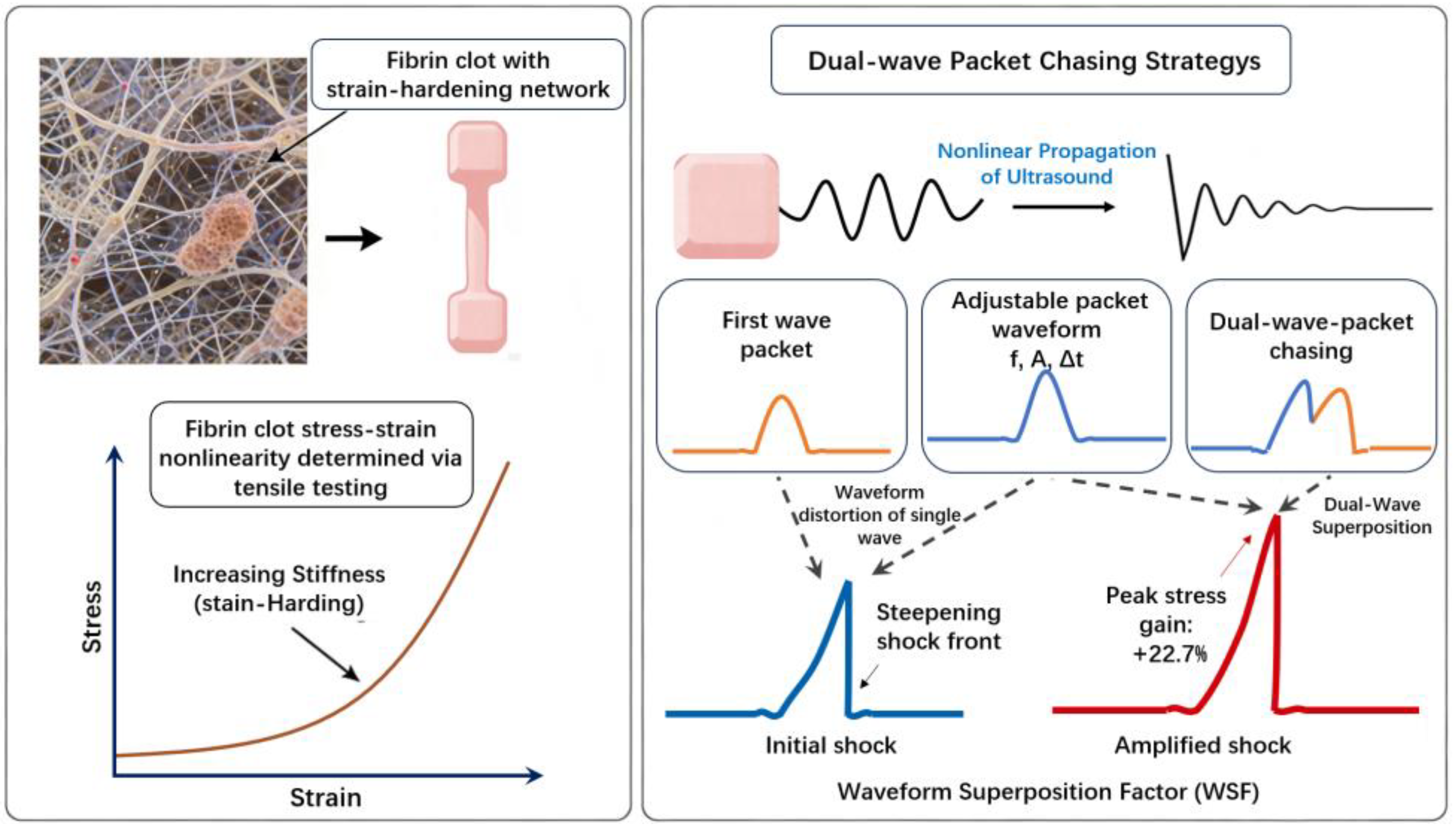
Figure 1. Schematic of the dual-wave-packet chasing strategy for shock wave amplification in a strain-hardening fibrin clot.
Applsci 16 01137 g001
Figure 2. Validation of the power-law constitutive model against experimental stress–strain data for the fibrin clot.
Applsci 16 01137 g002
Figure 3. Shock wave formation during the propagation of a single wave packet in a one-dimensional fibrin clot model: (a) spatial lead of Point A located on the wavefront over Point B on the wave crest; (b) decreasing distance between Point A and Point B; (c) Point B has caught up with Point A.
Applsci 16 01137 g003
Figure 4. Calculation diagram of waveform superposition factor (WSF) at the location x0 = 3.927 × 10⁻³ m) during the dual-wave-packet chasing process.
Applsci 16 01137 g004
Figure 5. Schematic diagram of two-dimensional fibrin clot model.
Applsci 16 01137 g005
Figure 6. Schematic of the input waveforms at the excitation boundary for dual-wave-packet chasing.
Applsci 16 01137 g006
Figure 7. Temporal evolution of the WSF and relative WSF change rate.
Applsci 16 01137 g007
Figure 8. Diagram of dual-wave-packet chasing: (a) the stress nephograms at different moments during wave propagation; (b) the corresponding line plots at t = 5.6 × 10⁻⁵ s; (c) the corresponding line plots at t = 1.0 × 10⁻⁴ s; (d) the corresponding line plots at t = 1.516 × 10⁻⁴ s; (e) the corresponding line plots at t = 2.0 × 10⁻⁴ s.
Applsci 16 01137 g008
Figure 9. Influence of frequency on shock-wave peak stress and its location.
Applsci 16 01137 g009
Figure 10. Influence of amplitude on shock-wave peak stress and its location.
Applsci 16 01137 g010
Figure 11. Influence Influence of duty cycle on shock-wave peak stress and its location.
Applsci 16 01137 g011
Figure 12. Relative influence of normalized parameters on WSF.
Applsci 16 01137 g012
Table 1. Physical property parameters of the model.
ParametersDescriptionValue
ρ0Density1050/(kg/m3)
U0Amplitude1 × 10−4/m
μDimensionless coefficient6.1
φProportionality factor1.2/(kPa/(mg/mL))
mExponent1.8
Disclaimer/Publisher’s Note: The statements, opinions and data contained in all publications are solely those of the individual author(s) and contributor(s) and not of MDPI and/or the editor(s). MDPI and/or the editor(s) disclaim responsibility for any injury to people or property resulting from any ideas, methods, instructions or products referred to in the content.