Article

Alginate-Based Bioformulation with Four Plant Growth-Promoting Bacteria for Sustainable Biostimulation of Spearmint Growth and Natural Defense

by
Zahra Imehli
1,*,
Anouar Mouhoub
2,
Abderrazak Ait Bihi
1,
Salma Oulad Ziane
1,
Soukaina Elkadaoui
1,
Zainab El Alaoui-Talibi
1 and
Cherkaoui El Modafar
1
1
Centre AgroBiotech, Laboratoire d’Excellence d’Agrobiotechnologie et Bioingénierie, Unité de Recherche Labellisée CNRST (URL05-CNRST), Université Cadi Ayyad, Marrakech 40000, Morocco
2
High Throughput Multidisciplinary Research Laboratory (HTMR), College of Chemical Sciences and Engineering (CCSE), Mohammed VI Polytechnic University (UM6P), Ben Guerir 43150, Morocco
*
Author to whom correspondence should be addressed.
Appl. Sci. 2026, 16(18), 9091; https://doi.org/10.3390/app16189091 (registering DOI)
Submission received: 25 July 2026 / Revised: 7 September 2026 / Accepted: 10 September 2026 / Published: 13 September 2026
(This article belongs to the Special Issue Biotechnological and Biostimulant Approaches in Plant Physiology)

Abstract

Spearmint is a medicinal and aromatic crop vulnerable to several diseases, and its cultivation relies heavily on chemical inputs, which can alter phytonutrient quality and raise environmental and health concerns. To address this, the current study focused on developing a safe bioformulation to promote growth and natural defense in spearmint. The bioformulation is based on four plant growth-promoting rhizobacteria (PGPR), Bacillus aryabhathai, Brevibacterium frigoritolerans, Bacillus vallismortis, and Pseudomonas frederiksbergensis, encapsulated in 2% natural alginate. The compositional, structural, and morphological properties of alginate powder and beads were characterized. The beads were evaluated for encapsulation efficiency, bacterial survival, release, and degradation in soil. The bioformulation was applied to the rhizosphere of spearmint and was assessed for its effects on plant growth and natural defenses. Extracted alginate exhibited high purity and structural integrity, and encapsulation efficiency reached 99.99%, providing long-term protection and bacterial viability while regulating their release. The encapsulated consortium notably increased the number of branches, height, and biomass in spearmint. Additionally, a potential induction of plant defense response was observed, as indicated by increased phenylalanine ammonia-lyase activity, higher phenolic compound levels, and lignin accumulation in the roots and shoots of treated plants. These findings highlight the biostimulating effects of these four PGPR encapsulated in alginate on spearmint’s defense and growth.

1. Introduction

Crop protection is increasingly challenging due to multiple threats arising from climate change [1]. The constant evolution of pests and pathogens necessitates effective phytosanitary management, both preventive and curative, that is appropriate to the different stages of crop development. Mentha spicata L. cv. viridis or Mentha viridis L. is a perennial herbaceous plant widely exploited for its aromatic and medicinal properties due to its richness in essential oils. Peppermint production in 2022 was 51,081 tons, with Morocco contributing to 84% of the total production [2]. However, this crop is sensitive and threatened by various pests (insects, mollusks) and pathogens (fungi, nematodes, viruses, etc.), which cause economic damage and affect plant growth and quality. Chemical fertilizers and pesticides are mostly used in plant cultivation. While this is a common approach among farmers across most regions of Morocco, several pesticides are not intended for mint protection, and some farmers do not follow pesticide application guidelines [3]. Although chemical pesticides remain common due to their effectiveness, growing concerns about health risks and environmental impacts have led to increased restrictions on their use. This underlines the need to develop safe alternatives to protect consumers’ health and the environment.
The application of Plant Growth-Promoting Rhizobacteria (PGPR) is widely adopted, and microbial richness confers beneficial effects on agricultural soils and the growth of a wide variety of crops [4]. Studies have highlighted the involvement of various PGPR in stimulating plant growth and natural defenses, as well as controlling pathogens through microbial antagonism [5,6]. In mint, microbial isolates from PGPR strains, particularly Bacillus subtilis and Pseudomonas fluorescens [7], have shown biostimulant effects on plants. However, when free PGPR are introduced into the soil without a carrier, they often fail to compete with the native microflora, leading to a prompt decline in their populations [8,9,10]. This highlights the importance of using an appropriate delivery technique to maintain bacterial survival and ensure their effective colonization of the rhizosphere. Encapsulation of PGPR in a stable carrier has become the most effective technique, ensuring prolonged survival and protection against external factors [11]. Among various encapsulation polymers, alginate stands out as a promising biosourced polymer for its biocompatibility, non-toxicity, biodegradability, and ability to form a stable matrix under mild conditions [11,12]. These properties make alginate an ideal carrier for the controlled release and protection of beneficial bacteria and fungi in agricultural applications. Previous studies have shown that alginate is an algal polysaccharide with a strong stimulating effect on plants’ natural defenses [13,14,15,16]. Additionally, Bifurcaria bifurcata is one of the most abundant brown algae along the Moroccan Atlantic coastline, specifically at the Sidi Bouzid station [17]. This seaweed is an excellent source of alginate in Morocco. The sustainability of alginate production depends on responsible biomass sourcing. The chemical composition of alginate, relative proportions of mannuronic and guluronic acids, and structural properties (molecular weight, viscosity, etc.) strongly influence its rheological behavior, gel stability, and functional properties. Therefore, a comprehensive chemical and structural characterization is essential to assess the suitability of the extracted alginate for optimal PGPR encapsulation.
Few studies have documented foliar treatment of mint plants with chitosan combined with gibberellic acid or algal polysaccharide extracts, such as alginate and carrageenan. Interestingly, results showed that plants treated with chitosan had relatively enhanced yield and biomass accumulation, and better essential oil quality, notably through increased photosynthetic activity and pigments, enzyme activity, and nutrient uptake [18,19,20]. This dual role makes sodium alginate a suitable component for enhancing both plant growth and resilience, in addition to its role as a protective encapsulating matrix for PGPR. Subsequently, combining four PGPR and alginate constitutes a potentially biological approach that induces plant growth and natural defense [11]. Given the reported improvements in physiological response and growth following the application of algal polysaccharides and single or consortia of PGPR in common plant models, limited attention has been given to perennial aromatic plants, such as spearmint, despite their sensitivity to pathogens and strong chemical dependence. To our knowledge, this is the first study reporting the development of an alginate-PGPR-based formulation for biostimulating spearmint growth and defense; this aromatic crop heavily relies on toxic chemical inputs. The objectives of this study were to develop a safe bio-formulation based on local algae-sourced alginate and a consortium of four PGPR, and to assess the biostimulant effects of the bio-formulation on spearmint’s responses.

2. Materials and Methods

2.1. Preparation of Bio-Formulation Components

2.1.1. Seaweed Collection

Bifurcaria bifurcata was chosen as a source of alginate due to its abundance on the Moroccan coast and its alginate yield. B. bifurcata was harvested from Sidi Bouzid on the Atlantic coast of El Jadida (33.2312° N; 8.5474° W) in May 2023, the harvest period of this seaweed in Morocco. After being washed several times with water, the seaweed samples were dried in the shade for 15 days, oven-dried at 50 °C overnight, and ground into powder using a spice grinder.

2.1.2. Extraction and Purification of Sodium Alginate

The algal powder was depigmented with 96% ethanol under agitation at room temperature for 48 h. After centrifugation at 9000 rpm for 10 min, the pellet was dried at 50 °C. The depigmented powder was acidified with a solution of HCl (0.1 N) at 60 °C with stirring for 3 h. The mixture was then centrifuged at 9000 rpm for 10 min at 25 °C, and the alginic acid pellet was dried at 50 °C. The latter was then converted to sodium alginate by carbonation in a 2% sodium carbonate solution (500 mL for 25 g of powder) under stirring at 80 °C for 2 h. After centrifugation at 9000 rpm for 10 min at 25 °C, the pellet was returned to the Na2CO3 solution (second carbonation under the same conditions as the previous carbonation) to maximize alginic acid recovery and optimize extraction yield. The supernatant, corresponding to sodium alginate, was precipitated with 96% ethanol cooled to −20 °C overnight, recovered by centrifugation (9000 rpm for 10 min at 25 °C), and then solubilized in distilled water. The alginate solution was neutralized with HCl (6 N) and then purified using a Vivaflow 50R200 (Sartorius Lab Instruments GmbH & Co. KG, Goettingen, Germany). Sodium alginate was precipitated with 98% ethanol at −20 °C and centrifuged to remove the supernatant. Finally, the purified sodium alginate pellet was solubilized in distilled water at −20 °C, and then freeze-dried [13]. The amount of sodium alginate was calculated per g of dry algal powder.

2.2. Characterization of Sodium Alginate

2.2.1. Biochemical Characterization

The biochemical composition of the extracted sodium alginate was evaluated through spectrophotometric analysis. First, the uronic acid content was measured following the method described by Bouissil et al. [15]. A volume of 1 mL of Borax was added to 200 µL samples. After stirring, the solutions were placed in a water bath at 90 °C for 1 h. Following that, 200 µL of meta-hydroxydiphenyl (m-HDP) solution was added immediately to the mixture, and the samples were returned to the water bath at 90 °C for 2 min. After cooling, absorbance was determined at 520 nm. The glucuronic acid was used as a standard.
Neutral sugar content was determined by adding 200 µL of the alginate samples to 200 µL of resorcinol and 1 mL of H2SO4 80%. After stirring, the mixture was placed in a water bath at 90 °C for 30 min. After cooling in the dark, the solutions were diluted by adding 1.4 mL of deionized water. Absorbance was measured with a spectrophotometer at 450 nm. A standard range was generated using glucose [15].
Total sugar content was measured using Dubois’s method [21]. Alginate samples (1 mL) were mixed with 1 mL of a 5% phenol solution and 5 mL of 80% sulfuric acid. After incubation for 5 min in a water bath at 90 °C, samples were placed in the dark at room temperature for 30 min. Optical density was measured at 492 nm, and a standard was using glucose. Additionally, we assessed the contamination of the extracted alginate by measuring protein and total polyphenols using Lowry’s method [22] and the Folin–Ciocalteu method [23], respectively.

2.2.2. Morphological and Chemical Structure Characterization

The alginate powder was visualized using a TESCAN VEGA 3 scanning electron microscope (TESCAN, Brno, Czech Republic) under vacuum at an accelerating voltage of 10 kV.
The chemical structure of the extracted alginate and commercialized alginate (Sodium Alginate Low viscosity; Loba Chemie KMB0BG37J1K1, Mumbai, India) was investigated using FTIR and NMR analyses. For FTIR, a sample of 1 mg of alginate was mixed with 199 mg of dried spectroscopic-grade potassium bromide. The mixture was compressed before analysis using an FTIR spectrometer (PerkinElmer, Shelton, CT, USA) with a frequency range of 400–4000 cm−1. The data were processed using Origin Pro 9.0. Program. For NMR analysis, the freeze-dried and commercialized alginates were reconstituted in deuterated water (20 mg/L). The 1H NMR and 13C NMR spectra were recorded at 80 °C using a spectrometer (Bruker 300 MHz, Billerica, MA, USA). The acquired data were treated using MestReNova software version 6.0.2.
The viscosity was analyzed using a Ubbelohde capillary viscometer (0.5–3 mm2.s−1, 15–20 mL) at 25.0 ± 0.1 °C. The intrinsic viscosity [η] is calculated from the extrapolation to zero concentration of the reduced viscosity curve. The viscometric molar mass Mv is then determined from the Mark–Houwink equation [24]:
[ η ] = K × M v × a
where [η] is the intrinsic viscosity (mL.g−1), K and a are constants that depend on the solvent and the temperature, and Mv is the viscometric molar mass (g.mol−1).

2.3. Preparation of the Bioformulation

2.3.1. Bacterial Strains

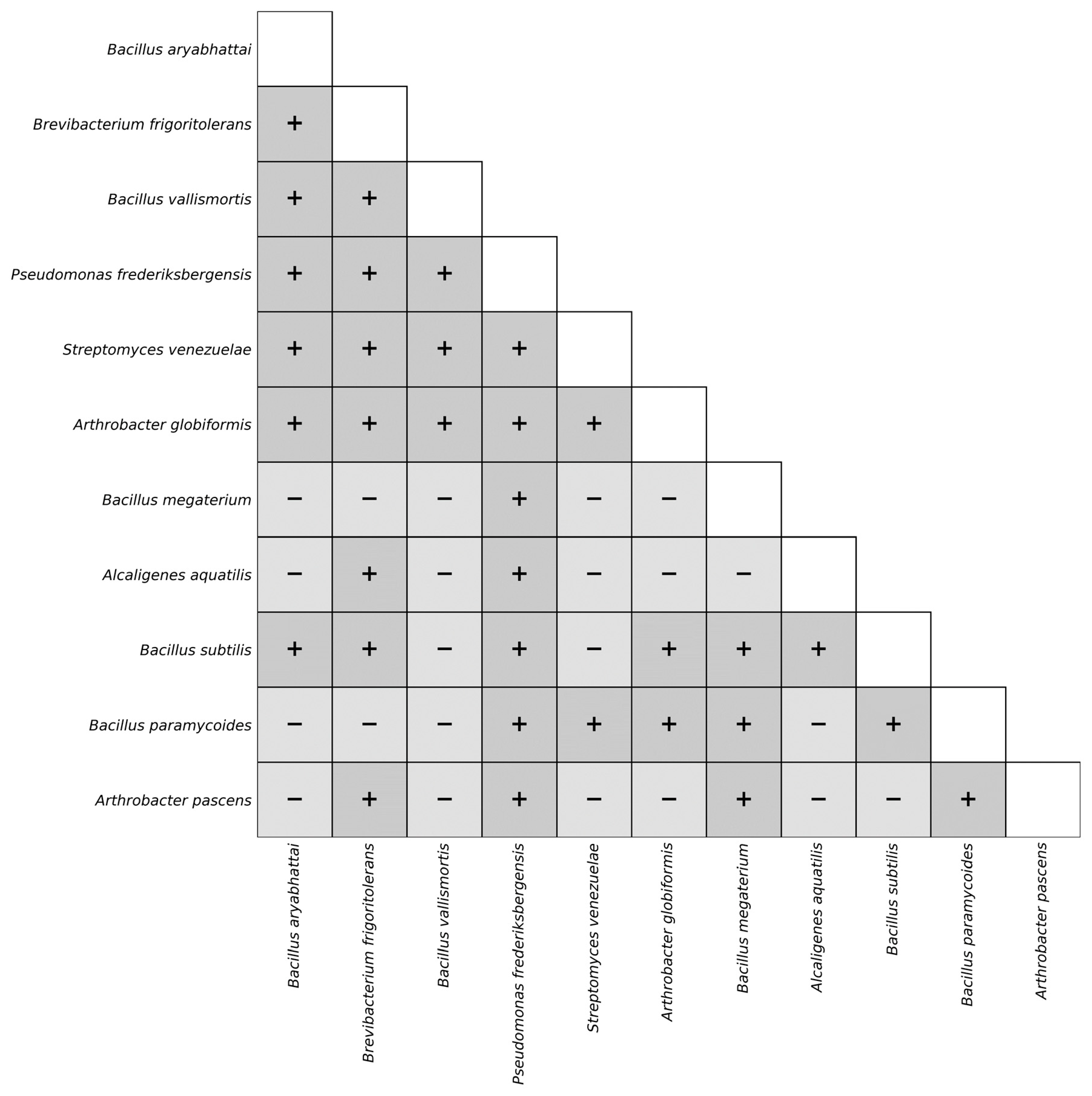
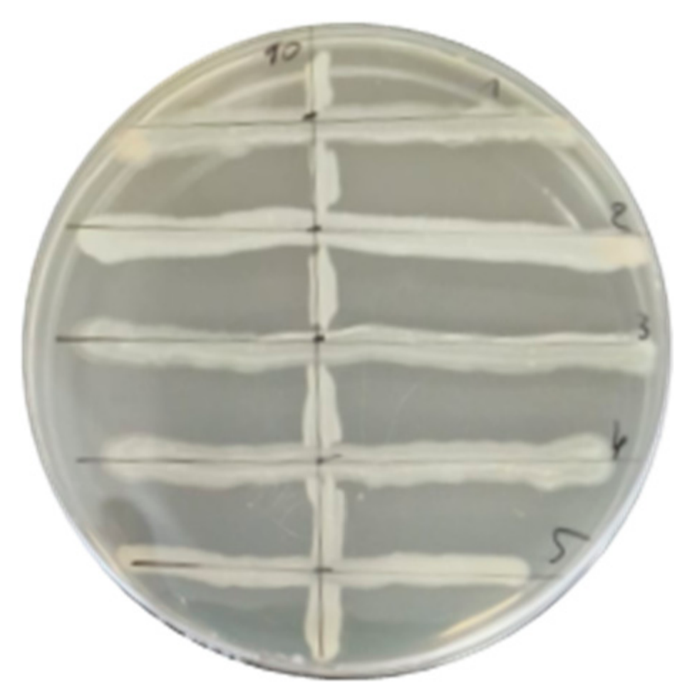
A set of 11 PGPR (Bacillus aryabhattai, Brevibacterium frigoritolerans, Bacillus vallimortis, Pseudomonas frederiksbergensis, Streptomyces venezuelae, Arthrobacter globiformis, Bacillus megaterium, Alcaligenes aquatilis, Bacillus subtilis, Bacillus paramycoides, and Arthrobacter pascens) was assessed for compatibility using the cross-streak method. These PGPR have been isolated from different soils in Morocco and have previously been identified and evaluated for their plant growth-promoting traits [25,26,27]. Briefly, a 24 h culture of each PGPR was streaked as a line on Petri dishes of Mueller-Hinton medium and incubated at 28 °C for 24 h; the other 10 strains were then streaked perpendicularly to the first streak and incubated at 28 °C for 48 h (Appendix A, Figure A1). The bacterial growth in the intersection zone was evaluated; two bacterial strains were considered compatible (+) when there was no visible inhibition zone, and non-compatible (−) strains were those that showed a clear inhibition zone in their intersection. Results of all eleven PGPR were presented in a compatibility matrix (Appendix A, Figure A2).
Four compatible PGPR strains were selected for the study: Bacillus aryabhathai, Brevibacterium frigoritolerans, Bacillus vallismortis, and Pseudomonas frederiksbergensis. These four strains were selected for their compatibility and complementarity with their PGPR and physiological traits. Appendix A (Table A1) provides the PGPR and physiological traits used to select the four bacteria for this study.

2.3.2. Encapsulation of the PGPR Consortium

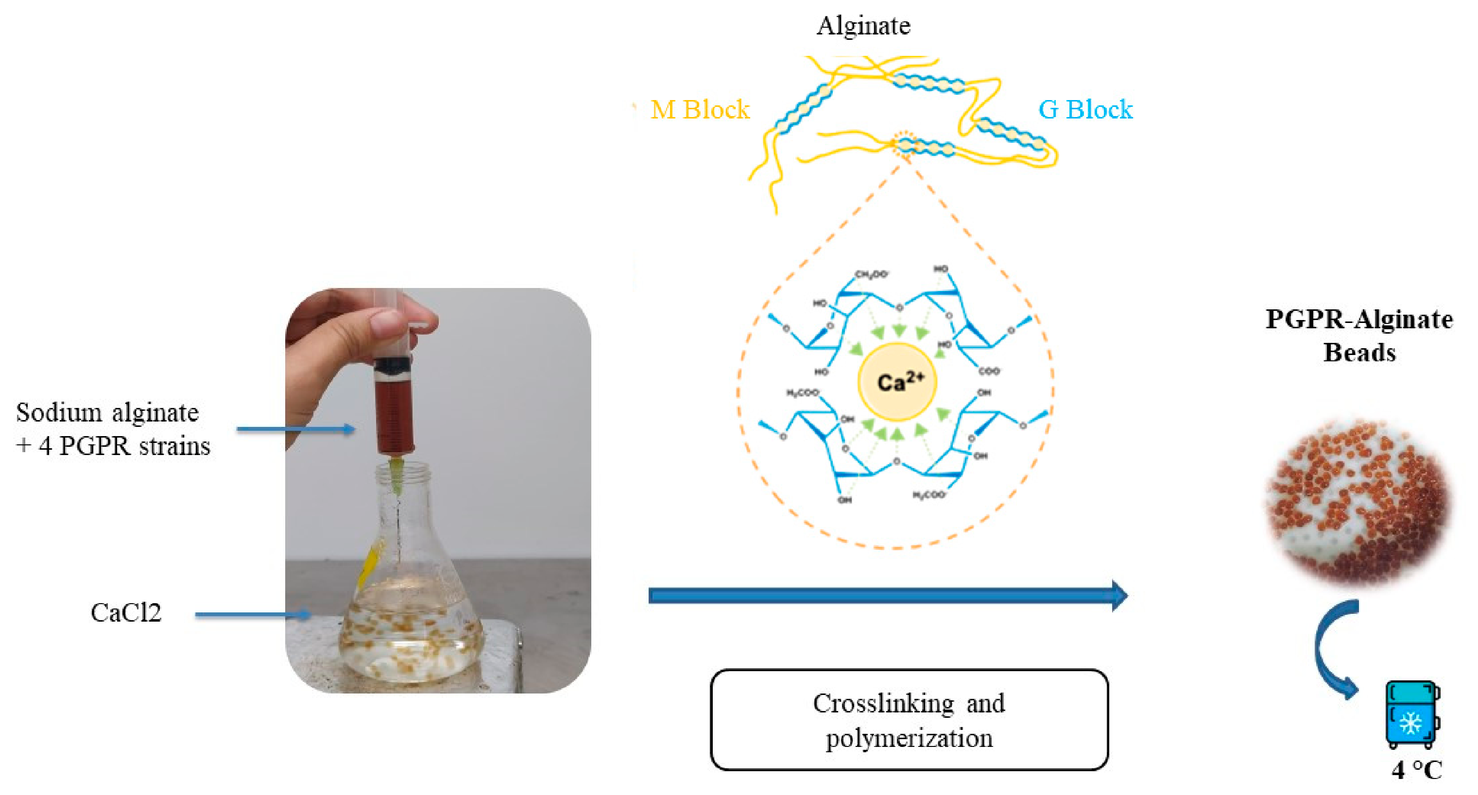
The PGPR consortium was entrapped in the sodium alginate beads using the extrusion method. Fresh cultures of each PGPR were separately grown until the early stationary phase to reach maximum biomass production before encapsulation. A suspension of each PGPR strain was separately prepared at 108 CFU/mL in sterile physiological water. Equal volumes of the individual PGPR suspensions were then mixed to obtain the bacterial consortium. The algal polysaccharide was solubilized in sterile distilled water and added to a similar volume of the PGPR consortium. The mixture was gently added to a sterile 2% CaCl2 solution using a sterile syringe (Figure 1). This contact between the sodium alginate solution and the calcium chloride solution induces the progressive formation of egg-box ionic bonds between the carboxylate groups of the alginate chains and divalent calcium ions, leading to alginate bead formation. After stirring for 30 min, the beads were filtered through a Whatman filter, washed three times with sterile distilled water, and stored at 4 °C. The bead size and mass were measured.

2.3.3. Encapsulation Efficiency Assessment

Encapsulation efficiency (E.E) was assessed by counting total bacteria before and after encapsulation. Three samples of 1 mL of free-suspended PGPR before encapsulation were collected, serially diluted, and inoculated onto Petri dishes containing nutrient agar medium (Tryptone 5 g/L, Meat extract 3 g/L, Bacteriological Agar 12 g/L). After encapsulation, one gram of beads was placed in a tube containing 9 mL of sterile 0.1 M sodium citrate. The tubes were then placed in a shaker at 25 °C until the beads were completely dissolved [28]. The dissolution procedures rely on gentle shaking in a buffer, facilitating matrix dissolution without decreasing the viability of the encapsulated bacterial population. Serial dilutions were then prepared, and the suspension was spread onto agar medium and incubated at 30 °C for 48 h. Three replicates per dilution were prepared. The total bacterial population, including the four PGPRs, was calculated before and after encapsulation. The following formula was used to calculate the encapsulation efficiency of the total bacterial population:
E E % = ( N / N 0 ) × 100
N0: Total bacterial population before encapsulation, and N: Total bacterial population after encapsulation.

2.3.4. Viability Assessment of PGPR Encapsulated in Sodium Alginate

The viability of the total encapsulated bacterial population was assessed using the previously described method for the encapsulation efficiency with three replicates. At each time point, one gram of PGPR-alginate beads was solubilized in sterile sodium citrate (0.1 M) [28]. The results are expressed as the total viable bacterial population per one gram of wet beads.

2.3.5. Evaluation of Progressive Release of PGPR Encapsulated in Sodium Alginate Beads

One gram of beads containing the PGPR consortium was placed in 9 mL of sterile physiological water (0.9% NaCl), under aseptic conditions. The samples were then placed in an oven at 28 °C. The release of the bacterial inoculum was monitored kinetically for up to 30 days by collecting 100 µL of physiological water (0.9% NaCl) and calculating the number of viable bacterial cells per gram of beads using the serial dilution method. Three replicates were prepared, and the number of released bacterial cells was determined for one gram of wet beads [29].

2.3.6. Degradation Assessment of Alginate Beads

The degradability of alginate beads was assessed in soil by placing 10 g of wet alginate beads on a small alveolus containing 100 g of dry soil. The beads were placed directly in the soil, and 50 g of dry soil was added to ensure the beads were covered. The soil was then regularly watered, and the experiment was conducted in greenhouse conditions for 7 weeks. The beads were removed from the soil, washed with water, and then left to dry in an oven set at 60 °C [30]. The dry weight of the beads was measured at different time points: 2, 4, 5, 6 and 7 weeks. The following formula is used to calculate the degradation rate of alginate beads:
D e g r a d a t i o n   ( % ) = D W 0 D W t D W 0 × 100
DW0: Dry weight of the beads at t = 0 (Dry weight of 10 g of wet beads before the experiment), and DWt: Dry weight of the beads collected at different stages of degradation.

2.3.7. Scanning Electron Microscopy of Alginate Beads

The alginate beads with encapsulated PGPR consortium were observed before and after degradation assessment using scanning electron microscopy TESCAN VEGA 3 (TESCAN, Brno, Czech Republic).

2.4. Evaluation of the Bioformulation’s Ability to Stimulate Spearmint’s Growth and Natural Defenses

2.4.1. Spearmint Plant Growth Response to Bioformulation Application

For the greenhouse trial, we used the Mentha spicata variety (green mint), which was grown from stem cuttings obtained from a local nursery in Marrakesh. Spearmint plants were grown in a greenhouse under the following conditions: temperature = 28 ± 2, photoperiod = 14 h, and plants were irrigated regularly to maintain soil moisture. After two weeks, the plants were divided into four groups to investigate the potential role of the bioformulation in stimulating plant growth and natural defenses. The four groups (one control and 3 treatments) consisted of a total of 53 plants per group:
  • (control) plants treated with 10 mL of distilled water only,
  • (Free PGPR) plants inoculated with a 10 mL suspension containing 108 CFU/mL of each of the four PGPR strains that was prepared from freshly cultivated PGPR and suspended in sterile saline water to obtain a liquid suspension of the consortium after adjusting the concentration by measuring optical density at 600 nm,
  • (Alginate) plants treated with 20 g of alginate-only beads,
  • (PGPR-Alginate) plants inoculated with 20 g of alginate beads containing 108 CFU/g of the PGPR consortium.
The treatments were applied to the plants’ rhizosphere. The experiment allowed us to evaluate the effects of this application on biochemical parameters by collecting 3 plants randomly from each group at different time points (0, 2 days, 4 days, 7 days, 14, 21, and 30 days) to assess early and late plant responses, and 5 plants from each group were collected for growth parameter measurements after 6 weeks. Plant growth was assessed by measuring plant height, branching, leaf number, and biomass.

2.4.2. Stimulation of Natural Defenses in the Spearmint Plant

  • Enzymatic activity of phenylalanine ammonia-lyase
The fresh spearmint leaves or roots were weighed and crushed in a mortar using liquid nitrogen, then mixed with 2 mL of cooled acetone (80%). After centrifugation at 14,000 rpm, the supernatant was stored at −20 °C, and the pellet was dried at room temperature. An aliquot of 25 mg of acetone extract was mixed with 1.5 mL of 0.1 M sodium borate buffer (pH 8.5), containing 20 mM β-mercaptoethanol. The enzyme extract was transferred to an Eppendorf tube and vortexed. Phenylalanine ammonia-lyase (PAL) activity was determined using the method described by Ballester et al. [31] with modifications. The reaction medium consisted of 1.5 mL sodium borate buffer, 0.2 mL of 0.1 M L-phenylalanine, and 0.3 mL of enzyme extract. The reaction was monitored for 45 min at 40 °C, and PAL activity was estimated by measuring trans-cinnamic acid concentration at 270 nm [32]. The specific activity of PAL was calculated after measuring total protein in the samples using the Bradford method [33]. Results are expressed as the percentage of PAL activity relative to the control.
  • Total polyphenol content
The reagent mixture was prepared with 1.5 mL of distilled water, to which 250 µL of Folin–Ciocalteu reagent and 200 µL of diluted acetone extract of mint leaves or roots were added. After 3 min, 500 µL of 20% Sodium Carbonate (Na2CO3) was then added. Samples were incubated at 40 °C for 30 min. Absorbance was measured at 760 nm. Total polyphenol content was determined in mg of gallic acid per g of fresh leaves or roots and expressed as a percentage relative to the control.
  • Lignin content
The acetone extract of mint leaves or roots was dried at 25 °C before lignin quantification. The recovered dry powder was treated with thioglycolic acid (1 M) and HCl (2 M). After 4 h of heating at 100 °C, the mixtures were centrifuged. The pellets were suspended in a solution of NaOH (1 M) after being washed with 1 mL of distilled water to eliminate residual acids. The samples were shaken for 18 h at room temperature. Following centrifugation, the recovered supernatants were treated with concentrated HCl (12 N). The mixtures were then diluted in NaOH (1 M) after being placed overnight at 4 °C to precipitate the thioglycolic acid-lignin complex [34]. After measuring absorbance at 280 nm, lignin amounts were represented as the mean percentage of A280/g of root or leaf dry weight relative to the control.

2.5. Statistical Analysis

Statistical analysis was performed using R version 4.2.1. Group means were compared using one-way ANOVA, followed by the Tukey HSD test. Results are expressed as the mean value of 3 replicates ± standard error (SE), and differences were considered significant at p < 0.05.

3. Results

3.1. Characterization of PGPR-Alginate-Based Bioformulation

3.1.1. Properties and Composition of Extracted Sodium Alginate

The sodium alginate was extracted from Bifurcaria bifurcata. The results of the colorimetric assays indicated that the extracted alginate contains 27.41% ± 3.09 total sugars, including 77.86% ± 1.16 uronic acids and 13.80% ± 4.97 neutral sugars. Minor concentrations of impurities, such as proteins, were present at 0.16% ± 0.02, whereas trace amounts of total polyphenols were detected (<0.001%) (Table 1).

3.1.2. Morphological and Chemical Structure of Alginate

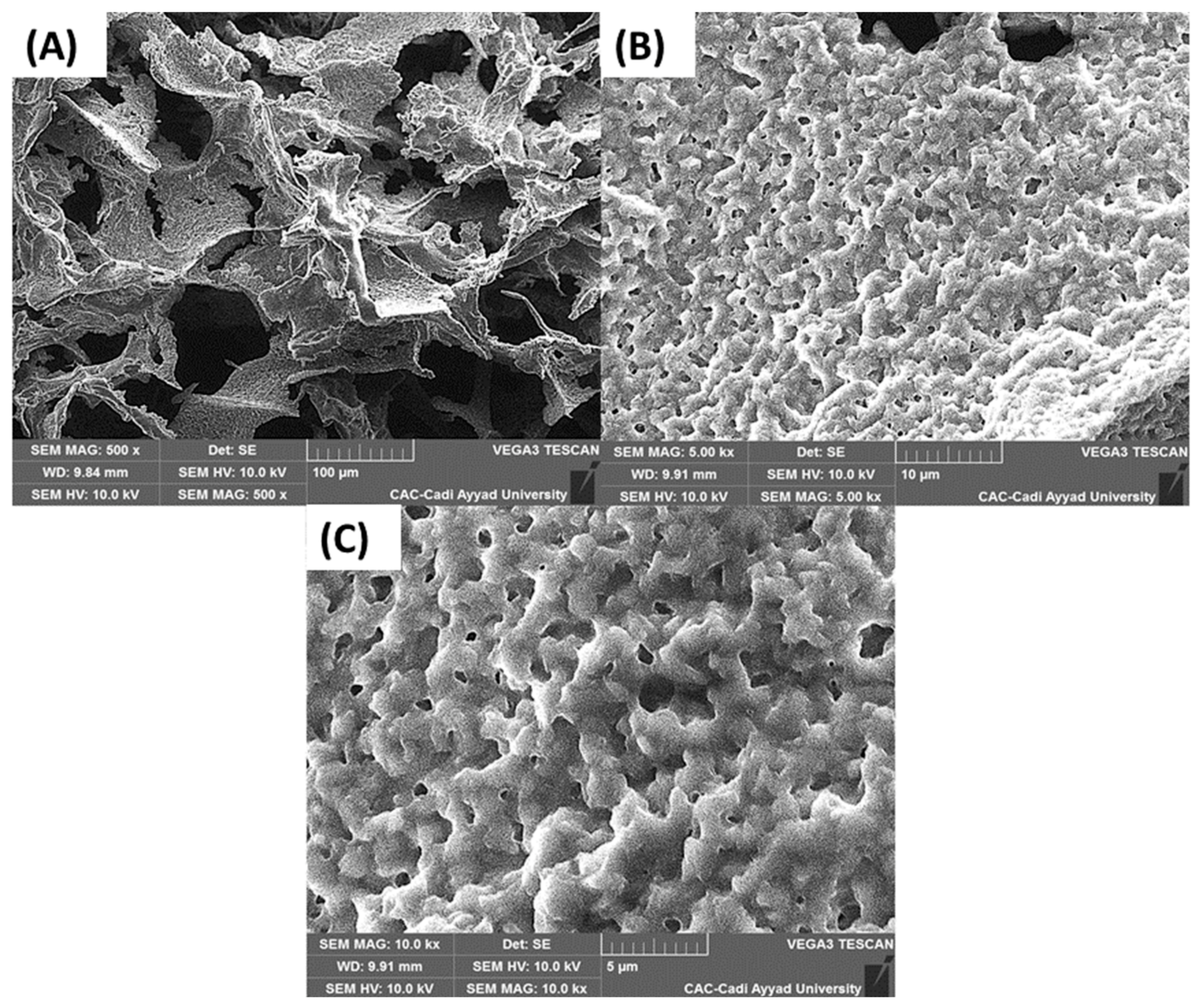
Scanning electron microscopy (SEM) imaging at 500× magnification of the extracted alginate powder showed a flake-like, fibrous structure typical of freeze-dried polysaccharides (Figure 2A). Nonetheless, at higher magnifications (5000× and 10,000×), the lyophilized alginate appears as a compact, granular matrix, consistent with its hierarchical structure (Figure 2B,C).
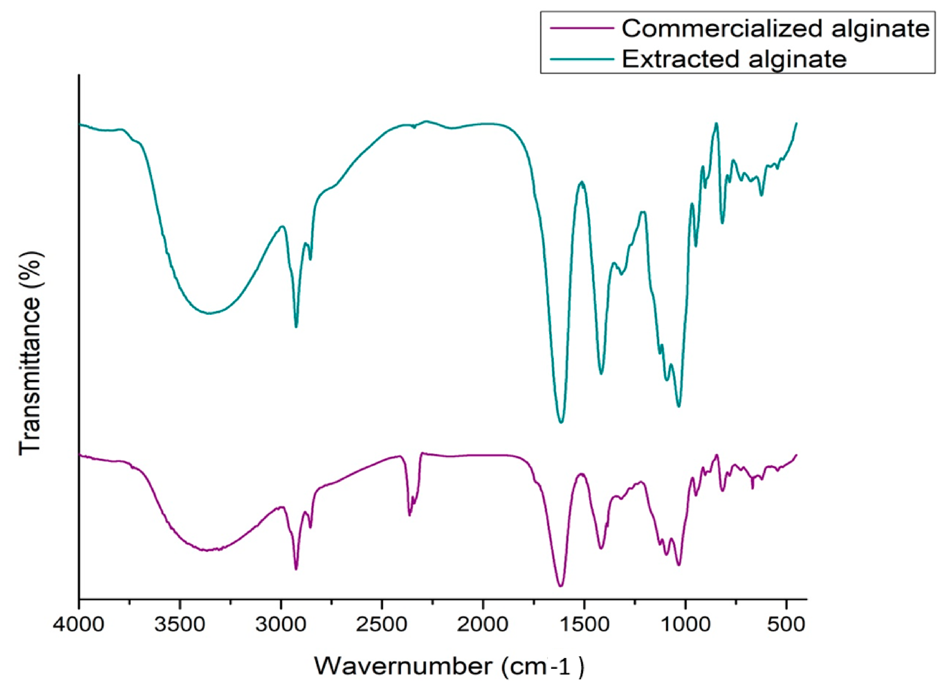
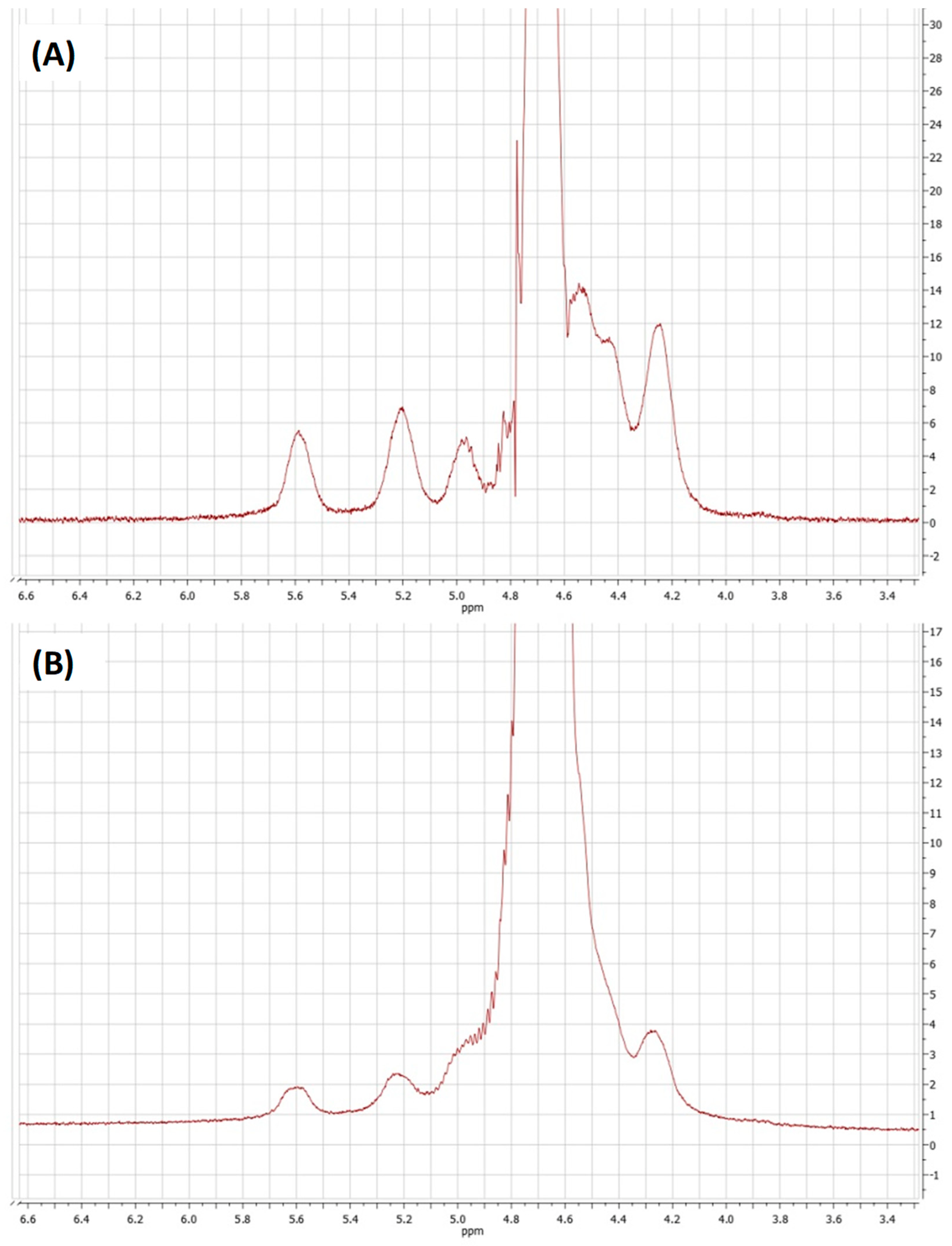
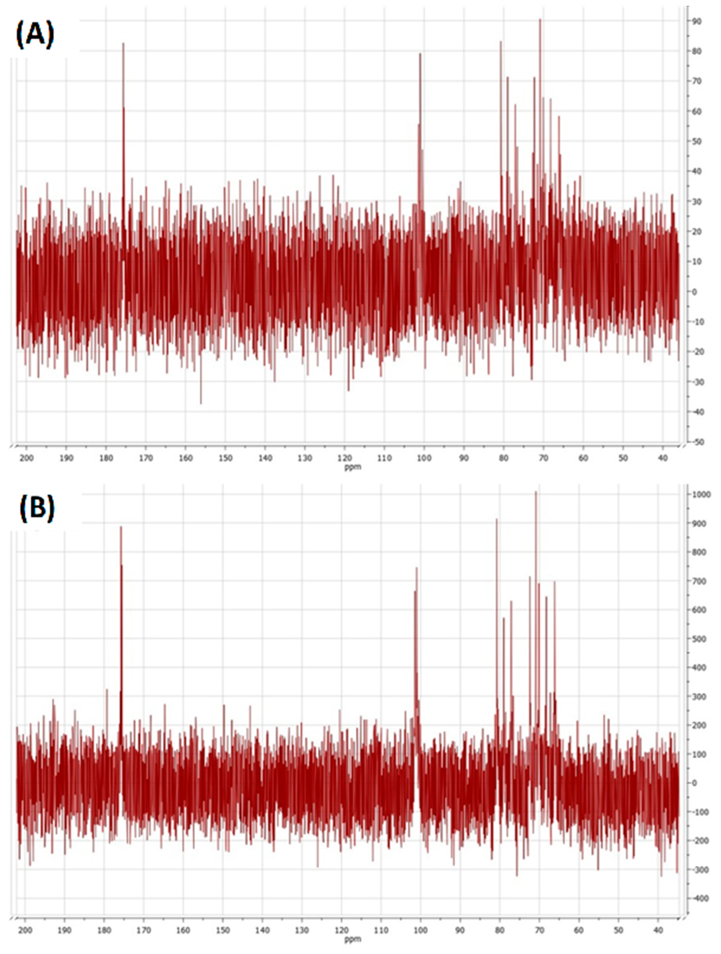
Chemical structure analyses confirmed the high quality and structural integrity of the extracted alginate. According to FTIR, the two spectra showed a similar trend (Figure 3), except for the peak at around 2350 cm−1. Overall, the spectrum represented a broad band at 3370 cm−1 (O–H stretching vibrations of hydroxyl groups), and peaks at 2910 (C–H stretching from aliphatic CH and CH2 groups), 1625 (asymmetric stretching vibration of carboxylate groups), 1410 (symmetric stretching vibration of carboxylate groups), 1045 (elongation of C-O groups), and 940 cm−1 (sugar ring vibration) [35,36]. As illustrated in Figure 4, the 1H NMR spectra of the extracted alginate show a typical spectrum of alginate [37,38]. However, the commercialized alginate spectrum showed overlapping peaks with reduced intensity compared to the extracted alginate. Distinct anomeric proton signals were noticed at 5.6, 5.2, and 5 ppm, which may be attributed to guluronic (G1) and mannuronic (M1) acid units in different sequence environments (GM, GG, or MM). The broad signal between 4.4 and 4.8 ppm may refer to the overlapping ring protons (H2 to H5) of both monomers. Ultimately, the peak at 4.2 ppm corresponds to more shielded ring protons (H5 or H6) of G and M units. The 13C NMR spectra of extracted and commercialized alginate showed a peak located at 175 ppm, indicating the carboxyl carbon (C6) (Figure 5). In contrast, the signal at 101 ppm refers to the anomeric carbon (C1) [39]. The remaining ring carbons (C2 to C5) appeared as partially overlapping signals between 66 and 81 ppm. These spectral features follow the known chemical structure of alginate, confirming the identity of the extracted biopolymer. The average molecular weight was calculated for alginate in distilled water, and Mark–Houwink constants were: K = 3.2 × 10⁻³ mL/g, and a = 0.85. The calculated intrinsic viscosity was [η] = 11,233 mL/g, and sodium alginate obtained in this study showed a viscometric molecular mass (Mv) of 222,569 g·mol−1. The value of Mv is related to the algae used and the extraction conditions.

3.1.3. Characterization of the Bioformulation: Encapsulation Efficiency, Viability and Bacterial Release of Encapsulated PGPR Population

The percentage of total bacterial population encapsulated in the beads reached 99.9%. Because the initial concentrations of the four strains were equivalent, the bacterial cells of the four strains entrapped within the alginate matrix were observed on the counting plates, although this test is not strain-specific. The encapsulation process using 2% sodium alginate was highly efficient. In addition, the beads had a uniform size of 3 mm, with an average wet mass of 8.08 mg.
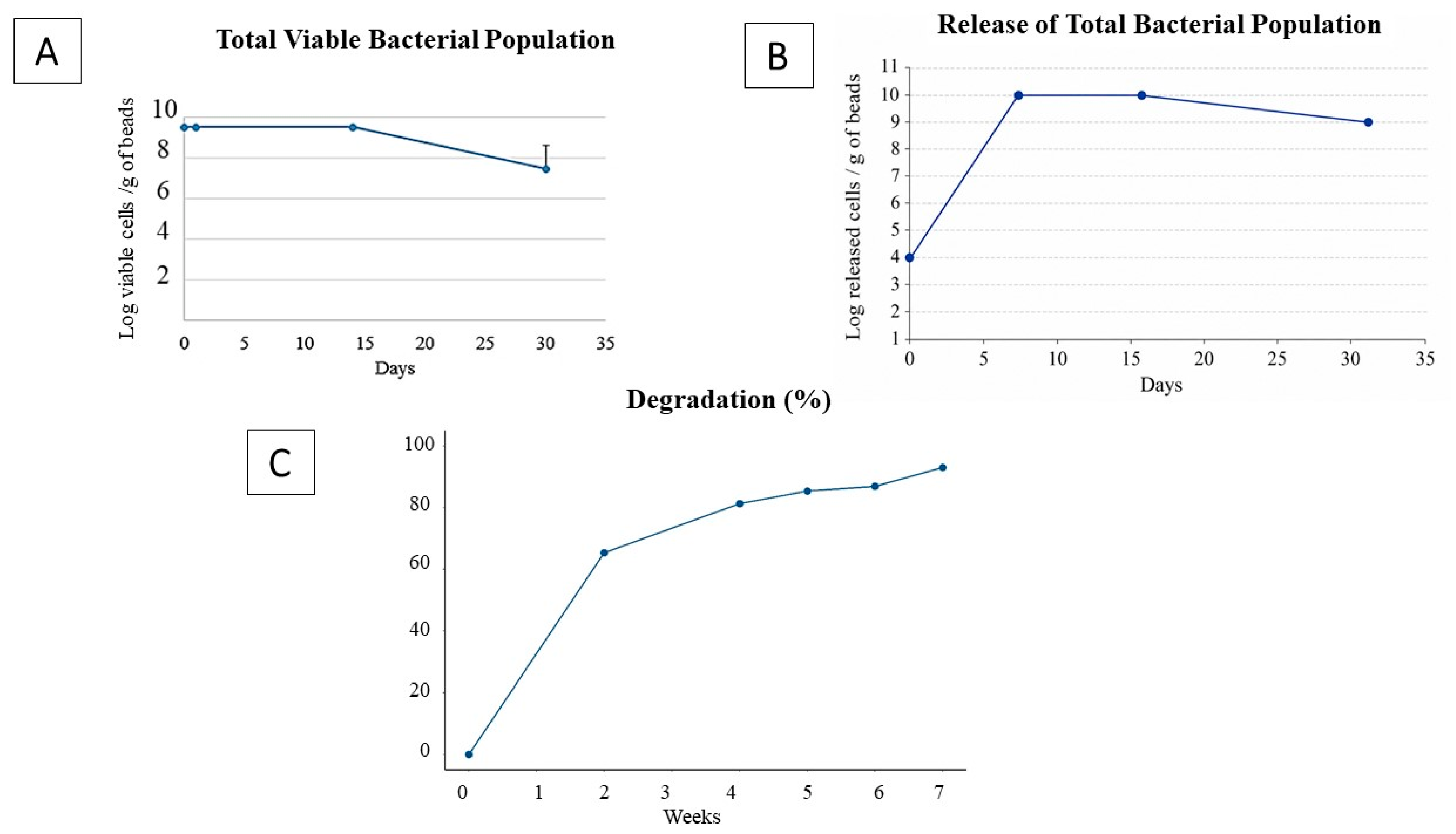
The total population of PGPR cells remained viable inside the beads after encapsulation and storage at 4 °C. During the first 14 days of storage, the total number of viable cells entrapped in the beads did not change significantly. However, the number of viable bacterial cells progressively decreased in the following days to reach 108 per gram of wet beads (Figure 6A). On the other hand, the release of the PGPR cells in physiological water increased significantly in the beads in the first two weeks, reaching its peak (1011 CFU) after 14 days of storage. Following that, the number of released bacterial cells decreased to 1010 CFU after 30 days at 28 °C (Figure 6B).

3.1.4. Degradation of Alginate Beads

The wet alginate beads containing the PGPR consortium were buried in soil. After two weeks, the collected beads were less than 40%. After four weeks, a high percentage was degraded (81.3%). Only a small number of beads were detected in the soil after 7 weeks, corresponding to a 93% degradation rate (Figure 6C).

3.1.5. Scanning Electron Microscope Images of PGPR-Alginate Beads

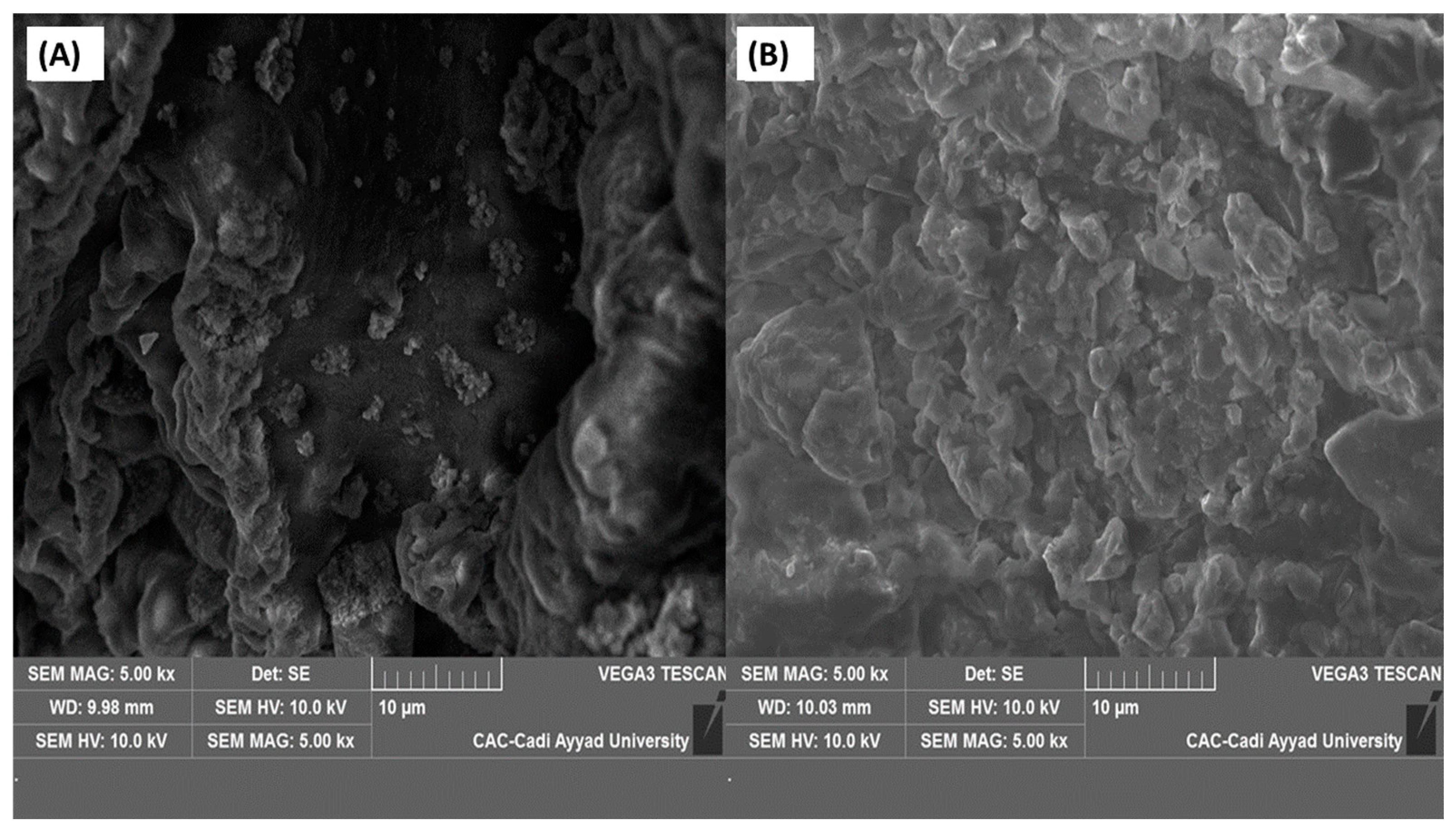
The alginate beads encapsulating the PGPR consortium were observed under a scanning electron microscope (SEM), showing CaCl2 integration in the matrix and bacterial agglomerations (Figure 7A). Alginate beads were also visualized after degradation assessment in soil; the image revealed structural disintegration in the matrix (Figure 7B).

3.2. Application of the Bioformulation on Spearmint Plants

3.2.1. Effects of the Bioformulation on Growth Parameters in Spearmint Plants

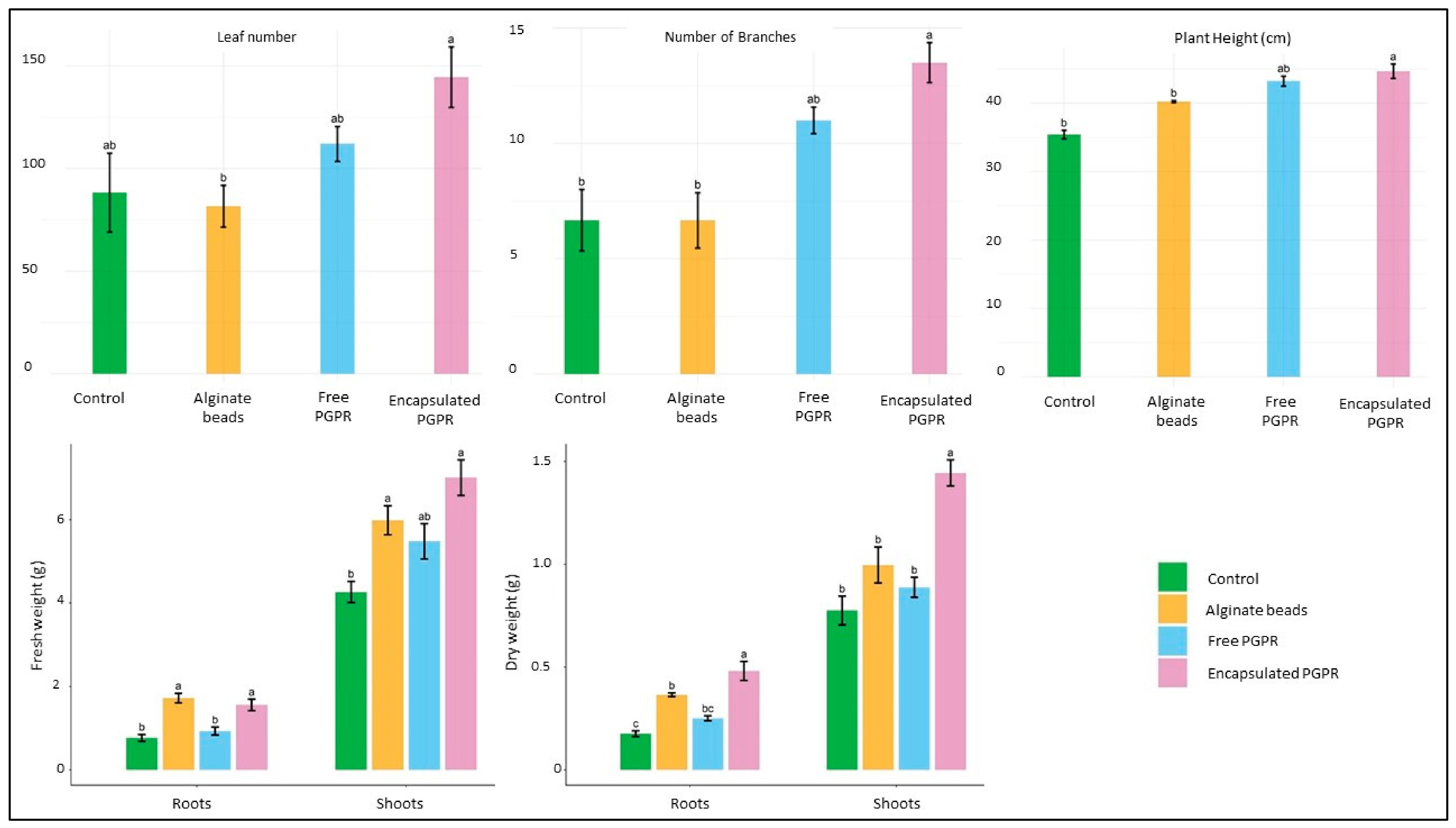
The bioformulation developed in this study was applied to the rhizosphere of mint plants for six weeks. The developed bioformulation, PGPR encapsulated in sodium alginate, enhanced plant growth, with significant increases observed in all measured parameters compared to the control. After six weeks, treatment with free PGPR and encapsulated PGPR promoted the main plant growth parameters, the number of branches and the height of spearmint, compared to untreated plants. Encapsulated PGPR performed better than free PGPR in terms of shoot and root dry weight, increasing by 53.4% and 37.5%, respectively, relative to the control. Alginate beads only showed an increase in biomass compared to the control (Figure 8).

3.2.2. Effects of the Bioformulation on Spearmint Plants’ Natural Defenses

  • PAL activity in spearmint
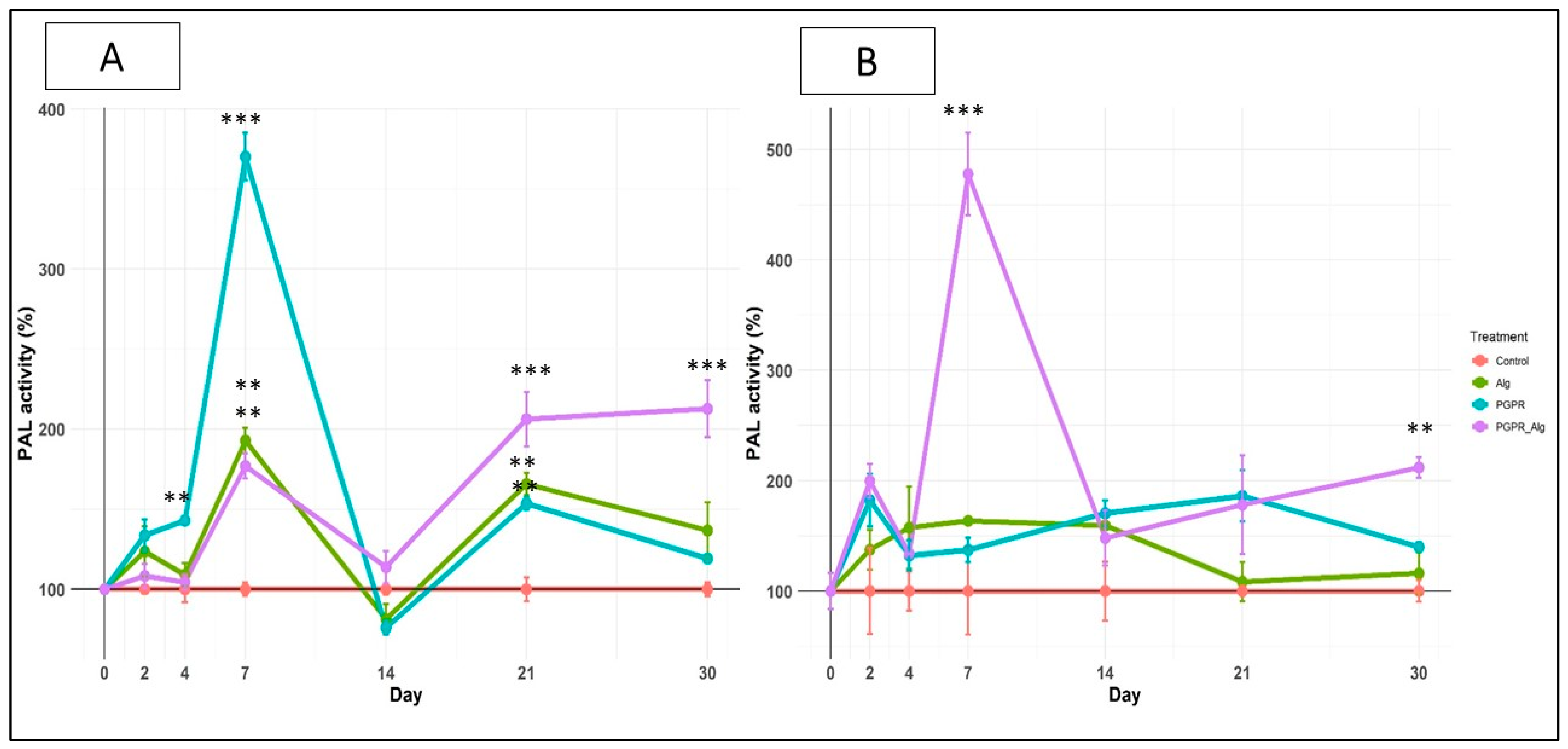
Phenylalanine ammonia lyase activity was measured in the roots and leaves of spearmint plants. Treatment with free PGPR significantly enhanced PAL activity in spearmint roots two days after treatment. After 7 days, PAL activity attained its highest peak in the roots of all three treatments compared with the control. Afterward, the activity decreased in the roots of these plants. However, PAL activity had increased again on the 21st day in all treatments. A significant increase in PAL activity was then observed in plants treated with PGPR encapsulated in alginate beads after three weeks, followed by a highly significant enhancement after one month compared with both the control and plants treated with free PGPR and plants treated with alginate only (Figure 9A). After two days, a significant increase in PAL activity was observed in the leaves of plants treated with encapsulated PGPR. The enzyme activity doubled, reaching its highest value in the leaves for the same treatment after 7 days, with a 5-fold increase compared to the control and to plants treated with free PGPR and the alginate beads group, a highly significant difference. After this peak, activity decreased in the following week in the leaves (Figure 9B). Moreover, plants treated with encapsulated PGPR showed a high PAL activity comparable to that observed in other groups after one month.
  • Total phenolic content in spearmint
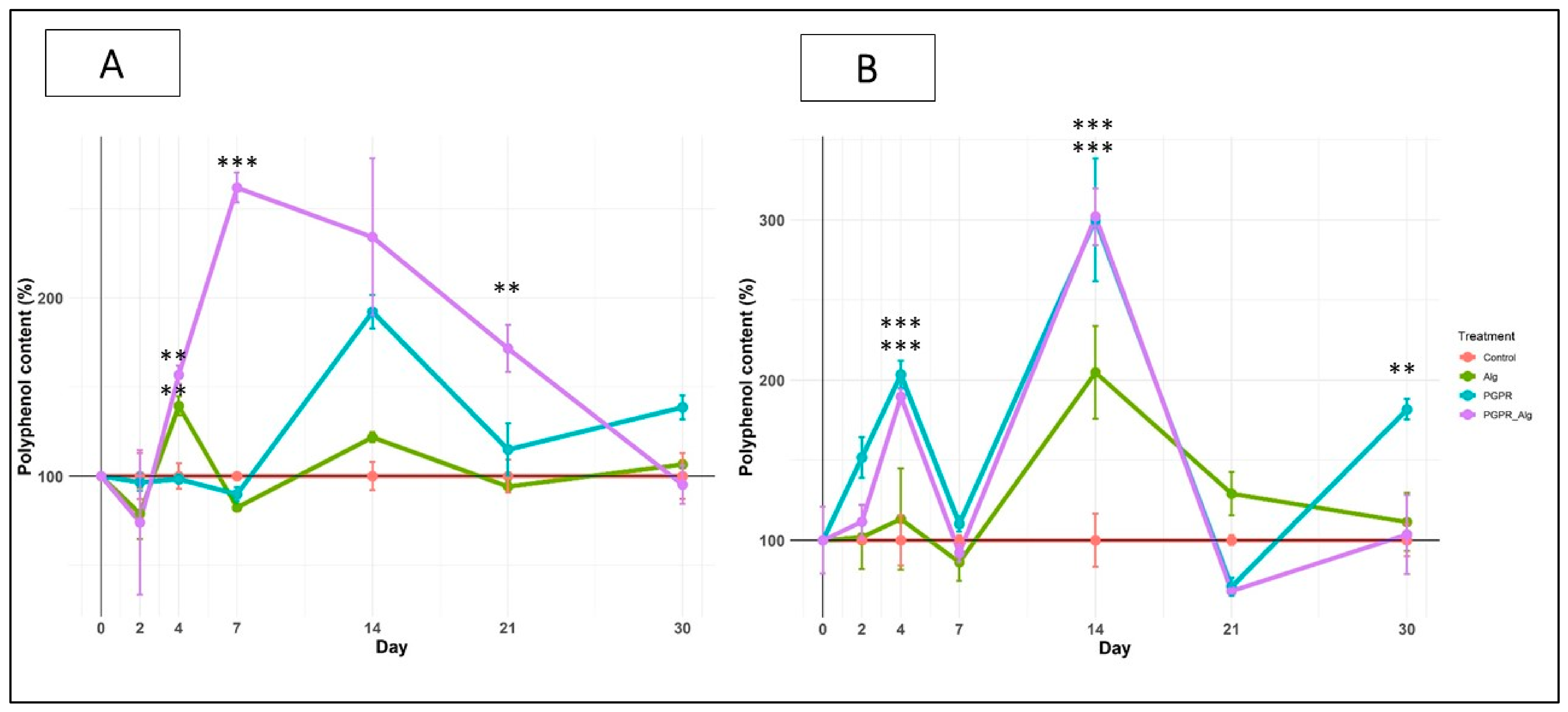
The total phenolic content was quantified in both the leaves and roots of treated and untreated spearmint plants. On the one hand, the total polyphenol content in the roots increased over the following four days of treatment; the difference was significant in plants inoculated with encapsulated PGPR and plants treated with alginate beads. This enhancement reached its highest level after seven days for the plants treated with encapsulated PGPR, showing a highly significant difference (p-value ≤ 0.05) compared to other plants. Afterward, the total phenolic content in spearmint roots decreased gradually over the following two weeks (Figure 10A). In contrast, after four days, a significant increase was observed in phenolic compound content in leaves from plants treated with encapsulated PGPR and plants treated with free PGPR (p-value ≤ 0.05). Afterward, a second peak was observed on day 14 in the same plants, as well as in plants treated with alginate beads, indicating significant production of phenolic compounds in the leaves following application of PGPR and alginate (Figure 10B).
  • Lignin content in spearmint
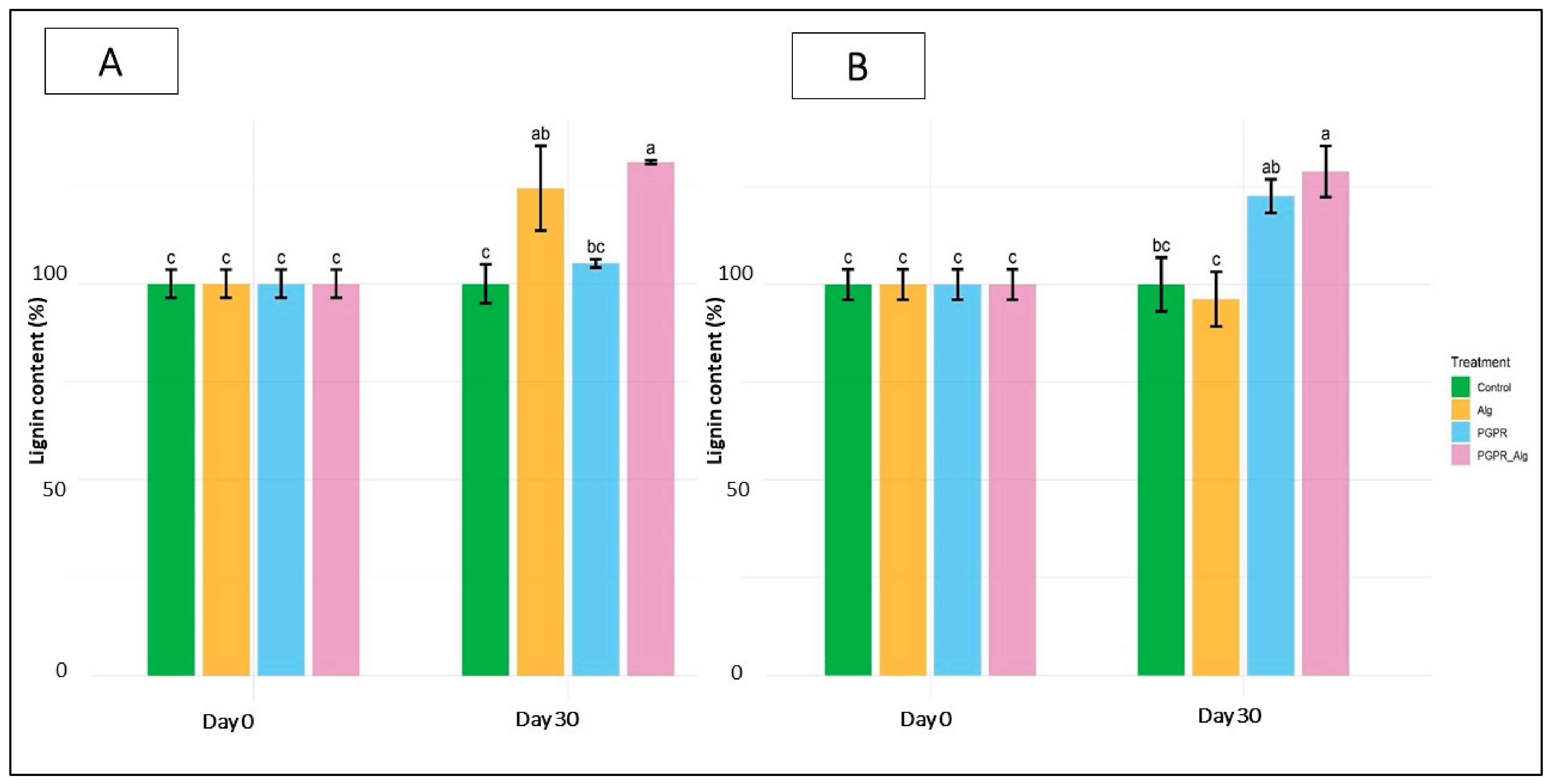
After a month, lignin content had significantly increased in the roots of mint plants after application of the encapsulated PGPR consortium and in plants treated with alginate. However, no significant difference was observed in the roots of mint plants treated with the free PGPR consortium compared to untreated plants. Furthermore, treatment with a PGPR consortium encapsulated in alginate beads had significantly enhanced (p-value ≤ 0.05) lignin content in mint leaves after one month, compared to untreated plants. However, no significant difference was observed between the application of free PGPR consortium and the treatment with encapsulated PGPR in lignin production in mint leaves (Figure 11).

4. Discussion

This study aimed to investigate the effect of a bioformulation based on a consortium of four PGPR encapsulated in alginate extracted from brown seaweed, combining both the beneficial effects of PGPR and alginate on spearmint growth parameters and defense.
Although we had not assessed disease suppression in this study, the observed stimulation of defense-related biochemical signals indicates the potential of the encapsulated PGPR consortium to enhance spearmint’s ability to respond to biotic stress. The results showed that Bifurcaria bifurcata constitutes an excellent local source of alginate; the extracted alginate, with a 27.41% yield, displayed a composition rich in guluronic acid blocks (mannuronic acid/glucuronic acid ratio of 0.47 M/G) and very minor impurities (Table 1) in line with previous findings [13,15]. This composition is advantageous for encapsulation, as G-blocks are principally responsible for forming strong ionic cross-links with calcium ions (Ca2+) through the “egg-box” model. Alginates with a low M/G ratio tend to form stable hydrogels with enhanced mechanical strength and lower permeability [40]. Characterization of alginate indicated that FTIR, 1H-NMR, and 13C-NMR spectra of the alginate extracted from Bifurcaria Bifurcata were similar to those documented in earlier studies of the structural properties of alginate from the same seaweed [13,15]. The sodium alginate extracted in this work exhibited good quality (Figure 3, Figure 4 and Figure 5). However, additional absorption bands observed in the FTIR spectra may be attributed to the adsorption of atmospheric gases. The signals observed in the 1H-NMR spectra suggest a decrease in uronic acid content or lower polymer purity in the commercial alginate. These structural characteristics of alginate are essential for ensuring physical protection and structural stability, particularly when used to encapsulate sensitive microorganisms such as plant growth-promoting rhizobacteria (PGPR). The molecular mass of alginate depends on the algae species, its maturity stage, and the environmental growth conditions. However, extraction conditions can also affect the polymer’s molar mass [24,41]. The average viscometric molecular weight of the alginate extracted in this work was 222,569 g·mol−1. This indicates that the extraction method preserved the polymer’s integrity without significant degradation. This value falls within the range reported in the literature for alginates extracted from brown algae (32,000 and 400,000 g·mol−1), which depends on the species and extraction conditions [15,42]. The composition and chemical properties of the extracted alginate indicate that this natural algal polysaccharide from B. bifurcata constitutes an appropriate matrix for bacterial encapsulation, owing to its purity, safety, and biodegradability, as indicated by previous studies [11,43]. The comparison between extracted and commercialized alginate indicates whether locally seaweed-sourced alginate could serve as an effective alternative to industrial alginate for microbial encapsulation, reducing reliance on processed polymers, lowering production costs, and supporting local inputs. This supports the circular bio-economy by transforming natural, unvalued biomass into value-added products.
The bioformulation showed an excellent encapsulation efficiency of 99.9% for the PGPR populations, consistent with a previous study on encapsulation of Lactobacillus acidophilus using different concentrations of alginate and CaCl2; all combinations showed an encapsulation efficiency higher than 98% [44]. Encapsulation of the selected consortium using 2% alginate and 2% CaCl2 maintained the viability of the bacterial population after storage at 4 °C for one month. Cross-linking strengthened the egg-box network formed inside the beads; consequently, the beads maintained their stability and prolonged protection for the bacteria during storage. The entrapment ensures optimal protection of bacteria against environmental factors, including UV, temperature fluctuations, pH variations, and microbial competition in soil, enhancing their shelf life compared to free bacteria [11,45]. Other studies demonstrated that encapsulating PGPR in 2% sodium alginate and storing the beads at 4 °C increases the survival of entrapped bacteria [46]. Moreover, our study revealed a progressive release of PGPR from the beads into water over 30 days, consistent with previous studies demonstrating similar release of encapsulated bacteria in water and soil [11]. Although the initial concentrations of the four strains were equal and the beads were preserved at 4 °C, a common temperature for medium-term storage and maintaining bacterial viability, the plate-count approach estimates the overall bacterial population; strain-selective approaches could provide more evidence on the encapsulation efficiency, viability, and release of each strain. While in vitro assays confirmed the gradual release of PGPR cells over time, understanding the degradation dynamics of alginate beads in soil conditions is critical. Degradation of alginate in soil was assessed by weight loss. SEM images also revealed morphological changes. The degradation of sodium alginate beads depends on soil physicochemical properties (soil pressure, soil type, and density), changes in soil conditions (especially water content, moisture level, and pH), microbial enzyme activity, the cross-linking, and additives used in the beads [47]. The biodegradation of alginate beads is associated with the capacity of soil microbes to break the egg box conformation by cleaving the β-1,4-glycosidic bonds in alginate polymers. Soil microorganisms gradually destabilize the three-dimensional “egg box”-shaped network and depolymerize the alginate chains. Alginate lyases, the main enzymes known to degrade this polysaccharide, cleave the β-(1,4) glycosidic bonds between β-D-mannuronate (M) and α-L-guluronate (G) residues via a β-elimination mechanism, thereby generating unsaturated alginate oligosaccharides [48]. A recent study also revealed that Paenibacillus elgii can produce alginate lyase capable of degrading sodium alginate into oligosaccharides, contributing to the biodegradation of alginate matrices in soil and facilitating the release of encapsulated bacterial cells [49]. The study of Mohapatra et al. [50] revealed that alginate lyases isolated from terrestrial bacteria exhibit optimal activity at neutral pH, moderate temperatures, and non-saline soil conditions, and are more effective against mannuronate blocks.
The application of alginate beads containing Bacillus aryabhathai, Brevibacterium frigoritolerans, Bacillus vallismortis, and Pseudomonas frederiksbergensis has significantly boosted spearmint plant growth and development as indicated by the measured morphological parameters, compared to the control. The application of free PGPR yielded similar results, with less significant effects on the dry weight of both shoots and roots. Earlier characterization of the bacterial strains in our study has shown strong plant growth-promoting traits, notably phosphate solubilization and Indole-3-acetic acid (IAA) production, in addition to their ability to tolerate salinity and hot temperatures between 45 °C and 50 °C [25,27]. Treatment with Bacillus aryabhattai promoted common bean growth by increasing shoot and root length [25]. In mint species, beneficial effects of PGPR application on plant growth and development have been documented, where Bacillus spp. and Pseudomonas spp. are well-known. Del Rosario Cappellari et al. [51] tested Bacillus subtilis, Pseudomonas fluorescens, and Pseudomonas putida strains freely and in combination on peppermint plants, resulting in overall growth enhancement through increased biomass, leaf area, and node number compared to the control. A dual treatment with Pseudomonas putida and Curtobacterium sp. strain LUW enhanced height, number of stems and leaves, and fresh and dry weight in peppermint [52]. Similar results were obtained in spearmint plants where the application of a free consortium of four PGPR: Pseudomonas sp., Bacillus cereus, Bacillus pumilus, and Sphingobacterium suaeda increased the plants’ morphological parameters, notably the number of leaves, shoot dry biomass, shoot height, and root biomass [53]. PGPR inoculation even mitigated the reduced growth rate under drought stress conditions, as shown by the application of two strains (Pseudomonas fluorescens and Bacillus amyloliquefaciens) on peppermint plants [54]. Our results indicate that encapsulation not only maintained the beneficial effects of the tested PGPR strains but also enhanced their overall performance on plant growth. Multiple studies have evaluated the impact of bacterial encapsulation by comparing the beneficial effects of PGPR strains in both free and encapsulated-state applications [55,56,57]. Overall, results indicated that encapsulation sustained the growth-promoting traits of PGPR using different matrices, including alginate. In their review, Riseh et al. [58] indicated the central role of alginate as an encapsulation biopolymer ensuring the protection of PGPR while preserving their plant-promoting traits. Direct application of free PGPR in soil has temporary effects due to environmental factors: Soil conditions, indigenous microorganisms, and surface and groundwater in the field shorten the duration of free PGPR efficacy [11,12]. Encapsulation in a stable, safe matrix such as alginate protects PGPRs and prolongs their survival in soil compared to unprotected free PGPR. Therefore, maintaining the PGPR population and providing the required nutrients and growth hormones in the rhizosphere could stimulate spearmint development. In addition to serving as an encapsulating matrix, sodium alginate has also been reported to directly promote plant growth, independently of PGPR action [20].
The induced natural defense is a multilayered process activated through rhizosphere colonization by beneficial microorganisms, mainly PGPR, followed by activation of pathogen-related genes (PR proteins), enzyme activity, and hormonal signaling via salicylic acid, jasmonic acid, and ethylene [59,60]. PGPRs, including Pseudomonas and Bacillus strains, are known for their strong ability to induce plant defense via multiple interactions with plant roots [59]. Some bacteria among these genera have been reported to enhance the production of enzymes such as phenylalanine ammonia-lyase, peroxidases, and phenol oxidases [61]. In our work, we assessed the effect of inoculating spearmint plants with four PGPR encapsulated in alginate beads on the natural defenses, including phenylalanine ammonia-lyase kinetics, a key enzyme in the plant’s defense mechanism [62]. This enzyme plays a principal role in the phenylpropanoid pathway, inducing the synthesis of secondary defense-related metabolites, mainly polyphenols, a broad class of phenolic compounds, including flavonoids, phenolic acids, and tannins. Their accumulation is often induced in response to pathogenic attack or environmental stress. These compounds have a dual function: on one hand, they possess antioxidant properties that limit cellular damage induced by reactive oxygen species (ROS) generated during oxidative stress; on the other hand, they have direct antimicrobial activity, inhibiting the growth of fungi and bacteria. In addition, several polyphenols are involved in signaling defense responses by modulating the expression of genes associated with plant immunity [63]. The observed enhancement in PAL enzyme activity after spearmint plants were treated with Bacillus aryabhathai, Brevibacterium frigoritolerans, Bacillus vallismortis, and Pseudomonas frederiksbergensis indicates a putative implication of these PGPRs in the defense mechanism induction in spearmint. Upon perception of the signals after PGPR colonization through root-to-shoot signaling, shoot tissues rapidly activate the phenylpropanoid pathway, resulting in enhanced synthesis of phenolic compounds [59]. The earlier response observed in leaves reflects their principal role in antioxidant protection and photosynthesis, whereas phenolic accumulation in roots occurs subsequently as local defense mechanisms progressively establish, which is consistent with induced natural defense priming [59,60]. An earlier study showed that inoculating spearmint with other PGPR strains increases PAL activity in the roots and leaves of spearmint in the few days following the treatment; furthermore, the activity was boosted after a second and third inoculation [53]. This indicates that a single application of free PGPR is insufficient because they are not protected and leach after soil irrigation, as suggested by Salisu et al. [64]. Thereby, other inoculations are required for sustained PAL activity. Oulad Ziane et al. [16] applied the same encapsulated strains used in this study in tomato, resulting in a prolonged stimulation of PAL enzyme activity in roots and leaves. Other studies have demonstrated the positive effect of inoculation with bacterial strains as biocontrol agents, indicated by increased stress enzyme activity, including PAL, catalase, and polyphenol oxidase [65,66,67]. According to these studies, inoculating mint plants with the studied PGPR consortium, including two similar genera, Bacillus and Pseudomonas, resulted in a significant increase in PAL activity, which leads to the accumulation of phenolic content in leaves and roots, thereby inducing the defense mechanism. This result corroborates previous work that reported increased phenol biosynthesis in peppermint and spearmint leaves and roots following treatment with consortia of Pseudomonas sp. [52,65,68]. In this context, a recent study on Bacillus aryabhattai has shown the capacity of this PGPR to participate in the biosynthesis and transformation of Tyrosine-derived metabolites through enzymes such as polyphenol oxidase and Tyrosine decarboxylase. Although this study was assessed in a microbial co-fermentation system, the findings support the metabolic versatility of B. aryabhattai and indicate its contribution to complex biochemical PGPR-plant interactions influencing the secondary metabolism and plant defense responses [69].
Furthermore, sodium alginate extracted from brown seaweed has demonstrated its potential as a biostimulator of PAL enzyme activity and polyphenol biosynthesis, which induced natural defense in the treated plants [13,14,15,16]. It is also suggested that degradation of sodium alginate beads by rhizosphere bacteria could enhance polyphenol biosynthesis in mint plants; this enhancement was revealed by the application of irradiated sodium alginate on Mentha arvensis [20]. Therefore, we hypothesize that the alginate beads underwent gradual degradation due to the combined effects of the soil’s physicochemical environment and microbial enzymatic activity [47], which led to the release of oligosaccharide motifs derived from alginate. These molecules may act as plant immunity elicitors by being recognized by pattern recognition receptors (PRRs) located on the plasma membrane, thereby triggering intracellular immune signaling pathways [13,70]. However, delayed activation of the phenylpropanoid pathway was observed after treatment with alginate beads, with limited capacity to stimulate growth and defense-related enzymes in mint plants, unlike studies in which soluble alginate or oligo-alginate were applied directly via stem injection or foliar application [13,71]. The relatively weaker response may be attributed to the slow, gradual, and sustained release of alginate degradation products from the beads into the soil, thereby delaying the immediate availability of potentially bioactive oligosaccharides in the rhizosphere. Before these molecules can be released and function as damage-associated molecular patterns (DAMPs) and trigger the plant’s immune responses [13,70], the alginate beads must undergo physical disintegration followed by enzymatic depolymerization by alginate lyases derived from the rhizosphere microbiota [72]. Consequently, during the four-week experiment, the concentrations of these elicitors in the rhizosphere may not have reached the threshold necessary to induce an SDN response as intense as that observed following the direct application of hydrolyzed alginate or alginate oligosaccharides.
Lignin is an essential component of the plant cell wall and is synthesized throughout the phenylpropanoid pathway. Polymerization of monolignols derived from phenylpropanoic acids produces lignin, which plays a fundamental structural role in reinforcing cell walls. This hydrophobic biopolymer is deposited in secondary walls, increasing their rigidity and impermeability. In the face of pathogenic aggression, localized lignification acts as a physical barrier, preventing the progression of the infectious agent [73]. It is principally accumulated in roots, which explains its critical role in preventing infection by soil-borne pathogens. An increased lignin content was observed in both leaves and roots of spearmint one month after treatment with encapsulated PGPR, particularly at the end of the PAL activity, leading to the transformation of phenolic compounds synthesized in response to spearmint’s natural defense induction. A previous study reported similar results, where treatment of spearmint with a consortium of PGPR showed an increased polyphenol content in both roots and leaves of treated plants [53]. Correspondingly, another study revealed that chemical pesticides, especially insecticides, enhance growth hormones, cinnamic acid, and lignin synthesis in peppermint. However, chemical pesticides have altered the physiological parameters and medicinal value of peppermint [74].
The findings of the present study demonstrate that encapsulating the studied consortium within sodium alginate beads is an effective method for enhancing spearmint growth and strengthening its natural defense system, as evidenced by increased PAL activity and polyphenol accumulation. To our knowledge, this has not been previously reported. Nevertheless, studies monitoring the biodegradation pathway of alginate in the rhizosphere are also required to understand the correlation between alginate biodegradation, bacterial release dynamics, and the timing of plant response. Likewise, the in situ application of this bioformulation in the presence of spearmint pathogens is required to assess its protective potential and to validate the proposed mechanisms underlying its biostimulatory and defense-inducing effects.

5. Conclusions

This work represents a specific contribution by providing a promising approach, based on a safe and effective bio-formulation of four PGPR: Bacillus aryabhathai, Brevibacterium frigoritolerans, Bacillus vallismortis, and Pseudomonas frederiksbergensis encapsulated in natural alginate beads, exhibiting both plant growth-promoting and defense-inducing potential in spearmint. Entrapping the four PGPR strains using 2% biosourced alginate showed high encapsulation efficiency, maintained survival, and enabled the release of the encapsulated bacterial population. Furthermore, applying this bio-formulation in the rhizosphere of Mentha spicata plants improved their growth parameters, stimulated the phenylpropanoid biosynthetic pathway, a key component producing defense-related compounds, and induced a defense response in treated plants. This encapsulated consortium could serve as a safe foundation for enhancing the growth and resilience of medicinal and aromatic plants such as spearmint. Based on these findings, the following implications should focus on optimizing bio-formulation properties, assessing stability under storage conditions, and evaluating the potential protective effect of spearmint against major soil-borne diseases.

Author Contributions

Conceptualization, Z.I., S.O.Z., Z.E.A.-T. and C.E.M.; Methodology, Z.I., A.M., A.A.B., S.O.Z., S.E., Z.E.A.-T. and C.E.M.; Software, Z.I. and A.M.; Validation, Z.E.A.-T. and C.E.M.; Formal Analysis, Z.I.; Investigation, Z.I., A.A.B., S.O.Z. and S.E.; Resources, A.A.B. and C.E.M.; Data Curation, Z.I.; Writing—Original Draft Preparation, Z.I.; Writing—Review & Editing, Z.I., A.M., S.E., Z.E.A.-T. and C.E.M.; Visualization, Z.I. and A.M.; Supervision, Z.E.A.-T. and C.E.M.; Project Administration, C.E.M.; Funding Acquisition, C.E.M. All authors have read and agreed to the published version of the manuscript.

Funding

This research received no external funding.

Institutional Review Board Statement

Not applicable.

Informed Consent Statement

Not applicable.

Data Availability Statement

The original contributions presented in this study are included in the article. Further inquiries can be directed to the corresponding author.

Acknowledgments

The authors acknowledge all the members of the Agrobiotech team, Cadi Ayyad University-Marrakech, the National Agency for Medicinal and Aromatic Plants (ANPMA)-Taounate, and CNRST-Morocco for their support through the 4th Edition of the Research Program in the field of Valorization of Medicinal and Aromatic Plants (VPMA4/2022/10). They also recognize the additional support provided by CNRST-Morocco through the PhD-Associate Scholarship (PASS).

Conflicts of Interest

The authors declare no conflicts of interest.

Abbreviations

The following abbreviations are used in this manuscript:
CFUColony Forming Unit
DAMPDamage-Associated Molecular Patterns
DWDry Weight
EEEncapsulation Efficiency
FTIRFourier Transform InfraRed
IAAIndole-3 acetic acid
MvViscometric Molar Mass
NMRNuclear Magnetic Resonance
PALPhenylalanine Ammonia Lyase
PGPRPlant Growth-Promoting Rhizobacteria
PRRPattern Recognition Receptors
ROSReactive Oxygen Species
SDNNatural Defense Stimulating
SEMScanning Electron Microscopy

Appendix A

Figure A1. Example of an in vitro compatibility assay for one PGPR.
Applsci 16 09091 g0a1
Figure A2. Compatibility matrix of the 11 PGPR strains. +: compatible, −: not compatible.
Applsci 16 09091 g0a2
Table A1. PGPR and physiological traits of the four selected PGPRs.
PGPR StrainPhosphate
Solubilization
IAA
Production
Siderophores ProductionThermo-ToleranceHalotoleranceHydrogen Cyanide
Production
References
Pseudomonas
frideriksbergensis
++++40 °C4%[27]
Bacillus
aryabhattai
++++++45 °C12%+++[25]
Brevibacterium
frigorotolerans
++++50 °C8%+[26]
Bacillus
vallismortis
+++++45 °C7%++[26]
−: No activity; +: Weak activity; ++: Moderate activity; +++: Strong activity.

References

  1. Singh, A.K.; Jyoti, B. A Review for Assessing the Impact of Climate Change on the Agricultural Sector, Food Security, and Pests in India. In Advances in Environmental Engineering and Green Technologies; Zia Ul Haq, M., Ali, I., Eds.; IGI Global: Hershey, PA, USA, 2024; pp. 53–84. [Google Scholar] [CrossRef] [Scilit]
  2. FAOSTAT. 2024. Available online: https://www.fao.org/faostat (accessed on 8 October 2025).
  3. Eddaya, T.; Boughdad, A.; Becker, L.; Chaimbault, P.; Zaïd, A. Utilisation et risques des pesticides en protection sanitaire de la menthe verte dans le Centre-Sud du Maroc (Use and risks of pesticides in sanitary protection of spearmint in south-central Morocco). J. Mater. Environ. Sci. 2015, 6, 656–665. [Google Scholar]
  4. Khatoon, Z.; Huang, S.; Rafique, M.; Fakhar, A.; Kamran, M.A.; Santoyo, G. Unlocking the potential of plant growth-promoting rhizobacteria on soil health and the sustainability of agricultural systems. J. Environ. Manag. 2020, 273, 111118. [Google Scholar] [CrossRef] [Scilit]
  5. El-Saadony, M.T.; Saad, A.M.; Soliman, S.M.; Salem, H.M.; Ahmed, A.I.; Mahmood, M.; El-Tahan, A.M.; Ebrahim, A.A.M.; Abd El-Mageed, T.A.; Negm, S.H.; et al. Plant growth-promoting microorganisms as biocontrol agents of plant diseases: Mechanisms, challenges and future perspectives. Front. Plant Sci. 2022, 13, 923880. [Google Scholar] [CrossRef] [Scilit]
  6. Jiao, X.; Takishita, Y.; Zhou, G.; Smith, D.L. Plant Associated Rhizobacteria for Biocontrol and Plant Growth Enhancement. Front. Plant Sci. 2021, 12, 634796. [Google Scholar] [CrossRef] [Scilit]
  7. Morsy, E.M.; El-Said, N.A. Efficiency of Bacillus subtilis and Pseudomonas fluorescence as Biocontrol Agents Against Rhizoctonia Solani on Spearmint Plants (Mentha viridis L.). Sci. J. Flowers Ornam. Plants 2015, 2, 213–225. [Google Scholar] [CrossRef] [Scilit]
  8. Bashan, Y.; De-Bashan, L.E.; Prabhu, S.R.; Hernandez, J.-P. Advances in plant growth-promoting bacterial inoculant technology: Formulations and practical perspectives (1998–2013). Plant Soil 2014, 378, 1–33. [Google Scholar] [CrossRef] [Scilit]
  9. Korir, H.; Mungai, N.W.; Wasike, V.W. Influence of native rhizobacteria co-inoculation and formulation of bacterial inoculants on the growth and yield of common bean (Phaseolus vulgaris L.). J. Sustain. Agric. Environ. 2024, 3, e12095. [Google Scholar] [CrossRef] [Scilit]
  10. Papin, M.; Philippot, L.; Breuil, M.C.; Bru, D.; Dreux-Zigha, A.; Mounier, A.; Le Roux, X.; Rouard, N.; Spor, A. Survival of a microbial inoculant in soil after recurrent inoculations. Sci. Rep. 2024, 14, 4177. [Google Scholar] [CrossRef] [Scilit]
  11. Szopa, D.; Mielczarek, M.; Skrzypczak, D.; Izydorczyk, G.; Mikula, K.; Chojnacka, K.; Witek-Krowiak, A. Encapsulation efficiency and survival of plant growth-promoting microorganisms in an alginate-based matrix—A systematic review and protocol for a practical approach. Ind. Crops Prod. 2022, 181, 114846. [Google Scholar] [CrossRef] [Scilit]
  12. Saberi Riseh, R.; Hassanisaadi, M.; Vatankhah, M.; Soroush, F.; Varma, R.S. Nano/microencapsulation of plant biocontrol agents by chitosan, alginate, and other important biopolymers as a novel strategy for alleviating plant biotic stresses. Int. J. Biol. Macromol. 2022, 222, 1589–1604. [Google Scholar] [CrossRef] [Scilit]
  13. Aitouguinane, M.; Bouissil, S.; Mouhoub, A.; Rchid, H.; Fendri, I.; Abdelkafi, S.; Ould El-Hadj, M.D.; Boual, Z.; Dubessay, P.; Gardarin, C.; et al. Induction of Natural Defenses in Tomato Seedlings by Using Alginate and Oligoalginates Derivatives Extracted from Moroccan Brown Algae. Mar. Drugs 2020, 18, 521. [Google Scholar] [CrossRef] [Scilit]
  14. Aitouguinane, M.; El Alaoui-Talibi, Z.; Rchid, H.; Fendri, I.; Abdelkafi, S.; El-Hadj, M.D.O.; Boual, Z.; Le Cerf, D.; Rihouey, C.; Gardarin, C.; et al. Elicitor Activity of Low-Molecular-Weight Alginates Obtained by Oxidative Degradation of Alginates Extracted from Sargassum muticum and Cystoseira myriophylloides. Mar. Drugs 2023, 21, 301. [Google Scholar] [CrossRef] [Scilit]
  15. Bouissil, S.; El Alaoui-Talibi, Z.; Pierre, G.; Michaud, P.; El Modafar, C.; Delattre, C. Use of Alginate Extracted from Moroccan Brown Algae to Stimulate Natural Defense in Date Palm Roots. Molecules 2020, 25, 720. [Google Scholar] [CrossRef] [Scilit]
  16. Oulad Ziane, S.; Imehli, Z.; El Alaoui Talibi, Z.; Ibnsouda Koraichi, S.; Meddich, A.; El Modafar, C. Biocontrol of tomato Verticillium wilt disease by plant growth-promoting bacteria encapsulated in alginate extracted from brown seaweed. Int. J. Biol. Macromol. 2024, 276, 133800. [Google Scholar] [CrossRef] [Scilit]
  17. Bahammou, N.; Sabri, H.; Cherifi, O.; Bouamama, H. Seasonal Variation, The Ecological Index And The Potential Use Of Chlorophyceae And Phaeophyceae Of The Moroccan Coast Sidi Bouzid Eljadida In The Agricultural Field. J. Anal. Sci. Appl. Biotechnol. 2021, 3, 1. [Google Scholar] [CrossRef]
  18. Ahmad, B.; Khan, M.M.A.; Jaleel, H.; Sadiq, Y.; Shabbir, A.; Uddin, M. Exogenously sourced γ-irradiated chitosan-mediated regulation of growth, physiology, quality attributes, and yield in Mentha piperita L. Turk. J. Biol. 2017, 41, 388–401. [Google Scholar] [CrossRef] [Scilit]
  19. Ahmad, B.; Jaleel, H.; Shabbir, A.; Khan, M.M.A.; Sadiq, Y. Concomitant application of depolymerized chitosan and GA3 modulates photosynthesis, essential oil and menthol production in peppermint (Mentha piperita L.). Sci. Hortic. 2019, 246, 371–379. [Google Scholar] [CrossRef] [Scilit]
  20. Naeem, M.; Idrees, M.; Aftab, T.; Khan, M.M.A.M.; Varshney, L. Irradiated sodium alginate improves plant growth, physiological activities and active constituents in Mentha arvensis L. J. Appl. Pharm. Sci. 2012, 2, 28–35. [Google Scholar] [CrossRef] [Scilit]
  21. DuBois, M.; Gilles, K.A.; Hamilton, J.K.; Rebers, P.A.; Smith, F. Colorimetric Method for Determination of Sugars and Related Substances. Anal. Chem. 1956, 28, 350–356. [Google Scholar] [CrossRef] [Scilit]
  22. Lowry, O.; Rosebrough, N.; Farr, A.L.; Randall, R. PROTEIN MEASUREMENT WITH THE FOLIN PHENOL REAGENT. J. Biol. Chem. 1951, 193, 265–275. [Google Scholar] [CrossRef] [Scilit]
  23. Singleton, V.L.; Orthofer, R.; Lamuela-Raventós, R.M. Analysis of total phenols and other oxidation substrates and antioxidants by means of folin-ciocalteu reagent. In Methods in Enzymology; Elsevier: Amsterdam, The Netherlands, 1999; Volume 299, pp. 152–178. [Google Scholar] [CrossRef] [Scilit]
  24. Masuelli, M.A. Mark-Houwink Parameters for Aqueous-Soluble Polymers and Biopolymers at Various Temperatures. J. Polym. Biopolym. Phys. Chem. 2014. Available online: http://pubs.sciepub.com/jpbpc/2/2/2 (accessed on 9 September 2026).
  25. Ait-Ouakrim, E.H.; Chakhchar, A.; El Modafar, C.; Douira, A.; Amir, S.; Ibnsouda-Koraichi, S.; Belkadi, B.; Filali-Maltouf, A. Assessment of Potent Phosphate-Solubilizing Bacteria Isolated from the Olive Tree Rhizosphere Grown on Phosphate Sludge and Their Effect on Common Bean Growth. Geomicrobiol. J. 2023, 40, 605–617. [Google Scholar] [CrossRef] [Scilit]
  26. Ait-Ouakrim, E.H.; Chakhchar, A.; El Modafar, C.; Douira, A.; Amir, S.; Ibnsouda-Koraichi, S.; Belkadi, B.; Filali-Maltouf, A. Valorization of Moroccan Phosphate Sludge Through Isolation and Characterization of Phosphate Solubilizing Bacteria and Assessment of Their Growth Promotion Effect on Phaseolus vulgaris. Waste Biomass Valorization 2023, 14, 2673–2690. [Google Scholar] [CrossRef] [Scilit]
  27. Sallami, A.; Rabeh, K.; Idrissi Lahsini, A.; El Khedri, H.; Douira, A.; El Modafar, C.; Medraoui, L.; Filali-Maltouf, A. The ability of two indigenous bacteria isolated from Moroccan olive tree to promote the growth of olive seedlings in the presence of the pathogen Verticillium dahlia. Biocontrol Sci. Technol. 2023, 33, 963–984. [Google Scholar] [CrossRef] [Scilit]
  28. Chanratana, M.; Han, G.H.; Melvin Joe, M.; Roy Choudhury, A.; Sundaram, S.; Halim, M.A.; Sa, T. Evaluation of chitosan and alginate immobilized Methylobacterium oryzae CBMB20 on tomato plant growth. Arch. Agron. Soil Sci. 2018, 64, 1489–1502. [Google Scholar] [CrossRef] [Scilit]
  29. He, Y.; Wu, Z.; Ye, B.-C.; Wang, J.; Guan, X.; Zhang, J. Viability evaluation of alginate-encapsulated Pseudomonas putida Rs-198 under simulated salt-stress conditions and its effect on cotton growth. Eur. J. Soil Biol. 2016, 75, 135–141. [Google Scholar] [CrossRef] [Scilit]
  30. Sharma, K.; Kaith, B.S.; Kumar, V.; Kalia, S.; Kumar, V.; Swart, H.C. Synthesis and biodegradation studies of gamma irradiated electrically conductive hydrogels. Polym. Degrad. Stab. 2014, 107, 166–177. [Google Scholar] [CrossRef] [Scilit]
  31. Ballester, A.R.; Lafuente, M.T.; González-Candelas, L. Spatial study of antioxidant enzymes, peroxidase and phenylalanine ammonia-lyase in the citrus fruit–Penicillium digitatum interaction. Postharvest Biol. Technol. 2006, 39, 115–124. [Google Scholar] [CrossRef] [Scilit]
  32. Medda, S.; Dessena, L.; Mulas, M. Monitoring of the PAL Enzymatic Activity and Polyphenolic Compounds in Leaves and Fruits of Two Myrtle Cultivars during Maturation. Agriculture 2020, 10, 389. [Google Scholar] [CrossRef] [Scilit]
  33. Bradford, M.M. A Rapid and Sensitive Method for the Quantitation of Microgram Quantities of Protein Utilizing the Principle of Protein-Dye Binding. Anal. Biochem. 1976, 72, 248–254. [Google Scholar] [CrossRef]
  34. Boutaj, H.; Meddich, A.; Chakhchar, A.; Wahbi, S.; Alaoui-Talibi, Z.E.; Douira, A.; Filali-Maltouf, A.; El Modafar, C. Induction of early oxidative events in mycorrhizal olive tree in response to Verticillium wilt. Arch. Phytopathol. Plant Prot. 2021, 54, 1323–1345. [Google Scholar] [CrossRef] [Scilit]
  35. Pereira, R.; Tojeira, A.; Vaz, D.C.; Mendes, A.; Bártolo, P. Preparation and Characterization of Films Based on Alginate and Aloe Vera. Int. J. Polym. Anal. Charact. 2011, 16, 449–464. [Google Scholar] [CrossRef] [Scilit]
  36. Rhimi, A.; Zlaoui, K.; Horchani-Naifer, K.; Ennigrou, D.J. Characterization and extraction of sodium alginate from Tunisian algae: Synthesizing a cross-linked ultrafiltration membrane. Iran. Polym. J. 2022, 31, 367–382. [Google Scholar] [CrossRef] [Scilit]
  37. Fenoradosoa, T.A.; Ali, G.; Delattre, C.; Laroche, C.; Petit, E.; Wadouachi, A.; Michaud, P. Extraction and characterization of an alginate from the brown seaweed Sargassum turbinarioides Grunow. J. Appl. Phycol. 2010, 22, 131–137. [Google Scholar] [CrossRef] [Scilit]
  38. Park, Y.; Malgas, S.; Krause, R.W.M.; Pletschke, B.I. Extraction and characterisation of sodium alginate from the Southern African seaweed Ecklonia maxima. Botan. Mar. 2024, 67, 513–523. [Google Scholar] [CrossRef] [Scilit]
  39. DeRamos, C.M.; Irwin, A.E.; Nauss, J.L.; Stout, B.E. 13C NMR and molecular modeling studies of alginic acid binding with alkaline earth and lanthanide metal ions. Inorganica Chim. Acta 1997, 256, 69–75. [Google Scholar] [CrossRef] [Scilit]
  40. Braccini, I.; Pérez, S. Molecular Basis of Ca2+ -Induced Gelation in Alginates and Pectins: The Egg-Box Model Revisited. Biomacromolecules 2001, 2, 1089–1096. [Google Scholar] [CrossRef] [Scilit]
  41. Masuelli, M.A.; Illanes, C.O. Review of the characterization of sodium alginate by intrinsic viscosity measurements. Comparative analysis between conventional and single point methods. Mater. Eng. 2014, 1, 1–11. [Google Scholar]
  42. Lee, K.Y.; Mooney, D.J. Alginate: Properties and biomedical applications. Prog. Polym. Sci. 2012, 37, 106–126. [Google Scholar] [CrossRef] [Scilit]
  43. Meftah Kadmiri, I.; El Mernissi, N.; Azaroual, S.E.; Mekhzoum, M.E.M.; Qaiss, A.E.K.; Bouhfid, R. Bioformulation of Microbial Fertilizer Based on Clay and Alginate Encapsulation. Curr. Microbiol. 2021, 78, 86–94. [Google Scholar] [CrossRef] [Scilit]
  44. Lotfipour, F.; Mirzaeei, S.; Maghsoodi, M. Evaluation of the effect of CaCl2 and alginate concentrations and hardening time on the characteristics of Lactobacillus acidophilus loaded alginate beads using response surface analysis. Adv. Pharm. Bull. 2012, 2, 2251–7308. [Google Scholar] [CrossRef] [Scilit]
  45. Ngulela, A.I.; Bengharez, Z.; Slamani, I.; Mahboubi, S. Formulation of Alginate- and Pectin-Based Beads Encapsulating Trichoderma for Sustainable and Efficient Agriculture. ECSOC 2025, 16, 64. [Google Scholar] [CrossRef] [Scilit]
  46. Liffourrena, A.S.; Lucchesi, G.I. Alginate-perlite encapsulated Pseudomonas putida A (ATCC 12633) cells: Preparation, characterization and potential use as plant inoculants. J. Biotechnol. 2018, 278, 28–33. [Google Scholar] [CrossRef] [Scilit]
  47. Machida-Sano, I.; Koizumi, H.; Yoshitake, S. A novel scaffold for biofilm formation by soil microbes using iron-cross-linked alginate gels. Biosci. Biotechnol. Biochem. 2025, 89, 473–479. [Google Scholar] [CrossRef] [Scilit]
  48. Cheng, D.; Jiang, C.; Xu, J.; Liu, Z.; Mao, X. Characteristics and applications of alginate lyases: A review. Int. J. Biol. Macromol. 2020, 164, 1304–1320. [Google Scholar] [CrossRef] [Scilit]
  49. Wei, Q.; Li, Z.; Liang, M.; Shen, N.; Pan, L.; Jiang, M.; Yang, D. Discovery and characterization of a novel alginate lyase PeAly15 and its truncated protein DUF4962 domain from Paenibacillus elgii HSFD1: Elucidation the molecular mechanism of endolytic mode. Int. J. Biol. Macromol. 2025, 306, 141822. [Google Scholar] [CrossRef] [Scilit]
  50. Mohapatra, B.R. Biocatalytic traits of a new nanobiocatalyst prepared via immobilization of alginatelyase from a novel Streptomyces sp. LB55 onto silicon dioxide nanoparticles. Biocatal. Biotransform. 2025, 43, 146–157. [Google Scholar] [CrossRef] [Scilit]
  51. Del Rosario Cappellari, L.; Santoro, M.V.; Reinoso, H.; Travaglia, C.; Giordano, W.; Banchio, E. Anatomical, morphological, and phytochemical effects of inoculation with plant growth-promoting rhizobacteria on peppermint (Mentha piperita). J. Chem. Ecol. 2015, 41, 149–158. [Google Scholar] [CrossRef] [Scilit]
  52. Tajzadah, A.W.; Rahmani, S.; Faqir, G.M. Effects of Plant Growth Promoting Bacteria on Growth and Essential Oil Production of Peppermint. Proc. Pak. Acad. Sci. B. Life Environ. Sci. 2024, 61, 3. [Google Scholar] [CrossRef] [Scilit]
  53. Koraichi, S.I.S.; Cherradi, O.; Ziane, S.O.; El Abed, S.; Talibi, Z.E.A.; El Ghachtouli, N.; El Modafar, C. Improving Growth and Stimulating Natural Defenses in Spearmint with Four Compatible PGPR Strains. J. Pure Appl. Microbiol. 2025, 19, 333–344. [Google Scholar] [CrossRef] [Scilit]
  54. Chiappero, J.; del Rosario Cappellari, L.; Alderete, L.G.S.; Palermo, T.B.; Banchio, E. Plant growth promoting rhizobacteria improve the antioxidant status in Mentha piperita grown under drought stress leading to an enhancement of plant growth and total phenolic content. Ind. Crops Prod. 2019, 139, 111553. [Google Scholar] [CrossRef] [Scilit]
  55. Young, C.C.; Rekha, P.D.; Lai, W.A.; Arun, A.B. Encapsulation of plant growth-promoting bacteria in alginate beads enriched with humic acid. Biotechnol. Bioeng. 2006, 95, 76–83. [Google Scholar] [CrossRef] [Scilit]
  56. Rekha, P.D.; Lai, W.A.; Arun, A.B.; Young, C.C. Effect of free and encapsulated Pseudomonas putida CC-FR2-4 and Bacillus subtilis CC-pg104 on plant growth under gnotobiotic conditions. Bioresour. Technol. 2007, 98, 447–451. [Google Scholar] [CrossRef] [Scilit]
  57. Balla, A.; Silini, A.; Cherif-Silini, H.; Mapelli, F.; Borin, S. Root colonization dynamics of alginate encapsulated rhizobacteria: Implications for Arabidopsis thaliana root growth and durum wheat performance. AIMS Microbiol. 2025, 11, 87. [Google Scholar] [CrossRef] [Scilit]
  58. Riseh, R.S.; Fathi, F.; Vatankhah, M.; Kennedy, J.F. Alginate supramolecular for encapsulation of plant biocontrol bacteria: A review. Carbohydr. Polym. 2025, 358, 123511. [Google Scholar] [CrossRef] [Scilit]
  59. Jung, J.; Ahn, S.; Kim, D.-H.; Riu, M. Triple interactions for induced systemic resistance in plants. Front. Plant Sci. 2024, 15, 1464710. [Google Scholar] [CrossRef] [Scilit]
  60. Salwan, R.; Sharma, M.; Sharma, A.; Sharma, V. Insights into plant beneficial microorganism-triggered induced systemic resistance. Plant Stress 2023, 7, 100140. [Google Scholar] [CrossRef] [Scilit]
  61. Annapurna, K.; Kumar, A.; Kumar, L.V.; Govindasamy, V.; Bose, P.; Ramadoss, D. PGPR-Induced Systemic Resistance (ISR) in Plant Disease Management. In Bacteria in Agrobiology: Disease Management; Maheshwari, D.K., Ed.; Springer: Berlin/Heidelberg, Germany, 2013; pp. 405–425. [Google Scholar] [CrossRef] [Scilit]
  62. Appu, M.; Ramalingam, P.; Sathiyanarayanan, A.; Huang, J. An overview of plant defense-related enzymes responses to biotic stresses. Plant Gene 2021, 27, 100302. [Google Scholar] [CrossRef] [Scilit]
  63. Yadav, V.; Wang, Z.; Wei, C.; Amo, A.; Ahmed, B.; Yang, X.; Zhang, X. Phenylpropanoid Pathway Engineering: An Emerging Approach towards Plant Defense. Pathogens 2020, 9, 312. [Google Scholar] [CrossRef] [Scilit]
  64. Salisu, A.; Sanagi, M.M.; Abu Naim, A.; Abd Karim, K.J.; Wan Ibrahim, W.A.; Abdulganiyu, U. Alginate graft polyacrylonitrile beads for the removal of lead from aqueous solutions. Polym. Bull. 2016, 73, 519–537. [Google Scholar] [CrossRef] [Scilit]
  65. Adhipathi, P.; Nakkeeran, S.; Devi, P.R.; Velazhahan, R.; Raguchander, T. PGPR-induced differential expression of Defence Enzymes Regulating Resistance Against Colletotrichum capsici in Turmeric. J. Adv. Biotechnol. 2014, 4, 358–371. [Google Scholar] [CrossRef] [Scilit]
  66. Kisiel, A.; Miller, T.; Łobodzińska, A.; Rybak, K. Biosynthesis of Phenolic Compounds of Medicago truncatula After Inoculation with Selected PGPR Strains. Int. J. Mol. Sci. 2024, 25, 12684. [Google Scholar] [CrossRef] [Scilit]
  67. Roy, T.; Bandopadhyay, A.; Majumdar, S.; Alam, S.; Das, N. Induced Systemic Resistance-Mediated Defense Against Alternaria Blight Disease in Lentil by Pesticide Degrading Plant Growth-Promoting Rhizobacteria. Curr. Microbiol. 2025, 82, 109. [Google Scholar] [CrossRef] [Scilit]
  68. Cappellari, L.D.R.; Chiappero, J.; Santoro, M.V.; Giordano, W.; Banchio, E. Inducing phenolic production and volatile organic compounds emission by inoculating Mentha piperita with plant growth-promoting rhizobacteria. Sci. Hortic. 2017, 220, 193–198. [Google Scholar] [CrossRef] [Scilit]
  69. Sun, H.; Wei, R.; Yin, K.; Meng, D.; Wu, S.; Bai, H.; Li, Z.; Kashif, M.; Liang, Z.; Chen, S.; et al. Discovery and interaction of four key biosynthetic enzymes under co-regulation for dopamine biosynthesis with marine Meyerozyma guilliermondii GXDK6 and Bacillus aryabhattai NM1-A2. Int. J. Biol. Macromol. 2025, 309, 142821. [Google Scholar] [CrossRef] [Scilit]
  70. Sun, G.; Xiao, Y.; Yin, H.; Yu, K.; Wang, Y.; Wang, Y. Oligosaccharide elicitors in plant immunity: Molecular mechanisms and disease resistance strategies. Plant Commun. 2025, 6, 101469. [Google Scholar] [CrossRef] [Scilit]
  71. Peng, C.; Xu, W.; Wang, X.; Meng, F.; Zhao, Y.; Wang, Q.; Wang, X.; Lodi, R.S.; Dong, X.; Zhu, C.; et al. Alginate oligosaccharides trigger multiple defense responses in tobacco and induce resistance to Phytophthora infestans. Front. Plant Sci. 2025, 16, 1506873. [Google Scholar] [CrossRef] [Scilit]
  72. Zhu, B.; Yin, H. Alginate lyase: Review of major sources and classification, properties, structure function analysis and applications. Bioengineered 2015, 6, 125–131. [Google Scholar] [CrossRef] [Scilit]
  73. Ninkuu, V.; Yan, J.; Fu, Z.; Yang, T.; Ziemah, J.; Ullrich, M.S.; Kuhnert, N.; Zeng, H. Lignin and Its Pathway-Associated Phytoalexins Modulate Plant Defense against Fungi. J. Fungi 2022, 9, 52. [Google Scholar] [CrossRef] [Scilit]
  74. Lin, Y.; Li, D.; Zhou, C.; Wu, Y.; Miao, P.; Dong, Q.; Zhu, S.; Pan, C. Application of insecticides on peppermint (Mentha × piperita L.) induces lignin accumulation in leaves by consuming phenolic acids and thus potentially deteriorates quality. J. Plant Physiol. 2022, 279, 153836. [Google Scholar] [CrossRef] [Scilit]
Figure 1. Overview of the encapsulation process of PGPR in alginate beads with CaCl2 as a cross-linker using the extrusion method.
Applsci 16 09091 g001
Figure 2. SEM micrographs of the extracted alginate at (A) 500×, (B) 5000×, and (C) 10,000× magnifications.
Applsci 16 09091 g002
Figure 3. FTIR spectra of alginate extracted from B. bifurcata and commercialized alginate.
Applsci 16 09091 g003
Figure 4. 1H NMR spectra of (A) alginate extracted from B. bifurcata and (B) commercialized alginate.
Applsci 16 09091 g004
Figure 5. 13C NMR spectra of (A) alginate extracted from B. bifurcata and (B) commercialized alginate.
Applsci 16 09091 g005
Figure 6. Characterization of the Alginate-PGPR bio-formulation: (A) Viability of total PGPR population in one gram of alginate beads stored at 4 °C for one month. (B) Release kinetics of total PGPR population from one gram of alginate beads in physiological water. (C) Degradation kinetics of alginate beads in soil over time. Data are expressed as mean ± SE (n = 3). The graphs were generated using RStudio version 4.2.1.
Applsci 16 09091 g006
Figure 7. SEM images of (A) alginate beads containing the PGPR strains and (B) degraded alginate beads buried in soil for 7 weeks.
Applsci 16 09091 g007
Figure 8. Growth parameters of spearmint 6 weeks after treatment. Data are expressed as mean ± SE (n = 5). The letters (a, b, c) indicate statistically significant differences between treatments according to one-way ANOVA at p < 0.05. Means sharing the same letters are not significantly different. The graphs were generated using RStudio version 4.2.1.
Applsci 16 09091 g008
Figure 9. Phenylalanine ammonia-lyase specific activity kinetics % relative to the control in the roots (A) and leaves (B) of spearmint plants after treatment. Data are expressed as mean ± SE (n = 3). The stars indicate statistically significant differences between treatments according to one-way ANOVA followed by Tukey’s HSD test at p < 0.05 (**: Highly significant; ***: Extremely significant). The graphs were generated using RStudio version 4.2.1.
Applsci 16 09091 g009
Figure 10. Total polyphenol percentages relative to the control in spearmint plants’ roots (A) and leaves (B) after treatment. Data are expressed as mean ± SE (n = 3). The stars indicate statistically significant differences between treatments according to one-way ANOVA followed by Tukey’s HSD test at p < 0.05 (**: Highly significant; ***: Extremely significant). The graphs were generated using RStudio version 4.2.1.
Applsci 16 09091 g010
Figure 11. Lignin content percentages relative to the control in spearmint plants’ roots (A) and leaves (B) one month after treatment. Data are expressed as mean ± SE (n = 3). The letters (a, b, c) indicate statistically significant differences between treatments according to one-way ANOVA at p < 0.05. Means sharing the same letters are not significantly different. The graphs were generated using RStudio version 4.2.1.
Applsci 16 09091 g011
Table 1. Yield and biochemical composition of extracted alginate. Data are expressed as mean ± SE (n = 3).
Extraction Yield (%)Uronic Acids % (w/w)Neutral Sugars % (w/w)Total Sugars % (w/w)Proteins % (w/w)Total Polyphenols % (w/w)
27.41 ± 3.0977.86 ± 1.1613.80 ± 4.9723.70 ± 3.670.16 ± 0.02Traces (<0.001%)
Disclaimer/Publisher’s Note: The statements, opinions and data contained in all publications are solely those of the individual author(s) and contributor(s) and not of MDPI and/or the editor(s). MDPI and/or the editor(s) disclaim responsibility for any injury to people or property resulting from any ideas, methods, instructions or products referred to in the content.