Article

Risk of Malignancy by Cytological Category in Oral Brush Liquid-Based Cytology: Baseline Data Toward a Structured Reporting System


1
Department of Oral and Maxillofacial Surgery, College of Dentistry, Chosun University, Gwangju 61452, Republic of Korea
2
Yonsei Oral Pathology Clinic, Daejeon 34176, Republic of Korea
*
Author to whom correspondence should be addressed.
Appl. Sci. 2026, 16(17), 8882; https://doi.org/10.3390/app16178882
Submission received: 1 July 2026 / Revised: 1 September 2026 / Accepted: 3 September 2026 / Published: 7 September 2026

Abstract

Background/Objectives: Structured cytopathology reporting systems attach an evidence-based risk of malignancy (ROM) to every diagnostic category for the thyroid, salivary gland, urinary tract, lung and other sites. None exists for the oral cavity, and the ROM of an oral brush cytology category has not, to our knowledge, been reported. Methods: All oral liquid-based cytology specimens accessioned at one tertiary dental hospital between 2015 and 2024 were reviewed; those with a site-concordant histopathological diagnosis obtained within 90 days formed the analytic cohort (n = 137). Cytological reports were recovered from primary laboratory records and assigned to a five-tier ordinal scale. The target condition was malignancy or high-grade dysplasia, and diagnostic indices were calculated at every ordinal threshold with Wilson 95% confidence intervals. Results: ROM rose monotonically: 15.6% (95% CI 8.7–26.4) for negative, 53.3% (30.1–75.2) for atypical favoring reactive, 76.9% (61.7–87.4) for atypical cells of undetermined significance (ACUS), 87.5% (52.9–97.8) for atypical favoring neoplastic and 100% (74.1–100) for malignancy. The ACUS value far exceeded that of the similarly named tier in the thyroid (22%) and salivary gland (30.5%) systems and fell within the range spanned by their suspicious-for-malignancy tiers (74% and 83.8%). Malignancy was never reported in a histologically benign or low-grade lesions. Sensitivity at the ACUS threshold was 25.0% in keratotic against 79.3% in non-keratotic lesions (p = 0.004). Conclusions: These are, to our knowledge, the first category-specific ROM estimates for oral brush cytology, and they identify a nomenclature problem: a category named for the pathologist’s uncertainty carried a risk equivalent to suspicious for malignancy elsewhere; while a negative report was followed by disease in one lesion in six. Any oral reporting system should attach an explicit ROM to each category; our exploratory data further suggest that the surface keratinization of the sampled lesion should be recorded.

1. Introduction

Oral squamous cell carcinoma (OSCC) accounts for more than 90% of all malignancies of the oral cavity and is among the most common cancers worldwide, with a high incidence in regions where tobacco and alcohol use, betel quid chewing, and high-risk human papillomavirus infection are prevalent [1,2,3]. Despite advances in surgical, radiation, and systemic therapy, the overall five-year survival rate for OSCC has remained at approximately 50–65%, largely because most cases are diagnosed at an advanced (T3–T4 or node-positive) stage [4,5,6]. In contrast, when the disease is detected at stage I or as carcinoma in situ, five-year survival exceeds 80–90%, with substantially reduced functional and esthetic morbidity [7,8].
The oral cavity is uniquely accessible to direct clinical examination, yet a diagnostic gap between visible mucosal change and histological confirmation persists. Visual inspection alone has limited sensitivity for distinguishing dysplasia and early carcinoma from reactive or benign lesions, and incisional biopsy—the diagnostic gold standard—requires local anesthesia, causes transient morbidity, and is poorly suited to large-scale or repeated screening [9,10]. Several adjunctive modalities have been proposed to bridge this gap, including toluidine blue vital staining, autofluorescence imaging, narrow-band imaging, brush biopsy with computer-assisted analysis (OralCDx), and salivary biomarker assays [11,12,13,14,15]. Each is limited in sensitivity, specificity, accessibility, or cost, and none has replaced scalpel biopsy as the definitive diagnostic procedure.
Liquid-based cytology (LBC), originally developed for cervical cancer screening, provides reproducible improvements in specimen adequacy, cellular preservation, and ease of interpretation compared with conventional smear cytology [16,17]. Early reports of its application to oral lesions described superior cellular yield and reduced obscuring background relative to conventional exfoliative cytology, with sensitivities of 70–95% and specificities of 80–99% [18,19,20,21,22]. Cytology-based oral screening is therefore not a new proposition: oral brush LBC has been evaluated as a cost-effective screening strategy [23], and cytological sampling has been extended by DNA-ploidy analysis for the detection of oral cancer and the prediction of lesion progression [24]. A more basic limitation is common to all of these series. In every other site where cytology has entered routine practice, results are issued within a structured reporting system: a small number of named categories, each anchored to an explicit risk of malignancy (ROM) and a linked management recommendation. The Bethesda System for the thyroid [25], the Milan System for the salivary gland [26,27], the Paris System for urinary cytology [28] and the WHO reporting systems for other sites [29] all share this design. The oral cavity has none. Oral brush cytology is reported in laboratory-specific formats, and the ROM of a given category has not been established. A clinician who receives a report of atypical cells therefore has no published figure with which to interpret it.
We therefore did not set out to demonstrate that oral LBC is accurate; that has been reported repeatedly. We set out to supply the quantity that a reporting system requires and that the existing literature does not contain. We retrospectively reviewed all oral LBC specimens submitted to the cytopathology archive of a single tertiary dental hospital over a 10-year period (2015–2024) and assembled a cohort of consecutive cases with a site-concordant histopathological diagnosis, with three objectives. The first was to determine the ROM of each cytological category as it was actually reported and to place those values beside the corresponding tiers of the established systems. The second was to report the operating characteristics of every threshold on the ordinal scale rather than of a single arbitrary cut-off. The third was to test whether the risk attached to a category depends on the surface keratinization of the lesion sampled. Because every patient underwent both tests, the design can describe how cytological categories map onto histopathology. It cannot establish what cytology adds to clinical examination alone, and no such claim is made.

2. Materials and Methods

2.1. Study Design and Setting

This investigation was a single-center retrospective observational study conducted at the Department of Oral and Maxillofacial Surgery and the Department of Oral Pathology, Chosun University Dental Hospital. The institutional pathology information system was queried for every oral LBC specimen submitted to the cytopathology archive between 1 January 2015 and 31 December 2024. The study protocol was approved by the Institutional Review Board (IRB) of Chosun University Dental Hospital (approval number CUDHIRB 2605 001). At the index institution, brush LBC was ordinarily obtained during the initial clinical assessment of a suspicious oral mucosal lesion, before the decision to biopsy had been taken. Scalpel biopsy followed where the treating surgeon judged it indicated, at the same visit or subsequently; in a minority of cases the order was reversed. The observed sequence and interval of every pair are reported in Section 3.1. Both procedures were performed as part of routine clinical care and not under a research protocol. No cytological sampling was undertaken, and no biopsy deferred, for the purposes of this study. Given the retrospective design and the exclusive use of de-identified archival data, the requirement for informed consent was waived by the IRB. The study was conducted in accordance with the Declaration of Helsinki and the relevant national regulations for retrospective clinical research.

2.2. Source Population and Eligibility

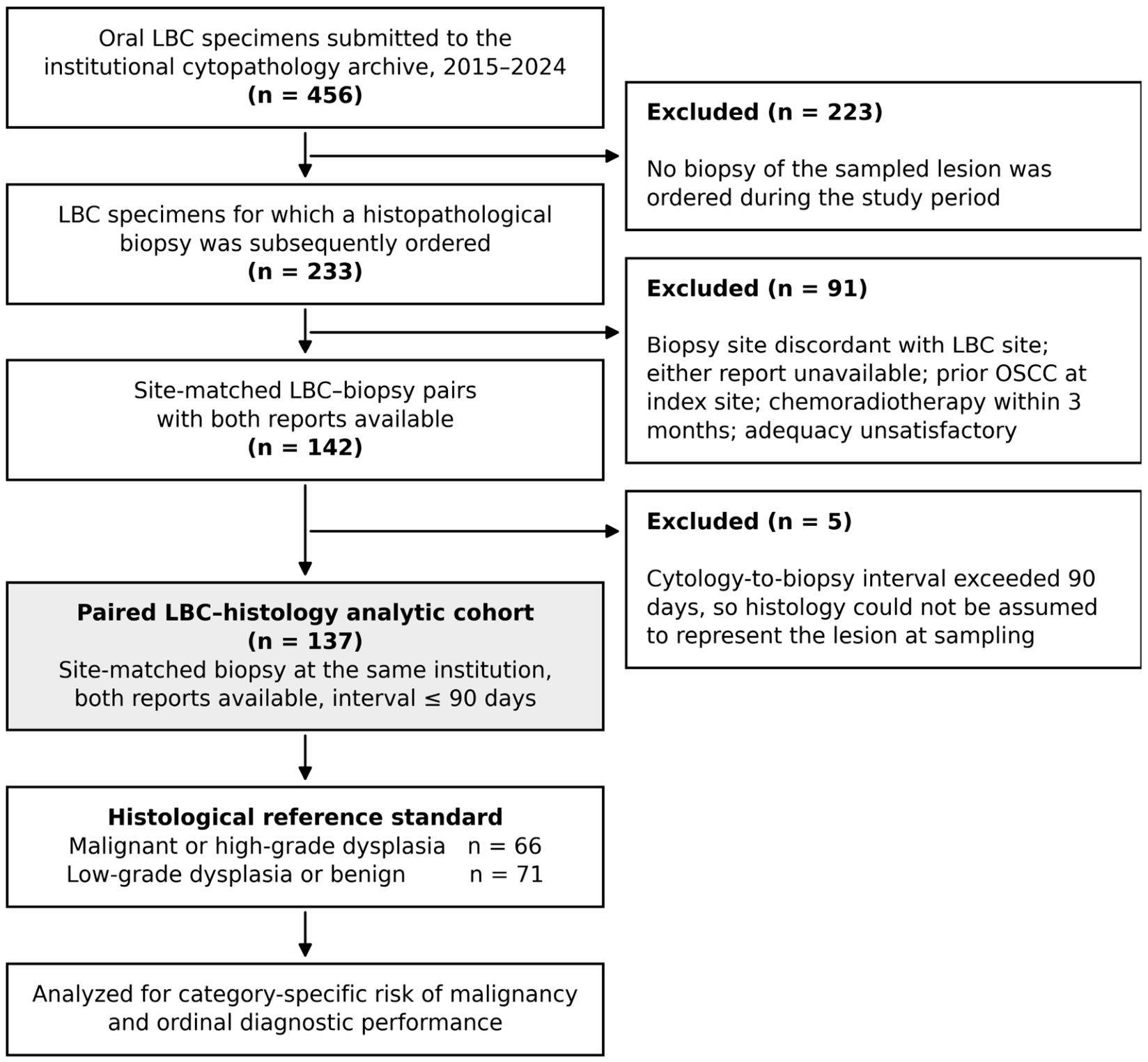
The source population comprised all adult patients (age ≥ 19 years) who underwent oral LBC sampling for a clinically suspicious mucosal lesion at the index institution during the study window. From an initial pool of 456 retrieved LBC specimens, the analytic cohort was restricted to specimens with (i) an intraoral mucosal sampling site clearly documented in the pathology report, (ii) a final cytology report available in the institutional archive, and (iii) a verifiable, site-matched histological biopsy report for the same patient and anatomical site, generated at the same institution. These criteria yielded 142 unique paired LBC–histology cases. Five pairs in which cytology and biopsy were separated by more than 90 days were then excluded, leaving 137 cases as the analytic cohort. Patients with a prior histological diagnosis of OSCC at the index site, current or recent (≤3 months) chemoradiotherapy, or an LBC specimen reported as unsatisfactory were also ineligible. These exclusions are contained within the step from 233 to 142 shown in Figure 1.

2.3. LBC Sampling and Processing

All LBC specimens included in the analytic cohort were obtained using a uniform institutional protocol. After removal of superficial debris with sterile saline, the suspicious lesion was sampled by firm, multidirectional brushing using a sterile cytobrush (Rovers Cervex-Brush; Rovers Medical Devices, Oss, The Netherlands) until pinpoint capillary bleeding was observed, ensuring transepithelial sampling. The brush was immediately immersed and vigorously rinsed in 20 mL of a methanol-based fixative (ThinPrep PreservCyt; Hologic, Marlborough, MA, USA). Specimens were processed on the ThinPrep 2000 processor (Hologic, Marlborough, MA, USA) using the standard non-gynecological protocol. Slides were stained with the Papanicolaou stain and reviewed by board-certified cytopathologists according to the institutional standard operating procedure.

2.4. Reference Standard: Scalpel Biopsy

The paired histological biopsy served as the reference standard. Biopsy specimens had been obtained under local anesthesia (2% lidocaine with 1:100,000 epinephrine) as part of the routine diagnostic workflow, fixed in 10% neutral-buffered formalin, paraffin-embedded, sectioned at 4 μm, and stained with hematoxylin and eosin. All histopathological diagnoses had been rendered by board-certified oral pathologists according to the World Health Organization classification of head and neck tumors in force at the time of reporting [30]. Each biopsy diagnosis was assigned to one of four levels, and the two target conditions were built from them, as defined in Section 2.6.

2.5. Cytological Categorization

Cytological reports were issued on a five-tier ordinal scale adapted from the Bethesda system [31], as applied by the reporting laboratory: (i) negative for malignancy, (ii) atypical, favoring reactive, (iii) atypical cells of undetermined significance (ACUS), (iv) atypical, favoring neoplastic, and (v) malignancy. Diagnostic indices were calculated against the histological reference standard under two pre-specified positivity definitions: a strict definition, in which only the malignancy category was treated as LBC-positive, and an expanded definition, in which every non-negative category was treated as LBC-positive. For the present analysis the original cytopathology reports for all 137 cases were retrieved from the primary laboratory records and assigned to that scale exactly as rendered. No slide was reinterpreted and no new cytological judgment was made. The study database had carried a reduced set of labels in which the three intermediate grades were collapsed under the single label atypical. Every analysis reported below is based on the retrieved primary reports, not the reduced labels (one further label discrepancy is documented in Supplementary Methods S1). Reporting in 2015, the first year of the series, was not uniform: 13 of the 22 cases from that year were issued in an earlier single-line format and were assigned to the ordinal scale from that diagnostic line verbatim, without slide review (mapping rules in Supplementary Methods S1). Because the format was mixed in that year, a sensitivity analysis excluding all 22 cases from 2015 is reported in Section 3.5. No written morphological criteria for these categories were published by the reporting laboratory, and none existed for the oral cavity during the study period; that absence is part of the problem this study addresses. The reports themselves, however, use stereotyped descriptive wording. An explicit microscopic description accompanied the categorical diagnosis in 203 of the 453 archived specimens carrying one (44.8%), and its content was consistent within each category (Supplementary Methods S2). We report these observed proportions rather than a criteria table, because a criteria table constructed after the fact would not describe how the categories were actually applied.

2.6. Variables Extracted

For each paired case the following variables were abstracted from the institutional records: anonymized chart identifier, date of LBC, year, specimen adequacy, cytological category, specific cytological diagnosis, reported anatomical location, and the final histopathological diagnosis from the paired biopsy. The surface character of the lesion was not a field of the clinical record and was derived post hoc, for the exploratory stratification in Section 3.5 only: a lesion was classified as keratotic when the histopathological or clinical diagnostic line recorded leukoplakia, hyperkeratosis, keratosis, or a verrucous lesion, and as non-keratotic otherwise. The histopathological diagnosis served as the reference standard. It was assigned to one of four levels from the verbatim diagnostic line of the biopsy report, without slide review: (i) malignant, comprising any invasive or in situ carcinoma or other malignancy; (ii) high-grade dysplasia, comprising moderate or severe epithelial dysplasia; (iii) low-grade dysplasia, comprising mild epithelial dysplasia; and (iv) benign, comprising all remaining diagnoses (assignments that required a decision beyond the verbatim line are documented in Supplementary Methods S1). Two target conditions were used: malignancy alone (level i), and the expanded target condition of malignancy or high-grade dysplasia (levels i and ii). ROM is reported under both. All remaining analyses use the expanded target condition unless malignancy alone is specified. Levels (iii) and (iv) were treated as disease-negative throughout, so a report of atypia in a mildly dysplastic lesion counts as a false positive.

2.7. Statistical Analysis

Categorical variables are presented as frequencies and percentages. The ROM of each cytological category was calculated as the proportion of specimens in that category meeting the target condition, and diagnostic indices were calculated at each of the four ordinal thresholds. The histological verification rate of each category—the proportion of all specimens reported in that category during the study period with a paired histopathological diagnosis within 90 days—is reported alongside the ROM, so that the direction of partial verification bias can be judged. Proportions were compared with the two-sided Fisher exact test and category distributions with the chi-square test. No adjustment was made for multiple comparisons, and the stratification by lesion surface character is exploratory. Sensitivity, specificity, positive predictive value (PPV), negative predictive value (NPV), overall accuracy, and their 95% confidence intervals were calculated using the Wilson score method. Analyses were performed in Python 3.13 (SciPy 1.17). A two-sided p-value < 0.05 was considered statistically significant.

3. Results

3.1. Cohort Overview

Of 456 oral LBC specimens identified in the institutional archive between 2015 and 2024, 137 met the inclusion criteria for paired analysis (Figure 1). A histopathological biopsy was ordered for the sampled lesion in 233 of the 456 specimens (51.1%). Of these, 142 had a site-matched biopsy with both reports retrievable from the electronic medical record, and five pairs separated by more than 90 days were excluded, leaving 137 pairs. Cytology and biopsy were performed on the same day in 102 pairs (74.5%); cytology preceded biopsy in 25 (18.2%; median 14 days, range 1–84) and followed it in 10 (7.3%; median 13 days, range 2–90). All 137 specimens were oral brush LBC preparations. Adequacy was recorded as satisfactory in 124 of 137 cases (90.5%); the remaining 13 reports, all issued in 2015, were produced in a format that contained no adequacy field (Section 2.5). Histopathology confirmed a malignant or high-grade dysplastic lesion in 66 cases (48.2%) and a benign or low-grade lesion in 71 (51.8%). Annual accrual of paired cases was uneven (range 1–39 per year; Supplementary Table S1). No paired case arose in 2021 or 2022: none of the 67 specimens accessioned in those two years had a site-concordant histopathological diagnosis. Given the small and uneven annual denominators, year-to-year differences are reported descriptively and are not interpreted as a temporal trend. The specimens that did not enter the analytic cohort are accounted for by cytological category in Section 3.5.

3.2. Distribution of Cytological Categories

Across the 137 paired cases, the distribution of ordinal cytological categories was: negative for malignancy 64 (46.7%), atypical favoring reactive 15 (10.9%), ACUS 39 (28.5%), atypical favoring neoplastic 8 (5.8%), and malignancy 11 (8.0%) (Table 1). The combined non-negative fraction accounted for 53.3% of all paired specimens.

3.3. Anatomical Location of Sampled Lesions

Among the 137 paired specimens, the anatomical site was specified in 78 cases (56.9%) and reported as “not specified” in the remaining 59 (43.1%). When recorded, the tongue (28 cases, 20.4%) and buccal mucosa (19, 13.9%) were the most frequently sampled subsites, followed by multiple-site sampling (13, 9.5%), gingiva/alveolar ridge (10, 7.3%), palate (7, 5.1%), and floor of mouth (1, 0.7%). The site distribution is summarized in Supplementary Table S2.

3.4. Diagnostic Performance Against Paired Histology

Diagnostic-performance indices were calculated under both pre-specified positivity definitions and are summarized in Table 2. The underlying cross-tabulation of the ordinal cytological categories against the histological reference is presented in Table 3.
Under the strict definition, in which only the LBC “Malignancy” category was considered positive, the sensitivity, specificity, PPV, NPV, and overall accuracy were 16.7%, 100.0%, 100.0%, 56.3%, and 59.9%, respectively. A cytological diagnosis of overt malignancy was therefore essentially confirmatory—every such read corresponded to a histologically malignant lesion—but identified only a small minority of histologically confirmed malignant cases.
Under the expanded definition, in which any non-negative cytological category was treated as positive, the corresponding values were 84.8%, 76.1%, 76.7%, 84.4%, and 80.3%. Across both definitions no false-positive read arose in the “Malignancy” category specifically (0 of 11 cytological malignancy reads were histologically benign or low-grade), while the three atypical categories together captured the majority of histologically confirmed malignant or high-grade lesions at the cost of 17 false-positive reads.

3.5. Risk of Malignancy by Cytological Category

The ROM attached to each reporting category increased monotonically across the ordinal scale (Table 4). For histologically confirmed malignancy, ROM was 9.4%, 26.7%, 66.7%, 87.5% and 100% for negative, atypical favoring reactive, ACUS, atypical favoring neoplastic and malignancy, respectively; for the expanded target condition, 15.6%, 53.3%, 76.9%, 87.5% and 100%. No cytological diagnosis of malignancy was rendered in a histologically benign or low-grade lesion. The proportion of specimens in each category that proceeded to histological verification was much lower in the two lowest categories (25.6% and 15.8%) than in the three highest (48.8%, 57.1% and 78.6%; Table 4, final column). The category distribution of verified and unverified specimens differed accordingly (chi-square = 45.3, df = 4, p < 0.001): the analytic cohort is enriched for the higher categories relative to the workload from which it was drawn. This partial verification has a known direction and is considered in Section 4.
Diagnostic indices at each of the four ordinal thresholds are given in Table 5; its first and last rows correspond to the expanded and strict positivity definitions of Table 2. At the ACUS threshold, sensitivity was 72.7% (95% CI 61.0–82.0) and specificity 85.9% (76.0–92.2) for the expanded target condition, and 81.5% (69.2–89.6) and 83.1% (73.7–89.7) for malignancy alone. Sensitivity at this threshold differed markedly by the surface character of the sampled lesion: 25.0% (7.1–59.1) in the 32 keratotic lesions (leukoplakia, hyperkeratosis, verrucous hyperplasia; 8 disease-positive) against 79.3% (67.2–87.7) in the 105 non-keratotic lesions (58 disease-positive; Fisher exact test, two-sided p = 0.004). Six of the eight disease-positive keratotic lesions were reported below the ACUS threshold, including one with severe epithelial dysplasia and one invasive carcinoma.
Two sensitivity analyses were performed (Supplementary Table S3). Excluding all 22 cases from 2015, in which the reporting format was mixed (Section 2.5), no diagnostic index shifted by more than 5.4 percentage points and every confidence interval overlapped that of the full cohort. The findings are therefore not an artifact of the retrospective category assignment. Excluding the two histologically confirmed malignancies that were not unequivocally squamous—a low-grade mucoepidermoid carcinoma reported as ACUS, and a gingival malignant tumor with a differential of spindle cell carcinoma, squamous cell carcinoma and sarcoma, reported as atypical favoring reactive—sensitivity at the ACUS threshold changed by less than two percentage points and specificity was unchanged.
All ten specimens reported at or above the ACUS threshold in lesions histologically negative for the expanded target condition are listed in Table 6. Seven arose in lesions showing epithelial reparative or reactive change: four ulcerated lesions (two with inflamed granulation tissue), one pseudoepitheliomatous hyperplasia, one lichenoid mucositis, and one subepithelial bulla with necrosis of the basal cell layer. An eighth arose in a hyperkeratotic lesion with acanthosis clinically diagnosed as leukoplakia. Of the remaining two, one was a pleomorphic adenoma reported as atypical favoring neoplastic, in which the identification of a neoplastic process was correct and the discordance is definitional. The other carried low-grade epithelial dysplasia, disease-negative by the same definition.

3.6. Representative Cytological and Histological Findings

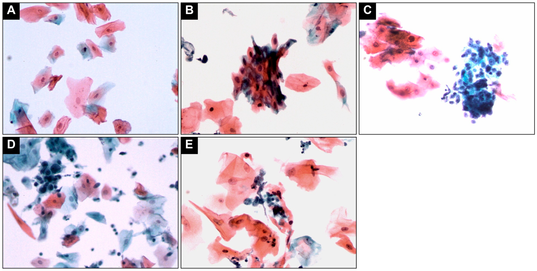
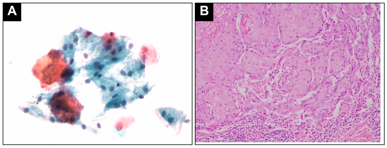
Figure 2 shows one representative preparation for each of the five ordinal categories, and Figure 3 the single case for which both a cytological and a histological photomicrograph were retained. Preparations reported as malignant showed cellular crowding, anisonucleosis, irregular nuclear contours, hyperchromasia and an increased nuclear-to-cytoplasmic ratio against a relatively clean background. Preparations reported as negative that proved malignant on biopsy were typically dominated by anucleate squames and keratinaceous debris with only sparse dysplastic cells. The ACUS and atypical-favoring-neoplastic categories were separated by the extent and distribution of the atypical population rather than by any single qualitatively distinct feature.

4. Discussion

Structured reporting is now the norm in cytopathology. The Bethesda, Milan, Paris and WHO systems each attach a published ROM and a linked management recommendation to a small set of defined categories; that attachment converts a descriptive report into a decision. The oral cavity has no such system, and the ROM of an oral brush cytology category has not been established. The present series was reported by its own laboratory on a five-tier scale that maps directly onto that common structure, and every case had a site-concordant histopathological diagnosis. It therefore permits the calculation that a reporting system requires. We present these values not as a proposal for a system, which would need multi-institutional data and formal reproducibility testing, but as a first empirical benchmark against which such a proposal could be framed.
Three findings warrant emphasis. First, the probability of malignancy or high-grade dysplasia rose monotonically across the five reporting categories, from 15.6% for a negative report to 100% for a report of malignancy; the categories are therefore separable in a way that could support graded management. Second, the category denoting the pathologist’s uncertainty carried a risk that in other organ systems belongs to the suspicious-for-malignancy tier. Third, these probabilities were not a property of the test alone: in keratotic lesions, a report below the ACUS threshold retained almost no exclusionary value.
Placing these values beside the established systems exposes two problems of different kinds. The malignant tier behaves as it does elsewhere: no cytological diagnosis of malignancy was rendered in a histologically benign or low-grade lesion. The negative tier does not. A negative report carries a low residual risk in the established systems, whereas here it was followed by malignancy or high-grade dysplasia in one lesion in six—a question of sampling rather than of nomenclature, taken up below. The divergence that inverts the meaning of a label is confined to the atypical range. ACUS carried a ROM of 76.9% (66.7% for malignancy alone), whereas the similarly named tier carries a pooled risk of 22% (95% CI 13–30) in the thyroid [25] and 30.5% in the salivary gland [27]; the oral value instead falls within the range spanned by their suspicious-for-malignancy tiers (74% and 83.8%). Stricter thresholding of the ACUS label and the high pre-test probability of a biopsy-selected cohort are both plausible explanations. They operate in the same direction and cannot be separated here, but the practical consequence is not in doubt: the same words denote a fundamentally different probability in this organ.
The threshold consequences are made explicit in Table 5. Moving the positivity threshold from malignancy alone down to the lowest atypical category raises sensitivity from 16.7% to 84.8% and lowers specificity from 100.0% to 76.1%. Which threshold is appropriate is a clinical judgment that these data do not settle. Of the 66 histologically confirmed malignant or high-grade lesions, 45 (68.2%) were reported in one of the three atypical categories rather than as malignant. The threshold at which oral LBC operates as a triage to biopsy is therefore the lowest atypical read, and at that threshold its performance (sensitivity 84.8%, specificity 76.1%) lies within the range of published oral LBC series (70–95% and 80–99% [18,19,20,21,22]), which differ from the present series in case mix, positivity definition and completeness of verification, and it is consistent with brush-based series reporting high accuracy for early detection [32]. The absence of false positives in the malignancy category rests on a small denominator: its PPV of 100.0% carries a 95% confidence interval of 74.1–100.0 and is compatible with a false-positive rate of up to one in four. A cytological diagnosis of malignancy therefore justifies proceeding to confirmatory biopsy without delay, not definitive treatment planning, and a negative or atypical read should not by itself defer biopsy of a clinically suspicious lesion. Collapsing all atypical reads into a single label, as the study database itself had done, discards decisive information: the single word atypical spanned lesions whose probability of disease ranged from 53% to 88%.
The designation atypical cells of undetermined significance describes the pathologist’s degree of certainty, not the patient’s risk. In this cohort the two diverge sharply: a read of undetermined significance was followed by malignancy or high-grade dysplasia in 77% of lesions. A nomenclature that signals uncertainty while denoting high risk is liable to be misinterpreted as grounds for observation, and we would argue that the reported category should be accompanied by its observed post-test probability.
Sensitivity was not a property of the test alone but of the lesion sampled. In keratotic lesions—leukoplakia, hyperkeratosis and verrucous hyperplasia—sensitivity at the ACUS threshold fell to 25.0% against 79.3% elsewhere (p = 0.004), and six of eight diseased keratotic lesions were reported below that threshold, including one with severe dysplasia and one invasive carcinoma. The most plausible explanation is mechanical: a keratinized surface obstructs brush sampling, so the preparation is dominated by anucleate squames while the basal cells carrying the dysplastic change remain unsampled, and the pinpoint-bleeding endpoint of the protocol does not guarantee that basal cells were recovered from the keratinized area itself. With only eight disease-positive keratotic lesions the observation is exploratory, but its practical consequence is immediate: a negative cytological report should not defer biopsy of a clinically suspicious leukoplakia. Whether surface decortication before brushing recovers the lost sensitivity is a question for prospective study.
The false positives at the ACUS threshold were likewise patterned rather than random (Table 6): seven of the ten arose in lesions showing epithelial repair or reactive change. Pseudoepitheliomatous hyperplasia, granulation tissue at the base of an ulcer and regenerating epithelium all yield enlarged, hyperchromatic nuclei against an inflammatory background; and an exfoliative preparation strips these cells of the architectural context on which the histological recognition of their benign nature depends. A report of atypia in a recently ulcerated, traumatized or acutely inflamed lesion therefore carries a lower probability of malignancy than the same report in an intact lesion, and repeat sampling after the inflammation has resolved may be more proportionate than immediate escalation—a qualification any oral reporting system would need to state explicitly.
Only lesions that proceeded to biopsy could enter this analysis, and the probability of biopsy was itself related to the cytological read (25.6% and 15.8% verification for the two lowest categories against 48.8–78.6% for the three highest). This partial verification bias operates in a predictable direction, inflating sensitivity and deflating specificity, and it makes the reported probabilities conditional on the decision to biopsy having been made. That conditioning is appropriate to the intended use—the interpretation of a report in a patient for whom biopsy is already being recommended—and the same constraint applies to the comparator systems, whose ROM figures likewise derive from verified subsets. We report the verification rate of every category so that the direction and size of the bias can be judged. Extrapolation to unselected populations is nonetheless precluded: at a disease prevalence of 1%, the same operating characteristics would yield a positive predictive value below 10%, and we do not present these data as support for population screening.
Oral LBC is one of several non-invasive approaches to the detection of oral cancer, and the present data do not compare it with the alternatives, which include organized screening programs, adjunctive diagnostic technologies and DNA-image cytometry [33,34,35,36,37]. A reporting system built on morphology alone also has a ceiling: the sensitivity obtained here is lower than that reported for brush sampling augmented by DNA-ploidy analysis of the same material [24], and the gain in that setting came from an ancillary measurement rather than from any change in morphological interpretation. Molecular characterization offers complementary axes. Transcriptomic signatures, including lipid-metabolism gene panels, stratify prognosis and immunotherapy response in OSCC independently of histological grading [38], and salivary biomarkers, including the protein cargo of small extracellular vesicles, distinguish OSCC from healthy mucosa non-invasively [39,40]. None of these yet attach an established ROM to a defined result category, so they are complementary to, rather than competitive with, a graded morphological report. Because liquid-based preparations leave a residual cell pellet that routinely supports ancillary testing in cervical cytology, applying a molecular assay to the residual sample of an atypical oral read is a plausible route to resolving precisely the category this study identifies as problematic. Computational reading is a second extension—deep-learning models approach subspecialty performance in cervical, urothelial and thyroid cytology [41,42,43,44,45]—but it will require prospectively collected, unselected whole-slide material with paired histological outcomes, which the present archive does not provide.
Several limitations should be acknowledged. The study is single-center and reflects local cytopathology reading conventions, referral patterns, and biopsy thresholds. The retrospective design inherits the data quality limits of clinical archives, including absent anatomical-site annotation in 43.1% of cases, and inter-observer reproducibility was not assessed. The cohort is enriched for clinically suspicious lesions and subject to the partial verification bias set out above, so the indices reflect a referred, biopsy-eligible population rather than a screening setting. Histopathological biopsy is itself subject to sampling error in heterogeneous lesions, and a small number of biopsies may not have co-registered exactly with the LBC sampling field. The case mix shifted over the window—ten of the eleven cytologically malignant reads were issued in 2015–2016—so the risk attached to the uppermost category should not be read as time-invariant. Finally, the archived photomicrographs in Figure 2 and Figure 3 were stored at 640 × 480 pixels without a recorded objective magnification, which suffices for cellular arrangement and staining pattern but not for fine chromatin detail, and paired histological images were retained for only one case.
Against these limitations, the cohort spans ten years of uniform sampling and processing at a single institution and is among the largest retrospective paired analyses of oral LBC reported from a Korean tertiary dental hospital. The dual positivity definitions make the effect of the threshold visible rather than fixing it by convention.

5. Conclusions

Every patient in this series underwent both cytology and biopsy, so the design cannot establish what oral liquid-based cytology adds to a diagnostic pathway, and we claim no such addition. What it does establish is the ROM attached to each reporting category—16%, 53%, 77%, 88% and 100% for negative, atypical-favoring-reactive, ACUS, atypical-favoring-neoplastic and malignant reads, respectively—derived in a population for which biopsy had already been judged necessary, to which setting these values apply. Two categories are liable to be misread in routine practice. A report of ACUS denotes the pathologist’s uncertainty, not the patient’s: 77% of these lesions were malignant or high-grade, and the category should be acted upon as a high-risk result rather than as grounds for observation. A negative report was followed by disease in 16% of lesions overall, and in keratotic lesions six of the eight diseased lesions were reported below the ACUS threshold, so in that setting a report below the threshold does not exclude disease. A cytological diagnosis of malignancy, conversely, was never rendered in a histologically benign or low-grade lesion and justifies confirmatory biopsy without delay, though not definitive treatment planning. These are single-institution estimates from 137 retrospective pairs and should be read as a first benchmark rather than as settled values. If a structured reporting system for oral cytology is to be developed, each category should carry an explicit ROM, because the terminology inherited from other organ systems does not transfer, and these data suggest that the surface keratinization of the sampled lesion should be recorded with the report. Prospective multicenter series, with the ordinal scale applied concurrently and histological verification not conditional on the cytological read, are needed to establish these values and to confirm the keratotic sensitivity deficit.

Supplementary Materials

The following supporting information can be downloaded at: https://www.mdpi.com/article/10.3390/app16178882/s1: Supplementary Methods S1, Assignment of the 2015 single-line reports to the ordinal scale and related labeling decisions; Supplementary Methods S2, Descriptive wording of the cytopathology reports by category; Table S1, Annual distribution of ordinal cytological categories (n = 137); Table S2, Anatomical-location distribution of sampled lesions (n = 137); Table S3, Sensitivity analyses of the diagnostic indices at the ACUS threshold.

Author Contributions

Conceptualization, S.-Y.M. and J.-H.Y.; methodology, J.-S.J., H.-J.K. and S.-Y.M.; software, J.-S.J.; validation, J.-H.Y., H.-J.K. and S.-Y.M.; formal analysis, J.-S.J. and H.-J.K.; investigation, J.-S.J., K.-C.P. and H.-J.K.; resources, S.-Y.M. and J.-H.Y.; data curation, J.-S.J. and K.-C.P.; writing—original draft preparation, J.-S.J. and H.-J.K.; writing—review and editing, S.-Y.M., J.-H.Y., K.-C.P. and H.-J.K.; visualization, J.-S.J.; supervision, S.-Y.M.; project administration, S.-Y.M.; funding acquisition, S.-Y.M. All authors have read and agreed to the published version of the manuscript.

Funding

This work was supported by the Institute of Information & Communications Technology Planning & Evaluation (IITP) grant funded by the Korea government (MSIT) (RS-2022-II220951).

Institutional Review Board Statement

The study was conducted in accordance with the Declaration of Helsinki and approved by the Institutional Review Board of Chosun University Dental Hospital (CUDHIRB 2605 001, 16 June 2026). Given the retrospective design and the exclusive use of de-identified archival data, the requirement for informed consent was waived by the IRB.

Informed Consent Statement

Patient consent was waived by the Institutional Review Board owing to the retrospective design and the use of de-identified archival data.

Data Availability Statement

The de-identified data supporting the findings of this study are available from the corresponding author upon reasonable request, subject to institutional and IRB approval.

Acknowledgments

During the preparation of this manuscript, the authors used Claude (Anthropic, Claude Opus 4.8) for the purposes of English editing. The authors have reviewed and edited the output and take full responsibility for the content of this publication.

Conflicts of Interest

Author Jung-Hoon Yoon was employed by the Yonsei Oral Pathology Clinic. The remaining authors declare that the research was conducted in the absence of any commercial or financial relationships that could be construed as a potential conflict of interest. The funders had no role in the design of the study; in the collection, analyses, or interpretation of data; in the writing of the manuscript; or in the decision to publish the results.

References

  1. Sung, H.; Ferlay, J.; Siegel, R.L.; Laversanne, M.; Soerjomataram, I.; Jemal, A.; Bray, F. Global cancer statistics 2020: GLOBOCAN estimates of incidence and mortality worldwide for 36 cancers in 185 countries. CA Cancer J. Clin. 2021, 71, 209–249. [Google Scholar] [CrossRef] [Scilit] [PubMed]
  2. Warnakulasuriya, S. Global epidemiology of oral and oropharyngeal cancer. Oral Oncol. 2009, 45, 309–316. [Google Scholar] [CrossRef] [Scilit] [PubMed]
  3. Chi, A.C.; Day, T.A.; Neville, B.W. Oral cavity and oropharyngeal squamous cell carcinoma—An update. CA Cancer J. Clin. 2015, 65, 401–421. [Google Scholar] [CrossRef] [Scilit] [PubMed]
  4. Siegel, R.L.; Miller, K.D.; Wagle, N.S.; Jemal, A. Cancer statistics, 2023. CA Cancer J. Clin. 2023, 73, 17–48. [Google Scholar] [CrossRef] [Scilit] [PubMed]
  5. Ferlay, J.; Colombet, M.; Soerjomataram, I.; Parkin, D.M.; Piñeros, M.; Znaor, A.; Bray, F. Cancer statistics for the year 2020: An overview. Int. J. Cancer 2021, 149, 778–789. [Google Scholar] [CrossRef] [Scilit] [PubMed]
  6. Petersen, P.E. Oral cancer prevention and control—The approach of the World Health Organization. Oral Oncol. 2009, 45, 454–460. [Google Scholar] [CrossRef] [Scilit] [PubMed]
  7. Listl, S.; Jansen, L.; Stenzinger, A.; Freier, K.; Emrich, K.; Holleczek, B.; Katalinic, A.; Gondos, A.; Brenner, H. Survival of patients with oral cavity cancer in Germany. PLoS ONE 2013, 8, e53415. [Google Scholar] [CrossRef] [Scilit] [PubMed]
  8. Sciubba, J.J. Oral cancer. The importance of early diagnosis and treatment. Am. J. Clin. Dermatol. 2001, 2, 239–251. [Google Scholar] [CrossRef] [Scilit] [PubMed]
  9. Lingen, M.W.; Kalmar, J.R.; Karrison, T.; Speight, P.M. Critical evaluation of diagnostic aids for the detection of oral cancer. Oral Oncol. 2008, 44, 10–22. [Google Scholar] [CrossRef] [Scilit] [PubMed]
  10. Speight, P.M.; Khurram, S.A.; Kujan, O. Oral potentially malignant disorders: Risk of progression to malignancy. Oral Surg. Oral Med. Oral Pathol. Oral Radiol. 2018, 125, 612–627. [Google Scholar] [CrossRef] [Scilit] [PubMed]
  11. Mashberg, A.; Samit, A. Early diagnosis of asymptomatic oral and oropharyngeal squamous cancers. CA Cancer J. Clin. 1995, 45, 328–351. [Google Scholar] [CrossRef] [Scilit] [PubMed]
  12. Lane, P.M.; Gilhuly, T.; Whitehead, P.; Zeng, H.; Poh, C.F.; Ng, S.; Williams, P.M.; Zhang, L.; Rosin, M.P.; MacAulay, C.E. Simple device for the direct visualization of oral cavity tissue fluorescence. J. Biomed. Opt. 2006, 11, 024006. [Google Scholar] [CrossRef] [Scilit] [PubMed]
  13. Sciubba, J.J. Improving detection of precancerous and cancerous oral lesions: Computer-assisted analysis of the oral brush biopsy. J. Am. Dent. Assoc. 1999, 130, 1445–1457. [Google Scholar] [CrossRef] [Scilit] [PubMed]
  14. Gillenwater, A.M.; Vigneswaran, N.; Fatani, H.; Saintigny, P.; El-Naggar, A.K. Proliferative verrucous leukoplakia: Recognition and differentiation from conventional leukoplakia and mimics. Head Neck 2014, 36, 1662–1668. [Google Scholar] [CrossRef] [Scilit] [PubMed]
  15. Wong, D.T.W. Salivary Diagnostics. Oper. Dent. 2012, 37, 562–570. [Google Scholar] [CrossRef] [Scilit] [PubMed]
  16. Hutchinson, M.L.; Zahniser, D.J.; Sherman, M.E.; Herrero, R.; Alfaro, M.; Bratti, M.C.; Hildesheim, A.; Lorincz, A.T.; Greenberg, M.D.; Morales, J.; et al. Utility of liquid-based cytology for cervical carcinoma screening: Results of a population-based study conducted in a region of Costa Rica with a high incidence of cervical carcinoma. Cancer 1999, 87, 48–55. [Google Scholar] [CrossRef] [Scilit]
  17. Bernstein, S.J.; Sanchez-Ramos, L.; Ndubisi, B. Liquid-based cervical cytologic smear study and conventional Papanicolaou smears: A metaanalysis of prospective studies comparing cytologic diagnosis and sample adequacy. Am. J. Obstet. Gynecol. 2001, 185, 308–317. [Google Scholar] [CrossRef] [Scilit] [PubMed]
  18. Navone, R.; Burlo, P.; Pich, A.; Pentenero, M.; Broccoletti, R.; Marsico, A.; Gandolfo, S. The impact of liquid-based oral cytology on the diagnosis of oral squamous dysplasia and carcinoma. Cytopathology 2007, 18, 356–360. [Google Scholar] [CrossRef] [Scilit] [PubMed]
  19. Hayama, F.H.; Motta, A.C.; Silva, A.P.; Migliari, D.A. Liquid-based preparations versus conventional cytology: Specimen adequacy and diagnostic agreement in oral lesions. Med. Oral Patol. Oral Cir. Bucal 2005, 10, 115–122. [Google Scholar] [PubMed]
  20. Kujan, O.; Desai, M.; Sargent, A.; Bailey, A.; Turner, A.; Sloan, P. Potential applications of oral brush cytology with liquid-based technology: Results from a cohort of normal oral mucosa. Oral Oncol. 2006, 42, 810–818. [Google Scholar] [CrossRef] [Scilit] [PubMed]
  21. Mehrotra, R.; Singh, M.; Thomas, S.; Nair, P.; Pandya, S.; Nigam, N.S.; Shukla, P. A cross-sectional study evaluating chemiluminescence and autofluorescence in the detection of clinically innocuous precancerous and cancerous oral lesions. J. Am. Dent. Assoc. 2010, 141, 151–156. [Google Scholar] [CrossRef] [Scilit] [PubMed]
  22. Mehrotra, R.; Hullmann, M.; Smeets, R.; Reichert, T.E.; Driemel, O. Oral cytology revisited. J. Oral Pathol. Med. 2009, 38, 161–166. [Google Scholar] [CrossRef] [Scilit] [PubMed]
  23. Idrees, M.; Farah, C.S.; Sloan, P.; Kujan, O. Oral brush biopsy using liquid-based cytology is a reliable tool for oral cancer screening: A cost-utility analysis. Cancer Cytopathol. 2022, 130, 740–748. [Google Scholar] [CrossRef] [Scilit] [PubMed]
  24. Liu, K.Y.P.; Ng, S.; Taleghani, M.; Zhu, S.Y.; Carraro, A.; Chen, Z.; Palcic, B.; Poh, C.F.; Guillaud, M. Oral cancer detection and progression prediction using noninvasive cytology-based DNA ploidy approach. J. Oral Pathol. Med. 2024, 53, 434–443. [Google Scholar] [CrossRef] [Scilit] [PubMed]
  25. Ali, S.Z.; Baloch, Z.W.; Cochand-Priollet, B.; Schmitt, F.C.; Vielh, P.; VanderLaan, P.A. The 2023 Bethesda System for Reporting Thyroid Cytopathology. Thyroid 2023, 33, 1039–1044. [Google Scholar] [CrossRef] [Scilit] [PubMed]
  26. Faquin, W.C.; Rossi, E.D.; Baloch, Z.; Barkan, G.A.; Foschini, M.P.; Kurtycz, D.F.I.; Pusztaszeri, M.P.; Vielh, P. (Eds.) The Milan System for Reporting Salivary Gland Cytopathology, 2nd ed.; Springer: Cham, Switzerland, 2023. [Google Scholar]
  27. Wang, Z.; Zhao, H.; Guo, H.; An, C. Application of the Milan System for Reporting Salivary Gland Cytopathology: A systematic review and meta-analysis. Cancer Cytopathol. 2022, 130, 849–859. [Google Scholar] [CrossRef] [Scilit] [PubMed]
  28. Wojcik, E.M.; Kurtycz, D.F.I.; Rosenthal, D.L. (Eds.) The Paris System for Reporting Urinary Cytology, 2nd ed.; Springer: Cham, Switzerland, 2022. [Google Scholar]
  29. IAC-IARC-WHO Joint Editorial Board. WHO Reporting System for Lung Cytopathology, 1st ed.; IAC-IARC-WHO Cytopathology Reporting Systems Series; International Agency for Research on Cancer: Lyon, France, 2022; Volume 1. [Google Scholar]
  30. El-Naggar, A.K.; Chan, J.K.C.; Grandis, J.R.; Takata, T.; Slootweg, P.J. (Eds.) WHO Classification of Head and Neck Tumours, 4th ed.; IARC Press: Lyon, France, 2017. [Google Scholar]
  31. Nayar, R.; Wilbur, D.C. (Eds.) The Bethesda System for Reporting Cervical Cytology: Definitions, Criteria, and Explanatory Notes, 3rd ed.; Springer: New York, NY, USA, 2015. [Google Scholar]
  32. Velleuer, E.; Dietrich, R.; Pomjanski, N.; de Santana Almeida Araujo, I.K.; Silva de Araujo, B.E.; Sroka, I.; Biesterfeld, S.; Bocking, A.; Schramm, M. Diagnostic accuracy of brush biopsy–based cytology for the early detection of oral cancer and precursors in Fanconi anemia. Cancer Cytopathol. 2020, 128, 403–413. [Google Scholar] [CrossRef] [Scilit] [PubMed]
  33. Speight, P.M.; Epstein, J.; Kujan, O.; Lingen, M.W.; Nagao, T.; Ranganathan, K.; Vargas, P. Screening for oral cancer—A perspective from the Global Oral Cancer Forum. Oral Surg. Oral Med. Oral Pathol. Oral Radiol. 2017, 123, 680–687. [Google Scholar] [CrossRef] [Scilit] [PubMed]
  34. Mehrotra, R.; Gupta, D.K. Exciting new advances in oral cancer diagnosis: Avenues to early detection. Head Neck Oncol. 2011, 3, 33. [Google Scholar] [CrossRef] [Scilit] [PubMed]
  35. Maraki, D.; Becker, J.; Boecking, A. Cytologic and DNA-cytometric very early diagnosis of oral cancer. J. Oral Pathol. Med. 2004, 33, 398–404. [Google Scholar] [CrossRef] [Scilit] [PubMed]
  36. Olms, C.; Hix, N.; Neumann, H.; Yahiaoui-Doktor, M.; Remmerbach, T.W. Clinical comparison of liquid-based and conventional cytology of oral brush biopsies: A randomized controlled trial. Head Face Med. 2018, 14, 9. [Google Scholar] [CrossRef] [Scilit] [PubMed]
  37. Kokubun, K.; Nakajima, K.; Yamamoto, K.; Akashi, Y.; Matsuzaka, K. Evaluation of oral brush liquid-based cytology for oral squamous cell carcinoma: A comparative study of cytological and histological diagnoses at a single center. BMC Oral Health 2023, 23, 145. [Google Scholar] [CrossRef] [Scilit] [PubMed]
  38. Xie, Y.; Chen, Z.; Chen, Z.; Yang, Y. Identification and validation of a lipid metabolism-related gene signature for predicting prognosis and immunotherapy response in oral squamous cell carcinoma. Metabolites 2026, 16, 455. [Google Scholar] [CrossRef] [Scilit] [PubMed]
  39. Fontana, S.; Mauceri, R.; Novara, M.E.; Alessandro, R.; Campisi, G. Protein cargo of salivary small extracellular vesicles as potential functional signature of oral squamous cell carcinoma. Int. J. Mol. Sci. 2021, 22, 11160. [Google Scholar] [CrossRef] [Scilit] [PubMed]
  40. Li, C.; Deng, S. Salivary biomarkers for early detection of oral squamous cell carcinoma (OSCC) and head/neck squamous cell carcinoma (HNSCC). Jpn. Dent. Sci. Rev. 2025, 61, 3–4. [Google Scholar] [CrossRef] [Scilit] [PubMed]
  41. Hamdi, M.; Senan, E.M.; Awaji, B.; Olayah, F.; Jadhav, M.E.; Alalayah, K.M. Analysis of WSI images by hybrid systems with fusion features for early diagnosis of cervical cancer. Diagnostics 2023, 13, 2538. [Google Scholar] [CrossRef] [Scilit] [PubMed]
  42. Sanghvi, A.B.; Allen, E.Z.; Callenberg, K.M.; Pantanowitz, L. Performance of an artificial intelligence algorithm for reporting urine cytopathology. Cancer Cytopathol. 2019, 127, 658–666. [Google Scholar] [CrossRef] [Scilit] [PubMed]
  43. Bychkov, D.; Linder, N.; Tiulpin, A.; Küçükel, H.; Lundin, M.; Nordling, S.; Sihto, H.; Isola, J.; Lehtimäki, T.; Kellokumpu-Lehtinen, P.-L.; et al. Deep learning identifies morphological features in breast cancer predictive of cancer ERBB2 status and trastuzumab treatment efficacy. Sci. Rep. 2021, 11, 4037. [Google Scholar] [CrossRef] [Scilit] [PubMed]
  44. Dov, D.; Kovalsky, S.Z.; Cohen, J.; Range, D.E.; Henao, R.; Carin, L. Thyroid cancer malignancy prediction from whole slide cytopathology images. Proc. Mach. Learn. Healthc. 2019, 106, 553–570. [Google Scholar]
  45. Kim, S.; Lee, H.; Lee, S.; Song, J.Y.; Lee, J.K.; Lee, N.W. Role of artificial intelligence interpretation of colposcopic images in cervical cancer screening. Healthcare 2022, 10, 468. [Google Scholar] [CrossRef] [Scilit] [PubMed]
Figure 1. Flow of oral liquid-based cytology (LBC) specimens through the study. Eligibility required a histopathological biopsy ordered for the cytologically sampled lesion, performed at the same site and institution, with both reports retrievable from the electronic medical record and the two procedures within 90 days of one another; the further ineligibility criteria shown in the exclusion boxes are defined in Section 2.2.
Applsci 16 08882 g001
Figure 2. Representative liquid-based cytology preparations across the five ordinal reporting categories (Papanicolaou stain). For each panel the features on which the category was assigned are described in place of superimposed markers, so that the entire field remains available for inspection, and the paired histopathological diagnosis is given. (A) Negative for malignancy: mature superficial and intermediate squamous cells with small pyknotic nuclei and low nuclear-to-cytoplasmic ratios, together with anucleate keratinized squames in a clean background; histopathology, hyperkeratosis and acanthosis of the left tongue (clinically leukoplakia). (B) Atypical, favoring reactive: flat cohesive sheets of mildly enlarged, evenly spaced nuclei with preserved cytoplasm among squamous and inflammatory cells; histopathology, hyperkeratosis and acanthosis with moderate epithelial dysplasia of the right lateral tongue (clinically leukoplakia). (C) Atypical cells of undetermined significance: a single discrete crowded group of cells with enlarged, overlapping hyperchromatic nuclei in an otherwise unremarkable background of mature squamous cells; histopathology, invasive squamous cell carcinoma of the left lateral tongue. (D) Atypical, favoring neoplastic: multiple dispersed three-dimensional clusters with scant cytoplasm, high nuclear-to-cytoplasmic ratios and marked variation in nuclear size; histopathology, poorly differentiated squamous cell carcinoma of the ventral tongue. (E) Malignancy: numerous atypical cells with irregular, angular, densely hyperchromatic nuclei; histopathology, invasive squamous cell carcinoma of the right buccal gingiva. Paired histological photomicrographs were not retained in the institutional archive for these five cases; a case for which both preparations were retained is shown in Figure 3.
Applsci 16 08882 g002
Figure 3. Paired cytological and histological preparations from a single case reported as negative for malignancy—the only case in the cohort for which both photomicrographs were retained. (A) Liquid-based cytology of a lesion of the right ventral tongue: cohesive sheets of squamous cells with regular nuclei and preserved cytoplasm, together with keratinized orangeophilic cells; no atypical population was identified and the case was reported as negative for malignancy (Papanicolaou stain). (B) Hematoxylin-and-eosin section of the scalpel biopsy from the same lesion, showing invasive squamous cell carcinoma with keratin pearl formation and stromal infiltration.
Applsci 16 08882 g003
Table 1. Distribution of ordinal cytological categories in the retrospective paired cohort (n = 137). Percentages do not total 100.0 because of rounding.
Cytological Categoryn%
Negative for malignancy6446.7
Atypical, favoring reactive1510.9
ACUS3928.5
Atypical, favoring neoplastic85.8
Malignancy118.0
Total13799.9
Table 2. Diagnostic-performance indices of LBC against paired histology (n = 137), reported under two pre-specified positivity definitions. Target condition: malignancy or high-grade (moderate-to-severe) epithelial dysplasia.
IndexStrict (Mal+ Only)Expanded (Any Non-Negative Category)
Sensitivity16.7% (9.6–27.4)84.8% (74.3–91.6)
Specificity100.0% (94.9–100.0)76.1% (65.0–84.5)
Positive predictive value100.0% (74.1–100.0)76.7% (65.8–84.9)
Negative predictive value56.3% (47.6–64.7)84.4% (73.6–91.3)
Overall accuracy59.9% (51.5–67.7)80.3% (72.8–86.1)
Table 3. Cross-tabulation of the ordinal cytological category against paired histological diagnosis (n = 137). The malignant or high-grade column comprises levels (i) and (ii) of the histological reference standard defined in Section 2.6, that is all invasive and in situ carcinomas and other malignancies together with moderate-to-severe epithelial dysplasia.
Cytological CategoryHistology Malignant or High-Grade (n = 66)Histology Benign or Low-Grade (n = 71)
Malignancy110
Atypical, favoring neoplastic71
ACUS309
Atypical, favoring reactive87
Negative for malignancy1054
Table 4. Risk of malignancy (ROM) by cytological reporting category (n = 137), with the corresponding tier of established WHO-type reporting systems. ROM is given for two target conditions: histologically confirmed malignancy, and malignancy or high-grade (moderate-to-severe) epithelial dysplasia. Wilson 95% confidence intervals. The final column gives the proportion of all specimens reported in that category during the study period that proceeded to histological verification (453 of the 456 archived specimens carried a categorical diagnosis; three were reported as unsatisfactory).
Cytological CategoryEquivalent Tier in WHO-Type Reporting SystemsnROM, Malignancy Only, % (95% CI)ROM, Malignancy or High-Grade Dysplasia, % (95% CI)Histological Verification Rate, %
Negative for malignancyBenign/negative for malignancy649.4 (4.4–19.0)15.6 (8.7–26.4)25.6 (64/250)
Atypical, favoring reactiveAtypical1526.7 (10.9–52.0)53.3 (30.1–75.2)15.8 (15/95)
Atypical cells of undetermined significance (ACUS)Atypical3966.7 (51.0–79.4)76.9 (61.7–87.4)48.8 (39/80)
Atypical, favoring neoplasticSuspicious for malignancy887.5 (52.9–97.8)87.5 (52.9–97.8)57.1 (8/14)
MalignancyMalignant11100.0 (74.1–100.0)100.0 (74.1–100.0)78.6 (11/14)
Table 5. Diagnostic indices at each ordinal cytological threshold (n = 137). Target condition: malignancy or high-grade (moderate-to-severe) epithelial dysplasia. TP, true positive; FP, false positive; FN, false negative; TN, true negative; PPV, positive predictive value; NPV, negative predictive value. Wilson 95% confidence intervals.
Threshold (Test Positive at or Above)TPFPFNTNSensitivity % (95% CI)Specificity % (95% CI)PPV % (95% CI)NPV % (95% CI)
Atypical, favoring reactive5617105484.8 (74.3–91.6)76.1 (65.0–84.5)76.7 (65.8–84.9)84.4 (73.6–91.3)
Atypical cells of undetermined significance (ACUS)4810186172.7 (61.0–82.0)85.9 (76.0–92.2)82.8 (71.1–90.4)77.2 (66.8–85.1)
Atypical, favoring neoplastic181487027.3 (18.0–39.0)98.6 (92.4–99.8)94.7 (75.4–99.1)59.3 (50.3–67.8)
Malignancy110557116.7 (9.6–27.4)100.0 (94.9–100.0)100.0 (74.1–100.0)56.3 (47.6–64.7)
Table 6. Specimens reported at or above the ACUS threshold in lesions that were histologically negative for the expanded target condition (n = 10). The final column gives the most probable source of the discordance. Case numbers are arbitrary and do not correspond to any institutional identifier.
CaseCytological CategoryHistopathological DiagnosisProbable Source of Discordance
1ACUSPseudoepitheliomatous hyperplasia with inflamed granulation tissuePseudoepitheliomatous hyperplasia
2ACUSTraumatic ulcerReparative atypia
3ACUSUlceration with inflamed granulation tissueReparative atypia
4ACUSUlceration with inflamed granulation tissueReparative atypia
5ACUSUlceration with surface exogenous pigmentation and chronic inflammationReparative atypia
6ACUSHyperkeratosis and acanthosis, clinically leukoplakiaKeratotic surface epithelium
7ACUSLichenoid mucositis with lymphoplasmacytic infiltrateLichenoid interface change
8ACUSSubepithelial and intraepithelial bulla with necrosis of the basal cell layerVesiculobullous disease with basal cell damage
9Atypical, favoring neoplasticPleomorphic adenomaBenign neoplasm; neoplastic reading correct, discordance definitional
10ACUSChronic inflammation with hyperkeratosis; low-grade squamous dysplasia at a second siteLow-grade dysplasia, disease-negative by definition
Disclaimer/Publisher’s Note: The statements, opinions and data contained in all publications are solely those of the individual author(s) and contributor(s) and not of MDPI and/or the editor(s). MDPI and/or the editor(s) disclaim responsibility for any injury to people or property resulting from any ideas, methods, instructions or products referred to in the content.