Article

Reliability and Agreement of Digital Caliper and Ultrasound Measurements for Inter-Recti Distance in Postpartum Women


1
Faculty of Health Sciences, University of Primorska, Polje 42, SI-6310 Izola, Slovenia
2
Ludwig Boltzmann Institute for Rehabilitation Research, 3100 St. Pölten, Austria
*
Author to whom correspondence should be addressed.
Appl. Sci. 2026, 16(17), 8705; https://doi.org/10.3390/app16178705
Submission received: 20 July 2026 / Revised: 29 August 2026 / Accepted: 31 August 2026 / Published: 1 September 2026
(This article belongs to the Special Issue Advanced Technologies in Rehabilitation Medicine)

Abstract

Diastasis Recti Abdominis is a common postpartum condition characterized by separation of the rectus abdominis muscles. Accurate measurement of inter-recti distance (IRD) is important for diagnosis and longitudinal monitoring, but the extent to which digital caliper and ultrasound measurements agree across abdominal sites remains uncertain. This study examined between-visit test–retest reliability within each method, caliper–ultrasound agreement, and associations with palpation at 4.5 cm above the umbilicus, at the umbilicus, and 4.5 cm below the umbilicus. Thirty healthy primiparous women 8–16 weeks after vaginal delivery completed two visits three days apart. Test–retest reliability was good to excellent (absolute-agreement ICC = 0.83–0.98; TE = 0.09–0.25 cm; CV = 2.84–8.28%; MDC = 0.24–0.70 cm). Caliper–ultrasound agreement varied by site and visit (absolute-agreement ICC = 0.72–0.95; TE = 0.12–0.30 cm; CV = 3.89–9.91%). Lin’s concordance correlation coefficient (CCC) ranged from 0.72 to 0.95, and inter-method minimal detectable change percentages (MDC%) ranged from 10.8% to 27.5%. Caliper values were systematically lower than ultrasound values above the umbilicus by 0.081 cm at Visit 1 and 0.068 cm at Visit 2 (both p = 0.031), while the widest limits of agreement occurred at the umbilicus and below the umbilicus at Visit 1. Visit 1 correlations between palpation and instrument-based measurements were moderate to very strong (Spearman’s ρ = 0.641–0.920). Thus, each instrument was sufficiently stable for repeated assessment under this protocol, but cross-method disagreement was site-dependent, and the data did not permit a definitive judgment of clinical interchangeability without a validated acceptability threshold. Using the same method across follow-up assessments is therefore recommended.

1. Introduction

Pregnancy induces physiological and biomechanical adaptations (including hormonal changes, weight gain, increased intra-abdominal pressure, and abdominal wall stretching) that support fetal development but may also impact musculoskeletal health and postpartum quality of life [1,2]. While the body gradually returns toward its pre-pregnancy state postpartum, many physiological changes (particularly those affecting the abdominal wall and pelvic region) may persist long after childbirth. One of the most common such conditions is Diastasis Recti Abdominis (DRA) [3,4,5]. DRA is characterized by a separation of the two bellies of the rectus abdominis muscle along the linea alba, resulting in an increased inter-recti distance (IRD) [6,7]. This separation typically develops in response to hormonal changes, mechanical stretching of the abdominal wall, and increased intra-abdominal pressure during pregnancy. Several predictors of increased risk for DRA have been identified, including high BMI, higher parity, twin pregnancy, and diabetes [8]. Research indicates that up to 60% of women experience DRA in late pregnancy, with approximately 30–40% remaining affected postpartum [9,10,11]. While not life-threatening, DRA can carry significant functional and psychosocial consequences, including altered trunk mechanics, reduced lumbopelvic stability, decreased core strength, pelvic floor dysfunction, low back pain, and cosmetic concerns, all of which may affect the physical and emotional well-being of postpartum women [2,12,13,14].
Given these consequences, accurate identification and quantification of IRD is essential. Ideally, IRD should be monitored during the third trimester and postpartum to determine whether natural recovery is occurring or whether intervention is needed. Assessment methods available in clinical and research settings include palpation, tape measure, calipers, ultrasound, computed tomography, and magnetic resonance imaging [15]. The choice of the measurement method should be based on the purpose of measurement, measurement properties, accessibility, cost-effectiveness, and the clinical situation [16]. A widening in the linea alba of greater than 2 cm [17,18] has been most typically considered clinically important IRD, while consensus is still lacking. The normal range can vary significantly depending on individual anatomical properties, parity, and individual factors [6,19]. Manual palpation was used first, and diastasis recti was considered if the IRD was greater than 2 finger widths [12,20]. However, individual factors such as subcutaneous fat and the examiner’s finger width make palpation an unreliable method [21,22]. Digital calipers, where the tips are fitted across the width of the diastasis, are another simple, low-cost measurement option. This method has been used in several studies [23,24,25]. Calipers have been shown to be comparable to ultrasound measurement and are clinically feasible [15]. Ultrasound measurements, as well as measurements from other imaging techniques, are done based on “ruler/caliper functions” within the software displaying the images. Measurements based on ultrasound or magnetic resonance imaging can be considered the ‘gold standard’, because they allow direct visualization of the rectus abdominis muscles and linea alba [22,24,26]. However, high cost, limited equipment availability, and the need for a skilled operator limit the widespread adoption of these methods [18,22,27].
Understanding the strengths and limitations of measurement approaches is critical for establishing consistent diagnostic criteria and improving the evaluation of treatment outcomes. Across the literature, most comparative studies on measuring DRA focus on simple clinical tools (palpation, tape measure, calipers) and compare them to ultrasound imaging. Ultrasound imaging has been extensively studied for validity [15,26,28,29,30] and for inter-rater, intra-rater, and retest reliability [15,18,22,27,28,31]. Two systematic reviews concluded that ultrasound and calipers are adequate methods for assessing DRA; both show reliability and validity, while other methods have limited or low-to-moderate quality evidence [15,32]. Original studies largely confirm this pattern, showing that simpler methods are reliable but less accurate than ultrasound [18,21,22,23,24,33,34]. Digital calipers typically perform well above the umbilicus but lose validity below it, limiting their ability to capture full-length separation [35,36]. Studies in postpartum populations show good reliability for palpation and caliper methods, though agreement between them remains only moderate [37,38]. In clinical and physiotherapy settings, practical applicability also matters; therefore, methods like calipers and palpation, which are easy to use and quick to record, can potentially have good clinical utility. Published studies have small sample sizes [21,39] and vary in participant positioning and measurement locations [21,24]. It is suggested that comprehensive assessment of DRA must include measurements at the borders of the upper and lower umbilicus where the linea alba appears the most vulnerable to stretch [40].
Previous work already provides important comparisons of clinical IRD measures with ultrasound. In particular, Benjamin et al. assessed the criterion validity and reliability of calipers, tape measure, and finger-width assessment against ultrasound in 50 postnatal women [38]. The present study is therefore best considered a focused replication and methodological extension rather than a first comparison of calipers with ultrasound. It examines a more homogeneous early-postpartum cohort of healthy primiparous women after vaginal delivery, uses three fixed locations including the umbilicus, repeats both caliper and ultrasound assessment across two visits, and reports site- and visit-specific relative reliability, absolute measurement error, and Bland–Altman agreement. This design provides an independent estimate of whether previously reported measurement properties extend to this narrowly defined population and protocol. Clinical assessment also occurs within a broader behavioral context, as pregnancy-related beliefs about physical activity and provider counseling are associated with activity behavior [41]. Accordingly, the aims were to determine (i) between-visit test–retest reliability of digital caliper and ultrasound IRD measurements, (ii) agreement and systematic bias between the two methods at each site and visit, and (iii) the strength of association of Visit 1 palpation scores with each instrument-based measure.

2. Materials and Methods

The study was designed and reported in accordance with the Guidelines for Reporting Reliability and Agreement Studies [42]. As this was a non-interventional methodological study evaluating the reliability, agreement, and validity of measurement procedures, prospective clinical trial registration was not sought.

2.1. Participants

Thirty healthy postpartum primiparous women participated in this study. Participants were recruited using a convenience sampling approach and were referred by the Department of Gynecology and Obstetrics at General Hospital Murska Sobota. Participants were between 8 and 16 weeks postpartum and were recruited via social media advertisements, online registration forms, and referrals from gynecologists. Inclusion criteria were: (1) primiparous status, (2) vaginal delivery, (3) 8–16 weeks postpartum, (4) ability to attend two testing sessions separated by three days, and (5) ability to perform the standardized abdominal contraction required for IRD assessment. Exclusion criteria included: (1) Cesarean section delivery, (2) abdominal surgery within the previous six months, (3) current pregnancy, (4) acute abdominal or lumbopelvic pain limiting movement, (5) diagnosed connective tissue disorders, and (6) any neurological or musculoskeletal condition that could affect abdominal wall function. All participants provided written informed consent prior to inclusion in the study, and the study received approval from the National Medical Ethics Committee of the Republic of Slovenia (approval No. 0120-158/2025-2711).

2.2. Sample Size

Sample size was justified using the precision-based approach for ICC estimation described by Bonett [43]. The calculation assumed two repeated visit-level measurements per participant, an expected ICC of 0.75, a two-sided 95% confidence interval, and a target total confidence-interval width of 0.35. For these inputs, the approximate requirement was 26 participants; the achieved sample of 30 exceeded this value. Note that this is a precision-based justification rather than a hypothesis-testing power calculation.

2.3. Study Design

This study employed a test–retest and method-comparison design. Each participant completed two testing visits three days apart under the same protocol. At each visit, IRD was measured by digital caliper and ultrasound at three predefined sites, with three consecutive readings per site and method; the three readings were averaged to obtain the visit-level value used in the analyses. Palpation was also recorded for the association analysis. All measurements were performed by the same examiner. The examiner did not review Visit 1 values before Visit 2.

2.4. Procedures

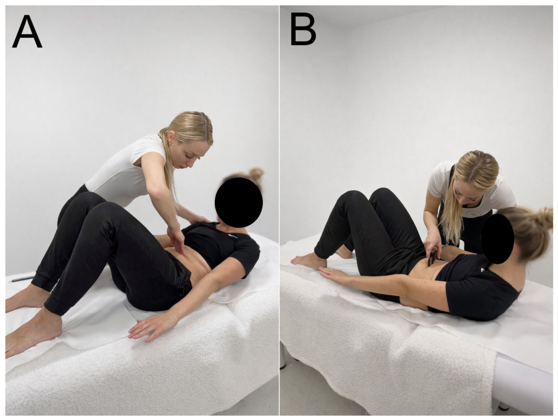
Participants were positioned in a supine crook-lying position on an examination table, with hips flexed to approximately 45° and knees flexed to approximately 90°. The arms were placed alongside the body, and the head rested in a neutral position (Figure 1). Before each measurement, the examiner instructed the participant to contract the abdominal wall by lifting the head and shoulders until the inferior angles of the scapulae were elevated from the table. This partial curl-up is consistent with head-and-shoulder-lift procedures used in previous IRD protocols [29,34,38], although contraction intensity was not quantified objectively. Measurements were obtained at three predefined locations: 4.5 cm above the umbilicus, at the umbilicus, and 4.5 cm below the umbilicus. The 4.5-cm supra- and infraumbilical sites are consistent with an established caliper–ultrasound protocol [34] and with later syntheses identifying 4.5–5 cm as commonly used fixed distances [29]. These locations sample regions on either side of the umbilicus, where linea alba anatomy differs, but a shared external landmark does not by itself ensure identical tissue depth or measurement-plane correspondence across techniques. Palpation was performed first (Figure 1A), followed by digital caliper (Figure 1B) and ultrasound measurements. For palpation, the examiner placed the fingertips perpendicular to the linea alba and estimated IRD in finger widths. Digital caliper measurements were taken with the tips positioned at the medial borders of the rectus abdominis muscles. Three consecutive measurements were obtained at each location and averaged.
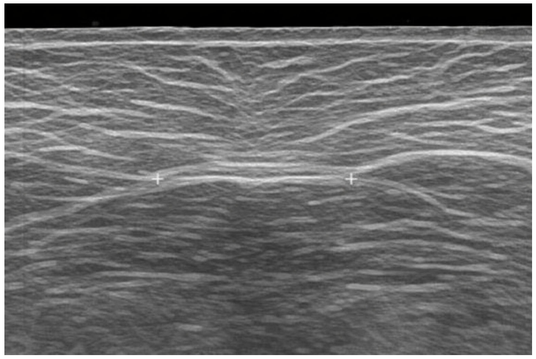
Ultrasound measurements were performed using a Mindray M8 portable ultrasound system (Mindray, Shenhzen, China), equipped with a linear-array transducer (L12-4s; bandwidth 3–13 MHz). Imaging depth and gain were adjusted individually to ensure clear visualization of the medial borders of the rectus abdominis muscles, and minimal probe pressure was applied. The transducer was placed transversely over the abdomen at the same predefined measurement sites. A sufficient amount of ultrasound gel was applied to minimize tissue deformation. The transducer was positioned without exerting excessive pressure, and IRD was defined as the distance between the medial borders of the rectus abdominis muscles (Figure 2), measured using the ultrasound system’s built-in caliper function. Each site was measured three times. All measurements were repeated during the second testing session using the identical protocol, including participant positioning, palpation technique, measurement order, and examiner. For each method and site, three repeated measures were obtained, and their mean was used for statistical analysis.

2.5. Statistical Analysis

Primary statistical analyses were performed using IBM SPSS Statistics (version 26.0, IBM Corp., Armonk, NY, USA). Supplementary CCC, outlier-diagnostic, and sensitivity analyses were conducted in Python 3.12. Descriptive statistics (mean ± SD) were calculated for all sites and methods, and normality was assessed using the Shapiro–Wilk test. Analyses used all available observations; paired analyses used complete pairs, resulting in n = 29 for analyses involving the missing Visit 2 umbilical ultrasound value and n = 30 otherwise. Caliper–ultrasound agreement was evaluated primarily using Bland–Altman mean bias and 95% limits of agreement (LoA = bias ± 1.96 SD of the paired differences) [44]. Linear regression of paired differences on pair means assessed proportional bias. For test–retest reliability, the unit of analysis was the visit-level mean of three consecutive measurements. Between-visit reliability within each method and agreement between the two fixed methods were quantified using a two-way mixed-effects, single-measure, absolute-agreement ICC, denoted ICC(A,1) in the McGraw–Wong framework [45,46]. Absolute agreement was selected because systematic differences between visits or methods were considered measurement disagreement rather than being ignored as in a consistency ICC. ICC values were interpreted as poor (<0.50), moderate (0.50–0.75), good (0.75–0.90), or excellent (>0.90) [45]. Absolute measurement error was summarized using typical error (TE), coefficient of variation (CV), and minimal detectable change at the 95% confidence level (MDC = TE × √2 × 1.96). Lin’s CCC was additionally calculated for each caliper–ultrasound comparison [47]. MDC was standardized to the paired grand mean (MDC% = MDC/paired grand mean × 100). Paired-samples t-tests assessed systematic mean differences; a non-significant p value was not interpreted as evidence of agreement. Spearman’s rank correlations described associations between Visit 1 palpation scores and the corresponding caliper and ultrasound measurements; these correlations were not interpreted as method agreement. Paired infraumbilical ultrasound between-visit differences were examined post hoc using Tukey’s outer fences (Q1 − 3 × IQR, Q3 + 3 × IQR) and modified z scores (|z| > 3.5); a leave-one-out sensitivity analysis assessed influence on ICC, TE, and MDC. Full-sample estimates were retained as primary because no outlier-exclusion rule was prespecified. Statistical significance was set at p < 0.05.

3. Results

Participant characteristics were as follows: mean age was 32.1 ± 4.3 years (range 25–44); mean height was 168.1 ± 3.8 cm; mean body mass was 65.6 ± 5.7 kg; and mean BMI was 23.2 ± 1.9 kg/m2 (range 19.7–27.8). On average, participants were 11.5 ± 1.6 weeks postpartum (range 8–14 weeks) at the time of the first testing session.

3.1. Test–Retest Reliability

Descriptive statistics and between-visit comparisons are presented in Table 1. Mean between-visit differences were small for both methods (caliper, −0.017 to −0.027 cm; ultrasound, −0.002 to −0.060 cm), and none of the paired tests detected a systematic visit effect. The umbilical ultrasound comparison included 29 complete pairs; all other comparisons included 30. These tests address mean bias only and do not establish test–retest reliability, which is reported in Table 2.
Both methods demonstrated good-to-excellent test–retest reliability, although performance varied by site (Table 2). ICC(A,1) values ranged from 0.83 to 0.98. Caliper ICCs were 0.96–0.97 across sites, whereas ultrasound ICCs ranged from 0.83 below the umbilicus to 0.97 at the umbilicus. Absolute error was also greatest for ultrasound below the umbilicus (TE = 0.25 cm; CV = 8.28%; MDC = 0.70 cm). Across the remaining method–site combinations, TE ranged from 0.09 to 0.13 cm, CV from 2.84% to 4.44%, and MDC from 0.24 to 0.37 cm.
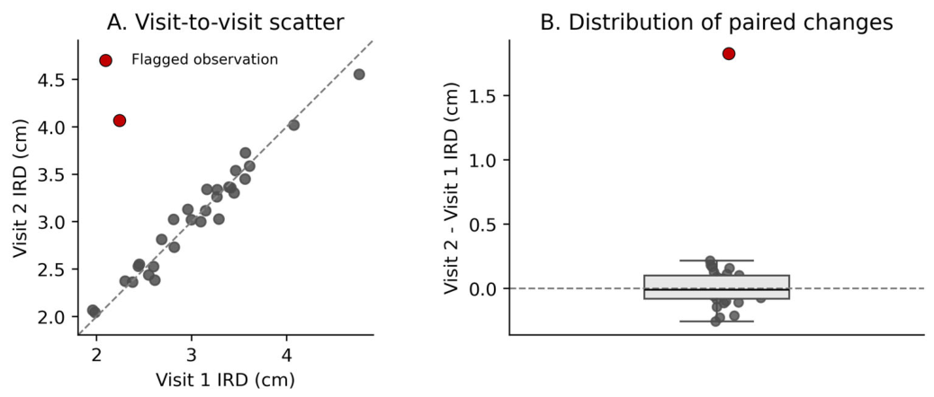
MDC values ranged from 0.24 to 0.70 cm. Caliper MDC values were 0.24–0.33 cm; ultrasound values were 0.27–0.70 cm. The comparatively large ultrasound MDC below the umbilicus indicates that a larger between-visit change would be required at that site to exceed measurement error under this protocol. Post hoc inspection identified one paired difference beyond Tukey’s outer fence (modified z = 13.26; Figure 3). Retaining all observations yielded ICC(A,1) = 0.83, TE = 0.25 cm, and MDC = 0.70 cm; excluding this observation in a sensitivity analysis yielded ICC(A,1) = 0.98, TE = 0.09 cm, and MDC = 0.25 cm. Excluding any other participant left ICC(A,1) between 0.78 and 0.83. The full-sample estimates were retained as primary.

3.2. Agreement Between Methods

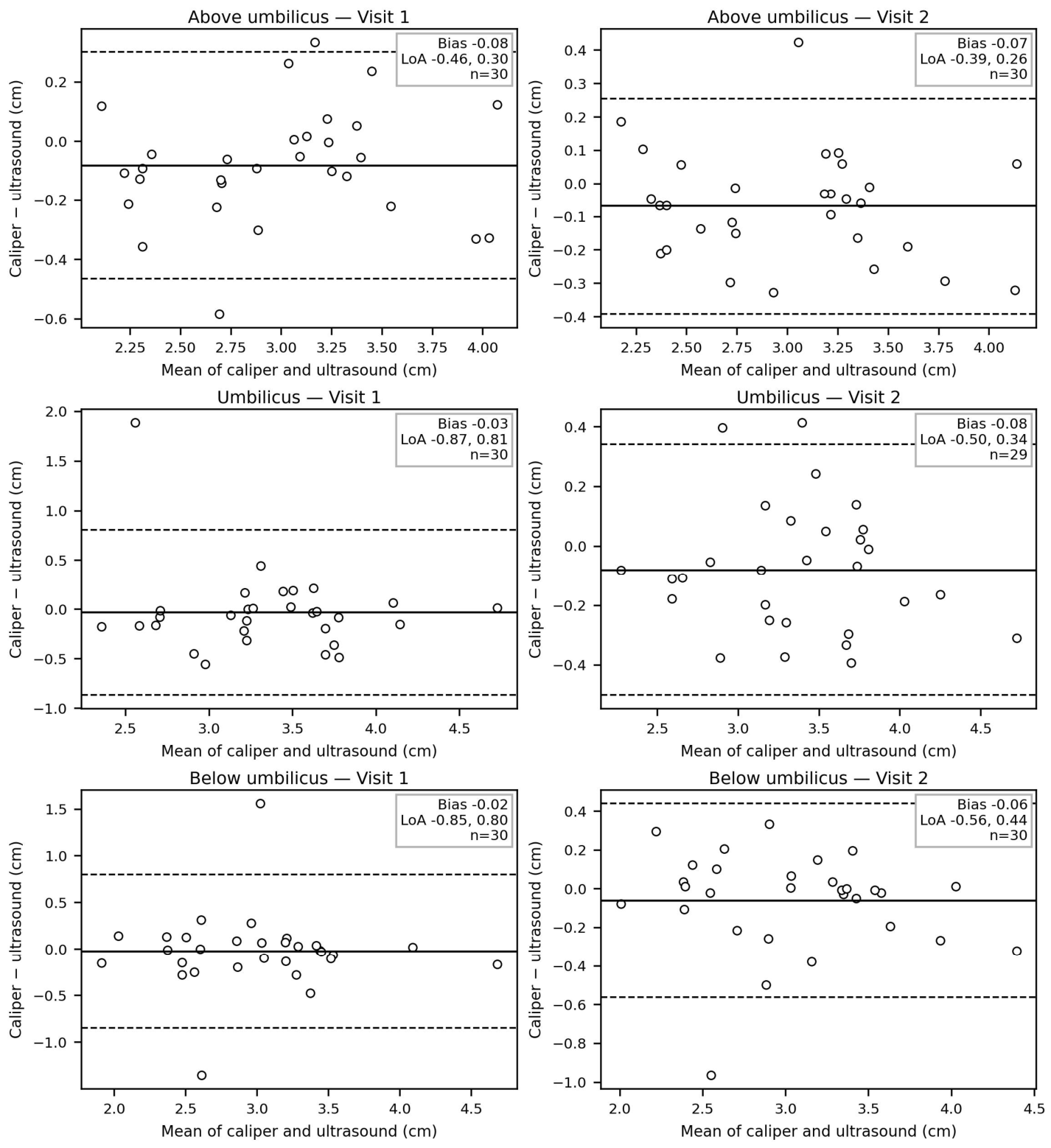
Caliper–ultrasound mean differences and paired tests are shown in Table 3. Calipers produced lower mean values at every site and visit. The mean bias reached statistical significance above the umbilicus at Visit 1 (−0.081 cm, 95% CI −0.154 to −0.008; p = 0.031) and Visit 2 (−0.068 cm, 95% CI −0.130 to −0.007; p = 0.031). The other paired tests were not significant, but this was not taken as evidence of agreement. Bland–Altman analysis (Figure 4) showed site- and visit-dependent disagreement. The 95% LoA were −0.464 to 0.302 cm above the umbilicus at Visit 1 and −0.392 to 0.255 cm at Visit 2; −0.866 to 0.808 cm at the umbilicus at Visit 1 and −0.501 to 0.341 cm at Visit 2; and −0.847 to 0.799 cm below the umbilicus at Visit 1 and −0.560 to 0.439 cm at Visit 2. No statistically significant proportional bias was detected (all p > 0.05).
Absolute-agreement ICCs between caliper and ultrasound ranged from 0.72 to 0.95 (Table 4), indicating moderate to excellent ICC estimates depending on site and visit. Agreement was highest above the umbilicus at Visit 2 (ICC(A,1) = 0.95) and lowest at the umbilicus at Visit 1 (ICC(A,1) = 0.72). TE ranged from 0.12 to 0.30 cm and CV from 3.89% to 9.91%, with the largest errors at the umbilicus and below the umbilicus at Visit 1. The more favorable Visit 2 estimates may reflect familiarization, positioning differences, soft-tissue conditions, or sampling variation; the design cannot distinguish among these possibilities. These ICCs quantify agreement relative to between-participant variability in the observed sample and, together with the limits of agreement, do not establish clinical interchangeability. Lin’s CCC ranged from 0.72 to 0.95, and MDC% from 10.8% to 27.5% (Table 5). The highest MDC% values occurred at the umbilicus (25.1%) and below the umbilicus (27.5%) at Visit 1. ICC and CCC depend partly on between-participant variability, whereas Bland–Altman LoA and MDC% are calculated from paired differences; therefore, wide absolute disagreement is not explained by between-participant dispersion alone.

3.3. Correlation with Palpation

Significant positive associations were found between Visit 1 palpation scores and both instrument-based measures. At the corresponding anatomical sites, Spearman’s ρ values were 0.918 for caliper and 0.920 for ultrasound above the umbilicus, 0.878 and 0.824 at the umbilicus, and 0.860 and 0.641 below the umbilicus, respectively. The palpation–ultrasound association below the umbilicus was therefore moderate and appreciably weaker than the other site-specific associations. Correlation quantifies monotonic association, not numerical agreement or criterion validity.

4. Discussion

This study examined between-visit test–retest reliability of digital caliper and ultrasound IRD measurements, caliper–ultrasound agreement, and associations with palpation at three abdominal sites. Both instruments were generally stable across visits, but ultrasound measurement below the umbilicus showed greater error than the other test–retest comparisons. Cross-method agreement varied by site and visit: the strongest estimates occurred above the umbilicus, whereas disagreement was wider at the umbilicus and below it during Visit 1. Palpation was positively associated with both instruments, although its association with infraumbilical ultrasound was only moderate. These findings support reliable repeated use of either instrument under a consistent protocol but require caution when methods are switched; without a validated acceptability threshold, the data do not permit a definitive judgment of clinical interchangeability.
The caliper ICCs were excellent, and their absolute errors were small, while ultrasound reliability ranged from good to excellent. The ultrasound result below the umbilicus (ICC(A,1) = 0.83; TE = 0.25 cm; MDC = 0.70 cm) was clearly less favorable than the other site-specific estimates. Post hoc diagnostics identified one markedly discordant observation. Excluding it increased ICC(A,1) to 0.98 and reduced TE and MDC to 0.09 and 0.25 cm, respectively, whereas excluding any other participant left ICC(A,1) between 0.78 and 0.83. The full-sample estimate was retained, but the sensitivity analysis shows that the lower reliability estimate was driven primarily by one individual deviation rather than a consistent pattern across participants. This deviation may reflect individual measurement difficulty or transient positioning or soft-tissue differences. The available data cannot distinguish these explanations. This site dependence is consistent with prior reports that measurement properties vary across abdominal locations [22,27,29]. The short interval and standardized examiner, posture, and task may have reduced biological and procedural variation; consequently, these estimates apply to repeat measurement under closely matched conditions and should not be generalized to longer follow-up intervals or different examiners.
The method-comparison results require a distinction between mean bias, relative agreement, and individual-level agreement. Calipers measured approximately 0.7–0.8 mm lower than ultrasound above the umbilicus at both visits. Although this bias is small relative to an average IRD of approximately 3 cm, its clinical importance cannot be declared negligible because no generally accepted, anchor-based clinically important difference or acceptable caliper–ultrasound LoA has been established for IRD [27,29]. Moreover, the Bland–Altman intervals indicate that individual caliper values could differ from ultrasound by several millimeters, with the widest intervals at the umbilicus and below it at Visit 1 (approximately −8.7 to +8.1 mm and −8.5 to +8.0 mm, respectively). The corresponding CCC values (0.72–0.95) and MDC% values (10.8–27.5%) provide standardized context and confirm that absolute disagreement was greatest at the umbilicus and below it at Visit 1. Although heterogeneous participant values can produce high ICC or CCC estimates, between-participant dispersion does not itself widen LoA or MDC%, which are based on paired differences. Thus, a non-significant paired test at other sites does not demonstrate agreement, and in the absence of a validated acceptability threshold, the present data cannot definitively confirm or exclude clinical interchangeability. The absence of proportional bias only indicates that the observed differences did not change systematically with measurement magnitude over the sampled range.
The sensitivity analysis cautions against attributing the lower infraumbilical ultrasound ICC solely to anatomy, because the full-sample estimate was dominated by one observation. Several defensible mechanisms may contribute to the less favorable umbilical and infraumbilical findings. The posterior rectus sheath becomes more difficult to visualize inferiorly, and regional differences in linea alba and fascial morphology may make muscle borders less distinct [6,29]. Calipers and palpation also compress superficial tissue, whereas ultrasound visualizes deeper borders; placing each device at the same external landmark therefore does not guarantee an identical depth or transverse plane when tissue is displaced. These factors could amplify small differences in probe angle, caliper placement, or contraction. The improvement in method agreement at Visit 2 is compatible with familiarization or more reproducible positioning, but random sampling variation or transient soft-tissue differences are equally plausible, and no learning effect was tested. The lower infraumbilical palpation–ultrasound correlation (ρ = 0.641) may similarly reflect the ordinal finger-width scale, tissue compression, border-identification difficulty, and plane mismatch. Palpation may remain useful for screening or coarse follow-up by the same clinician, but the correlation should not be interpreted as numerical agreement with ultrasound.
Clinically, the results favor consistency of method across serial assessments. Ultrasound remains the reference method when direct visualization and millimeter-level documentation are required. Digital calipers are practical and showed strong test–retest stability, particularly above the umbilicus, but their values should not be substituted directly for ultrasound values when changes of only a few millimeters could influence interpretation. The site-specific TE and MDC estimates can assist interpretation of longitudinal change within a method, while the Bland–Altman LoA and MDC% describe the additional uncertainty introduced by switching methods.
Several limitations should be acknowledged. All measurements were performed by one experienced examiner; this reduced between-examiner variation but precluded estimation of inter-rater reliability and does not exclude examiner-specific systematic effects. The examiner did not review Visit 1 results before Visit 2, but the three consecutive readings within each visit were not blinded from the immediately preceding readings, so recall or visual feedback could have reduced apparent within-visit variability. Abdominal contraction was standardized by a head-and-shoulder lift, but movement amplitude, force, and muscle activation were not objectively quantified; inter-individual or between-visit differences in contraction magnitude could therefore influence IRD. Common external landmarks were used across techniques, yet soft-tissue displacement and technique-specific depth mean that exact measurement-plane correspondence cannot be guaranteed. No accepted clinical threshold was available against which to judge the observed method bias and LoA. The three-day interval limits inference about longer-term monitoring. Finally, the sample consisted of healthy primiparous women after vaginal delivery, approximately 8–16 weeks postpartum, with BMI values of 19.7–27.8 kg/m2. Generalization to multiparous women, women after Cesarean delivery or multiple pregnancy, people with obesity or substantially larger DRA, and other pregnancy or postpartum stages should be made cautiously.

5. Conclusions

Digital caliper and ultrasound IRD measurements showed good-to-excellent between-visit reliability under a standardized, single-examiner protocol, although ultrasound error was greater below the umbilicus. Caliper–ultrasound agreement was site- and visit-dependent, with a reproducible 0.7–0.8 mm mean bias above the umbilicus and substantially wider individual differences at the umbilicus and below it during Visit 1. Because no accepted clinical acceptability threshold is available, the present data do not permit a definitive judgment of clinical interchangeability. Nevertheless, the several-millimeter LoA and MDC% values of 10.8–27.5% indicate that switching methods may introduce clinically relevant uncertainty, particularly at the umbilical and infraumbilical sites. Ultrasound remains the reference method, while digital calipers may provide a practical option for repeated assessment when the same method, site, and protocol are maintained.

Author Contributions

Conceptualization, I.W., N.Š. and Ž.K.; methodology, I.W., N.Š. and Ž.K.; software, Ž.K.; validation, I.W., N.Š. and Ž.K.; formal analysis, I.W. and Ž.K.; investigation, I.W.; resources, I.W. and N.Š.; data curation, I.W.; writing—original draft preparation, I.W. and Ž.K.; writing—review and editing, I.W., N.Š. and Ž.K.; visualization, I.W. and Ž.K.; supervision, N.Š. and Ž.K.; project administration, I.W. and Ž.K.; funding acquisition, N.Š. and Ž.K. All authors have read and agreed to the published version of the manuscript.

Funding

This work was supported by the Slovenian Research and Innovation Agency under Grant P5-0443. The funder had no role in study conceptualization, data collection, analysis, interpretation, or manuscript preparation.

Institutional Review Board Statement

The study was approved by the National Medical Ethics Committee of the Republic of Slovenia (approval No. 0120-158/2025-2711; date of approval: 17 June 2025).

Informed Consent Statement

Informed consent was obtained from all participants involved in the study. Written informed consent has been obtained from the participants to publish this paper.

Data Availability Statement

The raw data are available in Zenodo: https://zenodo.org/records/19133133.

Acknowledgments

During the preparation of this manuscript/study, the authors used ChatGPT 5.5 (OpenAI) to check the grammar. The authors have reviewed and edited the output and take full responsibility for the content of this publication.

Conflicts of Interest

The authors declare no conflicts of interest.

Abbreviations

The following abbreviations are used in this manuscript:
BMIBody mass index
CCCConcordance correlation coefficient
CIConfidence interval
CVCoefficient of variation
DRADiastasis recti abdominis
GRRASGuidelines for Reporting Reliability and Agreement Studies
ICCIntraclass correlation coefficient
IRDInter-recti distance
MDMean difference
MDCMinimal detectable change
MDC%Minimal detectable change percentage
SDStandard deviation
SPSSStatistical Package for the Social Sciences
TETypical error
LoALimits of agreement

References

  1. Hills, N.F.; Graham, R.B.; McLean, L. Comparison of trunk muscle function between women with and without diastasis recti abdominis at 1 year postpartum. Phys. Ther. 2018, 98, 891–901. [Google Scholar] [CrossRef] [Scilit] [PubMed]
  2. Benjamin, D.R.; Frawley, H.C.; Shields, N.; van de Water, A.T.M.; Taylor, N.F. Relationship between diastasis of the rectus abdominis muscle (DRAM) and musculoskeletal dysfunctions, pain and quality of life: A systematic review. Physiotherapy 2019, 105, 24–34. [Google Scholar] [CrossRef] [Scilit] [PubMed]
  3. Gilleard, W.L.; Brown, J.M.M. Structure and function of the abdominal muscles in primigravid subjects during pregnancy and the immediate postbirth period. Phys. Ther. 1996, 76, 750–762. [Google Scholar] [CrossRef] [Scilit] [PubMed]
  4. Lee, D.G.; Lee, L.J.; McLaughlin, L. Stability, continence and breathing: The role of fascia following pregnancy and delivery. J. Bodyw. Mov. Ther. 2008, 12, 333–348. [Google Scholar] [CrossRef] [Scilit] [PubMed]
  5. Keeler, J.; Albrecht, M.; Eberhardt, L.; Horn, L.; Donnelly, C.; Lowe, D. Diastasis recti abdominis: A survey of women’s health specialists for current physical therapy clinical practice for postpartum women. J. Women’s Health Phys. Ther. 2012, 36, 131–142. [Google Scholar] [CrossRef] [Scilit]
  6. Rath, A.M.; Attali, P.; Dumas, J.L.; Goldlust, D.; Zhang, J.; Chevrel, J.P. The abdominal linea alba: An anatomo-radiologic and biomechanical study. Surg. Radiol. Anat. 1996, 18, 281–288. [Google Scholar] [CrossRef] [Scilit] [PubMed]
  7. Hsia, M.; Jones, S. Natural resolution of rectus abdominis diastasis. Two single case studies. Aust. J. Physiother. 2000, 46, 301–307. [Google Scholar] [CrossRef] [Scilit] [PubMed]
  8. Sperstad, J.B.; Tennfjord, M.K.; Hilde, G.; Ellström-Engh, M.; Bø, K. Diastasis recti abdominis during pregnancy and 12 months after childbirth: Prevalence, risk factors and report of lumbopelvic pain. Br. J. Sports Med. 2016, 50, 1092–1096. [Google Scholar] [CrossRef] [Scilit] [PubMed]
  9. Šerbec Medjimurec, M.; Mihelj, E.; Šćepanović, D. The incidence and location of diastasis rectus abdominis during the childbearing year and puerperium at the Maternity Hospital Ljubljana. Zdr. Vestn. 2018, 87, 215–222. [Google Scholar] [CrossRef] [Scilit]
  10. Tuominen, R.; Jahkola, T.; Saisto, T.; Arokoski, J.; Vironen, J. The prevalence and consequences of abdominal rectus muscle diastasis among Finnish women: An epidemiological cohort study. Hernia 2022, 26, 599–608. [Google Scholar] [CrossRef] [Scilit] [PubMed]
  11. Lin, S.; Lu, J.; Wang, L.; Zhang, Y.; Zhu, C.; Qian, S.; Xu, H.; Gu, Y. Prevalence and risk factors of diastasis recti abdominis in the long-term postpartum: A cross-sectional study. Sci. Rep. 2024, 14, 25640. [Google Scholar] [CrossRef] [Scilit] [PubMed]
  12. Spitznagle, T.M.; Leong, F.C.; Van Dillen, L.R. Prevalence of diastasis recti abdominis in a urogynecological patient population. Int. Urogynecology J. 2007, 18, 321–328. [Google Scholar] [CrossRef] [Scilit] [PubMed]
  13. Fei, H.; Liu, Y.; Li, M.; He, J.; Liu, L.; Li, J.; Wan, Y.; Li, T. The relationship of severity in diastasis recti abdominis and pelvic floor dysfunction: A retrospective cohort study. BMC Women’s Health 2021, 21, 68. [Google Scholar] [CrossRef] [Scilit] [PubMed]
  14. Wu, L.; Gu, Y.; Gu, Y.; Wang, Y.; Lu, X.; Zhu, C.; Lu, Z.; Xu, H. Diastasis recti abdominis in adult women based on abdominal computed tomography imaging: Prevalence, risk factors and its impact on life. J. Clin. Nurs. 2021, 30, 518–527. [Google Scholar] [CrossRef] [Scilit] [PubMed]
  15. van de Water, A.T.M.; Benjamin, D.R. Measurement methods to assess diastasis of the rectus abdominis muscle (DRAM): A systematic review of their measurement properties and meta-analytic reliability generalisation. Man. Ther. 2016, 21, 41–53. [Google Scholar] [CrossRef] [Scilit] [PubMed]
  16. Streiner, D.L.; Norman, G.R. Health Measurement Scales: A Practical Guide to Their Development and Use, 4th ed.; Oxford University Press: Oxford, UK, 2008. [Google Scholar]
  17. Coldron, Y.; Stokes, M.J.; Newham, D.J.; Cook, K. Postpartum characteristics of rectus abdominis on ultrasound imaging. Man. Ther. 2008, 13, 112–121. [Google Scholar] [CrossRef] [Scilit] [PubMed]
  18. Liaw, L.-J.; Hsu, M.-J.; Liao, C.-F.; Liu, M.-F.; Hsu, A.-T. The relationships between inter-recti distance measured by ultrasound imaging and abdominal muscle function in postpartum women: A 6-month follow-up study. J. Orthop. Sports Phys. Ther. 2011, 41, 435–443. [Google Scholar] [CrossRef] [Scilit] [PubMed]
  19. Beer, G.M.; Schuster, A.; Seifert, B.; Manestar, M.; Mihic-Probst, D.; Weber, S.A. The normal width of the linea alba in nulliparous women. Clin. Anat. 2009, 22, 706–711. [Google Scholar] [CrossRef] [Scilit] [PubMed]
  20. Boissonnault, J.S.; Blaschak, M.J. Incidence of diastasis recti abdominis during the childbearing year. Phys. Ther. 1988, 68, 1082–1086. [Google Scholar] [CrossRef] [Scilit] [PubMed]
  21. Bursch, S.G. Interrater reliability of diastasis recti abdominis measurement. Phys. Ther. 1987, 67, 1077–1079. [Google Scholar] [CrossRef] [Scilit] [PubMed]
  22. Mota, P.; Pascoal, A.G.; Sancho, F.; Bø, K. Test–retest and intrarater reliability of 2-dimensional ultrasound measurements of distance between rectus abdominis in women. J. Orthop. Sports Phys. Ther. 2012, 42, 940–946. [Google Scholar] [CrossRef] [Scilit] [PubMed]
  23. Boxer, S.; Jones, S. Intra-rater reliability of rectus abdominis diastasis measurement using dial calipers. Aust. J. Physiother. 1997, 43, 109–114. [Google Scholar] [CrossRef] [Scilit] [PubMed]
  24. Barbosa, S.; de Sá, R.A.M.; Coca Velarde, L.G. Diastasis of rectus abdominis in the immediate puerperium: Correlation between imaging diagnosis and clinical examination. Arch. Gynecol. Obstet. 2013, 288, 299–303. [Google Scholar] [CrossRef] [Scilit] [PubMed]
  25. Parker, M.A.; Millar, L.A.; Dugan, S.A. Diastasis rectus abdominis and lumbo-pelvic pain and dysfunction—are they related? J. Women’s Health Phys. Ther. 2009, 33, 15–22. [Google Scholar] [CrossRef] [Scilit]
  26. Mendes, D.d.A.; Nahas, F.X.; Veiga, D.F.; Mendes, F.V.; Figueiras, R.G.; Gomes, H.C.; Ely, P.B.; Novo, N.F.; Ferreira, L.M. Ultrasonography for measuring rectus abdominis muscles diastasis. Acta Cir. Bras. 2007, 22, 182–186. [Google Scholar] [CrossRef] [Scilit] [PubMed]
  27. Keshwani, N.; McLean, L. Ultrasound imaging in postpartum women with diastasis recti: Intrarater between-session reliability. J. Orthop. Sports Phys. Ther. 2015, 45, 713–718. [Google Scholar] [CrossRef] [Scilit] [PubMed]
  28. Iwan, T.; Garton, B.; Ellis, R. The reliability of measuring the inter-recti distance using high-resolution and low-resolution ultrasound imaging comparing a novice to an experienced sonographer. N. Z. J. Physiother. 2014, 42, 154–162. [Google Scholar] [CrossRef] [Scilit]
  29. Opala-Berdzik, A.; Rudek-Zeprzałka, M.; Niesporek, J.; Cebula, M.; Baron, J.; Gruszczyńska, K.; Pascoal, A.G.; Mota, P.; Chmielewska, D. Technical aspects of inter-recti distance measurement with ultrasonographic imaging for physiotherapy purposes: The scoping review. Insights Imaging 2023, 14, 92. [Google Scholar] [CrossRef] [Scilit] [PubMed]
  30. Wang, Y.; Wang, H. Systematic review and meta-analysis of the inter-recti distance on ultrasound measurement in nulliparas. J. Plast. Surg. Hand Surg. 2023, 57, 22–28. [Google Scholar] [CrossRef] [Scilit] [PubMed]
  31. Soleimanzadeh, E.; Adigozali, H.; Salehi-Pourmehr, H.; Ghojazadeh, M.; Ghaderi, F.; Ahadi, J. A multidimensional investigation of diastasis recti abdominis treatment and assessment in postpartum women: A systematic review and meta-analysis. J. Mod. Rehabil. 2024, 18, 407–420. [Google Scholar] [CrossRef] [Scilit]
  32. van de Water, A.T.M.; Benjamin, D.R. Measure DRAM with a purpose: Diagnose or evaluate. Arch. Gynecol. Obstet. 2014, 289, 3–4. [Google Scholar] [CrossRef] [Scilit] [PubMed]
  33. Ojukwu, C.P.; Ezeigwe, A.U.; Ezeigwe, C.F.; Ugwuonye, C.L. Assessment of diastasis recti abdominis: Clinimetric properties of four measuring instruments. Int. J. Health Sci. 2023, 17, 6–14. [Google Scholar]
  34. Chiarello, C.M.; McAuley, J.A. Concurrent validity of calipers and ultrasound imaging to measure inter-recti distance. J. Orthop. Sports Phys. Ther. 2013, 43, 495–503. [Google Scholar] [CrossRef] [Scilit] [PubMed]
  35. Belo, M.; Melo, A.; Delgado, A.; Costa, A.; Anísio, V.; Lemos, A. The digital caliper’s interrater reliability in measuring the interrecti distance and its accuracy in diagnosing the diastasis of rectus abdominis muscle in the third trimester of pregnancy. J. Chiropr. Med. 2020, 19, 136–144. [Google Scholar] [CrossRef] [Scilit] [PubMed]
  36. Mota, P.; Pascoal, A.G.; Sancho, F.; Carita, A.I.; Bø, K. Reliability of the inter-rectus distance measured by palpation. Comparison of palpation and ultrasound measurements. Man. Ther. 2013, 18, 294–298. [Google Scholar] [CrossRef] [Scilit] [PubMed]
  37. Pitangui, A.C.R.; Fukagawa, L.K.; Barbosa, C.S.; Lima, A.S.; Gomes, M.R.d.A.; de Araújo, R.C. Analysis of reliability and agreement of the methods caliper and fingerbreadths to measure the rectus abdominis diastasis. ABCS Health Sci. 2016, 41, 150–155. [Google Scholar] [CrossRef] [Scilit]
  38. Benjamin, D.R.; Frawley, H.C.; Shields, N.; Georgiou, C.; Taylor, N.F. Establishing measurement properties in the assessment of inter-recti distance of the abdominal muscles in a postnatal women. Musculoskelet. Sci. Pract. 2020, 49, 102202. [Google Scholar] [CrossRef] [Scilit] [PubMed]
  39. Roehling, T.L. Are Finger Width Palpation, Tape Measure, and Caliper Reliable, Valid, and Accurate to Diagnose Diastasis Recti Abdominis? Doctoral Dissertation, Nova Southeastern University, Fort Lauderdale, FL, USA, 2020. [Google Scholar]
  40. Keshwani, N.; Mathur, S.; McLean, L. Validity of inter-rectus distance measurement in postpartum women using extended field-of-view ultrasound imaging techniques. J. Orthop. Sports Phys. Ther. 2015, 45, 808–813. [Google Scholar] [CrossRef] [Scilit] [PubMed]
  41. Rebelle, C.C.; Jette, S.L.; Mills, J.M.; Tinius, R.A. Physical activity beliefs and behaviors during pregnancy and their association with provider counseling among women in the Southern United States. Phys. Act. Health 2022, 6, 287–298. [Google Scholar] [CrossRef] [Scilit]
  42. Kottner, J.; Audigé, L.; Brorson, S.; Donner, A.; Gajewski, B.J.; Hróbjartsson, A.; Roberts, C.; Shoukri, M.; Streiner, D.L. Guidelines for reporting reliability and agreement studies (GRRAS) were proposed. Int. J. Nurs. Stud. 2011, 48, 661–671. [Google Scholar] [CrossRef] [Scilit] [PubMed]
  43. Bonett, D.G. Sample size requirements for estimating intraclass correlations with desired precision. Stat. Med. 2002, 21, 1331–1335. [Google Scholar] [CrossRef] [Scilit] [PubMed]
  44. Bland, J.M.; Altman, D.G. Statistical methods for assessing agreement between two methods of clinical measurement. Lancet 1986, 327, 307–310. [Google Scholar] [CrossRef] [Scilit]
  45. McGraw, K.O.; Wong, S.P. Forming inferences about some intraclass correlation coefficients. Psychol. Methods 1996, 1, 30–46. [Google Scholar] [CrossRef]
  46. Koo, T.K.; Li, M.Y. A guideline of selecting and reporting intraclass correlation coefficients for reliability research. J. Chiropr. Med. 2016, 15, 155–163. [Google Scholar] [CrossRef] [Scilit] [PubMed]
  47. Lin, L.I.-K. A concordance correlation coefficient to evaluate reproducibility. Biometrics 1989, 45, 255–268. [Google Scholar] [CrossRef] [Scilit]
Figure 1. Palpation (A) and digital caliper (B) measurements.
Applsci 16 08705 g001
Figure 2. A snapshot of ultrasound measurement of inter-recti distance.
Applsci 16 08705 g002
Figure 3. Robustness assessment for ultrasound IRD remeasurement below the umbilicus. (A) Visit 1 versus Visit 2 values with the line of identity; (B) distribution of paired changes. The red point denotes the observation that exceeded Tukey’s outer fence and the modified-z criterion. This observation was retained in the primary analysis.
Applsci 16 08705 g003
Figure 4. Bland–Altman analysis of agreement between caliper and ultrasound measurements at three anatomical levels (above umbilicus, at umbilicus, and below umbilicus) across two visits. The solid horizontal line denotes mean bias, and dashed lines denote the 95% limits of agreement; differences are caliper minus ultrasound.
Applsci 16 08705 g004
Table 1. Test–retest descriptive statistics and mean differences for caliper and ultrasound measurements of inter-recti distance at three locations.
Method/LocationVisit 1Visit 2Mean Difference
MeanSDMeanSDMD95% CItSig. (p)
Caliper, Above umbilicus2.940.562.970.53−0.027−0.0720.019−1.190.242
Caliper, Umbilicus3.330.533.340.53−0.017−0.0770.043−0.580.560
Caliper, Below umbilicus2.990.633.010.57−0.023−0.0870.040−0.750.459
Ultrasound, Above umbilicus3.020.543.040.56−0.014−0.0850.057−0.400.692
Ultrasound, Umbilicus3.420.513.420.56−0.002−0.0540.050−0.070.943
Ultrasound, Below umbilicus3.010.623.070.60−0.060−0.1920.073−0.910.367
MD—mean difference; SD—standard deviation; CI—confidence interval. Umbilical ultrasound comparison: n = 29 complete pairs; all other comparisons: n = 30.
Table 2. Absolute-agreement ICC and absolute measurement-error indices for test–retest measurements of inter-recti distance at three locations.
Method/LocationAbsolute-Agreement ICCAbsolute Measurement ErrorMDC
ICC95% CITE95% CICV95% CI
Caliper,
Above umbilicus
0.970.950.990.090.070.122.932.333.940.24
Caliper,
Umbilicus
0.960.910.980.110.090.153.392.704.560.31
Caliper,
Below umbilicus
0.960.920.980.120.100.164.023.205.400.33
Ultrasound, Above umbilicus0.940.880.970.130.110.184.443.535.970.37
Ultrasound,
Umbilicus
0.970.930.990.100.080.132.842.263.850.27
Ultrasound,
Below umbilicus
0.830.680.920.250.200.348.286.5911.130.70
ICC(A,1)—two-way mixed-effects, single-measure, absolute-agreement intraclass correlation coefficient; TE—typical error; CV—coefficient of variation; CI—confidence interval; MDC—minimal detectable change. Umbilical ultrasound: n = 29; all other estimates: n = 30.
Table 3. Agreement between caliper and ultrasound measurements of inter-recti distance at three locations across two visits.
Location/VisitCaliperUltrasoundMean Difference
MeanSDMeanSDMean95% CItSig. (p)
Above umbilicus, Visit 12.940.563.020.54−0.081−0.154−0.008−2.270.031
Above umbilicus, Visit 22.970.533.040.56−0.068−0.130−0.007−2.270.031
Umbilicus, Visit 13.330.533.360.60−0.029−0.1880.131−0.370.715
Umbilicus, Visit 23.340.533.420.56−0.080−0.1620.002−2.010.055
Below umbilicus, Visit 12.990.633.010.62−0.024−0.1810.132−0.320.753
Below umbilicus, Visit 23.010.573.070.60−0.061−0.1560.035−1.300.204
MD—mean difference (caliper minus ultrasound); SD—standard deviation; CI—confidence interval. Umbilicus, Visit 2: n = 29 complete pairs; all other comparisons: n = 30.
Table 4. Absolute-agreement ICC and absolute method error for caliper and ultrasound measurements of inter-recti distance at three locations across two visits.
Location/VisitAbsolute-Agreement ICCAbsolute Method Error
ICC95% CITE95% CICV95% CI
Above umbilicus, Visit 10.930.850.970.140.110.194.643.696.23
Above umbilicus, Visit 20.950.890.980.120.090.163.893.105.23
Umbilicus, Visit 10.720.500.860.300.240.419.047.2012.15
Umbilicus, Visit 20.920.820.960.150.120.214.493.576.08
Below umbilicus, Visit 10.780.580.890.300.240.409.917.8913.32
Below umbilicus, Visit 20.900.810.950.180.140.245.934.737.98
ICC(A,1)—two-way mixed-effects, single-measure, absolute-agreement intraclass correlation coefficient; TE—typical error of the paired method difference; CV—coefficient of variation; CI—confidence interval. Umbilicus, Visit 2: n = 29; all other estimates: n = 30.
Table 5. Lin’s concordance correlation coefficient and standardized minimal detectable change for caliper–ultrasound agreement.
Location/VisitCCCMDC%
Above umbilicus, Visit 10.9312.9
Above umbilicus, Visit 20.9510.8
Umbilicus, Visit 10.7225.1
Umbilicus, Visit 20.9112.5
Below umbilicus, Visit 10.7727.5
Below umbilicus, Visit 20.9016.4
CCC—Lin’s concordance correlation coefficient; MDC%—minimal detectable change at 95% confidence expressed as a percentage of the paired grand mean. Umbilicus, Visit 2: n = 29; all other estimates: n = 30.
Disclaimer/Publisher’s Note: The statements, opinions and data contained in all publications are solely those of the individual author(s) and contributor(s) and not of MDPI and/or the editor(s). MDPI and/or the editor(s) disclaim responsibility for any injury to people or property resulting from any ideas, methods, instructions or products referred to in the content.