1. Introduction
Quadriceps weakness is a well-recognized and persistent consequence of knee injury, particularly following ACL injury and subsequent reconstruction. Despite advances in surgical techniques and rehabilitation protocols, deficits in quadriceps strength and function can persist for months or even years after injury, often limiting functional recovery and increasing the risk of reinjury and long-term joint degeneration [
1,
2]. Traditionally, these deficits have been attributed primarily to muscle atrophy and disuse. However, accumulating evidence suggests that quadriceps weakness is not purely a muscular problem, but also a neurophysiological one [
3,
4].
A key mechanism underlying this phenomenon is Arthrogenic Muscle Inhibition (AMI), a neurally mediated impairment in the ability to voluntarily activate periarticular musculature following joint injury. AMI therefore refers specifically to a deficit in voluntary muscle activation rather than to quadriceps weakness itself. Although the two are closely related, quadriceps weakness is multifactorial and may reflect neural activation failure, muscle atrophy, structural changes, pain, and disuse [
5,
6]. Likewise, alterations in electromyographic activity, spinal reflex excitability, corticospinal excitability, and cortical activation should not be considered interchangeable measures of AMI; rather, they may represent proposed mechanisms, associated neurophysiological findings, or secondary adaptations [
6,
7,
8,
9]. Following joint injury, altered afferent input related to pain, joint effusion, and mechanoreceptor disruption may contribute to the inhibitory neural processes underlying AMI and impaired neuromuscular control [
3,
7,
10].
Although AMI has been described across a range of knee disorders and following joint surgery, the majority of contemporary mechanistic and neurophysiological evidence has been generated in patients with ACL injury and ACL reconstruction. Accordingly, ACL injury is used throughout this review as the principal clinical model for examining AMI, while evidence from other knee conditions is incorporated where relevant. This emphasis reflects the distribution of the available literature rather than an assumption that AMI is specific to ACL pathology.
At the peripheral level, injury-induced changes in joint sensory input initiate inhibitory reflex pathways that reduce motor neuron excitability. These effects are further modulated at the spinal level through alterations in reflex activity and motor unit recruitment. Beyond the spinal cord, growing evidence highlights the role of supraspinal mechanisms, including cortical reorganization and altered corticomotor excitability, in the persistence of neuromuscular deficits. Neuroimaging and electrophysiological studies have demonstrated that individuals with joint injury may rely on increased cortical activation and cognitive resources to maintain motor performance, suggesting a shift toward less efficient motor control strategies. Collectively, these findings support the concept that AMI occurs within a multi-level neurophysiological response to joint injury, rather than representing an isolated peripheral phenomenon [
7,
8,
9,
10,
11,
12,
13].
The clinical consequences of AMI extend beyond isolated strength deficits. Impaired voluntary activation and altered neuromuscular control may coexist with biomechanical asymmetries during functional tasks such as walking and sport-specific movements [
14,
15]. Such alterations may be associated with persistent functional deficits and altered joint-loading patterns; however, their independent contribution to subsequent injury or long-term joint degeneration remains uncertain. Furthermore, variability in patient outcomes, including the distinction between “copers” and “non-copers” following ACL injury, highlights the relationship between neuromuscular control and functional recovery [
16].
Despite substantial progress in understanding AMI, important controversies remain. In particular, the relative contributions of neural activation failure and structural muscular changes to persistent quadriceps weakness continue to be debated [
17,
18,
19]. Evidence suggests that persistent weakness may reflect different combinations of impaired voluntary activation and muscle atrophy across individuals and stages of recovery; however, the temporal relationship between these mechanisms has not been clearly established. Clarifying their relative contributions is important for appropriately targeting rehabilitation strategies.
Previous reviews have addressed important aspects of AMI from complementary perspectives. Rice and McNair reviewed the neural mechanisms underlying quadriceps AMI and their implications for treatment [
6], while subsequent reviews expanded this framework to include reflexive inhibition, descending motor pathways, higher-center adaptations, and broader central nervous system responses following ACL injury [
7,
9,
20]. More recently, the effectiveness of physiotherapy interventions targeting AMI after ACL injury or reconstruction has been systematically evaluated [
21], while assessment of persistent AMI and its implications for return-to-sport decision-making has also received specific attention [
22]. Building on this research, the present review seeks to integrate peripheral, spinal, and supraspinal mechanisms within a clinically oriented framework while explicitly distinguishing AMI itself from proposed mechanisms, secondary muscular adaptations, associated neurophysiological findings, and potential functional consequences. Particular emphasis is placed on linking this multilevel framework to clinical assessment and rehabilitation and on highlighting areas in which the available evidence remains limited or uncertain.
Therefore, the aim of this narrative review is to critically synthesize current evidence regarding the neurophysiological mechanisms, clinical manifestations, assessment, and rehabilitation implications of AMI. By integrating evidence across peripheral, spinal, and supraspinal levels, this review seeks to clarify the role of neural activation failure within the broader spectrum of persistent quadriceps dysfunction, critically examine the relationship between AMI and its associated neurophysiological and muscular adaptations, and identify clinically relevant implications and priorities for future research.
2. Materials and Methods
This narrative review was conducted to synthesize current evidence regarding the neurophysiological mechanisms, clinical assessment, functional correlates, and rehabilitation implications of arthrogenic muscle inhibition (AMI), with particular emphasis on anterior cruciate ligament (ACL) injury and reconstruction, which represent the most extensively investigated clinical models.
PubMed and Scopus were searched from database inception through June 2026. During manuscript revision, an additional targeted literature search was performed in August 2026 to identify recent publications addressing topics requiring further consideration, including AMI mechanisms and assessment, neuroplasticity, blood-flow restriction training, motor-learning and dual-task approaches, virtual reality, wearable-sensor systems, digital rehabilitation, and personalized neurorehabilitation. The reference lists of relevant original investigations and recent narrative, scoping, and systematic reviews were also screened manually to identify additional pertinent publications.
Searches combined controlled vocabulary, where available, with free-text terms. Principal search terms included “arthrogenic muscle inhibition,” “quadriceps activation failure,” “voluntary muscle activation,” “central activation ratio,” “interpolated twitch,” “anterior cruciate ligament,” “ACL reconstruction,” “quadriceps weakness,” “electromyography,” “H-reflex,” “spinal excitability,” “corticospinal excitability,” “transcranial magnetic stimulation,” “electroencephalography,” “functional magnetic resonance imaging,” “neuroplasticity,” “sensorimotor control,” “neuromuscular rehabilitation,” “blood flow restriction,” “dual task,” “motor learning,” “virtual reality,” “visual feedback,” “wearable sensors,” and “digital rehabilitation.”
English-language full-text publications were considered eligible. Included sources comprised original experimental, observational, electrophysiological, neuroimaging, and clinical intervention studies, together with relevant narrative, scoping, and systematic reviews. Studies were selected when they directly assessed voluntary activation failure or provided mechanistic, muscular, biomechanical, assessment-related, or rehabilitation evidence relevant to AMI following knee injury or surgery. Studies addressing quadriceps weakness or functional outcomes without a direct measure of AMI were retained only when they informed a clearly identified an associated mechanism, secondary adaptation, or clinical correlate. Duplicate publications, conference abstracts without sufficient full-text information, and studies that did not address the objectives of the review were excluded.
Titles, abstracts, and relevant full texts were evaluated by at least two authors. Disagreements regarding relevance or thematic placement were resolved through discussion and consensus. Selected studies were organized into peripheral, spinal, supraspinal, muscular, assessment, functional, and rehabilitation domains. Because this article was designed as a narrative review, no quantitative synthesis, formal risk-of-bias assessment, or certainty-of-evidence grading was undertaken. Instead, methodological limitations and the distinction between direct AMI measurements, proposed mechanisms, secondary adaptations, associated findings, and potential clinical consequences were considered during the qualitative synthesis.
3. Pathophysiology of Arthrogenic Muscle Inhibition
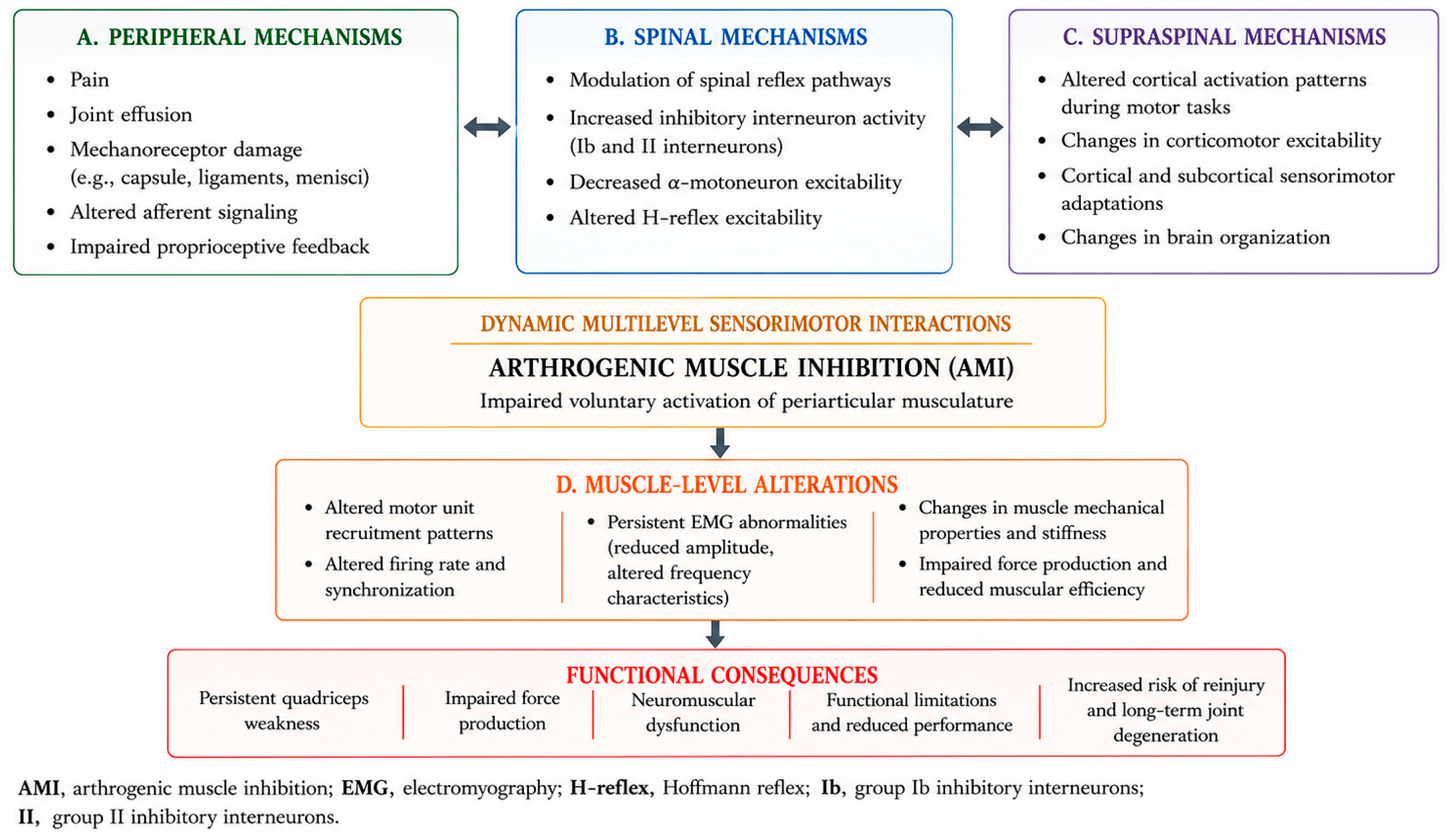
AMI represents a complex neurophysiological response to joint injury, characterized by an inability to fully activate periarticular musculature despite maximal voluntary effort. Rather than being a purely local phenomenon, AMI reflects an integrated response involving peripheral sensory alterations, spinal reflex modulation, and supraspinal adaptations. These mechanisms interact dynamically to produce persistent deficits in muscle activation, particularly affecting the quadriceps following injuries such as ACL injury [
7,
10]. The multilevel and interactive organization of these mechanisms, together with the associated muscle-level alterations and functional consequences, is summarized in
Figure 1.
3.1. Peripheral Mechanisms
Peripheral mechanisms are central to the initiation of AMI. Joint injury leads to disruption of normal afferent signaling due to pain, joint effusion, and damage to mechanoreceptors [
3,
7,
10,
23,
24]. These factors alter sensory input from the joint to the central nervous system, which plays a critical role in regulating motor output. Joint effusion increases intra-articular pressure, stimulating mechanosensitive receptors and eliciting inhibitory reflex responses that reduce alpha motor neuron excitability. Similarly, nociceptive input associated with pain contributes to the activation of inhibitory pathways, further limiting voluntary muscle contraction. Structural damage to joint mechanoreceptors, particularly following ligamentous injury, impairs proprioceptive feedback and disrupts sensorimotor integration [
25]. Importantly, the combined presence of pain and structural damage appears to exert a greater inhibitory effect than either factor alone, underscoring the multifactorial nature of AMI initiation. These peripheral alterations represent the primary trigger for the cascade of neurophysiological adaptations that follow.
3.2. Spinal Mechanisms
At the spinal level, altered afferent input from the injured joint modulates reflex pathways, leading to decreased excitability of alpha motor neurons innervating the quadriceps [
2,
3,
6,
7,
26,
27]. This process is mediated by increased activity of inhibitory interneurons within the spinal cord, which suppress motor output and limit voluntary activation. Changes in spinal reflex excitability have been demonstrated through measures such as the Hoffmann reflex (H-reflex), which reflects the integrity and responsiveness of the monosynaptic reflex arc. Reductions in H-reflex amplitude following joint injury indicate diminished motor neuron excitability and contribute to the persistence of activation deficits. These spinal adaptations likely serve a protective role by reducing joint loading and preventing further injury. However, when prolonged, they contribute to chronic neuromuscular dysfunction. Moreover, spinal inhibition does not occur in isolation but is influenced by both peripheral inputs and descending supraspinal control [
7,
27,
28].
3.3. Supraspinal Mechanisms
Increasing evidence highlights potential supraspinal contributions to persistent neuromuscular dysfunction associated with AMI. Altered afferent input from the injured joint has been associated with functional and structural changes within cortical and subcortical regions involved in motor control and proprioception [
4,
8,
9,
11,
12,
13,
25,
27,
29]. Neurophysiological and neuroimaging studies have demonstrated altered patterns of cortical activation during motor tasks following joint injury and reconstruction, together with changes in corticomotor excitability and brain organization [
8,
9,
11,
12,
13,
27,
29]. These findings support the presence of supraspinal adaptations within the broader sensorimotor response to joint injury.
3.4. Muscle-Level Alterations
The neurophysiological changes associated with AMI ultimately manifest at the level of the muscle. Alterations in motor unit recruitment patterns, firing rates, and synchronization contribute to impaired force production and reduced muscular efficiency [
30,
31]. Electromyographic studies have demonstrated persistent changes in muscle activation patterns following joint injury, including reduced signal amplitude and altered frequency characteristics, reflecting modifications in motor unit behavior. Notably, these abnormalities may persist even when voluntary activation appears to recover, indicating that neuromuscular function remains compromised. In addition to neural factors, changes in the mechanical properties of muscle tissue have been observed [
15,
30,
32,
33]. Altered muscle stiffness during contraction has been reported following anterior cruciate ligament injury, suggesting that neural adaptations influence not only activation but also the intrinsic properties of muscle. These changes may further impair force transmission and functional performance [
31]. Collectively, these findings demonstrate that AMI is not solely a neural phenomenon but also involves secondary adaptations at the muscular level, reinforcing its multifactorial nature [
30,
31,
33].
4. Assessment of Arthrogenic Muscle Inhibition
Assessment of AMI requires distinction between methods that directly estimate voluntary activation failure and techniques that characterize associated neuromuscular or neurophysiological alterations. Voluntary activation techniques, including the central activation ratio (CAR) derived from superimposed electrical stimulation and related interpolated-twitch approaches, provide the most direct experimental assessment of the inability to fully activate the quadriceps voluntarily [
1,
19,
20]. In contrast, electromyography (EMG), Hoffmann reflex (H-reflex) testing, transcranial magnetic stimulation (TMS), electroencephalography (EEG), and functional magnetic resonance imaging (fMRI) assess muscle activation patterns or neural processes that may contribute to, accompany, or result from AMI, but should not be interpreted as interchangeable direct measures of AMI. Accordingly, a multimodal assessment may provide complementary information regarding the mechanisms and functional correlates of quadriceps dysfunction, while preserving the distinction between voluntary activation failure and its associated neurophysiological findings.
4.1. Central Activation Ratio
The Central Activation Ratio (CAR) is among the most commonly utilized methods for evaluating voluntary quadriceps activation. CAR is typically measured using the superimposed burst technique, in which an electrical stimulus is applied during a maximal voluntary contraction. If additional force is generated following stimulation, incomplete voluntary activation is assumed to be present. Conceptually, CAR reflects the proportion of the muscle’s maximal activation capacity achieved voluntarily. Lower CAR values indicate greater voluntary activation failure and therefore provide a relatively direct estimate of the neural activation deficit that characterizes AMI [
1,
19,
20]. Reduced CAR values have been reported following ACL injury and reconstruction, indicating that voluntary activation failure may contribute to persistent quadriceps weakness in at least a subset of patients. Voluntary activation measurements can also help distinguish the contribution of neural activation failure from structural muscular factors contributing to weakness. Importantly, persistent quadriceps weakness has also been reported in the presence of relatively preserved voluntary activation, indicating that weakness after ACL reconstruction cannot be attributed to AMI alone and may also reflect structural muscular adaptations such as atrophy [
1,
17,
18]. Interpretation of CAR requires consideration of both methodological and participant-related factors. A CAR value below 95% has commonly been used in the knee-injury literature as an operational threshold for incomplete voluntary quadriceps activation; however, this value should not be interpreted as a universally validated pathological diagnostic cut-off for AMI. CAR estimates may be influenced by the stimulation protocol and calculation method, testing position and joint angle, isolation of the knee extensors, and the participant’s ability to generate a true maximal voluntary contraction. These methodological differences can complicate comparisons across studies and individuals. Accordingly, CAR values should be interpreted within the context of a standardized testing protocol and the broader clinical assessment rather than as an isolated diagnostic criterion [
20,
34].
4.2. Electromyography
Electromyography (EMG) is frequently used to evaluate muscle activation patterns and neuromuscular control following joint injury. Surface EMG allows assessment of activation timing, amplitude, and coordination between muscle groups during functional and isometric tasks. Unlike voluntary activation techniques such as CAR, EMG does not directly quantify the inability to achieve maximal voluntary muscle activation and therefore should not be considered a direct measure of AMI. Altered quadriceps EMG activity has been reported following ACL injury and reconstruction, including changes in activation amplitude, timing, and recruitment strategies. Such abnormalities may persist despite restoration of joint stability and may provide complementary information regarding ongoing neuromuscular dysfunction. EMG analysis has additionally demonstrated altered recruitment of the hamstrings and gastrocnemius musculature, which may represent compensatory neuromuscular strategies in the presence of quadriceps dysfunction [
15,
30,
32].
4.3. Hoffmann Reflex and Spinal Excitability
Assessment of spinal reflex excitability through the Hoffmann reflex (H-reflex) has provided important insights into the spinal mechanisms potentially associated with AMI. The H-reflex serves as an electrophysiological analogue of the monosynaptic stretch reflex and reflects alpha motor neuron excitability of the spinal reflex pathway. Unlike CAR or other voluntary activation techniques, however, the H-reflex does not directly quantify voluntary activation failure and should therefore be interpreted as a mechanistic measure rather than a direct measure of AMI. Experimental knee effusion models have demonstrated alterations in quadriceps spinal reflex excitability following joint distension, supporting the involvement of altered afferent input and spinal inhibitory mechanisms in AMI [
3,
28]. Together, these findings support spinal reflex modulation as one component of the neurophysiological response associated with AMI, while emphasizing that changes in H-reflex excitability should not be equated with voluntary activation failure itself [
3,
28].
4.4. Cortical and Neurophysiological Assessment
Neurophysiological and neuroimaging techniques can characterize supraspinal adaptations associated with ACL injury and reconstruction [
8,
9,
27]. Electroencephalography (EEG), transcranial magnetic stimulation (TMS), and functional magnetic resonance imaging (fMRI) provide complementary information regarding cortical activity, corticospinal excitability, and brain activation patterns, respectively; however, these techniques do not directly quantify voluntary activation failure and should therefore not be considered direct measures of AMI. EEG studies have reported increased frontal theta activity during force-control tasks in individuals following ACL reconstruction, a pattern interpreted as reflecting greater attentional demand during motor performance [
11]. TMS has been used to examine corticospinal and corticomotor excitability in both experimental joint-effusion models and individuals with ACL-related dysfunction [
29,
35,
36]. fMRI studies and neuroimaging reports have likewise identified altered patterns of brain activation associated with knee movement and sensorimotor processing following ACL injury or reconstruction [
8,
9,
12,
37]. Collectively, these methods provide complementary evidence of supraspinal adaptations following joint injury, but the clinical significance, temporal evolution, and relationship of these findings to directly measured voluntary activation failure remain incompletely established. At the supraspinal level, measurable parameters include motor threshold, motor-evoked potential amplitude, and intracortical inhibition assessed by TMS, whereas fMRI characterizes task-related changes in regional blood-oxygen-level-dependent (BOLD) activation. Although these measures can identify cortical and corticospinal alterations following ACL injury or reconstruction, validated quantitative thresholds that distinguish adaptive from maladaptive neuroplasticity have not been established. Their interpretation therefore remains dependent on the direction and context of the observed change and its relationship with motor or functional outcomes [
27,
35,
37].
The principal methods used to assess voluntary activation failure and associated neurophysiological alterations are summarized in
Table 1.
The assessment techniques summarized in
Table 1 interrogate different components of AMI-related neuromuscular dysfunction and should therefore be regarded as complementary rather than interchangeable. Voluntary activation techniques such as CAR estimate activation failure, whereas EMG, H-reflex, TMS, EEG, and fMRI characterize associated muscular, spinal, corticospinal, or cortical alterations; their findings should therefore be interpreted according to the specific physiological construct being assessed [
19,
20,
22].
5. Clinical Manifestations and Functional Correlates
Voluntary activation failure may contribute to persistent quadriceps dysfunction following joint injury, but the clinical phenotype observed after ACL injury and reconstruction is multifactorial. Strength deficits, altered force regulation, changes in muscle recruitment, and biomechanical asymmetries may reflect varying contributions from AMI, muscle atrophy, structural muscular adaptations, pain, altered sensorimotor input, and compensatory motor strategies. Accordingly, the functional abnormalities discussed in this section are considered within the broader clinical expression of AMI and post-injury neuromuscular dysfunction, while recognizing their multifactorial nature.
5.1. Quadriceps Weakness and Activation Failure
Persistent quadriceps weakness is common following ACL injury and reconstruction but should not be considered synonymous with AMI. Reduced force-generating capacity may reflect multiple interacting factors, including voluntary activation failure, muscle atrophy, and other structural or neuromuscular adaptations. Voluntary activation deficits have been reported following ACL injury and reconstruction and may contribute to persistent weakness in some individuals. However, quadriceps weakness can also remain evident when voluntary activation is relatively preserved, indicating an important contribution from muscular factors. The relative contributions of neural activation failure and structural muscle impairment therefore appear to vary between individuals and across stages of recovery [
1,
2,
17,
18].
5.2. Altered Motor Control and Force Regulation
Alterations in motor control and force regulation have been reported following ACL injury and may coexist with quadriceps activation and strength deficits. Individuals with ACL injury can demonstrate impaired regulation of submaximal force output and reduced adaptability of quadriceps force production [
33,
38]. Ward et al. identified deficits in quadriceps force control following ACL injury, while Hollman et al. reported altered variability and complexity of knee muscle force output, findings that may reflect broader disturbances in sensorimotor regulation rather than voluntary activation failure alone [
33,
38]. These findings provide complementary insight into the quality and adaptability of motor output associated with post-injury neuromuscular dysfunction.
Altered neuromuscular coordination has also been observed during multidirectional force-control tasks. Changes in the relative contribution of the quadriceps, hamstrings, and gastrocnemius may represent compensatory recruitment strategies in response to altered knee function [
32]. Although such strategies may help maintain task performance, their relationship to movement efficiency, joint loading, and long-term clinical outcomes remains incompletely established.
5.3. Biomechanical and Gait Alterations
Biomechanical alterations are frequently observed after ACL injury and reconstruction and may coexist with persistent strength and neuromuscular deficits. Individuals with ACL deficiency or following ACL reconstruction have demonstrated asymmetries in gait mechanics, including reduced knee flexion and diminished knee extensor moments during weight-bearing activities [
14,
39]. Altered muscle activation and coordination patterns have also been reported during functional tasks, indicating that restoration of mechanical joint stability does not necessarily normalize movement strategies [
15,
32].
These biomechanical findings should be interpreted as associated functional adaptations rather than direct manifestations of AMI. Persistent asymmetries may alter the distribution of joint loading and movement demands; however, the extent to which AMI independently contributes to these patterns, or whether such patterns directly increase the risk of subsequent injury or long-term joint degeneration, remains uncertain [
14,
15,
32,
39,
40].
5.4. “Copers” and “Non-Copers”
The distinction between “copers” and “non-copers” following ACL injury illustrates the heterogeneity of functional adaptation after ligament injury. “Copers” are individuals who maintain satisfactory functional stability despite ACL deficiency, whereas “non-copers” experience recurrent episodes of instability or functional limitation [
16,
41]. Differences between these groups cannot be explained by mechanical joint laxity alone and have been associated with differences in neuromuscular function, muscle activation, and movement strategies [
16,
41].
Importantly, coping status should not be interpreted as a direct clinical measure of AMI. Rather, the “coper” or “non-coper” paradigm illustrates how multiple factors, including neuromuscular control, muscular capacity, sensorimotor adaptation, and compensatory movement strategies, may influence functional performance after ACL injury. The ability of some individuals to maintain functional stability despite ligament deficiency further emphasizes that structural impairment, quadriceps weakness, voluntary activation failure, and functional disability are related but non-equivalent phenomena.
5.5. Functional Outcomes and Return to Sport
Persistent neuromuscular deficits following ACL injury and reconstruction may have important implications for long-term functional recovery and return to sport [
40]. Even after restoration of mechanical stability and completion of rehabilitation, alterations in muscle activation, force control, movement symmetry, and sensorimotor function may persist [
12,
13,
15,
38,
40]. These residual deficits can affect movement efficiency and athletic performance and highlight the importance of evaluating neuromuscular recovery alongside conventional measures of strength and joint stability.
Persistent biomechanical asymmetries and altered neuromuscular control have also been discussed in relation to subsequent injury and long-term joint health following ACL injury. However, these outcomes are influenced by multiple interacting factors, and the specific contribution of AMI remains incompletely understood. A broader assessment of neuromuscular function may therefore provide clinically relevant information during rehabilitation and return-to-sport decision-making.
6. Clinical and Functional Implications of Neuroplasticity
Central nervous system adaptations following ACL injury may have important implications for motor control and functional recovery. Neurophysiological and neuroimaging findings suggest that individuals following ACL injury or reconstruction may recruit different cortical strategies during motor tasks compared with uninjured individuals [
8,
9,
11,
12]. Increased reliance on attentional and cognitive resources during tasks that would normally require relatively automatic motor control may represent a compensatory response to altered sensorimotor input [
11,
12].
Whether these neuroplastic adaptations should be interpreted as beneficial compensation, maladaptive reorganization, or a combination of both remains uncertain [
8,
9,
27]. Alternative cortical recruitment strategies may help preserve task performance despite altered peripheral sensory information; however, greater cognitive dependence could potentially reduce movement efficiency or adaptability under complex or rapidly changing conditions. Importantly, most available evidence is associative and does not establish that altered cortical activation independently causes persistent functional impairment or subsequent injury.
The presence of bilateral neuromuscular deficits following unilateral ACL injury provides additional evidence that post-injury adaptations are not restricted to the injured joint. Alterations in force control and muscle activation have been identified in the involved and contralateral extremities, supporting the presence of broader sensorimotor adaptations [
12,
38]. These bilateral findings are consistent with the involvement of central nervous system mechanisms in the broader neuromuscular adaptations associated with AMI.
From a clinical perspective, these findings suggest that recovery of muscle strength and mechanical stability may not fully capture restoration of neuromuscular function. Persistent alterations in force regulation, muscle activation strategies, and neuromuscular adaptability have been reported following ACL injury or reconstruction [
15,
33,
38]. Assessment of movement quality, force control, sensorimotor performance, and performance under cognitively demanding conditions may therefore provide complementary information alongside conventional strength-based measures. However, the prognostic significance and clinically meaningful thresholds of these measures remain incompletely established.
7. Rehabilitation Strategies and Clinical Implications
Recognition of Arthrogenic Muscle Inhibition as a multilevel neurophysiological process has important implications for rehabilitation following joint injury. Traditional rehabilitation approaches have often focused primarily on restoring muscle strength and joint stability [
5,
10,
19]. However, addressing muscle atrophy alone may be insufficient when voluntary activation failure and altered sensorimotor control remain present. Because the mechanisms associated with AMI involve interactions among peripheral, spinal, and supraspinal processes, rehabilitation may benefit from combining interventions directed toward restoration of quadriceps activation and strength with strategies addressing pain, joint effusion, sensorimotor control, and movement quality [
5,
6,
7,
10,
19]. The level of clinical evidence supporting individual interventions varies, and established treatments should therefore be distinguished from emerging approaches that remain under investigation.
7.1. Early Phase: Pain, Effusion, and Quadriceps Activation
Because pain and joint effusion can sustain inhibitory joint-related input, their early management is an important component of AMI rehabilitation [
23,
24]. Neuromuscular electrical stimulation (NMES) has been widely used to facilitate quadriceps activation and counteract activation failure. By externally recruiting motor units, NMES can augment muscle contraction when voluntary activation is impaired and may be particularly useful during the early postoperative period, when AMI is frequently pronounced [
19,
42]. Cryotherapy has also been investigated as an adjunctive intervention, with potential effects on pain and inhibitory afferent signaling. Some studies suggest that cryotherapy can transiently improve quadriceps activation in the presence of AMI, although its effects should be considered primarily short-term and adjunctive rather than a substitute for active rehabilitation [
19]. Together, interventions aimed at reducing inhibitory joint-related input while facilitating quadriceps activation may provide an appropriate foundation for subsequent progressive rehabilitation.
7.2. Progressive Strengthening and Blood-Flow Restriction
Progressive resistance exercise remains a fundamental component of rehabilitation following ACL injury and reconstruction. As voluntary quadriceps activation improves and pain and joint effusion decrease, progressively increasing mechanical loading is required to address persistent deficits in muscle strength and muscle mass. However, substantial quadriceps weakness and atrophy may limit the ability to tolerate conventional high-load resistance exercise, particularly during the earlier stages of postoperative rehabilitation.
Blood-flow restriction (BFR) training has emerged as a potential adjunct to conventional strengthening in this setting. By partially restricting arterial inflow and venous outflow during low-load resistance exercise, BFR aims to promote muscular adaptations while using lower external loads. Following ACL reconstruction, clinical studies have reported improvements in quadriceps strength and physical function when BFR was incorporated into rehabilitation [
43]. A recent systematic review and meta-analysis also reported potential benefits for knee extensor and flexor isokinetic strength and quadriceps cross-sectional area; however, no significant improvements were identified for isometric knee extensor strength or knee-related function, and the overall certainty of evidence was rated as very low [
44]. BFR should nevertheless be applied with appropriate patient screening, individualized cuff pressure, and clinical supervision, particularly in individuals with relevant cardiovascular, peripheral vascular, or thromboembolic risk [
45]. Although serious adverse events appear uncommon in appropriately selected patients, safety reporting in the ACL rehabilitation literature remains limited [
44,
45].
7.3. Sensorimotor and Neuromuscular Retraining
Beyond restoration of quadriceps activation and strength, rehabilitation should address sensorimotor control and movement quality. Altered motor control, impaired force regulation, and compensatory movement strategies may persist despite recovery of isolated strength measures. Consequently, rehabilitation should incorporate exercises aimed at improving coordination, movement adaptability, proprioception, and dynamic joint stability. Perturbation training and proprioceptive exercises have been proposed to enhance sensorimotor integration and neuromuscular responsiveness during functional tasks [
3,
28]. By exposing patients to progressively challenging and less predictable movement conditions, these approaches may promote more adaptable motor strategies and improve the integration of sensory information during movement. Task-specific rehabilitation emphasizing dynamic movement control may therefore complement strength restoration in addressing the broader neuromuscular alterations associated with AMI.
7.4. Neurocognitive, Dual-Task, and Technology-Assisted Rehabilitation
Emerging evidence regarding cortical reorganization following ACL injury has increased interest in rehabilitation strategies that incorporate neurocognitive and visual-motor demands [
8,
9,
27]. Conventional rehabilitation is frequently performed under predictable conditions, whereas sport requires rapid integration of visual information, attention, decision-making, and motor responses. Accordingly, the progressive incorporation of dual-task and visual-cognitive challenges may help bridge the gap between controlled rehabilitation exercises and the more complex demands encountered during sport [
46]. Visual feedback and technology-assisted approaches may provide additional opportunities to challenge sensorimotor control and movement quality. Visual biofeedback has been investigated following ACL reconstruction, with reported improvements in balance-related outcomes [
47]. Virtual-reality-based and other technology-assisted interventions are also being explored as methods of providing interactive, task-specific, and progressively challenging rehabilitation environments. However, the clinical evidence supporting these approaches remains heterogeneous, and their specific effects on AMI or voluntary activation failure have not been established. Digital rehabilitation platforms and wearable technologies may further support remote supervision, objective monitoring, feedback, and individualized progression during ACL rehabilitation. Recent evidence suggests that digitally supported rehabilitation can achieve functional outcomes comparable with conventional approaches in selected settings, although substantial variation exists among technologies and rehabilitation protocols [
48]. These technologies should therefore be considered complementary tools within rehabilitation rather than established treatments for AMI itself.
7.5. Return to Sport and Individualized Rehabilitation
Persistent deficits in voluntary activation, motor control, and movement quality may remain present even after restoration of mechanical stability and apparent recovery of muscle strength, with important implications for return-to-sport decision-making [
12,
13,
15,
18]. Consequently, assessment during the later stages of rehabilitation should extend beyond time-based criteria and isolated strength measures to consider neuromuscular control, movement quality, task-specific performance, and the ability to respond to progressively complex motor demands. Rehabilitation progression should also account for the heterogeneity of AMI and post-injury neuromuscular recovery. The relative contributions of activation failure, muscular impairment, sensorimotor dysfunction, and compensatory motor strategies may differ among individuals and throughout rehabilitation. An individualized approach may therefore be appropriate, with rehabilitation priorities adapted according to the predominant deficits identified during clinical and functional assessment [
17,
18].
Overall, contemporary rehabilitation should recognize AMI as a multilevel neurophysiological phenomenon requiring a multidimensional approach. Integration of strategies targeting quadriceps activation and strength, sensorimotor control, movement quality, and, where appropriate, neurocognitive demands may provide a more comprehensive framework for functional recovery and return to sport.
For clinical clarity, the rehabilitation approaches discussed above are summarized in
Table 2 as a phase-based framework organized according to predominant rehabilitation targets, while recognizing that progression should remain individualized rather than determined exclusively by time from injury or surgery.
8. Current Controversies and Future Perspectives
Despite substantial advances in the understanding of AMI, important questions remain regarding its mechanisms, clinical significance, and optimal management.
8.1. Neural and Muscular Contributions to Persistent Weakness
One of the principal controversies concerns the relative contribution of voluntary activation failure, muscle atrophy, and other structural or neuromuscular adaptations to persistent quadriceps dysfunction following joint injury [
1,
17,
18,
49]. Longitudinal observations after ACL reconstruction indicate that voluntary activation may improve while substantial strength deficits persist, suggesting an increasing contribution of muscular hypotrophy during recovery [
17]. At approximately 6 months after reconstruction, quadriceps cross-sectional area explained substantially more variance in strength than CAR, further illustrating that persistent weakness cannot be attributed uniformly to activation failure alone [
49]. These findings do not, however, establish a fixed phase-specific trajectory or a reciprocal causal relationship between neural inhibition and muscle atrophy. Although reduced voluntary activation and altered neural drive have been demonstrated following ACL injury and reconstruction, persistent weakness may also occur when voluntary activation is relatively preserved, indicating that AMI and structural muscular impairment can contribute to recovery to varying degrees [
1,
17,
18,
49].
The relative importance of these mechanisms may also change throughout rehabilitation, but their temporal interaction remains incompletely characterized. Neural inhibition may be particularly relevant during periods of pronounced pain, effusion, and impaired voluntary activation, whereas muscle atrophy and morphological adaptations may become increasingly relevant with prolonged disuse and altered loading. However, current evidence does not support a uniform temporal sequence applicable to all patients. Longitudinal studies combining direct measures of voluntary activation with muscular, spinal, and supraspinal assessments are needed to determine how these mechanisms interact across different stages of recovery.
8.2. Interpretation and Temporal Evolution of Supraspinal Adaptations
Another unresolved question concerns the functional significance of supraspinal adaptations associated with AMI. Neuroimaging and electrophysiological studies have demonstrated altered cortical activation, corticomotor excitability, and sensorimotor processing following ACL injury and reconstruction [
8,
9,
11,
12,
13,
38]. However, whether these changes primarily represent beneficial compensation, maladaptive reorganization, or a combination of both remains uncertain. Increased recruitment of cortical and cognitive resources may initially represent an adaptive response that helps maintain motor performance in the presence of altered afferent input and impaired neuromuscular control. Conversely, persistent dependence on greater conscious control may be less efficient when movement must be rapid, automatic, or performed under complex environmental demands. The interpretation of neuroplasticity should therefore remain context-dependent: cortical reorganization is not inherently pathological, and its clinical significance may depend on the stage of recovery, task demands, and the individual’s ability to regain efficient and adaptable motor control [
11,
12,
13,
38].
The temporal development of supraspinal adaptations associated with AMI also remains incompletely understood. Experimental models of acute knee joint effusion have not consistently demonstrated immediate changes in corticomotor excitability, suggesting that alterations at the supraspinal level may not emerge uniformly or simultaneously with the peripheral and spinal responses to joint injury. However, these findings should not be interpreted as establishing a fixed temporal progression from peripheral inhibition to central reorganization. The development and persistence of supraspinal adaptations are likely influenced by multiple factors, including the nature and duration of altered afferent input, pain, changes in motor behavior, rehabilitation exposure, and repeated use of compensatory movement strategies. Longitudinal studies incorporating serial neurophysiological assessments are therefore needed to determine when these adaptations emerge, whether they resolve or persist during recovery, and how their evolution relates to voluntary activation and functional performance.
Another clinically relevant issue is the persistence of neuromuscular abnormalities despite successful restoration of mechanical stability following ACL reconstruction. Deficits in force regulation, movement coordination, muscle activation, and sensorimotor control may persist during later stages of recovery and even following return to sport [
14,
15,
32]. This dissociation between mechanical restoration and neuromuscular recovery reinforces the concept that AMI and its associated adaptations cannot be fully characterized by joint stability or isolated strength measures alone.
These persistent abnormalities also raise important questions regarding how recovery is assessed and how readiness for return to sport is determined. Conventional strength and functional measures remain essential, but complementary assessment of voluntary activation, movement quality, force regulation, and sensorimotor control may provide a more comprehensive representation of neuromuscular recovery. Future studies should investigate whether combining these measures with neurophysiological assessments can identify clinically meaningful recovery profiles and improve individualized rehabilitation decision-making.
8.3. Future Research and Individualized Recovery
Future research should prioritize prospective longitudinal studies with repeated assessments across clinically meaningful stages of recovery rather than relying predominantly on isolated cross-sectional measurements. Multimodal protocols combining direct measures of voluntary activation with electrophysiological, muscular, biomechanical, functional, and, where feasible, neuroimaging assessments may help clarify how peripheral, spinal, supraspinal, and structural adaptations evolve and interact over time [
8,
9,
11,
12,
13,
17,
18,
37]. Future interventional studies should also account for clinically relevant sources of heterogeneity, including injury stage, postoperative status, baseline activation failure, and rehabilitation exposure, to determine whether treatment effects differ across distinct recovery profiles. A further priority is the development of individualized rehabilitation strategies based on the predominant mechanisms contributing to dysfunction. Patients in whom voluntary activation failure remains prominent may require different therapeutic priorities from those whose limitations are predominantly related to muscle atrophy, sensorimotor dysfunction, or persistent alterations in motor control [
17,
18]. Repeated assessment across rehabilitation may therefore be more informative than reliance on a single measure or fixed time-based progression.
Emerging technologies may facilitate this more individualized approach. Wearable sensors, digital rehabilitation platforms, and advanced movement-analysis systems offer opportunities for repeated monitoring of motor performance and rehabilitation progress outside traditional laboratory environments [
48]. Neurophysiological techniques such as transcranial magnetic stimulation, electroencephalography, and functional neuroimaging may further contribute to characterization of central adaptations associated with AMI [
8,
9,
11,
12,
13,
37]. Data-driven and artificial-intelligence-based approaches may eventually assist in integrating multidimensional clinical and neurophysiological information and identifying patient-specific recovery patterns. However, their role in AMI remains exploratory, and prospective validation is required before such approaches can inform routine clinical decision-making.
Ultimately, progress in AMI research will depend not only on identifying additional neurophysiological abnormalities but also on determining which abnormalities are clinically meaningful and modifiable through rehabilitation. Establishing standardized assessment approaches and clinically relevant thresholds would facilitate comparison across studies and may ultimately support more individualized rehabilitation and return-to-sport decision-making.
9. Discussion
The evidence synthesized in this review supports AMI as a multilevel neurophysiological phenomenon in which altered joint-related afferent input interacts with spinal reflex pathways and supraspinal adaptations to impair voluntary quadriceps activation following knee injury [
3,
4,
5,
6,
7,
8,
9,
10,
11,
12,
13]. Although ACL injury and reconstruction provide the principal clinical model examined in the available literature, the mechanisms underlying AMI extend beyond structural ligament injury and reflect broader interactions between the injured joint and the nervous system. This framework helps explain why restoration of mechanical stability or muscle strength alone may not fully represent neuromuscular recovery.
An important implication of the present synthesis is that AMI should be considered within the broader context of post-injury quadriceps dysfunction without being equated with every accompanying neuromuscular abnormality. Voluntary activation failure represents the defining feature of AMI, whereas muscle atrophy, altered force regulation, biomechanical asymmetry, changes in spinal or corticospinal excitability, and cortical reorganization provide complementary information regarding its mechanisms, associated adaptations, and functional expression [
1,
2,
6,
7,
8,
9,
10,
11,
12,
13,
17,
18,
23,
24]. Integrating these levels of evidence provides a more complete interpretation of persistent dysfunction than reliance on any single physiological or functional measure.
From a clinical perspective, this multilevel framework supports assessment and rehabilitation strategies that extend beyond isolated strength restoration. Direct assessment of voluntary activation can help characterize activation failure, while electrophysiological, biomechanical, and functional measures may provide additional information regarding the mechanisms and consequences accompanying AMI [
1,
3,
8,
9,
11,
12,
15,
17,
18,
28,
29,
30,
32,
35,
36,
37,
38,
39,
40]. Similarly, rehabilitation may require different priorities across recovery, ranging from management of pain and effusion and restoration of quadriceps activation to progressive strengthening, sensorimotor retraining, and later integration of complex motor and neurocognitive demands [
3,
8,
9,
19,
23,
24,
27,
28,
42,
43,
44,
46,
47,
48]. Importantly, the evidence supporting these interventions is not uniform, and emerging approaches should complement rather than replace established rehabilitation principles.
Relation to Previous Reviews. Previous reviews have examined AMI following ACL injury from complementary perspectives, including its neurophysiological mechanisms and clinical consequences, the effectiveness of physiotherapeutic interventions, and the assessment of persistent neuromuscular deficits in relation to return-to-sport decision-making [
20,
21,
22]. The present review extends this literature by integrating peripheral, spinal, supraspinal, muscular, functional, and rehabilitation evidence within a single clinically oriented framework. Particular emphasis is placed on distinguishing direct assessment of voluntary activation from mechanistic or associated neurophysiological measures, linking these levels of evidence to clinical assessment, and organizing rehabilitation according to predominant recovery targets. This integrated approach may facilitate translation of neurophysiological findings into clinically meaningful assessment and rehabilitation strategies while also highlighting areas in which current evidence remains uncertain.
10. Limitations
Several limitations should be considered when interpreting this narrative review. First, although a structured literature search was performed and supplemented during manuscript revision, the narrative design did not involve a formal systematic-review protocol, quantitative synthesis, risk-of-bias assessment, or certainty-of-evidence grading. Consequently, selection bias cannot be completely excluded, and the relative strength of evidence supporting individual mechanisms and interventions should be interpreted with appropriate caution. Because no formal certainty-of-evidence assessment was performed, intervention-specific evidence grades were not assigned; where published systematic reviews reported certainty ratings, these were described narratively in the relevant sections. In addition, retrieval was limited to PubMed and Scopus and to the predefined search terminology, and relevant studies indexed elsewhere or described using different terminology may therefore have been missed.
Second, substantial methodological heterogeneity exists across the AMI literature. Studies differ in patient populations, injury and postoperative stages, rehabilitation exposure, and methods used to characterize neuromuscular dysfunction. In particular, voluntary activation techniques, electrophysiological measures, neuroimaging findings, biomechanical assessments, and functional outcomes evaluate different components of the post-injury neuromuscular response and should not be considered interchangeable. This heterogeneity limits direct comparison across studies and complicates the establishment of standardized clinical thresholds. The peripheral–spinal–supraspinal framework used in this review should therefore be interpreted as an integrative conceptual model rather than a fixed pathway or single mechanistic explanation applicable uniformly to all patients or stages of recovery.
Third, much of the available evidence derives from individuals with ACL injury or following ACL reconstruction. Although this provides a well-characterized clinical model for investigating AMI, the extent to which findings can be generalized to other joint injuries or musculoskeletal conditions remains uncertain. Furthermore, the predominance of cross-sectional studies in several areas, particularly those investigating supraspinal adaptations, limits conclusions regarding the temporal evolution and causal significance of observed neurophysiological changes. Accordingly, associations between voluntary activation failure, spinal or supraspinal alterations, muscle atrophy, and functional outcomes should not be interpreted as establishing a causal sequence.
Finally, only English-language full-text publications were considered, introducing the possibility of language bias. Emerging rehabilitation approaches and technologies are also supported by variable and, in some cases, limited evidence; therefore, their inclusion in this review should be interpreted as reflecting developing areas of investigation rather than established standards of care.
11. Conclusions
Arthrogenic Muscle Inhibition is a complex, multilevel neurophysiological phenomenon that can substantially influence quadriceps activation and functional recovery following knee injury, particularly after ACL injury and reconstruction [
3,
4,
5,
6,
7,
10]. Current evidence supports interactions among altered joint-related afferent input, spinal inhibitory mechanisms, supraspinal adaptations, and secondary muscular changes [
3,
4,
5,
6,
7,
8,
9,
10,
11,
12,
13,
17,
18,
27,
29,
37,
38]. AMI should therefore be considered within the broader context of post-injury neuromuscular dysfunction while remaining specifically defined by impaired voluntary muscle activation [
5,
7,
10].
Recognition of this multilevel framework has important implications for both assessment and rehabilitation. Direct measures of voluntary activation can help characterize AMI, whereas electrophysiological, neuroimaging, biomechanical, and functional assessments provide complementary information regarding its mechanisms, associated adaptations, and clinical expression [
6,
7,
8,
9,
10,
11,
12,
13,
14,
15,
27,
29,
33,
37,
38]. Rehabilitation should similarly address the predominant deficits identified throughout recovery, integrating restoration of quadriceps activation and strength with sensorimotor and functional retraining and, where appropriate, progressively complex neurocognitive demands [
5,
6,
10,
17,
18,
19,
28,
33,
49].
Important uncertainties remain regarding the temporal evolution of AMI, the clinical significance of neuroplastic adaptations, and the relative contribution of neural and structural muscular factors to persistent dysfunction [
8,
9,
11,
12,
13,
17,
18,
27,
29,
35,
36,
37]. Future longitudinal and multimodal studies are needed to clarify these relationships, establish clinically meaningful assessment thresholds, and determine whether mechanism-informed rehabilitation can improve functional recovery. A more precise understanding of AMI as an interaction among joint-related sensory alterations, neural activation failure, central adaptation, and secondary muscular changes may ultimately support more individualized assessment and rehabilitation following joint injury [
4,
7,
8,
9,
10,
11,
12,
13,
19,
27].





 ,
,