Article

Kinematic and Muscle Activity Differences During Change of Direction in Female Second Division Football Players Following ACL Reconstruction Compared with Uninjured Controls


by
Loreto Ferrández-Laliena
1,*,
Lucía Vicente-Pina
1,
Rocío Sánchez-Rodríguez
1,
Mira Ambrus
2,
Sofía Monti-Ballano
1,
Julián Müller-Thyssen-Uriarte
1,
César Hidalgo-García
1,
José Miguel Tricás-Moreno
1 and
María Orosia Lucha-López
1,*
1
Unidad de Investigación en Fisioterapia, Spin off Centro Clínico OMT-E Fisioterapia SLP, Universidad de Zaragoza, Domingo Miral s/n, 50009 Zaragoza, Spain
2
Research Center for Sport Physiology, Hungarian University of Sports Sciences, 1123 Budapest, Hungary
*
Authors to whom correspondence should be addressed.
Appl. Sci. 2026, 16(13), 6631; https://doi.org/10.3390/app16136631
Submission received: 15 May 2026 / Revised: 18 June 2026 / Accepted: 26 June 2026 / Published: 2 July 2026

Featured Application

This study supports the importance of integrating three-dimensional kinematics with muscle activity analysis during sport-specific functional tasks that replicate anterior cruciate ligament injury mechanisms to improve the identification of compensatory motor control strategies after anterior cruciate ligament reconstruction and provide a more robust basis for return to play decision-making, specific in female football players, thereby reducing the risk of secondary anterior cruciate ligament injury.

Abstract

The high rate of reinjury after anterior cruciate ligament reconstruction (ACLR), persistent sex-related disparity, and increased susceptibility during transitional stages in female football players highlight the need for more specific strategies to identify biomechanical parameters associated with valgus collapse. This study aimed to compare three-dimensional knee kinematic and synchronized electromyography (EMG) muscle activity of the medial and lateral thigh muscle complexes during a change of direction (COD) task, between ACLR and healthy controls players. A cross-sectional case–control study was conducted with 26 under-23 semiprofessional female football players (22.89 ± 2.68 years), divided into ACLR (n = 13) and control (n = 13) groups. The maximum and minimum peaks and range of knee angular velocity across three planes, along with the average and peak electromyography (EMG) muscle activity of the Biceps Femoris (BF), Semitendinosus (ST), Vastus Medialis (VM), and Vastus Lateralis (VL), were recorded during the preparation and load phases. Between-group differences were assessed using independent t-tests or Mann–Whitney U tests. Statistical significance after Holm–Bonferroni correction was established at p-Holm < 0.05. ACLR players demonstrated significant increased knee valgus angular velocity, alongside 4% reduced average ST muscle activity and 27% diminished peak BF muscle activity during the load phase, compared to controls. These findings indicate altered knee kinematic and muscle activity patterns during COD in ACLR players, suggesting persistent long-term functional adaptations in female football players after ACLR.

1. Introduction

The anterior cruciate ligament (ACL) injury is one of the most severe injuries in female football, due to its highly incidence, burden, rate of short-term reinjury and long-term impact on performance. In elite players, ACL injury incidence reaches 0.2 injuries per 1000 h of exposure, corresponding to 0.7 injuries per squad and season [1,2,3]. This incidence increases to 0.3–0.4 injuries per 1000 h of exposure in semiprofessional players, particularly during transitional stages in which the performance functional demands may exceed the functional capacity of the players [2,4]. In contrast, male players show lower ACL injury rates during comparable stages of development, 0–0.2 injuries per 1000 h of exposure [2]. This sex-related disparity highlights the necessity of more specific strategies to improve the identification of biomechanical parameters associated with ACL injury mechanism in female players during this key stage of performance development [5].
Although anterior cruciate ligament reconstruction (ACLR) is often recommended to restore knee stability, only 69% of pivoting sports players, such as football players, return to play after surgical intervention, and just 60% of them return to preinjury levels of competition [5,6,7,8]. Moreover, reinjury risk remains substantial, ranging from 24 to 30% within the first two years after return to play, increasing in female players injured during key academy stages to approximately 35% [5,6,9]. These persistent reinjury rates suggest that current clinical assessment post ACLR may not adequately detect biomechanical deficits required in high-intensity performance football environment [5,6]. Therefore, the integration of synchronized kinematic and muscle activity assessments during sport-specific tasks that replicate ACL injury mechanisms may improve the identification of dysfunctional compensatory strategies and provide a more robust basis for return-to-play decision-making, thereby reducing the risk of secondary ACL injury [3,6].
Approximately 59–64% of ACL injuries in female football occur via a non-contact mechanism associated with a proprioceptive motor control failure, therefore theoretically preventable by the identification of biomechanical parameters related to injury mechanisms [4,5,8,10,11]. However, most accepted return-to-play tests are based on hop tests or isometric strength measures, despite evidence that nearly 70% of ACL injuries are produced during high-speed change of direction (COD) tasks [3,5,6,10]. Epidemiological research indicates that these injuries most frequently occur when defending, in situations in which players must execute rapid and unanticipated reactions to an opponent’s movement [10]. Under these conditions, COD imposes highly multiplanar kinematic demands on the knee that may exceed the tensile capacity of the ACL, especially in players who exhibit dysfunctional patterns such as valgus collapse [3,11,12]. Therefore, the integration of muscle activity analysis, by electromyography (EMG), synchronized with three-dimensional knee kinematic analysis during COD tasks as clinical evaluation post ACLR, provides a new opportunity to identify residual neuromuscular deficits and compensatory strategies in otherwise conventional clinical assessment [3,5,6].
The valgus collapse mechanism, defined by knee abduction and excessive transverse motion, is widely recognized as a primary risk factor for ACL injury [10,11,13,14]. It is described as a sequence of interrelated multiplanar kinematic adjustments at lower knee flexion angles and joint position associated with increased ACL demands [11,15,16,17]. On the coronal plane, knee abduction involves a medialization of the knee relative to the lower-limb load axis, increasing compressive forces across the lateral compartment while simultaneously placing tensile stress on the medial stabilizing structures, such as ACL [11,16]. This kinematic perspective joined with increased knee transversal range of movement (ROM) facilitates pivot-shift development [11,18,19,20]. This intra-articular mechanism produces the medial and rotational displacement of lateral femoral condyle relative to the tibia surface, which remains fixed during ground contact in the COD task [18,21,22]. Consequently, the ACL may fail when strain exceeds its tensile capacity during femoral external rotation relative to the tibial segment, or through mechanical impingement when excessive internal rotation occurs [11,18,19,20].
Motor control, driven by muscle activity, directly influences the kinematic parameters associated with ACL injury mechanics [8,12,23,24]. From a functional perspective, hamstrings act as important ACL synergists by counteracting the anterior tibial translation generated by the quadriceps [12]. Among them, the Semitendinosus (ST) plays a main role as ‘knee adductor’, contributing to medial joint compartment compression and preventing valgus collapse [24,25,26]. However, most ACLR uses the ST as autograft, which may promote the acquisition of compensatory muscle function adaptations during the return-to-play process, predisposing to reinjury [8,23,27]. Recent evidence has highlighted the relevance of complementary latero-medial muscle disbalance, in favor of the lateral complex, as a precursor of ACL injury [19,28,29]. Specifically, the lateral muscle complex, composed of the Vastus Lateralis (VL) and Biceps Femoris (BF), predisposes to tibial abduction and external rotation, favoring valgus collapse [19], while the medial complex, integrated by the ST and Vastus Medialis (VM), performs the opposite function, offering a protective effect against ACL tensile overload [19]. This tendency toward lateral dominance may be exacerbated following ACLR, underscoring the need for more specific muscle activity analysis to ensure balanced integration of medial and lateral muscle complexes and to improve return-to-play decision-making [6,23,27].
The high rate of reinjury after ACLR, the persistent sex-related disparity and the increased susceptibility observed during transitional stages of development in female players underscore the need for more specific strategies to identify biomechanical parameters associated with valgus collapse [5,6,27]. To address this limitation, the present study integrated three-dimensional knee kinematic analysis with muscle activity of the medial and lateral thigh muscle complexes during a COD task, reproducing more prevalent tasks implicated in the ACL injury mechanism [3,5,6,10]. As a methodological alternative to optimize the specificity of kinematic analysis, angular velocity was selected. This parameter characterizes movement quality by the speed and direction of joint angular excursion, variables closely linked to motor control. Therefore, it may provide more specific representation of joint function control than the traditional range of angular motion measures [23,24,25]. To ensure specific muscle function analysis, surface electromyography (EMG) was used to quantify the isolated contribution of each muscle within both complexes [27]. Therefore, this study aimed to compare knee angular velocity and EMG activity during COD between under-23 semiprofessional female football players with ACLR and healthy controls. It was hypothesized that ACLR players would exhibit altered kinematic and muscle activity patterns compared with controls.

2. Materials and Methods

2.1. Participants

A cross-sectional case–control study was conducted. Potential participants were identified from professional female football teams in the northern group of the Spanish Second Division and were recruited during the 2022–2025 seasons. They were eligible if they held an active national football license, trained for over 8 h per week, competed in the Spanish Second National Football Division, had more than 10 years of football experience and had been actively participating in competition for at least four months prior to the study [3,6]. Participants were excluded from the study if they had any current lower limb injury which may affect the study outcomes. Goalkeepers were not included in this investigation [4,30].
Participants were allocated to groups based on their clinical history. The ACLR group comprised players who had previously sustained an isolated non-contact ACL injury resulting from a valgus collapse mechanism and subsequently underwent primary ACL surgical reconstruction using ST graph. Players presenting concomitant knee injuries, including medial collateral ligament injury, meniscal injury, previous contralateral ACL injury, or other knee conditions requiring surgical intervention, were excluded from the study. All players in this group had completed a physiotherapist-supervised rehabilitation program and received medical clearance to return for unrestricted participation in the Second National Football Division at least two years before testing [6,8,31]. As all participants competed within the same national competition level, rehabilitation was conducted according to contemporary clinical guidelines and consisted of progressive stages including restoration of knee function, neuromuscular control, strength development, and football-specific task reintegration [4,32]. The rehabilitation period ranged between 9 and 10 months [33]. The control group consisted of players that were injury-free and had not previously sustained a knee injury. A total of 26 players were included, with 13 participants in each group. The Research Ethics Committee of the Community of Aragón approved this study (code PI20/127), which adhered to the ethical principles of the Declaration of Helsinki [34]. All participants provided written informed consent prior to data collection commencement.

2.2. Procedure

Female players took part in a single testing session, during which knee kinematics, based on thigh segment angular velocity, as well as hamstring and quadriceps EMG muscle activity, were recorded. Prior to each session, a clinical history was completed in which height, mass, lower limb dominance, football experience in national division category and football position were registered to avoid physical and sport performance bias between players allocated in ACLR and control groups. Players from the ACLR group were inquired about injured limb and previous ACL injury mechanism and clinical intervention received to ensure eligibility criteria. Prior to data collection, the players completed a standardized 10 min warm-up consisting of mobility exercises and variable-intensity running with COD drills to ensure an adequate physical state for performing the maximal-intensity COD test [3,4,30]. After completion of the warm-up, the players were familiarized with the COD test and were provided with practice opportunities to avoid learning bias [6].
The Change of Direction and Acceleration Test (CODAT) was selected based on prior research indicating that 90° directional changes impose a great demanding challenge for players, providing a more effective assessment of knee stability [3,4]. The second 90° COD was selected for data analysis, following previous recommendations (see Figure 1) [3]. Each player completed three trials of the CODAT using the analyzed limb as the stance limb during the second 90° COD. To evaluate the potential influence of limb selection, additional paired comparisons were performed between the injured and non-injured limbs in the ACLR group and between the dominant and non-dominant limbs in the control group. As no significant between-limb differences were observed after Holm–Bonferroni adjustment, the injured limb of ACLR players and the dominant limb of healthy controls were retained for the primary between-group. All trials were performed at maximum effort, with 45 s of recovery between them [35]. Players were given a unique anonymized study code to minimize any bias during data analysis.

2.3. Sensor Placement

Surface EMG signals were recorded using four Trigno Avanti wireless sensors (Delsys Inc., Natick, MA, USA), sampled at 1000 Hz, and placed over the BF, ST, VM, and VL muscles, in accordance with SENIAM guidelines [4,24,35]. Throughout data collection, the signal-to-noise ratio was regularly monitored to ensure the quality of EMG recordings.
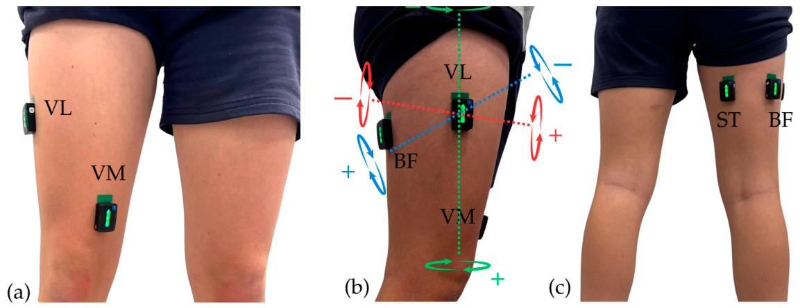
Knee kinematic data were quantified through segmental angular velocity, used herein as an indicator of movement quality. Angular velocity signals were acquired using the embedded triaxial gyroscopes integrated within the inertial measurement units (IMUs) of the Trigno Avanti sensors [4,36,37]. To characterize lower-limb segment kinematics, the Trigno Avanti sensor placed on the VL muscle was additionally used to record thigh angular velocity data [4,36,37]. All sensor placements are shown in Figure 2.

2.4. Data Analysis

EMG and angular velocity data were exported to C3D format and processed using Visual3D software v2024.08.3 (HAS Motion, Kingston, ON, Canada). EMG signals were filtered using a second-order Butterworth high-pass filter with a cut-off frequency of 40 Hz to minimize movement artifacts, then full-wave-rectified, and low-pass-filtered with a 15 Hz cut-off frequency [3,4]. The maximum observed signal from the filtered data across all trials and muscles was used to normalize the muscle activity during the preparation phase, defined as 100 ms prior to ground contact to the frame immediately before initial contact, and the load phase, defined as initial contact to final foot ground contact [8,12]. Accordingly, average and peak muscle activity outcomes were reported for analysis during both preparation and load phases, expressed as a percentage of maximum.
The peak maximum, peak minimum, and ROM angular velocities in the sagittal, coronal, and transverse planes were extracted for analysis during the load phase [4,37,38]. The peak maximum values correspond to the highest positive values for each plane: flexion in the sagittal, varus in the coronal, and external rotation in the transverse plane. In contrast, the peak minimum values correspond to the lowest negative values for each plane, extension in the sagittal, valgus in the coronal, and internal rotation in the transverse plane. The ROM angular velocity was defined as the difference between the maximum and minimum angular velocities for each plane. All angular velocity values were reported in degrees per second (°/s).

2.5. Sample Size

The sample size was estimated using the GRANMO 8.0 calculator (IMIM, Barcelona, Spain) [39]. Hamstring muscle activity was selected as the primary outcome, based on prior evidence supporting its key role as an ACL synergist during the stabilization process, and therefore as a critical variable expected to show significant differences between groups if ACLR players demonstrate dysfunctional muscle activity adaptations [12,19,25,27]. Based on the data reported by Markström et al., a minimum expected difference of 12% in average BF muscle activity, expressed as a percentage of EMG maximum capacity, during the LOAD phase of a jump test was assumed [8,40]. Considering an independent-samples two-sided t-test, an alpha risk of 0.05, and a statistical power of 80%, 13 participants per group were required. An estimated dropout rate of 10% was also considered.

2.6. Statistical Analysis

Data normality was assessed using the Shapiro–Wilk test. Normally distributed variables are presented as mean ± standard deviation, whereas non-normally distributed variables are reported as median and interquartile range [Q1–Q3]. Between-group comparisons, ACLR against control players, were performed to examine differences in knee angular velocity across the sagittal, coronal, and transverse planes during the LOAD phase, as well as differences in peak and average EMG muscle activity of the BF, ST, VL and VM during both the preparation and load phases.
For normally distributed variables, independent-samples t-tests were used. For non- normality distributed variables, Mann–Whitney U tests were performed. Statistical significance was established at p < 0.05. To account for multiple comparisons, Holm–Bonferroni corrections were applied within predefined families of biomechanically related variables. For knee kinematics, comparisons were grouped according to the sagittal, coronal, and transverse planes. For EMG outcomes, corrections were applied separately to average and peak muscle activity variables within the preparation and load phases. Adjusted p-values (p-Holm) were calculated and reported alongside unadjusted p-values. Statistical significance after correction was established at p-Holm < 0.05. Effect sizes were computed to quantify the magnitude of between-group differences. Cohen’s d was calculated for parametric analysis. For non-parametric analyses, the effect size r was computed as r = Z/√n. Effect sizes derived from parametric analysis, Cohen’s d, were interpreted as large when d > 0.8 according to conventional thresholds. Effect size r, derived from non-parametric analysis, was interpreted as large when r > 0.5. In addition, 95% confidence intervals were calculated. All analyses were performed using SPSS Statistics v25.0 (IBM Corp., Armonk, NY, USA).

3. Results

Thirteen participants were allocated to the ACLR group and thirteen to the control group, according to their clinical history. The average age was 22.89 ± 2.68 years, which defines players in the under-23 category, consistent with the aim of the study to evaluate players at a transitional stage toward elite football. Players in both groups had comparable experience competing at the national semiprofessional level, indicating similar sport-specific training exposure and competitive physical demands throughout the season. Demographic characteristics are presented in Table 1.
Table 2a,b summarize the descriptive statistics and between-group comparisons for normally and non-normally distributed knee kinematic variables, respectively, across the sagittal, coronal and transverse planes during the load phase of the COD. Compared with healthy controls, ACLR players exhibited significant knee kinematic differences in the coronal plane and high differences in the transverse plane. The Mann–Whitney U test, adjusted by Holm–Bonferroni corrections, demonstrated that ACLR group exhibited higher knee valgus angular velocity compared to control group, representing large effect size (p-Holm = 0.015; r = 0.54). No other kinematic variables remained statistically significant after correction. Nevertheless, transverse angular velocity ROM exhibited a large effect size (Cohen’s d = 0.83), indicating greater transverse-plane angular velocity in ACLR players compared to controls. No relevant between-group differences were observed in the sagittal plane variables.
Table 3a,b present the descriptive statistics and between-group comparisons for normally and non-normally distributed EMG muscle activity variables, respectively. The average and peak muscle activity of the BF, ST, VM and VL were recorded during the preparation and load phases of the COD. During the preparation phase, no significant differences in muscle activity were observed between groups after Holm–Bonferroni corrections. However, ACLR players exhibited lower average ST muscle activity than controls, with a large effect size (Cohen’s d = 0.88).
During the load phase, ACLR players exhibited significant differences which persisted for peak BF and average ST EMG muscle activity relative to healthy control players. The independent t-test, adjusted by Holm–Bonferroni corrections, revealed that the ACLR group demonstrated 4% lower average ST muscle activity, representing large effect size (p-Holm = 0.024; Cohen’s d = 1.00). In addition, the ACLR group demonstrated 27% higher peak BF muscle activity compared to the control group, representing large effect size (p-Holm < 0.001; Cohen’s d = 1.83). Additionally, lower average BF muscle activity (Cohen’s d = 0.88) and lower average VM muscle activity (Cohen’s d = 0.97) were observed in the ACLR group compared to controls, although these differences did not remain statistically significant after correction. No relevant differences between groups were observed in average or peak VL muscle activity during either the preparation or load phases.

4. Discussion

The aim of this study was to compare movement quality, quantified by knee angular velocity, and motor control, assessed through EMG thigh muscle activity, between under-23 semiprofessional female football players with ACLR and healthy controls. These biomechanical parameters were evaluated during a high-intensity COD test, to replicate the functional demands of the most common ACL injury mechanism [3,5,6,10]. The primary findings revealed that ACLR players exhibited significant differences in both kinematic and muscle activity parameters during COD tasks, supporting our hypothesis. Specifically, the ACLR group demonstrated altered knee kinematics in the coronal plane accompanied by modified motor control patterns in hamstrings during the load phase of the COD task.
The kinematic findings showed that players with ACLR exhibited significantly higher knee valgus angular velocity and a trend toward greater knee transverse ROM angular velocity during the COD task compared with controls. According to Budini et al., movement quality reflects the ability of the neuromuscular system to regulate the speed and direction of joint motion; therefore, higher angular velocity implies reduced control and stability [37]. Previous studies have reported altered angular velocity patterns in individuals with ACLR; however, these investigations were largely restricted to sagittal-plane analyses and included participants evaluated within 2–6 months after surgery, before completion of the return-to-play process [38,41]. In contrast, the present study identified persistent alterations toward increases in peak knee valgus angular velocity during COD. Given the established relationship between excessive knee valgus angular velocity and valgus collapse mechanisms, these findings may indicate the persistence of movement patterns associated with reduced control during high-intensity change of direction tasks [10,11,13,14]. These findings suggest that female football players with ACLR may retain, at least two years after surgery, coronal plane movement quality deficits and a trend toward transverse plane differences that could be associated with altered adaptations contributing to the elevated long-term likelihood of sustaining a secondary ACL injury.
These kinematic alterations of ACLR players were accompanied by significant differences in muscle activity characteristics in the load phase of the COD task. During this phase, the ACLR group demonstrated significantly 4% lower average ST muscle activity and 27% lower peak BF muscle activity. Furthermore, ACLR players exhibited a trend toward reduced average ST muscle activity during the preparation phase and lower average BF and VM muscle activity, although these differences did not remain statistically significant after adjustment for multiple comparisons. The present study only identified differences in muscle activity between groups during the load phase, and this fact may provide a biomechanical explanation for the kinematic alterations observed in the ACLR group. However, a tendency observed in ACLR toward reduced ST pre-load activity corresponds with previous research which explained that this neuromuscular alteration has previously been identified as a neuromuscular factor associated with ACL injury mechanisms [24,25]. This altered muscle activity suggests that insufficient preparatory activation may compromise motor control before initial contact and may influence subsequent loading responses, particularly during the early load phase of COD, which aligns with high prevalence timing of ACL injury [4,16].
The coexistence of altered hamstring activation and increased knee valgus angular velocity suggests a potential interaction between motor control and movement quality following ACLR. The reduced functional role of the hamstring during the stabilization load phase may predispose to quadriceps dominance favoring anterior tibial displacement, increasing ACL strain [12]. In addition, the specifically reduction in ST activity may indicate diminished contribution of the medial thigh muscle complex. Given their anatomical orientation and functional role in resisting knee valgus and transverse plane motion, this pattern may reduce the capacity to counteract valgus collapse [19]. Therefore, the findings relative to muscle activity motor patterns in ACLR female football players may describe a tendency toward hamstring and medial complex muscle reduced function, which may predispose to reinjury over a maximal physical demand context as is the case during ACL injury. Collectively, these findings suggest that female football players with ACLR exhibit persistent lateral to medial neuromuscular imbalance that may reflect long-term muscle compensatory adaptations and support the use of EMG-based assessments to improve return-to-play decision-making [6,23,27].

Limitations

Several limitations should be considered when interpreting these findings. The assessors in the present study were not blinded during data collection. However, participants were given a unique, anonymized study code to mitigate potential bias during data and statistical analyses. A further limitation of the present study is the relatively small sample size, which should be considered when interpreting the findings and deriving practical conclusions. The muscle activity assessment was limited to the hamstring and quadriceps, selected because of their functional relevance to the medial and lateral thigh muscle complexes. However, future studies should incorporate proximal and distal muscles involved in lower-limb load transfer, such as the gluteal musculature and gastrocnemius, to provide a more comprehensive characterization of neuromuscular compensatory strategies. Although playing position was considered during participant recruitment, the sample size did not allow meaningful comparisons between positional roles. This may be relevant because the functional demands associated with different playing positions influence movement patterns, exposure to COD actions, and overall biomechanical loading during match play. Future studies with larger cohorts should investigate whether positional role influences kinematic and neuromuscular adaptations following ACLR. A limitation of this study is that static coronal knee alignment and isolated muscle strength developed were not assessed. Although neither resting valgus angle nor muscle strength constituted primary outcomes, their measurement could have provided additional contextual information for interpreting the observed neuromuscular and kinematic differences between groups. Although the COD task was selected for its ability to reproduce the most common functional context of ACL injury, it did not include the unanticipated component frequently observed in match situations. Incorporating reactive decision-making conditions may further enhance the ecological validity and specificity of biomechanical assessment. Despite these limitations, the synchronized analysis of three-dimensional kinematics and surface EMG during a sport-specific task provides a clinically relevant framework for improving return-to-play evaluation after ACLR.

5. Conclusions

This study identified differences in knee angular velocity and muscle activity between under-23 semiprofessional female football players with ACLR and healthy controls during a COD task. Specifically, ACLR players may demonstrate increased knee valgus angular velocity, alongside reduced average ST and peak BF muscle activity, during the load phase of the change of direction task, compared to healthy control players.
Overall, the combined presence of increased angular velocity in the coronal and a tendency toward the transverse plane, along with reduced muscle activity of hamstrings and a tendency toward alterations in medial complex thigh muscles during the preparation and load phases of the COD task, may reflect persistent long-term dysfunctional motor pattern adaptations associated with valgus collapse mechanics. These findings, observed at least two years after ACLR, highlight the importance of integrating three-dimensional kinematics with muscle activity analysis during sport-specific functional tasks that replicate ACL injury mechanisms. This approach may improve the identification of compensatory motor control strategies and provide a more robust basis for return-to-play decision-making, specifically in female football players, thereby reducing the risk of secondary ACL injury.

Author Contributions

Conceptualization, L.F.-L., M.O.L.-L. and J.M.T.-M.; methodology, L.F.-L., M.O.L.-L. and C.H.-G.; software, L.F.-L. and J.M.-T.-U.; validation, L.V.-P., R.S.-R. and S.M.-B.; formal analysis, J.M.T.-M. and C.H.-G.; investigation, M.A., L.V.-P., R.S.-R. and S.M.-B.; resources, J.M.-T.-U.; data curation, M.A., L.V.-P. and R.S.-R.; writing—original draft preparation, L.F.-L.; writing—review and editing, M.O.L.-L., L.V.-P. and J.M.T.-M.; visualization, M.A.; supervision, M.O.L.-L. and C.H.-G.; project administration, M.O.L.-L. and J.M.T.-M. All authors have read and agreed to the published version of the manuscript.

Funding

Loreto Ferrández-Laliena received a PhD fellowship supported by Gobierno de Aragón (Spain), [ORDEN CUS/621/2023], 10 May 2023 (BOA 245 18 May 2023).

Institutional Review Board Statement

The study was conducted in accordance with the Declaration of Helsinki and approved by the Research Ethics Committee of the Community of Aragón (protocol code PI20/127; date of approval: 18 March 2020).

Informed Consent Statement

Informed consent was obtained from all subjects involved in the study. Written informed consent was obtained from the patient(s) to publish this paper.

Data Availability Statement

The datasets presented in this study are available on request from the corresponding author. All data covered by this study are included in this manuscript.

Acknowledgments

The authors wish to thank the Unidad de Investigación en Fisioterapia, Gobierno de Aragón, grant number B38_23R, for permission to use their infrastructure. We also acknowledge the participants who generously participated and collaborated in the research.

Conflicts of Interest

The authors declare no conflicts of interest.

Abbreviations

The following abbreviations are used in this manuscript:
ACLAnterior Cruciate Ligament
ACLRAnterior Cruciate Ligament Reconstruction
EMGElectromyography
STSemitendinosus
BFBiceps Femoris
VMVastus Medialis
VLVastus Lateralis
ROMRange of Movement
CODATChange of Direction and Acceleration Test
CODChange of Direction

References

  1. Hallén, A.; Tomás, R.; Ekstrand, J.; Bengtsson, H.; Van den Steen, E.; Hägglund, M.; Waldén, M. UEFA Women’s Elite Club Injury Study: A prospective study on 1527 injuries over four consecutive seasons 2018/2019 to 2021/2022 reveals thigh muscle injuries to be most common and ACL injuries most burdensome. Br. J. Sports Med. 2024, 58, 128–136. [Google Scholar] [CrossRef] [Scilit] [PubMed]
  2. Larruskain, J.; Lekue, J.A.; Angulo, P.; Santisteban, J.M.; Diaz-Beitia, G.; Martin-Garetxana, I.; Gil, S.M.; Bidaurrazaga-Letona, I.; Monasterio, X. An injury burden heat map of all men’s and women’s teams of a professional football club over a decade. Sports Med. 2024, 32, 740–750. [Google Scholar] [CrossRef] [Scilit] [PubMed]
  3. Ferrández-Laliena, L.; Vicente-Pina, L.; Sánchez-Rodríguez, R.; Chapman, G.J.; Heredia-Jimenez, J.; Hidalgo-García, C.; Tricás-Moreno, J.M.; Lucha-López, M.O. Differences in Kinematic and Muscle Activity Between ACL Injury Risk and Healthy Players in Female Football: Influence of Change of Direction Amplitude in a Cross-Sectional Case–Control Study. Medicina 2025, 61, 1259. [Google Scholar] [CrossRef] [Scilit] [PubMed]
  4. Ferrández-Laliena, L.; Vicente-Pina, L.; Sánchez-Rodríguez, R.; Chapman, G.J.; Soussi, B.; Hidalgo-García, C.; Lucha-López, M.O.; Tricás-Moreno, J.M.; Ambrus, M. Effects of a Multicomponent Periodized Program in Kinematic and Muscle Activity Characteristics Related to Anterior Cruciate Ligament Injury Mechanism in College Football Players—A Controlled Trial. J. Funct. Morphol. Kinesiol. 2025, 10, 412. [Google Scholar] [CrossRef] [Scilit] [PubMed]
  5. D’Ambrosi, R.; Baumert, P.; Fink, C. ACL injury in female soccer players: Risk, resilience, and prevention in the modern game. Sci. Prog. 2026, 109, 8994. [Google Scholar] [CrossRef] [Scilit] [PubMed]
  6. Murphy, C.; Landry, S.; Urquhart, N.; Coady, C.; Rutherford, D. Drop landing between subjects post anterior cruciate ligament reconstruction and uninjured controls: A biomechanical and neuromuscular analysis. Clin. Biomech. 2025, 124, 106504. [Google Scholar] [CrossRef] [Scilit] [PubMed]
  7. Liu, W.; Tan, Z.; Li, W.; Qin, Z.; Krustrup, P.; Li, W. High return to play but low return to preinjury level after anterior cruciate ligament reconstruction in soccer players: A systematic review with meta-analysis. Knee Surg. Sports Traumatol. Arthrosc. 2026, 34, 930–944. [Google Scholar] [CrossRef] [Scilit] [PubMed]
  8. Markström, J.L.; Grinberg, A.; Häger, C.K. Fear of Reinjury Following Anterior Cruciate Ligament Reconstruction Is Manifested in Muscle Activation Patterns of Single-Leg Side-Hop Landings. Phys. Ther. 2022, 102, pzab218. [Google Scholar] [CrossRef] [Scilit] [PubMed]
  9. Arundale, A.J.H.; Kvist, J.; Hägglund, M.; Fältström, A. Jumping performance based on duration of rehabilitation in female football players after anterior cruciate ligament reconstruction. Knee Surg. Sports Traumatol. Arthrosc. 2019, 27, 556–563. [Google Scholar] [CrossRef] [Scilit] [PubMed]
  10. Lucarno, S.; Zago, M.; Buckthorpe, M.; Grassi, A.; Tosarelli, F.; Smith, R.; Della Villa, F. Systematic Video Analysis of Anterior Cruciate Ligament Injuries in Professional Female Soccer Players. Am. J. Sports Med. 2021, 49, 1794–1802. [Google Scholar] [CrossRef] [Scilit] [PubMed]
  11. Beaulieu, M.L.; Ashton-Miller, J.A.; Wojtys, E.M. Loading mechanisms of the anterior cruciate ligament. Sports Biomech. 2023, 22, 1–29. [Google Scholar] [CrossRef] [Scilit] [PubMed]
  12. Nasseri, A.; Lloyd, D.G.; Bryant, A.L.; Headrick, J.; Sayer, T.A.; Saxby, D.J. Mechanism of Anterior Cruciate Ligament Loading during Dynamic Motor Tasks. Med. Sci. Sports Exerc. 2021, 53, 1235–1244. [Google Scholar] [CrossRef] [Scilit] [PubMed]
  13. Dix, C.; Arundale, A.; Silvers-Granelli, H.; Marmon, A.; Zarzycki, R.; Snyder-Mackler, L. Biomechanical measures during two sport-specific tasks differentiate between soccer players who go on to anterior cruciate ligament injury and those who do not: A prospective cohort analysis. Int. J. Sports Phys. Ther. 2020, 15, 928–935. [Google Scholar] [CrossRef] [Scilit] [PubMed]
  14. Hewett, T.E.; Myer, G.D.; Ford, K.R.; Heidt, R.S.; Colosimo, A.J.; McLean, S.G.; Van Den Bogert, A.J.; Paterno, M.V.; Succop, P. Biomechanical measures of neuromuscular control and valgus loading of the knee predict anterior cruciate ligament injury risk in female athletes: A prospective study. Am. J. Sports Med. 2005, 33, 492–501. [Google Scholar] [CrossRef] [Scilit] [PubMed]
  15. Larwa, J.; Stoy, C.; Chafetz, R.S.; Boniello, M.; Franklin, C. Stiff Landings, Core Stability, and Dynamic Knee Valgus: A Systematic Review on Documented Anterior Cruciate Ligament Ruptures in Male and Female Athletes. Int. J. Environ. Res. Public Health 2021, 18, 3826. [Google Scholar] [CrossRef] [Scilit] [PubMed]
  16. Ueno, R.; Navacchia, A.; Schilaty, N.D.; Myer, G.D.; Hewett, T.E.; Bates, N.A. Anterior Cruciate Ligament Loading Increases with Pivot-Shift Mechanism During Asymmetrical Drop Vertical Jump in Female Athletes. Orthop. J. Sports Med. 2021, 9, 2325967121989095. [Google Scholar] [CrossRef] [Scilit] [PubMed]
  17. Ishida, T.; Koshino, Y.; Yamanaka, M.; Ueno, R.; Taniguchi, S.; Ino, T.; Kasahara, S.; Samukawa, M.; Tohyama, H. Larger hip external rotation motion is associated with larger knee abduction and internal rotation motions during a drop vertical jump. Sports Biomech. 2021, 23, 640–654. [Google Scholar] [CrossRef] [Scilit] [PubMed]
  18. Navacchia, A.; Ueno, R.; Ford, K.R.; DiCesare, C.A.; Myer, G.D.; Hewett, T.E. EMG-Informed Musculoskeletal Modeling to Estimate Realistic Knee Anterior Shear Force During Drop Vertical Jump in Female Athletes. Ann. Biomed. Eng. 2019, 47, 2416–2430. [Google Scholar] [CrossRef] [Scilit] [PubMed]
  19. Jeong, J.; Choi, D.-H.; Shin, C.S. Association Between the Medial-Lateral Quadriceps and Hamstring Muscle Thickness and the Knee Kinematics and Kinetics During Single-Leg Landing. Sports Health 2023, 15, 519–526. [Google Scholar] [CrossRef] [Scilit] [PubMed]
  20. Bates, N.A.; Nesbitt, R.J.; Shearn, J.T.; Myer, G.D.; Hewett, T.E. The influence of internal and external tibial rotation offsets on knee joint and ligament biomechanics during simulated athletic tasks. Clin. Biomech. 2018, 52, 109–116. [Google Scholar] [CrossRef] [Scilit] [PubMed]
  21. Choi, W.R.; Yang, J.H.; Jeong, S.Y.; Lee, J.K. MRI comparison of injury mechanism and anatomical factors between sexes in noncontact anterior cruciate ligament injuries. PLoS ONE 2019, 14, e0219586. [Google Scholar] [CrossRef] [Scilit] [PubMed]
  22. Quatman, C.E.; Hewett, T.E. The anterior cruciate ligament injury controversy: Is “valgus collapse” a sex-specific mechanism? Br. J. Sports Med. 2009, 43, 328–335. [Google Scholar] [CrossRef] [Scilit] [PubMed]
  23. Behnke, A.L.; Parola, L.R.; Karamchedu, N.P.; Badger, G.J.; Fleming, B.C.; Beveridge, J.E. Neuromuscular function in anterior cruciate ligament reconstructed patients at long-term follow-up. Clin. Biomech. 2021, 81, 105231. [Google Scholar] [CrossRef] [Scilit] [PubMed]
  24. Picot, B.; Lempereur, M.; Morel, B.; Forestier, N.; Rémy-Néris, O. Lack of Proprioceptive Strategy Modulation Leads to At-Risk Biomechanics for Anterior Cruciate Ligament in Healthy Athletes. Med. Sci. Sports Exerc. 2024, 56, 942–952. [Google Scholar] [CrossRef] [Scilit] [PubMed]
  25. Zebis, M.K.; Aagaard, P.; Andersen, L.L.; Hölmich, P.; Clausen, M.B.; Brandt, M.; Husted, R.S.; Lauridsen, H.B.; Curtis, D.J.; Bencke, J. First-time anterior cruciate ligament injury in adolescent female elite athletes: A prospective cohort study to identify modifiable risk factors. Knee Surg. Sports Traumatol. Arthrosc. 2022, 30, 1341–1351. [Google Scholar] [CrossRef] [Scilit] [PubMed]
  26. Zebis, M.K.; Andersen, L.L.; Bencke, J.; Kjær, M.; Aagaard, P. Identification of athletes at future risk of anterior cruciate ligament ruptures by neuromuscular screening. Am. J. Sports Med. 2009, 37, 1967–1973. [Google Scholar] [CrossRef] [Scilit] [PubMed]
  27. Nutarelli, S.; Sangiorgio, A.; Legrand, T.; Monzoni, F.; Deabate, L.; Delcogliano, M.; Delahunt, E.; Filardo, G. Medial Hamstrings Used as Autograft for ACL Reconstruction Show an Abnormal Neuromuscular Pattern at the Time of Return to Sport. Am. J. Sports Med. 2026, 54, 569–579. [Google Scholar] [CrossRef] [Scilit] [PubMed]
  28. Jeong, J.; Choi, D.-H.; Shin, C.S. The Effect of Strength Training Targeting Medial Thigh Muscles on Neuromuscular and Biomechanical Risk Factors for Anterior Cruciate Ligament Injury: A Randomized Controlled Trial. Sports Med. Open 2025, 11, 5. [Google Scholar] [CrossRef] [Scilit] [PubMed]
  29. Maniar, N.; Cole, M.H.; Bryant, A.L.; Opar, D.A. Muscle Force Contributions to Anterior Cruciate Ligament Loading. Sports Med. 2022, 52, 1737–1750. [Google Scholar] [CrossRef] [Scilit] [PubMed]
  30. Dos’Santos, T.; McBurnie, A.; Comfort, P.; Jones, P.A. The effects of six-weeks change of direction speed and technique modification training on cutting performance and movement quality in male youth soccer players. Sports 2019, 7, 205. [Google Scholar] [CrossRef] [Scilit] [PubMed]
  31. Ardern, C.L.; Glasgow, P.; Schneiders, A.; Witvrouw, E.; Clarsen, B.; Cools, A.; Gojanovic, B.; Griffin, S.; Khan, K.M.; Moksnes, H.; et al. 2016 Consensus statement on return to sport from the First World Congress in Sports Physical Therapy, Bern. Br. J. Sports Med. 2016, 50, 853–864. [Google Scholar] [CrossRef] [Scilit] [PubMed]
  32. Zampogna, B.; Vasta, S.; Torre, G.; Gupta, A.; Hettrich, C.M.; Bollier, M.J.; Wolf, B.R.; Amendola, A. Return to Sport After Anterior Cruciate Ligament Reconstruction in a Cohort of Division I NCAA Athletes From a Single Institution. Orthop. J. Sports Med. 2021, 9, 2281. [Google Scholar] [CrossRef] [Scilit] [PubMed]
  33. Wan, Y.; Chen, S.; Wang, H.; Zhu, S.; Shuai, H.; Yan, Z. Return to sport after ACL reconstruction in female athletes: A meta-analysis of return rates and time to return. J. Orthop. Surg. Res. 2026, 21, 267. [Google Scholar] [CrossRef] [Scilit] [PubMed]
  34. World Medical Association. Ethical Principles for Medical Research Involving Human Subjects. Eur. J. Emerg. Med. 2001, 8, 221–223. [Google Scholar] [CrossRef] [Scilit] [PubMed]
  35. Hermens, H.J.; Freriks, B.; Disselhorst-Klug, C.; Rau, G. Development of recommendations for SEMG sensors and sensor placement procedures. J. Electromyogr. Kinesiol. 2000, 10, 361–374. [Google Scholar] [CrossRef] [Scilit] [PubMed]
  36. Costello, K.E.; Eigenbrot, S.; Geronimo, A.; Guermazi, A.; Felson, D.T.; Richards, J.; Kumar, D. Quantifying varus thrust in knee osteoarthritis using wearable inertial sensors: A proof of concept. Clin. Biomech. 2020, 80, 105232. [Google Scholar] [CrossRef] [Scilit] [PubMed]
  37. Budini, K.; Richards, J.; Cole, T.; Levine, D.; Trede, R.; George, L.S.; Selfe, J. An exploration of the use of Inertial Measurement Units in the assessment of dynamic postural control of the knee and the effect of bracing and taping. Physiother. Prac. Res. 2018, 39, 91–98. [Google Scholar] [CrossRef] [Scilit]
  38. Sigward, S.M.; Chan, M.S.M.; Lin, P.E. Characterizing knee loading asymmetry in individuals following anterior cruciate ligament reconstruction using inertial sensors. Gait Posture 2016, 49, 114–119. [Google Scholar] [CrossRef] [Scilit] [PubMed]
  39. Universitat de Barcelona. Institut Municipal d’Investigació Mèdica (IMIM). GRANMO: Sample Size Calculator; Version 8.0.; Universidad de Barcelona: Barcelona, Spain, 2012. [Google Scholar]
  40. Palmieri-Smith, R.M.; Strickland, M.; Lepley, L.K. Hamstring Muscle Activity After Primary Anterior Cruciate Ligament Reconstruction—A Protective Mechanism in Those Who Do Not Sustain a Secondary Injury? A Preliminary Study. Sports Health 2019, 11, 316–323. [Google Scholar] [CrossRef] [Scilit] [PubMed]
  41. Alshehri, Y.S.; Liu, W.; Mullen, S.; Phadnis, M.; Sharma, N.K.; Santos, M. Inertial sensors identified asymmetries in shank angular velocity at different gait speeds in individuals with anterior cruciate ligament reconstruction. Gait Posture 2020, 80, 302–307. [Google Scholar] [CrossRef] [Scilit] [PubMed]
Figure 1. CODAT. The starting position is indicated with the dark circle, and the cross identifies the change of direction task that was recorded.
Applsci 16 06631 g001
Figure 2. Sensor placement. The figure represents the projection of sensor placement in (a) anterior view, in which the Vastus Lateralis (VL) and Vastus Medialis (VM) sensors were placed; (b) lateral view, in which VL sensor was placed to provide thigh segment kinematic values; and (c) posterior view, in which the Semitendinosus (ST) and Biceps Femoris (BF) sensors were placed. In picture (b), the kinematic projection of the three-rotation axis of angular velocity, registered by triaxial gyroscope of IMU integrated in sensors, are represented with pointed lines. A blue pointed line represents the sagittal plane rotation axis, (+) knee flexion and (−) knee extension. A red pointed line represents the coronal plane rotation axis, (+) knee varus and (−) knee valgus. Finally, a green pointed line represents the transverse plane rotation axis, (+) knee external rotation and (−) knee internal rotation [4,36,37,38].
Applsci 16 06631 g002
Table 1. Demographic characteristics of the players.
Total (n = 26)ACLR (n = 13)Control (n = 13)
Age (years)22.89 ± 2.6822.86 ± 2.8022.93 ± 2.56
Position (Defender/Midfielder/Forward)3:14:91:7:52:7:4
Height (cm)165.44 ± 6.19164.34 ± 6.41166.55 ± 5.97
Limb dominance (Right/Left)20:610:310:3
National Football Experience (years)6.04 ± 2.476.00 ± 2.166.07 ± 2.79
Injury limb (Dominant Limb/Non-Dominant Limb)8:58:5
Table 2. (a) Mean (SDs) for normally distributed data and comparison analysis between ACLR and control players for peaks and range of angular velocity sagittal, coronal and transverse plane knee kinematics during the load phase of the CODAT. (b) Median and interquartile range [Q1, Q3] values for non-normally distributed data and comparison analysis between ACLR and control players for peaks and range of angular velocity sagittal, coronal and transverse plane knee kinematics during the load phase of the CODAT.
(a)
ACLRControlp Valuep-HolmEffect
Size
Cohen’s d
95% Confidence
Intervals for
Differences
Lower
Bound
Upper Bound
Variables (°/s)
Sagittal Plane
Extension Angular Velocity−370.31 ± 96.54−387.24 ± 129.470.7091.0000.15−75.51109.38
Flexion Angular Velocity243.39 ± 115.67280.08 ± 157.880.5061.0000.27−148.7275.34
Coronal Plane
Varus Angular Velocity334.10 ± 162.52340.24 ± 170.090.9260.9260.04−140.82128.52
Coronal Angular Velocity ROM723.64 ± 261.70590.46 ± 290.050.2310.4620.48−90.45356.80
Transverse Plane
Internal Rotation Angular Velocity−842.64 ± 430.10−575.62 ± 338.420.0910.1620.69−580.5046.05
Transverse Angular Velocity ROM1787.28 ± 725.421227.27 ± 617.710.045 *0.1350.83 14.611105.41
(b)
ACLRControlp Valuep-HolmEffect
Size
r
Variables (°/s)
Sagittal Plane
Sagittal Angular Velocity ROM642.25[451.70, 708.35]740.77[571.10, 811.44]0.2870.8610.22
Coronal Plane
Valgus Angular Velocity−368.45[−508.03, −309.06]−215.82[−247.00, −151.05]0.005 *0.015 **0.54
Transverse Plane
External Rotation Angular Velocity940.37[479.62, 1235.23]827.61[249.22, 880.06]0.0810.1620.35
* denotes significance (p < 0.05); ** denotes significance; Holm–Bonferroni adjusted p-value. denotes effect size d > 0.8; p-Holm: Holm–Bonferroni adjusted p-value.
Table 3. (a) Mean (SDs) and comparison analysis between ACLR and control players for peak and average muscle activity for Biceps Femoris, Semitendinosus, Vastus Medialis and Vastus Lateralis during the CODAT. (b) Median and interquartile range [Q1, Q3] values for non-normally distributed data and comparison analysis between ACLR and control players for peak muscle activity for Vastus Medialis during the CODAT.
(a)
ACLRControlp Valuep-HolmEffect
Size
95% Confidence
Intervals for
Differences
Lower
Bound
Upper Bound
Variables (% of Maximum)
Preparation Phase
Average Biceps Femoris12.69 ± 8.4013.77 ± 5.200.6981.0000.15−0.070.04
Peak Biceps Femoris54.85 ± 25.9166.08 ± 15.750.1940.7760.51−0.290.06
Average Semitendinosus11.62 ± 4.4815.77 ± 5.340.042 *0.1680.88−0.08−0.01
Peak Semitendinosus62.31 ± 18.4969.00 ± 12.440.2900.8700.46−0.190.06
Average Vastus Medialis14.69 ± 7.8216.23 ± 4.950.5551.0000.15−0.070.04
Peak Vastus Medialis61.85 ± 19.5266.92 ± 16.320.4790.9580.28−0.200.10
Average Vastus Lateralis15.15 ± 5.4316.15 ± 3.850.5931.0000.22−0.050.03
Peak Vastus Lateralis67.54 ± 15.3471.54 ± 13.650.4890.9580.28−0.160.08
Load Phase
Average Biceps Femoris9.46 ± 5.7813.31 ± 4.380.0680.1360.88 −0.080.03
Peak Biceps Femoris49.08 ± 17.1376.31 ± 11.70<0.001 *0.004 **1.83 −0.39−0.15
Average Semitendinosus8.54 ± 2.7612.00 ± 3.080.006 *0.024 **1.00 −0.06−0.01
Peak Semitendinosus52.54 ± 15.4465.46 ± 19.890.0770.2310.68−0.270.01
Average Vastus Medialis11.85 ± 4.9615.62 ± 3.360.033 *0.0990.97 −0.190.07
Average Vastus Lateralis13.62 ± 3.6415.54 ± 5.920.3290.3290.39−0.060.02
Peak Vastus Lateralis69.69 ± 17.7176.00 ± 14.660.3320.3380.36−0.190.07
(b)
ACLRControlp Valuep-HolmEffect
Size
r
Variables (% of Maximum)
Load Phase
Peak Vastus Medialis52.00[45.50, 80.00]77.00[70.50, 87.50]0.1690.3380.22
* denotes significance (p < 0.05); ** denotes significance; Holm–Bonferroni adjusted p-value. denotes effect size d > 0.8; p-Holm: Holm–Bonferroni adjusted p-value.
Disclaimer/Publisher’s Note: The statements, opinions and data contained in all publications are solely those of the individual author(s) and contributor(s) and not of MDPI and/or the editor(s). MDPI and/or the editor(s) disclaim responsibility for any injury to people or property resulting from any ideas, methods, instructions or products referred to in the content.