Article

Longitudinal Evaluation of Gingival Margin Modifications During Treatment with Clear Aligners: A Retrospective Clinical Study in Growing Patients


1
Department of Health Science, Unicamillus—Saint Camillus International Medical University, 00131 Rome, Italy
2
Department of Oral and Maxillofacial Sciences, Sapienza University of Rome, 00161 Rome, Italy
3
Department of Paediatric Dentistry (0–14 Years Old), Policlinico Roma Tor Vergata, 00133 Rome, Italy
*
Author to whom correspondence should be addressed.
Appl. Sci. 2026, 16(17), 8445; https://doi.org/10.3390/app16178445
Submission received: 6 July 2026 / Revised: 11 August 2026 / Accepted: 20 August 2026 / Published: 25 August 2026
(This article belongs to the Special Issue Advanced Studies in Orthodontics, 2nd Edition)

Abstract

Background: The aim of this study was to evaluate gingival margin modifications during and after orthodontic treatment with Clear Aligners in paediatric patients. Secondary objectives included assessing the relationship between gingival changes and different types of tooth movement and evaluating the stability of these changes after treatment completion. Methods: A total of 50 patients (26 f, 24 m, mean age: 7.6 ± 1.1 years) in mixed dentition were enrolled. Inclusion criteria were good general health, plaque and gingival indices below 20%, good compliance with aligners, and no prior orthodontic treatment. Patients with periodontal issues, medications influencing gingival response, or with poor expected compliance were excluded. Patients were treated using Clear Aligners and attachments to facilitate specific tooth movements (expansion, intrusion, and rotational corrections). Measurements were performed at four time points: before treatment (T0), after 6 months (T1), at the end of treatment (T2), and 12 months post-treatment (T3). At each time point, intraoral scans, clinical photographs, and periodontal measurements were recorded. Results: A statistically significant increase in gingival margin height was observed from baseline to T1 (+0.29 mm), T2 (+0.44 mm), and T3 (+0.39 mm). Mean Changes between T0 and T3 were +0.39 ± 0.22 (p ≤ 0.001). A total of 60% of patients demonstrated a coronal migration of the gingival margin. Gingival Index showed a non-significant increase at T1, followed by normalization at T2 (p = 0.481) and T3 (p = 0.637). Plaque Index (PI) remained stable throughout the observation period. Expansion was associated with the greatest gingival change (+0.52 mm), followed by intrusion (+0.45 mm). Conclusions: Clear aligner treatment in mixed dentition appears to produce favourable and stable gingival margin modifications, with minimal adverse periodontal effects when properly monitored.

1. Introduction

Interceptive orthodontics plays a fundamental role in the management of malocclusions in paediatric patients [1,2,3]. To avoid complex orthodontic therapy in the late stage of growth, early interceptive treatment should begin during the first or transitional dentition phase [4].
Early intervention aims not only to correct developing occlusal discrepancies, but also to guide craniofacial growth, to improve oral function and to solve any muscular imbalances, with the aim of enhancing chewing function and to improve the orofacial environment before the eruption of permanent teeth [4,5,6].
The mixed dentition phase is particularly important, as it allows clinicians to guide occlusal development to prevent the worsening of malocclusions, and to preserve arch length in cases of premature primary tooth loss [7,8]. For this reason, interceptive treatment helps to ensure the proper development of the dental arches and to support the transitional phase of dentition, managing the space for the permanent teeth [1,2,3,4,5,6,7,8].
In recent years, technological advancements have significantly influenced orthodontic approaches, with the introduction and widespread adoption of clear aligner systems. Clear aligners represent an alternative to conventional fixed appliances, offering advantages such as improved aesthetics, enhanced comfort, and better oral hygiene maintenance [9,10,11,12,13].
Previous studies [14,15,16] showed that Clear Aligners (CA) are effective in promoting dento-alveolar arch expansion, in turning the triangular maxillary arch form into a semicircular one, in increasing arch length and in regaining and maintaining space for the development of permanent dentition. CAs are designed to manage malocclusions in younger patients, who typically present teeth with shorter clinical crowns and still in eruption. The main advantages of CA over conventional techniques lie in their ability to achieve dentoalveolar expansion, tooth alignment, and restoration of the correct arch shape simultaneously [14,15,16].
While the biomechanical effectiveness of aligners has been extensively investigated, their impact on periodontal tissues—particularly in growing patients—remains an area of ongoing research. Orthodontic tooth movement is inherently associated with biological responses involving the periodontal ligament, alveolar bone, and gingival tissues [17,18,19,20]. These responses are typically adaptive; however, under certain conditions, they may lead to undesirable periodontal outcomes such as gingival inflammation and recession [21,22,23,24,25,26]. The gingival margin plays a crucial role in both periodontal health and dental aesthetics [27]. Its position relative to the tooth structure influences not only the biological stability of the periodontium but also the visual harmony of the smile [28,29,30,31]. Therefore, understanding how orthodontic treatment affects gingival margins is of significant clinical importance.
Clear aligners may offer advantages over fixed appliances in terms of periodontal health due to their removability, which allows for more effective oral hygiene and reduced plaque accumulation [32,33,34,35,36]. However, the specific effects of aligner therapy on gingival margin position, especially in pediatric populations, have not been extensively documented.
A preliminary investigation [37] suggested that treatment with Invisalign First® may be associated with measurable changes in gingival margin position, often indicating a coronal shift. Nevertheless, the study was limited by a small sample size and a short follow-up period. Given the importance of early orthodontic treatment and the need to ensure periodontal safety, further research is necessary to confirm and to expand upon these findings. It is essential to evaluate whether gingival changes observed during treatment are stable over time and how they relate to specific types of tooth movement [37].
The present study was therefore designed as a retrospective longitudinal clinical investigation involving a larger sample size. The primary aim was to evaluate gingival margin modifications during and after orthodontic treatment with clear aligners in pediatric patients. Secondary objectives included assessing the relationship between gingival changes and different types of tooth movement and evaluating the stability of these changes after treatment completion.

2. Materials and Methods

This study was designed as a retrospective longitudinal clinical investigation conducted at the University of Rome “UniCamillus”. The study project was approved by the Ethical Committee at the same university (protocol number E00176-2025, date approval 3 June 2025). The parents or legal guardians of all participants signed a written informed consent form and were informed that the anonymised clinical records of their children would be used for research purposes. A total of 50 patients were recruited for the study. The sample included 26 females and 24 males, with ages ranging from 6 to 10 years (mean age: 7.6 ± 1.1 years) and cervical stage maturation (CS) between CS1–CS2 [38]. Inclusion criteria were mixed dentition phase, absence of skeletal cross-bite and posterior transverse interarch discrepancy < 5 mm assessed on the digital dental casts, dental crowding and arch-length discrepancy, increased overjet and/or overbite, and rotations of the anterior teeth, requiring an interceptive orthodontic treatment suitable with Clear Aligners. Patients with periodontal issues, medications influencing gingival response, or poor expected compliance were excluded.
Inclusion criteria were as follows: good general health; a full-mouth plaque score (FMPS) [39] and a full-mouth bleeding score (FMBS) [40] below 20%, both recorded on four surfaces per tooth (mesial, distal, buccal and lingual/palatal) and expressed as the percentage of positive surfaces over the total number of surfaces examined; good compliance with aligners; and no prior orthodontic treatment.
Patients with systemic conditions affecting periodontal tissues, those taking medications influencing gingival response, or those with poor expected compliance were excluded.
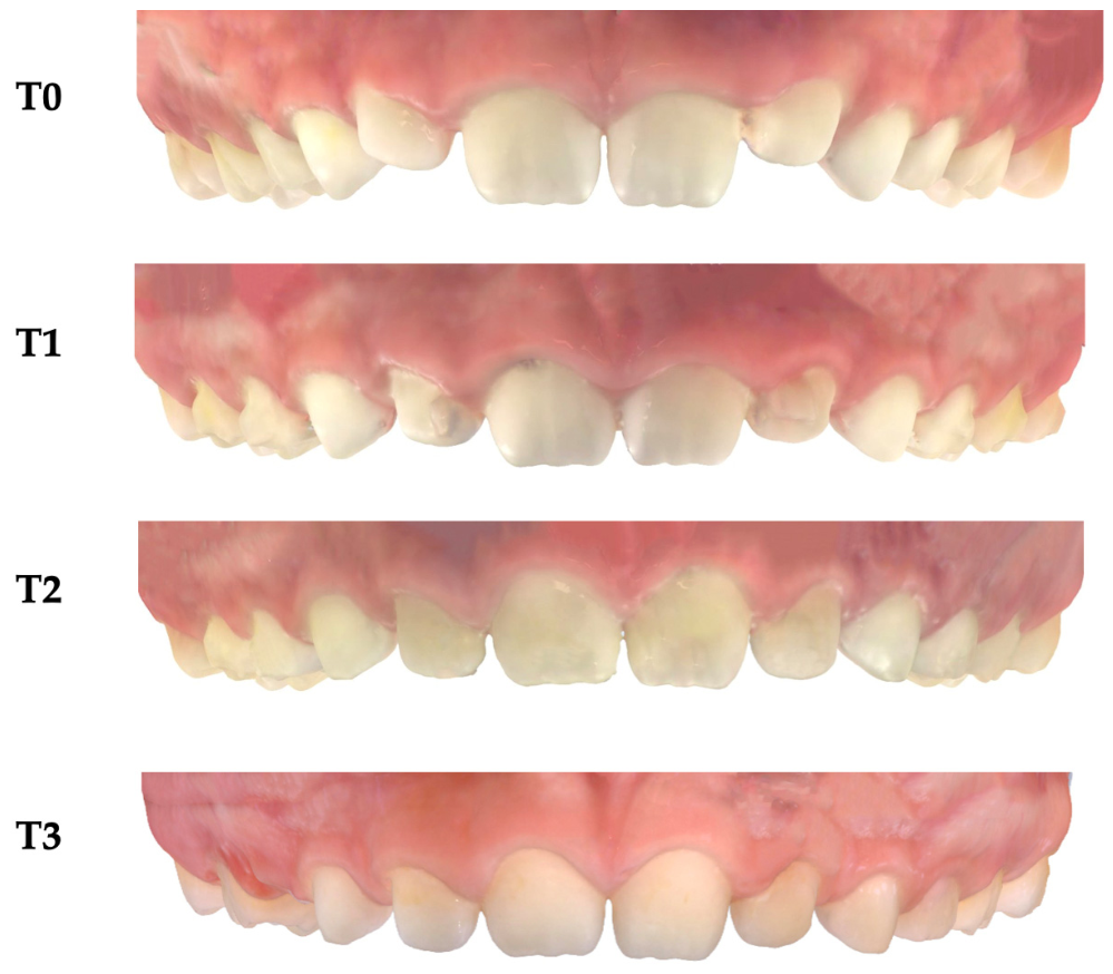
All patients were treated using Invisalign First® aligners (Align Technology, Santa Clara, CA, USA). Digital treatment planning was performed using ClinCheck® software. Aligners were prescribed for 20–22 h of daily wear and replaced every 7 days. Clinical check-ups were scheduled every 6–8 weeks. Attachments were placed when necessary to facilitate specific tooth movements, particularly expansion, intrusion, and rotational corrections. All patients received standardized oral hygiene instructions at each visit. Measurements were performed at four time points: before treatment (T0), after 6 months (T1), at the end of treatment (T2), and 12 months post-treatment (T3) (Figure 1). At each time point, intraoral scans, clinical photographs, and periodontal measurements were recorded. The gingival margin of each aligner extended to fully cover the clinical crown and terminated at the free gingival margin, with a scalloped/straight trim as prescribed by the digital planning, to limit any mechanical interaction between the aligner edge and the marginal gingival tissues. Given the mixed-dentition stage and the expansion-based nature of the treatment, interproximal reduction (IPR) was not performed in the anterior region under study.
The digital dental casts were collected for each patient involved in the study. Measurements were performed using Viewbox 4.0 (dHAL Software, Kifissia, Greece).
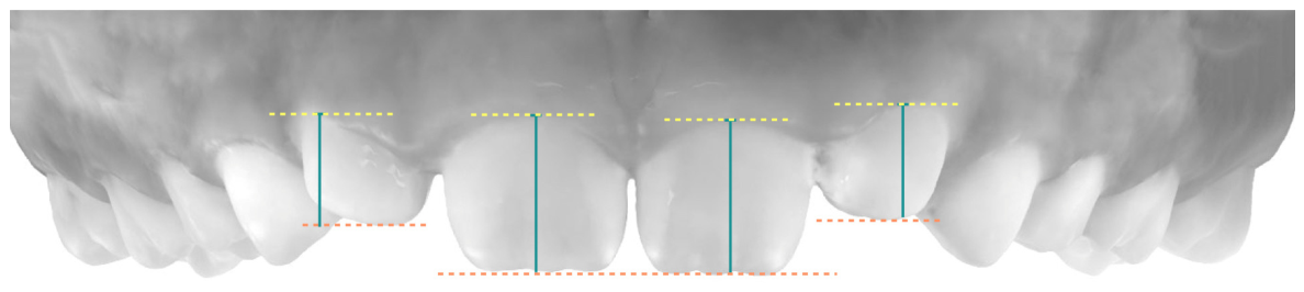
The primary outcome was the gingival margin position, digitally measured on the central and lateral incisors. It was measured as the vertical distance, parallel to the tooth axis, from the gingival margin to a fixed dental reference point set as the incisal line. The incisal line was set as a fixed dental reference located on the tooth itself, so that it moved together with the erupting tooth; the recorded value therefore reflects the relationship between the gingival margin and the crown rather than an absolute vertical position (Figure 2).
The upper incisors were selected because they represent the site of greatest aesthetic relevance and the region where gingival-margin discrepancies are most reliably perceived clinically; because the permanent upper central and lateral incisors were consistently erupted and identifiable at every time point, providing a standardized and reproducible measurement site; and because the anterior region concentrated the tooth movements of interest (expansion, intrusion, and rotation).
Secondary outcomes included two clinical periodontal measurements, recorded at the same incisor sites with a calibrated periodontal probe.
The Gingival Index (GI) was assessed according to Löe and Silness [41]. Four gingival areas of each tooth (distal, vestibular, mesial and palatal) were scored on a 0–3 ordinal scale: 0, normal gingiva; 1, mild inflammation with a slight change in color and slight edema, without bleeding on probing; 2, moderate inflammation with redness, edema and glazing, with bleeding on probing; and 3, severe inflammation with marked redness and edema, ulceration, and a tendency for spontaneous bleeding.
The Plaque Index (PI) was assessed according to Silness and Löe [42] on the same four surfaces, using a 0–3 ordinal scale that quantifies the thickness of plaque at the gingival margin: 0, no plaque; 1, a film of plaque adhering to the free gingival margin, detectable only by running the probe across the tooth surface; 2, a moderate accumulation of soft deposits within the gingival sulcus and on the gingival margin, visible to the naked eye; and 3, an abundance of soft matter within the gingival sulcus and on the gingival margin.
For GI and PI, the score of each tooth was calculated as the mean of the four areas, and the individual score as the mean of the scores of the examined teeth.
Tooth movements were categorized based on the digital treatment plan as expansion, rotation, intrusion, and torque. For each tooth, movement was recorded, and gingival margin was digitally measured at T0 and T2.

Statistical Analysis

Intra-examiner reliability for the digital measurement of the gingival margin was assessed by repeating all measurements on 20 randomly selected casts after an interval of 24 h and was expressed as the intraclass correlation coefficient (ICC, two-way mixed model, absolute agreement) with its 95% confidence interval; the method error was computed with the Dahlberg formula. Intra-examiner reliability for the ordinal periodontal indices was expressed as the weighted Cohen’s kappa coefficient.
The sample size was estimated a priori based on the primary outcome, i.e., the change in the position of the gingival margin of the permanent incisors between T0 and T2, analysed with a two-tailed paired t-test. The standard deviation of the paired difference was derived from a preliminary investigation [37] in which the T1–T0 changes were 0.39 ± 0.99 mm and 0.41 ± 1.07 mm for the right lateral and central incisors, and 0.44 ± 1.20 mm and 0.46 ± 0.96 mm for the left central and lateral incisors, giving a pooled standard deviation of the difference of 1.06 mm. The minimum clinically relevant difference was set at 0.50 mm, this being the smallest discrepancy of the gingival margin that clinicians have been shown to detect on the anterior dentition [28].
With δ = 0.50 mm, σd = 1.06 mm (standardised effect size dz = 0.47), a two-tailed α of 0.05 and a power (1 − β) of 0.90, a minimum of 50 subjects was required. The calculation was performed with G*Power 3.1.9.7 (Heinrich Heine University, Düsseldorf, Germany). Allowing for the three patients lost to follow-up, the 47 subjects who completed the study retained a power of 0.88 to detect the pre-specified difference of 0.50 mm.
Statistical analyses were performed with SPSS Statistics v.27 (IBM Corp., Armonk, NY, USA). Descriptive statistics (mean and standard deviation) were calculated for all variables at each time point. The normality of the distributions was tested with the Shapiro–Wilk test; the sphericity assumption was verified with Mauchly’s test and, when violated, the Greenhouse–Geisser correction was applied.
The effect of time on the gingival margin position, on GI and on PI was evaluated with a one-way repeated-measures ANOVA over the four time points. Pairwise comparisons between time points were performed with paired t-tests and the significance level was adjusted with the Bonferroni correction for multiple comparisons.
For each of the four categories of tooth movement, the gingival margin change between T0 and T2 was analysed with a paired t-test on the teeth in which that movement had been prescribed; the mean difference was reported with its 95% confidence interval. Significance was set at p < 0.05.

3. Results

Out of the 50 patients enrolled, 47 completed the study, resulting in a dropout rate of 6%. Compliance was high, with an average aligner wear adherence of approximately 91%. Intra-examiner reliability was excellent for the digital measurement of the gingival margin position (ICC = 0.96; 95% CI 0.92–0.98; method error 0.08 mm) and for the ordinal periodontal indices (weighted Cohen’s kappa = 0.88).
A progressive and statistically significant increase in gingival margin height was observed from baseline to T1 (+0.29 mm, p = 0.041), T2 (+0.44 mm, p = 0.008), and T3 (+0.39 mm, p = 0.015). Mean Changes between T0 and T3 were +0.39 ± 0.22 (p ≤ 0.001). Overall, 60% of patients demonstrated a coronal migration of the gingival margin, whereas 30% showed no significant variation and 10% exhibited an apical shift (<0.5 mm). Gingival Index (GI) values showed a slight, non-significant increase at T1 (p = 0.072), followed by normalization at T2 (p = 0.481) and T3 (p = 0.637). Plaque Index (PI) remained stable throughout the observation period (p = 0.276) (Table 1).
Among the different tooth movements, expansion was associated with the greatest gingival change (+0.52 mm, p < 0.001), followed by intrusion (+0.45 mm, p < 0.001) (Table 2).

4. Discussion

The results of this study confirm that orthodontic treatment with clear aligners in paediatric patients is associated with measurable and generally favourable modifications of the gingival margin. The observed coronal displacement of the gingival margin in most patients suggested a positive adaptive response of the periodontal tissues to aligner-based orthodontic forces, likely influenced by both biological and mechanical factors. The relatively young age of the study population may have contributed to the enhanced tissue responsiveness, as growth-related cellular activity and vascularization are known to facilitate periodontal remodelling during orthodontic treatment [17,32,43,44,45,46,47,48,49].
The mean age of the sample (approximately 7 years) corresponds to the period of active eruption of the permanent incisors used as reference. Consequently, part of the recorded change in gingival-margin position is likely to reflect physiologic eruption and the associated increase in clinical crown exposure rather than a treatment-induced effect. In the absence of an untreated, age-matched control group, the treatment effect cannot be disentangled from normal growth, and the recorded change should be interpreted as a description of gingival-margin behaviour during aligner therapy rather than as proof of a causally induced coronal migration.
In growing children, the mucogingival complex is not static: the width of the attached gingiva changes significantly during development, as shown in subjects aged 6 to 12 years [50,51], while the erupting tooth continuously moves relative to the surrounding soft tissues. Consequently, in the mixed dentition no soft-tissue landmark is entirely independent of developmental change: a dental reference such as the incisal line follows the erupting tooth, whereas a mucogingival reference is associated with a developmentally changing attached gingiva. Regardless of the reference adopted, the recorded change in gingival-margin position therefore reflects a combination of the treatment effect and physiological development, which can be separated only by means of an untreated control group or longitudinal normative data.
Within our aligner-treated cohort, the periodontal parameters remained favourable throughout the observation period. The stability of the plaque index values indicates that patients were able to maintain consistent oral-hygiene practices during treatment, which likely contributed to minimising inflammation and supporting gingival health [32,36]. As no comparison group treated with fixed appliances or left untreated was included, no comparative inference regarding the relative periodontal impact of different appliances can be drawn from these data.
The association between transverse expansion and increased gingival margin height observed in this study is consistent with previous research suggesting that lateral displacement of teeth can lead to soft tissue stretching and volumetric adaptation [43,47]. Similarly, intrusion movements may contribute to coronal repositioning of the gingival margin due to the relative displacement of the tooth within the alveolar bone [48,49]. These biomechanical relationships highlight the importance of treatment planning in predicting periodontal outcomes. In our interpretation, the movement-dependent pattern we observed—i.e., the greater changes recorded with transverse expansion and intrusion compared with rotation and torque—suggests that the amount of gingival-margin change is related to the magnitude of vertical and transverse crown displacement produced by each movement, an interpretation that is consistent with, and here precedes, the mechanistic explanations proposed in the literature.
The overall stability of gingival changes at the 12-month follow-up indicated that the modifications induced during treatment are maintained over time, suggesting a stable reorganization of the periodontal architecture [44]. This is particularly relevant in the context of early orthodontic treatment, where long-term outcomes are a primary concern.
Although a small percentage of patients experienced mild apical shift in the gingival margin, the extent was limited and clinically insignificant in most cases. This finding aligns with the existing literature indicating that orthodontic treatment carries a low, but present risk of apical shifting (recession), particularly in individuals with thin gingival biotypes or unfavourable tooth movements [22,26,49]. Careful patient selection and monitoring remain essential to minimize such risks.
This study presents some limitations that should be considered when interpreting the results. First, the retrospective, single-centre design of the study; second, the absence of an untreated control group, or of a group treated with a different appliance such as rapid maxillary expander. Prospective, controlled, multi-centre trials with longer follow-up and phenotype stratification are necessary to confirm the stability of these results. Most importantly, because the sample was in the active eruptive phase of the permanent incisors, physiologic eruption represents an unavoidable confounder that cannot be separated from the treatment effect without an untreated, age-matched control group; the observed changes should therefore be interpreted with this limitation in mind.
From a clinical standpoint, our findings support the periodontal safety of interceptive clear aligner therapy in the mixed dentition if patients are appropriately selected and periodontal parameters are monitored throughout treatment. On this basis, this treatment approach can be reasonably recommended to clinicians, with the type of prescribed movement considered when anticipating gingival outcomes at the treatment-planning stage.
Our data do not support any periodontal advantage of clear aligners over conventional therapy. The principal observed advantage of aligners is aesthetic—an aspect particularly valued by growing children. To preserve this favourable soft-tissue outcome, clinicians should respect the biomechanical limits of aligner therapy and ensure appropriate trimming of the aligner margin, to avoid traumatising the free gingival margin.

5. Conclusions

Clear aligner treatment in mixed dentition is associated with significant and stable gingival-margin modifications, with minimal adverse periodontal effects and aesthetic benefits for the patient.
A change in the recorded gingival-margin position, consistent with a coronal relationship, was observed during treatment and was maintained at the 12-month follow-up, while gingival and plaque indices remained within healthy ranges throughout the observation period.
The magnitude of the change was movement-dependent, being greatest for transverse expansion and intrusion, which suggests that the prescribed movement should be considered when anticipating periodontal outcomes at the treatment planning stage.

Author Contributions

Conceptualization, P.C. and R.L.; methodology, M.C.; software, M.C.; validation, M.C. and R.L.; formal analysis, A.M.; investigation, F.G.; resources, C.P.; data curation, F.C.D.R. and L.L.; writing—original draft preparation, L.L.; writing—review and editing, F.C.D.R.; supervision, P.C. All authors have read and agreed to the published version of the manuscript.

Funding

This research received no external funding.

Institutional Review Board Statement

This study was designed as a retrospective longitudinal clinical investigation conducted at the University of Rome “UniCamillus” and the study project was approved by the Ethical Committee at the same University (protocol number E00176-2025, date approval 3 June 2025).

Informed Consent Statement

Informed consent was obtained from all subjects involved in the study.

Data Availability Statement

The original contributions presented in this study are included in the article. Further inquiries can be directed to the corresponding author.

Conflicts of Interest

The authors declare no conflicts of interest.

References

  1. Zhou, C.; Duan, P.; He, H.; Song, J.; Hu, M.; Liu, Y.; Liu, Y.; Guo, J.; Jin, F.; Cao, Y.; et al. Expert consensus on pediatric orthodontic therapies of malocclusions in children. Int. J. Oral Sci. 2024, 16, 32. [Google Scholar] [CrossRef] [Scilit]
  2. Proffit, W.R.; Fields, H.W.; Sarver, D.M. Contemporary Orthodontics, 6th ed.; Elsevier: St. Louis, MO, USA, 2019. [Google Scholar]
  3. Graber, L.W.; Vanarsdall, R.L.; Vig, K.W.L.; Huang, G.J. Orthodontics: Current Principles and Techniques, 6th ed.; Elsevier: St. Louis, MO, USA, 2017. [Google Scholar]
  4. Ebrahimy, G.; Konermann, A.; El-Bialy, T.; Keilig, L.; Bourauel, C. Comparative experimental evaluation of orthodontic appliances for maxillary arch expansion. J. Clin. Med. 2024, 13, 6473. [Google Scholar] [CrossRef] [Scilit]
  5. Bishara, S.E. Facial and dental changes in adolescents and their clinical implications. Angle Orthod. 2000, 70, 471–483. [Google Scholar]
  6. Keski-Nisula, K.; Hernesniemi, R.; Heiskanen, M.; Keski-Nisula, L.; Varrela, J. Orthodontic intervention in the early mixed dentition: A prospective, controlled study on the effects of the eruption guidance appliance. Am. J. Orthod. Dentofac. Orthop. 2008, 133, 254–260. [Google Scholar] [CrossRef] [Scilit]
  7. Li, H. Reflections on the current state of early orthodontic treatment. Hua Xi Kou Qiang Yi Xue Za Zhi 2025, 43, 151–157. [Google Scholar] [CrossRef] [Scilit]
  8. Ngan, P. Contemporary orthodontics, 5th ed. Am. J. Orthod. Dentofac. Orthop. 2012, 142, 425. [Google Scholar]
  9. Ke, Y.; Zhu, Y.; Zhu, M. A comparison of treatment effectiveness between clear aligner and fixed appliance therapies. BMC Oral Health 2019, 19, 24. [Google Scholar] [CrossRef] [Scilit] [PubMed]
  10. Lione, R.; Buongiorno, M.; Laganà, G.; Cozza, P.; Franchi, L. Early treatment of Class III malocclusion with RME and facial mask: Evaluation of dentoalveolar effects on digital dental casts. Eur. J. Paediatr. Dent. 2015, 16, 217–220. [Google Scholar]
  11. Boyd, R.L. Esthetic orthodontic treatment using the Invisalign appliance for moderate to complex malocclusions. Semin. Orthod. 2008, 14, 97–119. [Google Scholar]
  12. Upadhyay, M.; Abu Arqub, S. Biomechanics of clear aligners: Hidden truths & first principles. J. World Fed. Orthod. 2022, 11, 12–21. [Google Scholar] [CrossRef] [Scilit]
  13. Rossini, G.; Parrini, S.; Castroflorio, T.; Deregibus, A.; Debernardi, C.L. Efficacy of clear aligners in controlling orthodontic tooth movement: A systematic review. Angle Orthod. 2015, 85, 881–889. [Google Scholar] [CrossRef] [Scilit]
  14. McNamara, J.A.; Brudon, W.L. Orthodontic and Orthopedic Treatment in the Mixed Dentition; Needham Press: Ann Harbor, MI, USA, 1993. [Google Scholar]
  15. Inchingolo, A.D.; Dipalma, G.; Ferrara, I.; Viapiano, F.; Netti, A.; Ciocia, A.M.; Mancini, A.; Malcangi, G.; Palermo, A.; Inchingolo, A.M.; et al. Clear aligners in the growing patient: A systematic review. Children 2024, 11, 385. [Google Scholar] [CrossRef] [Scilit]
  16. Levrini, L.; Zecca, P.A.; Borgese, M.; Scurati, E.I.; Deppieri, A.; Saran, S.; Caccia, M.; Carganico, A. Efficacy of maxillary expansion with clear aligner in the mixed dentition: A systematic review. Appl. Sci. 2025, 15, 7233. [Google Scholar] [CrossRef] [Scilit]
  17. Krishnan, V.; Davidovitch, Z. Cellular, molecular, and tissue-level reactions to orthodontic force. Am. J. Orthod. Dentofac. Orthop. 2006, 129, 469.e1–469.e32. [Google Scholar] [CrossRef] [Scilit]
  18. Meikle, M.C. The tissue, cellular, and molecular regulation of orthodontic tooth movement: 100 years after Carl Sandstedt. Eur. J. Orthod. 2006, 28, 221–240. [Google Scholar] [CrossRef] [Scilit]
  19. Ballanti, F.; Lione, R.; Fiaschetti, V.; Fanucci, E.; Cozza, P. Low-dose CT protocol for orthodontic diagnosis. Eur. J. Paediatr. Dent. 2008, 9, 65–70. [Google Scholar]
  20. Lione, R.; Franchi, L.; Fanucci, E.; Laganà, G.; Cozza, P. Three-dimensional densitometric analysis of maxillary sutural changes induced by rapid maxillary expansion. Dentomaxillofac. Radiol. 2013, 42, 71798010. [Google Scholar] [CrossRef] [Scilit]
  21. Zachrisson, S.; Zachrisson, B.U. Gingival condition associated with orthodontic treatment. Angle Orthod. 1972, 42, 26–34. [Google Scholar]
  22. Wennström, J.L. Mucogingival considerations in orthodontic treatment. Semin. Orthod. 1996, 2, 46–54. [Google Scholar] [CrossRef] [Scilit]
  23. Tugnait, A.; Clerehugh, V. Gingival recession—Its significance and management. J. Dent. 2001, 29, 381–394. [Google Scholar] [CrossRef] [Scilit]
  24. Joss-Vassalli, I.; Grebenstein, C.; Topouzelis, N.; Sculean, A.; Katsaros, C. Orthodontic therapy and gingival recession: A systematic review. Orthod. Craniofac. Res. 2010, 13, 127–141. [Google Scholar] [CrossRef] [Scilit]
  25. Vignoletti, F.; Di Martino, M.; Clementini, M.; Di Domenico, G.L.; de Sanctis, M. Prevalence and risk indicators of gingival recessions in an Italian school of dentistry and dental hygiene: A cross-sectional study. Clin. Oral Investig. 2020, 24, 991–1000. [Google Scholar] [CrossRef] [Scilit]
  26. Mandelli, G.; Mandelli, A.; Fiorillo, G.; Gastaldi, G.; Medoro, L.; Clementini, M. Annual Incidence and Pre-Treatment Prognostic Factors of Labial Gingival Recessions in Young Orthodontically Treated Patients. J. Periodontal Res. 2025, 60, 628–629. [Google Scholar] [CrossRef] [Scilit]
  27. Clementini, M.; Discepoli, N.; Danesi, C.; de Sanctis, M. Biologically guided flap stability: The role of flap thickness including periosteum retention on the performance of the coronally advanced flap-A double-blind randomized clinical trial. J. Clin. Periodontol. 2018, 45, 1238–1246. [Google Scholar] [CrossRef] [Scilit]
  28. Kokich, V.O., Jr.; Kiyak, H.A.; Shapiro, P.A. Comparing the perception of dentists and lay people to altered dental esthetics. J. Esthet. Dent. 1999, 11, 311–324. [Google Scholar] [CrossRef] [Scilit]
  29. Cairo, F.; Nieri, M.; Cincinelli, S.; Mervelt, J.; Pagliaro, U. The interproximal clinical attachment level to classify gingival recessions and predict root coverage outcomes. J. Clin. Periodontol. 2011, 38, 661–666. [Google Scholar] [CrossRef] [Scilit]
  30. Gazzani, F.; Bellisario, D.; Pavoni, C.; Santo, L.; Cozza, P.; Lione, R. Comparative In Vitro Analysis of Composite Resins Used in Clear Aligner Attachments. Appl. Sci. 2025, 15, 8698. [Google Scholar] [CrossRef] [Scilit]
  31. Chu, S.J.; Tan, J.H.; Stappert, C.F.J.; Tarnow, D.P. Gingival zenith positions and levels of the maxillary anterior dentition. J. Esthet. Restor. Dent. 2009, 21, 113–120. [Google Scholar] [CrossRef] [Scilit]
  32. Miethke, R.R.; Vogt, S. A comparison of the periodontal health of patients during treatment with the Invisalign system and with fixed lingual appliances. J. Orofac. Orthop. 2005, 66, 219–229. [Google Scholar] [CrossRef] [Scilit]
  33. Djeu, G.; Shelton, C.; Maganzini, A. Outcome assessment of Invisalign and traditional orthodontic treatment compared with the American Board of Orthodontics objective grading system. Am. J. Orthod. Dentofac. Orthop. 2005, 128, 292–298. [Google Scholar] [CrossRef] [Scilit]
  34. Lagravère, M.O.; Flores-Mir, C. The treatment effects of Invisalign orthodontic aligners: A systematic review. J. Am. Dent. Assoc. 2005, 136, 1724–1729. [Google Scholar] [CrossRef] [Scilit]
  35. Papageorgiou, S.N.; Koletsi, D.; Iliadi, A.; Peltomäki, T.; Eliades, T. Treatment outcome with orthodontic aligners and fixed appliances: A systematic review with meta-analyses. Eur. J. Orthod. 2020, 42, 331–343. [Google Scholar] [CrossRef] [Scilit]
  36. Azaripour, A.; Weusmann, J.; Mahmoodi, B.; Peppas, D.; Gerhold-Ay, A.; Van Noorden, C.J.F.; Willershausen, B. Braces versus Invisalign®: Gingival parameters and patients’ satisfaction during treatment: A cross-sectional study. BMC Oral Health 2015, 15, 69. [Google Scholar] [CrossRef] [Scilit]
  37. Lione, R.; Gazzani, F.; Moretti, S.; Danesi, C.; Cretella Lombardo, E.; Pavoni, C. Gingival Margins’ Modifications during Orthodontic Treatment with Invisalign First®: A Preliminary Study. Children 2022, 9, 1423. [Google Scholar] [CrossRef] [Scilit]
  38. Baccetti, T.; Franchi, L.; McNamara, J.A., Jr. The cervical vertebral maturation method for the assessment of optimal treatment timing in dentofacial orthopedics. Semin. Orthod. 2005, 11, 119–129. [Google Scholar] [CrossRef] [Scilit]
  39. O’Leary, T.J.; Drake, R.B.; Naylor, J.E. The plaque control record. J. Periodontol. 1972, 43, 38. [Google Scholar] [CrossRef] [Scilit]
  40. Ainamo, J.; Bay, I. Problems and proposals for recording gingivitis and plaque. Int. Dent. J. 1975, 25, 229–235. [Google Scholar]
  41. Löe, H.; Silness, J. Periodontal disease in pregnancy. I. Prevalence and severity. Acta Odontol. Scand. 1963, 21, 533–551. [Google Scholar] [CrossRef] [Scilit] [PubMed]
  42. Silness, J.; Löe, H. Periodontal disease in pregnancy. II. Correlation between oral hygiene and periodontal condition. Acta Odontol. Scand. 1964, 22, 121–135. [Google Scholar] [CrossRef] [Scilit] [PubMed]
  43. Garib, D.G.; Henriques, J.F.C.; Janson, G.; de Freitas, M.R.; Coelho, R.A. Rapid maxillary expansion—Tooth tissue-borne versus tooth-borne expanders: A computed tomography evaluation. Angle Orthod. 2006, 76, 813–820. [Google Scholar]
  44. Reitan, K. Clinical and histologic observations on tooth movement during and after orthodontic treatment. Am. J. Orthod. 1967, 53, 721–745. [Google Scholar] [CrossRef] [Scilit]
  45. Yared, K.F.; Zenobio, E.G.; Pacheco, W. Periodontal status of mandibular central incisors after orthodontic proclination in adults. Am. J. Orthod. Dentofac. Orthop. 2006, 130, 6.e1–6.e8. [Google Scholar] [CrossRef] [Scilit]
  46. Lione, R.; Franchi, L.; Laganà, G.; Cozza, P. Effects of cervical headgear and pendulum appliance on vertical dimension in growing subjects: A retrospective controlled clinical trial. Eur. J. Orthod. 2015, 37, 338–344. [Google Scholar] [CrossRef] [Scilit]
  47. Handelman, C.S. Nonsurgical rapid maxillary alveolar expansion in adults: A clinical evaluation. Angle Orthod. 1997, 67, 291–305. [Google Scholar]
  48. Melsen, B. Tissue reaction to orthodontic intrusion of teeth in adult monkeys. Am. J. Orthod. 1986, 89, 469–475. [Google Scholar] [CrossRef] [Scilit]
  49. Bollen, A.M.; Cunha-Cruz, J.; Bakko, D.W.; Huang, G.J.; Hujoel, P.P. The effects of orthodontic therapy on periodontal health: A systematic review of controlled evidence. J. Am. Dent. Assoc. 2008, 139, 413–422. [Google Scholar]
  50. Wyrebek, B.; Orzechowska, A.; Cudziło, D.; Plakwicz, P. Evaluation of changes in the width of gingiva in children and youth. Review of literature. Dev. Period Med. 2015, 19, 212–216. [Google Scholar]
  51. Saario, M.; Ainamo, A.; Mattila, K.; Ainamo, J. The width of radiologically defined attached gingiva over permanent teeth in children. J. Clin. Periodontol. 1994, 21, 666–669. [Google Scholar] [CrossRef] [Scilit]
Figure 1. Digital dental casts before treatment (T0), after 6 months (T1), at the end of treatment (T2), and 12 months post-treatment (T3).
Applsci 16 08445 g001
Figure 2. Gingival margin position of the central and lateral incisors was measured as the vertical distance, parallel to the tooth axis, from the gingival margin to a fixed dental reference point (the incisal line).
Applsci 16 08445 g002
Table 1. Descriptive statistics of periodontal parameters at T0, T1, T2, and T3 and overall comparisons (repeated measures ANOVA).
VariableT0 Mean ± SDT1 Mean ± SDT2 Mean ± SDT3 Mean ± SDMean Changep-Value
Gingival margin (mm)10.85 ± 0.4211.14 ± 0.4511.29 ± 0.4711.24 ± 0.460.39 ± 0.22<0.001 *
Gingival index (GI)0.42 ± 0.200.55 ± 0.250.44 ± 0.210.43 ± 0.200.01 ± 0.090.084
Plaque Index (PI)0.38 ± 0.180.40 ± 0.190.39 ± 0.170.38 ± 0.160.00 ± 0.060.276
* p-value Statistically significant.
Table 2. Gingival margin changes stratified by type of orthodontic movement (T2–T0).
MovementN (Teeth)T0 Mean ± SD (mm)T2 Mean ± SD (mm)Mean Diff T2–T0 ± SD
(mm)
95% CIp-Value
Expansion31210.80 ± 0.4011.32 ± 0.45+0.52 ± 0.050.47 to 0.57<0.001 *
Intrusion/Extrusion19810.88 ± 0.4311.33 ± 0.46+0.45 ± 0.030.40 to 0.50<0.001 *
Rotation25610.86 ± 0.4111.05 ± 0.44+0.19 ± 0.030.15 to 0.23<0.01 *
Torque17410.92 ± 0.4511.18 ± 0.48+0.26 ± 0.030.20 to 0.32<0.01 *
* p-value Statistically significant.
Disclaimer/Publisher’s Note: The statements, opinions and data contained in all publications are solely those of the individual author(s) and contributor(s) and not of MDPI and/or the editor(s). MDPI and/or the editor(s) disclaim responsibility for any injury to people or property resulting from any ideas, methods, instructions or products referred to in the content.