Article

Integrated Imaging and Spectroscopic Analysis of Residual Polychromy on the Roman Sculptures of the National Archaeological Museum of Formia (LT), Italy


1
Italian National Council of Research, Institute of Heritage Science (CNR-ISPC), 50019 Florence, Italy
2
Italian National Council of Research, Nello Carrara Institute of Applied Physics (CNR-IFAC), 50019 Florence, Italy
3
Dipartimento di Storia, Archeologia e Storia dell’Arte, Università Cattolica del Sacro Cuore, 20123 Milan, Italy
4
Dipartimento di Storia, Archeologia, Geografia, Arte e Spettacolo (SAGAS), University of Florence, 50121 Firenze, Italy
5
Italian National Council of Research, Institute of Chemistry of OrganoMetallic Compounds (CNR-ICCOM), 56124 Pisa, Italy
*
Author to whom correspondence should be addressed.
Appl. Sci. 2026, 16(15), 7399; https://doi.org/10.3390/app16157399
Submission received: 11 May 2026 / Revised: 15 July 2026 / Accepted: 18 July 2026 / Published: 23 July 2026

Abstract

The study of ancient sculptural polychromy increasingly relies on non-invasive analytical approaches capable of identifying pigments and reconstructing original decorative schemes while preserving the integrity of archaeological objects. This paper presents the investigation of the polychromy, extraordinarily preserved, on two Roman marble statues discovered in the forum of Formia (southern Latium) and currently housed in the National Archaeological Museum of Formia: a togate statue (inv. 147614) and a headless draped female figure (inv. 147680). Both sculptures retain exceptionally well-preserved traces of pigments, offering a rare opportunity to investigate materials and painting techniques applied to Roman marble statuary. The analytical protocol combined multiband imaging (Visible-Induced Luminescence and Ultraviolet Luminescence), optical microscopy, Fiber Optic Reflectance Spectroscopy (FORS), portable X-ray Fluorescence (XRF), and Surface Enhanced Raman Spectroscopy (Raman-SERS) applied to two micro-samples. The analyses allowed the identification of Egyptian blue, iron-based pigments, gilding, and an organic red lake on the palettes used for the statues. Raman-SERS measurements provided additional information on the composition of the organic lake detected on the female statue’s himation, supporting its attribution to a natural vegetal-derived dye, as madder lake. The results highlight the success of integrated non-destructive methodologies for the study of Roman sculptural polychromy and contribute to the reconstruction of complex decorative schemes on marble statuary.

1. Introduction

The assessment that ancient marble sculpture was originally richly polychrome has profoundly altered the interpretation of classical art. Analytical studies carried out over the last decades have demonstrated that Roman sculptures were frequently decorated with complex chromatic schemes involving mineral pigments, organic dyes, and metallic elements [1,2,3]. The identification and characterization of pigments and painting materials are therefore fundamental for reconstructing the original appearance of ancient statues, understanding the technological practices involved in their production, and interpreting the complex systems of color coding that structured ancient visual culture [4]. In recent decades, the study of ancient polychromy has significantly revised the traditional perception of classical sculpture as monochrome, demonstrating the extensive and sophisticated use of color in Greek and Roman contexts [2,4].
Within this field of research, a growing number of studies emphasize the importance of non-invasive and non-destructive analytical approaches [5,6]. The investigation of ancient polychromy presents several methodological challenges, primarily due to the extremely fragmentary preservation of pigments and the need to preserve the integrity of archaeological objects. For this reason, non-invasive analytical techniques have become essential tools for the study of sculptural surfaces. Multiband imaging (MBI) and portable spectroscopic methods allow researchers to detect, spatially map, and characterize pigments even when they survive only as microscopic residues [7,8,9].
Within this framework, the present study focuses on the polychromy preserved on two Roman marble statues discovered in the forum of Formia (Latina LT, Italy).
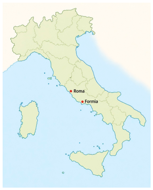
The modern city of Formia is located on the site of the ancient Roman city of Formiae, which was one of the major towns along the Appian Way between Rome and Capua (Figure 1).
The exceptional preservation of pigment traces on these sculptures (face and clothing in particular) provides a valuable opportunity to contribute to current research by applying an integrated non-invasive analytical protocol with the aim of improving our understanding of materials, painting techniques, and decorative schemes in Roman statuary.
This will also help to clarify historical and archaeological issues regarding the social significance of clothing.
The study aims to characterize the pigments and materials used in the decoration of the garments and to reconstruct the preserved decorative patterns. Attention was devoted to the identification of organic lake pigments, which are often difficult to characterize using exclusively non-invasive methods. For this reason, Raman-SERS was employed on microsamples to obtain additional molecular information on the organic dye detected on the female statue. The Raman technique has proven to be a valuable tool for identifying both organic and inorganic pigments [10]. In archaeometry, it has been used both in situ and on microsamples, thanks to its coupling with microscopes. However, for organic dyes, whether of animal or plant origin, the high intrinsic fluorescence of these compounds reduces their effectiveness. To amplify the signal, one of the most widespread approaches in the cultural heritage field is the SERS (Surface-Enhanced Raman Spectroscopy) technique [11]. In this case, the presence of metallic nanostructures allows for significant signal amplification. Numerous studies in literature demonstrate how this technique has allowed the presence of different dyes to be discriminated regardless of their nature. The use of spherical silver nanoparticles is the most widespread approach [12]; these can be deposited directly on the sample, or extraction techniques can be used to improve their effectiveness [13].

2. Materials and Methods

2.1. Archaeological Context and Objects

Some of the Roman remains of the city of Formiae were brought to light on different occasions in the past: in particular, the main road (the ancient Via Appia) with the portico, the theatre, and the area of the forum.
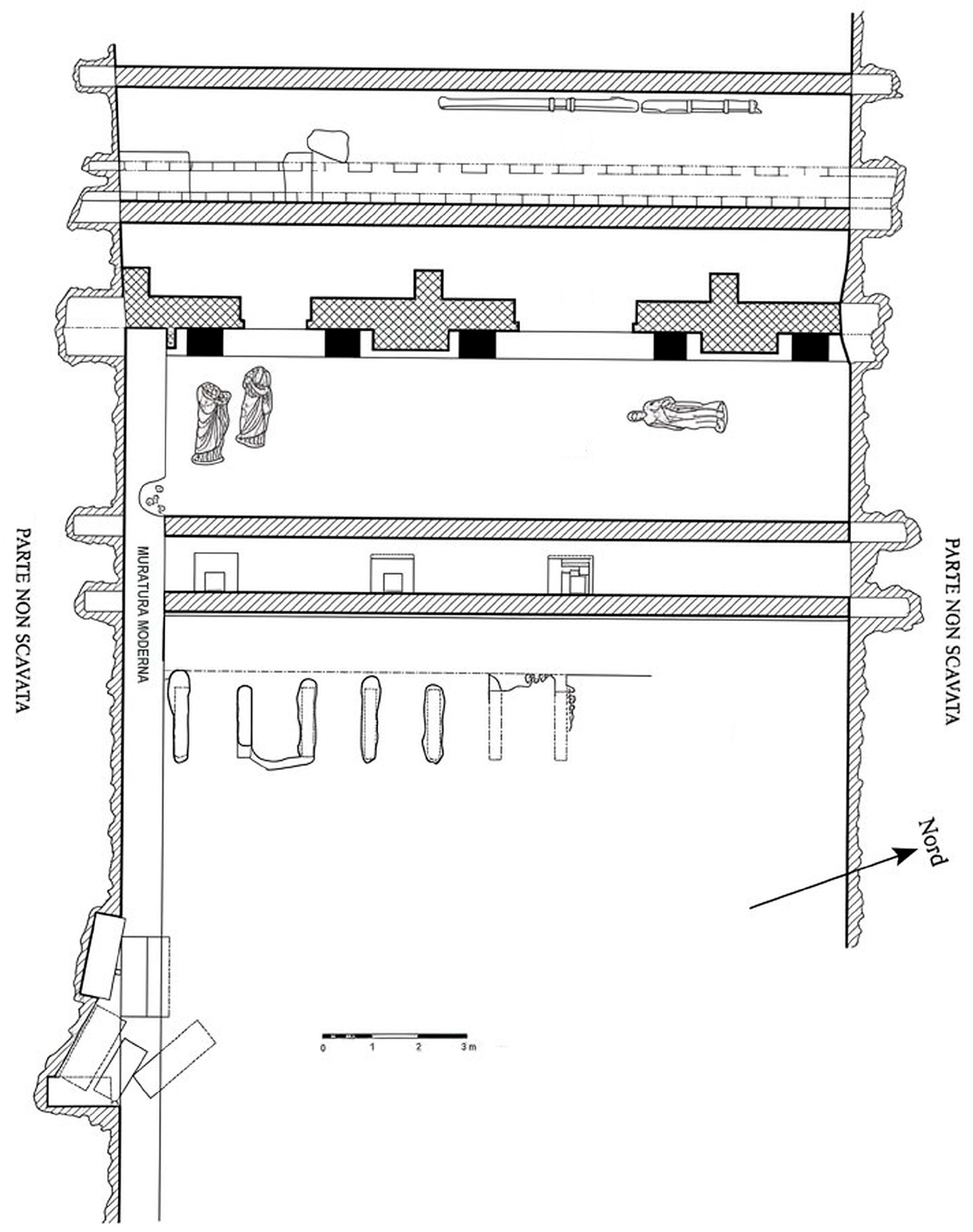
During the archaeological excavations in 1920–1923, eight statues were discovered (three Hüftmantel statues, one male statue with toga, one male head, two female statues, one female head) in Building A on the western side of the forum (Figure 2) [14]. According to the context and level of discovery, the statues are considered in secondary position, likely the result of accumulation after the Late Roman abandonment of the Forum buildings. Although there is no monographic study of the ensemble, they are dated between the late republican and proto-imperial period [15,16] and assigned to the probable decoration of the basilica. Both statues analyzed in this research came to light during the 1921 excavations.
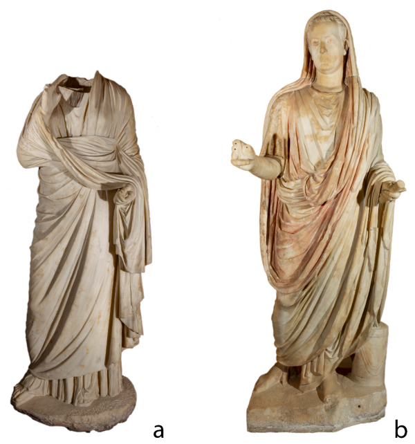
The female statue (Figure 3a) imitates the Hellenistic type of the “Small Herculaneum Woman” of which the best-known example is that of the archaeological museum of Athens, coming from Delos. The type had a notable diffusion in the Roman age from the Republican period to the Imperial one. It was used in portraiture until the Antonine period, such as the well-known example from the Nymphaeum of Herodes Atticus in Olympia. The example from Formia differs in the folding of the himation, in the gesture of the right hand, and in the uncovered left hand. It is the eponymous replica of the Formia Type [17,18,19,20], not very common in municipal contexts, which was more widespread in Greece, especially in the 2nd century AD. The type seems to be found in this statue, not only one of the highest quality examples, but also one of the oldest attestations. Recently, Stefania Tuccinardi [21] confirmed the Augustan chronology.
The togate statue (Figure 3b) has had a long critical history, which, however, agrees in attributing it to the late Augustan period [15,16,22,23,24,25,26,27,28,29,30,31,32,33,34,35] (see p. 24 for the Formia statue). The chronology can be confirmed according to the formal characteristics of the portrait and the type of toga. Aurigemma 1921 [22] mentions the presence of colors in the toga (purple-red), in the eyes (colored with red for the iris and dark red for the pupils), and supposed that his calcei were probably painted red.

2.2. Analytical Instrumentation

The investigation was conducted through a well-established multi-analytical protocol integrating imaging and spectroscopic techniques [36,37,38]. The study was performed in situ and prioritized non-destructive methodologies. The combined application of complementary non-invasive techniques enhances the reliability of pigment identification by integrating spatial, spectral, and compositional information, allowing for cross-validation of the analytical results and reducing the ambiguity associated with individual methods. Imaging techniques provide information on the distribution and localization of pigment traces across the surface, while spectroscopic analyses enable their chemical characterization. This integrated approach partially mitigates the intrinsic limitations of each method, such as restricted sensitivity, spectral overlaps, or limited spatial resolution. However, the results remain influenced by several factors, including the state of surface preservation, post-depositional alteration processes, the possible presence of contamination or restoration materials, and the often weak or degraded signals associated with residual pigments.
Multiband imaging (MBI) was employed as a preliminary survey method. Among the different techniques belonging to the MBI, Visible-Induced Luminescence (VIL) imaging was used to map Egyptian blue exploiting its characteristic infrared luminescence emission, while Ultraviolet-induced Luminescence (UVL) imaging was applied to localize organic materials and luminescent dyes. The analyses were conducted using two cameras for the two photographic techniques. For the VIL [39], the images were acquired with a modified Canon EOS 450D (12.2 Megapixel, CMOS sensor APS-c, maximum resolution of 4272 × 2848 pixels, Canon Europe, Amstelveen, The Netherlands), while for visible and UVL techniques a Canon EOS 7D (18 Megapixel CMOS sensor APS-c, maximum resolution of 5184 × 3456 pixels, Canon Europe, The Netherlands) was used. Both cameras can use the same lens: a Canon EFS 18–135 mm f/3,5–5,6 IS lens with different B + W filters on varying of every photographic technique applied. Different filters were also applied to the two Quantum Qflash T5dR (150 W/s, Quantum Instruments, Humbracht Circle, Bartlett, IL, USA) flashes, equipped with QF30 flashtubes and a filter adapter, thus providing proper radiation.
The following Table 1 summarizes the various setups of cameras and filters used for each technique.
Microscopic observations were carried out using digital optical microscopy (Dino-Lite AM4517MT-FUW (R4), 20–200× magnification, IDCP B.V., Almere, The Netherlands) under visible and UV illumination in order to document pigment morphology and stratigraphic relationships between paint layers. The optical microscopy was also used to document the measured points.
Point analyses were performed using Fiber Optic Reflectance Spectroscopy (FORS) and portable X-ray Fluorescence (XRF). FORS spectra were acquired in the 350–1000 nm range using a tungsten lamp (20 W) as source and the grating Ocean Optics (model HR2000, Ocean Optics, Duiven, The Netherlands) as detector. Optical fiber bundles were used both to deliver the light to the surface under analysis and to collect the reflected radiation. A Labsphere Spectralon® Diffuse Reflectance Standard (Labsphere Inc., North Sutton, NH, USA) was used as reference. Spectra were interpreted, with the help of available spectral databases [40,41], according to characteristic absorption features associated with specific pigments and dyes. The process also involved cross-referencing obtained data with extant bibliographic sources. XRF measurements provided elemental information useful for the identification of inorganic pigments and metallic components. The XRF analyses were performed using a portable device, Tracer III SD Bruker (Bruker, Rosenheim, Germany), equipped with a rhodium X-ray tube, a palladium anticathode, and a solid-state silicon detector energy dispersion system. The setup was as follows: 40 keV and 12 μA for 60 s. The measuring area was an elliptical spot of 4 mm × 7 mm. For data elaboration, ARTAX 7 software (Bruker, Rosenheim, Germany) was used. Spectra were normalized with respect to Rh Kα peak.
To complement the results obtained by the non-invasive measurements, and in accordance with the museum, 2 micro fragments from specific areas were sampled. The first micro sample was taken from the togatus, and it was collected from the left side of the toga. The second micro sample was taken from the female statue, and it was collected from the inner part of a himation’s fold. In both cases, the purpose of the sampling was the characterization of the organic pigments previously detected and mapped with the non-invasive techniques. The samples were analysed by means of Raman SERS using a Renishaw InVia Raman microscope coupled with a Leica DM2500M microscope (Renishaw plc, Kingswood, Wotton-under-Edge, UK), equipped with a CCD detector and a 1800 lines/mm grating. As the excitation source, a 532 nm Nd:YAG laser was used at 1% power (maximum output power 50 mW), with a 20× magnification, an acquisition time of 10 s, and 1 accumulation. Prior to SERS analyses, the silver colloid was prepared following the procedure of Lee and Meisel [42], then concentrated by centrifugation for 50 min at 10,000× g, after which the supernatant was removed. Before SERS analysis, the dyes were extracted by suspending the sample in 50 μL of 0.5 M oxalic acid/methanol/acetone/water (1:30:40:40 v/v/v/v) solution and sonicated for 30 min at 60 °C. For SERS analysis, 5 μL of the Ag colloidal solution (10×) was mixed with 1 μL of sample extract and 1 μL of 2 M KNO3 (aggregating agent), and then a droplet was deposited on a microscope slide. Spectra were acquired immediately by focusing on the top of the droplet.

3. Results

3.1. Female Statue

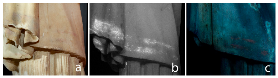
MBI revealed extensive pigment residues localized across the surface of the garments. VIL images clearly documented the presence of Egyptian blue on both the himation and the chiton, producing the characteristic intense luminescence signal in the near-infrared region, associated with this pigment. The application of VIL revealed hidden decorative details, providing a much clearer visualization of the polychromy, which remains imperceptible in standard visible-light photography.
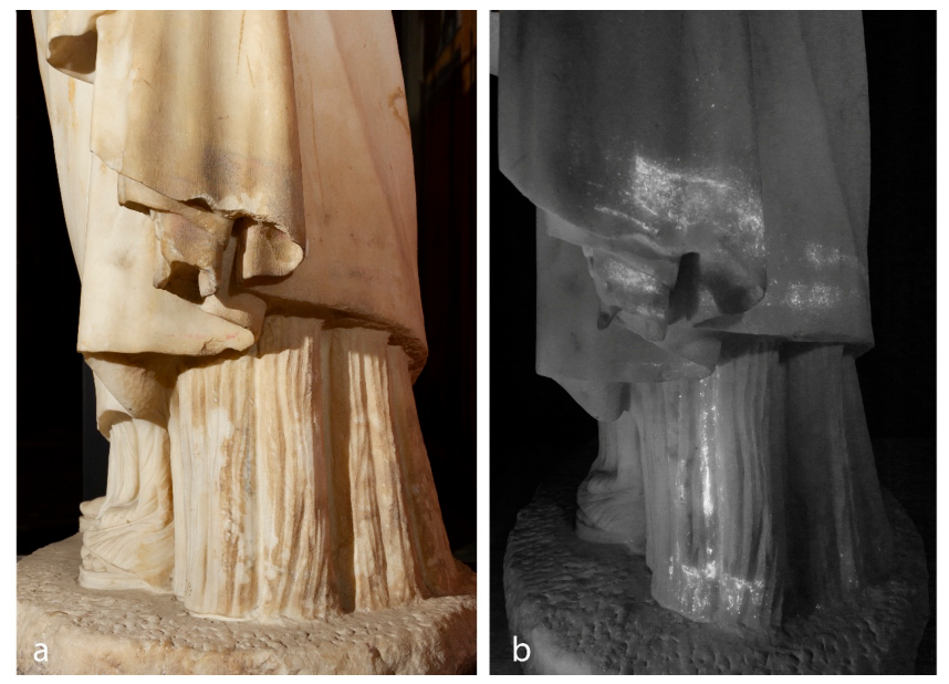
The VIL imaging (Figure 4b) reveals the presence of two decorative bands on the himation that are no longer visible on the front of the statue. The chiton also retains traces of Egyptian blue, specifically along a vertical band on the left side that ends in a decorated strip.
UVL technique highlighted further areas characterized by polychromy residuals, with a strong reddish luminescence, suggesting the presence of an organic lake. The distribution of this luminescence partially overlaps with areas where VIL shows the Egyptian blue emission, indicating either the superposition or intentional mixture of different pigments within the decorative scheme. While the two bands appear well-defined and separated on the right side of the image (Figure 5), the pink traces vanish as they move toward the left, where the Egyptian blue emerges as a singular, indistinct band.
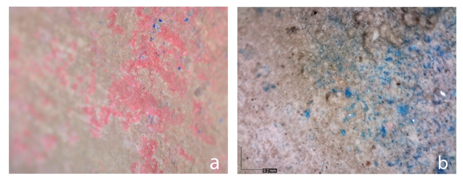
The presence of these decorations, together with the colors chosen on the himation and the chiton (Figure 6), led to a comparison with the iconographic sources and previous analyses on this type of representation. The dress color of the statue offers an element of comparison with the reconstruction presented by Blume [43], showing a marked contrast between the main color of the himation and the decoration, referring to elements associated with Hellenistic practice, such as the use of lake dye and Egyptian blue, and the decorative scheme. These schemes and colors can be seen, for example, in the Tanagra figurines [44] or in the painting of the catechesis scene in the Villa of the Mysteries (Pompei, around 60 BCE, [45] with previous bibliography).
As evidenced by the microscopy images, the traces of pink pigment consistently reveal the inclusion of blue particles. This observation suggests an intentional mixing of pigments, likely an organic lake and Egyptian blue, to achieve a specific purplish hue for the decoration of the himation. In contrast, the decorative elements on the chiton appear to follow a different technical logic, indicating the painter’s deliberate intent to produce a purely blue design.
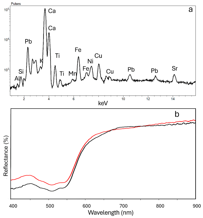
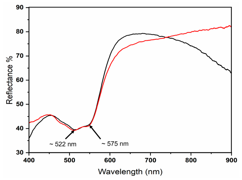
FORS measurements conducted on the pink areas revealed diagnostic absorption features consistent with the spectral behaviour of red organic lakes. The reflectance spectra are characterized by a broad, structured absorption band in the green region of the visible spectrum, typically extending between 500 and 560 nm (Figure 7).
Specifically, the spectral profile displays two characteristic reflectance minima located at approximately 515 nm and 540 nm. These features are attributed to the electronic transitions of hydroxyanthraquinone chromophores, the primary coloring agents in many organic reds. According to established literature [46,47,48], the presence of this double-minimum structure is strongly indicative of a madder-based lake. While alizarin and purpurin are the main components of madder, the precise positioning and relative intensity of these sub-bands are often influenced by the purpurin content, which contributes significantly to the absorption at 540 nm.
The observed absorption in the visible region is principally due to transitions of the carbonyl groups within the anthraquinone structure. Furthermore, the spectral data show a sharp increase in reflectance starting at approximately 600 nm and extending into the Near-Infrared (NIR) region, a characteristic “red rise” typical of these dyes. In the shorter wavelength range, a smaller reflectance peak is observed around 400–420 nm, situated between the main visible absorption and a secondary, intense transition occurring in the UV region (below 350 nm) [49].
The wavelength position and shape of these diagnostic bands are highly sensitive to the local chemical environment, including the nature of the mordant used (likely alum-based for organic lakes) and the presence of other functional groups. Based on these spectroscopic markers, the pink pigment can be conclusively identified as a red lake derived from the Rubia species, most likely Rubia tinctorum. This identification is further supported by the high degree of correlation between the experimental data and reference spectra for purpurin-rich madder lakes, which were traditionally utilized in the Hellenistic period to achieve sophisticated pink and purplish tints through varying concentrations or mixtures with inorganic blue pigments.
To complement the reflectance data, XRF measurements were conducted on the same areas, providing a clear elemental mapping of the inorganic components. The detection of significant copper (Cu) signals confirmed the presence of Egyptian blue, consistent with the VIL emissions observed and reflectance spectra acquired.
Furthermore, the analysis revealed the presence of iron (Fe), with peak intensities suggesting the use of iron-based earth pigments, such as red ochre (hematite). The co-presence of Cu and Fe in the purplish areas supports the hypothesis of a complex pictorial stratigraphy where inorganic earths were likely used as a base or mixed with organic components to adjust the final hue and opacity.
While non-invasive techniques provided a strong preliminary identification, Surface-Enhanced Raman Spectroscopy (SERS) was employed to achieve a definitive molecular characterization of the organic dyes. Informed and guided by the non-invasive mapping, a targeted micro-sampling was performed, and these micro-fragments underwent a SERS analysis.
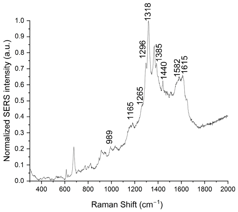
In Figure 8, one of the acquired spectra is reported, showing the typical vibrational “fingerprints” of anthraquinone derivatives contained in the roots of Rubia tinctorum, in particular alizarin and purpurin. The coexistence of these two chromophores represents a distinctive marker of natural madder lake as opposed to modern synthetic pigments.
Specifically, in the spectrum is visible the band at 989 cm−1 that is a diagnostic signal of purpurin, while the band at 1165 cm−1 corresponds to C–H bending of the aromatic ring and is attributable to alizarin. The band at 1265 cm−1 is due to C–C and C–O stretching, and it is very intense in purpurin but also present in alizarin. As reported in the literature [50,51,52,53,54,55], this band, together with the one at 1318 cm−1 (ring stretching), can be considered characteristic of madder. Also, the band at 1440 cm−1, corresponding to C–C stretching and C–H bending, is typical of the lake complex, while the bands at 1582 cm−1 and 1615 cm−1 are attributed, respectively, to stretching of the aromatic ring system and to stretching of the carbonyl group (C=O) and conjugated C=C bonds.
Microscopic examination identified a yellowish-brown paint layer applied across both the chiton and the himation. This layer was strategically thickened or darkened within the folds to create a sense of three-dimensional shading. This use of darker hues indicates a high level of technical skill in simulating the optical properties of fabric in relation to light.
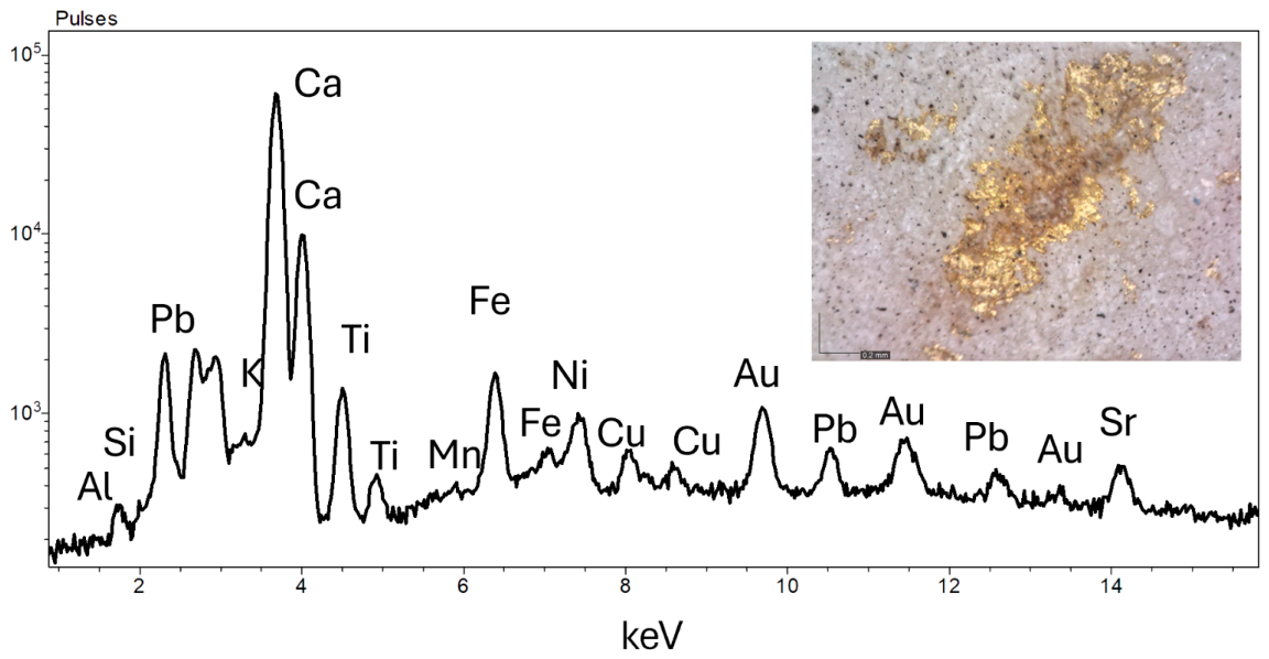
The most prestigious feature identified is the gilding along the neckline of the chiton. Microscopic analysis confirmed the survival of delicate fragments of gold leaf. To validate this, XRF analysis was employed, detecting the characteristic peaks for gold (Au) (Figure 9). The presence of minor elements found in the spectra could be significant, as it is consistent with the geochemical profile of ancient gold leaf rather than modern restorations, confirming the authenticity of the decorative scheme [56,57,58]. Nonetheless, this remains purely speculative, given the non-uniform thickness of the leaf in the measured area, which precludes quantitative analysis, and the possible presence of impurities from the surrounding environment or from having been buried.
The garment’s decoration is characterized by intricate, multi-pigment patterns that highlight the technical skill of the workshop. On the chiton is visible a series of blue vertical lines on the lower section, reinforcing the structured, classical silhouette of the dress. The himation displays a more complex arrangement along the edge, consisting of a pink band embellished with blue.
These details indicate that the painter did not simply apply pure colors but utilized complex mixtures and stratigraphic layering to achieve sophisticated visual textures. The juxtaposition of pink (madder lake) and blue (Egyptian blue) suggests a deliberate attempt to create a vibrant, high-contrast border that would have been highly visible in the original context.

3.2. Togate Statue

The analytical investigation of the togate statue revealed a similarly complex pigment system, indicative of high-level craftsmanship. This statue has notably better-preserved polychromy, particularly on the face and on the drapery of the toga.
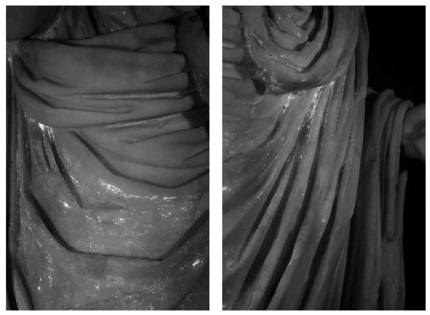
VIL imaging successfully detected widespread traces of Egyptian blue across multiple regions of the sculpture (Figure 10). Significant areas characterised by intense luminescence were documented within the deep folds of the toga, on the veil covering the head, and along the lower sections adjacent to the legs, highlighting an extensive use of this pigment.
Integrating these findings, UVL imaging revealed an intense, characteristic reddish-pink luminescence across extensive portions of the garment, a feature diagnostic of the presence of an organic red lake. The spatial distribution of the UVL signal suggests that the red colorant was either layered over or physically mixed with the Egyptian blue. Such a technical choice may have been intended to achieve specific chromatic effect, such as a rich purple hue associated with high-status Roman garments [59].
High-magnification documentation has provided physical confirmation of this pigment recipe. Microscopic observations of the surface of the garment confirmed the coexistence of blue and pink pigments within the same stratigraphic layer. This evidence supports the hypothesis of a deliberate blending of inorganic and organic materials, aimed at achieving a nuanced tonal range that would have enhanced the naturalistic rendering of the volume and texture of the toga.
The FORS spectra acquired from the reddish-pink areas exhibit absorption features characteristic of anthraquinone-based organic dyes (Figure 11).
Initial observation of the spectral profile, specifically the position of the main absorption bands, appears slightly shifted compared to standard madder references. As demonstrated by Fonseca et al. [47], the absorption mechanism in anthraquinones is strictly related to electronic transitions between delocalized molecular orbitals. Consequently, the presence of specific substituents on the aromatic rings, including various functional groups and mordanting ions, along with their relative positions and the overall chemical environment. These factors can significantly affect the position of the absorption bands both below 400 nm and within the 500–600 nm range. Therefore, while the current spectral data might tentatively point toward an animal-based lake, the observed shifts could also be interpreted as a variation of a vegetable-based lake (madder), where the specific mordanting process or the local chemical environment has altered the standard spectral behaviour.
While there is evidence about the potential use of an animal-derived lake on coeval artefacts [60,61], historical and archaeological evidence indicates that madder lake was significantly more common and more easily sourced during this period [62,63]. A definitive identification would require molecular-level analytical techniques. However, the micro-sample previously subjected to SERS analysis proved to be inconclusive, likely due to the low concentration of the organic chromophores. Consequently, further speculation regarding the origin of the lake, whether animal or vegetable, is currently not possible until new analytical evidence becomes available.
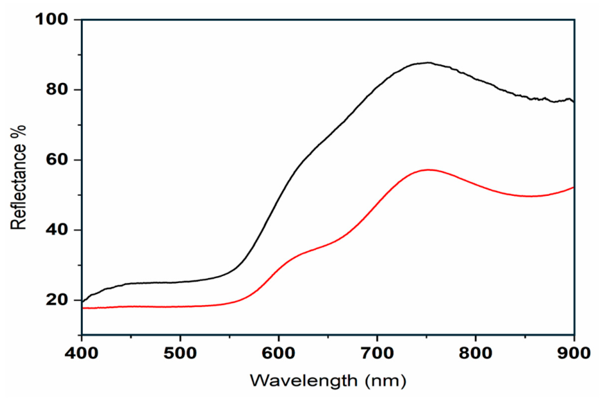
The XRF campaign provided essential elemental validation of the pigments identified through imaging techniques. Measurements performed confirmed concentration of copper (Cu), providing a direct elemental correlation with the presence of Egyptian blue. In the same areas, the detection of iron (Fe) peaks points toward the use of iron-based earth pigments. While iron is often ubiquitous in archaeological findings, due to burial, the specific correlation of these peaks with the darker or more opaque passages suggests the deliberate addition of ochre pigments, with different compositions, likely hematite-rich red ochre or goethite-based yellow ochre. FORS measurements on these areas confirm the hypothesis. In Figure 12, an example of a FORS spectrum, acquired on a red area, is reported in comparison with a reference spectrum of a hematite-rich iron-based pigment.
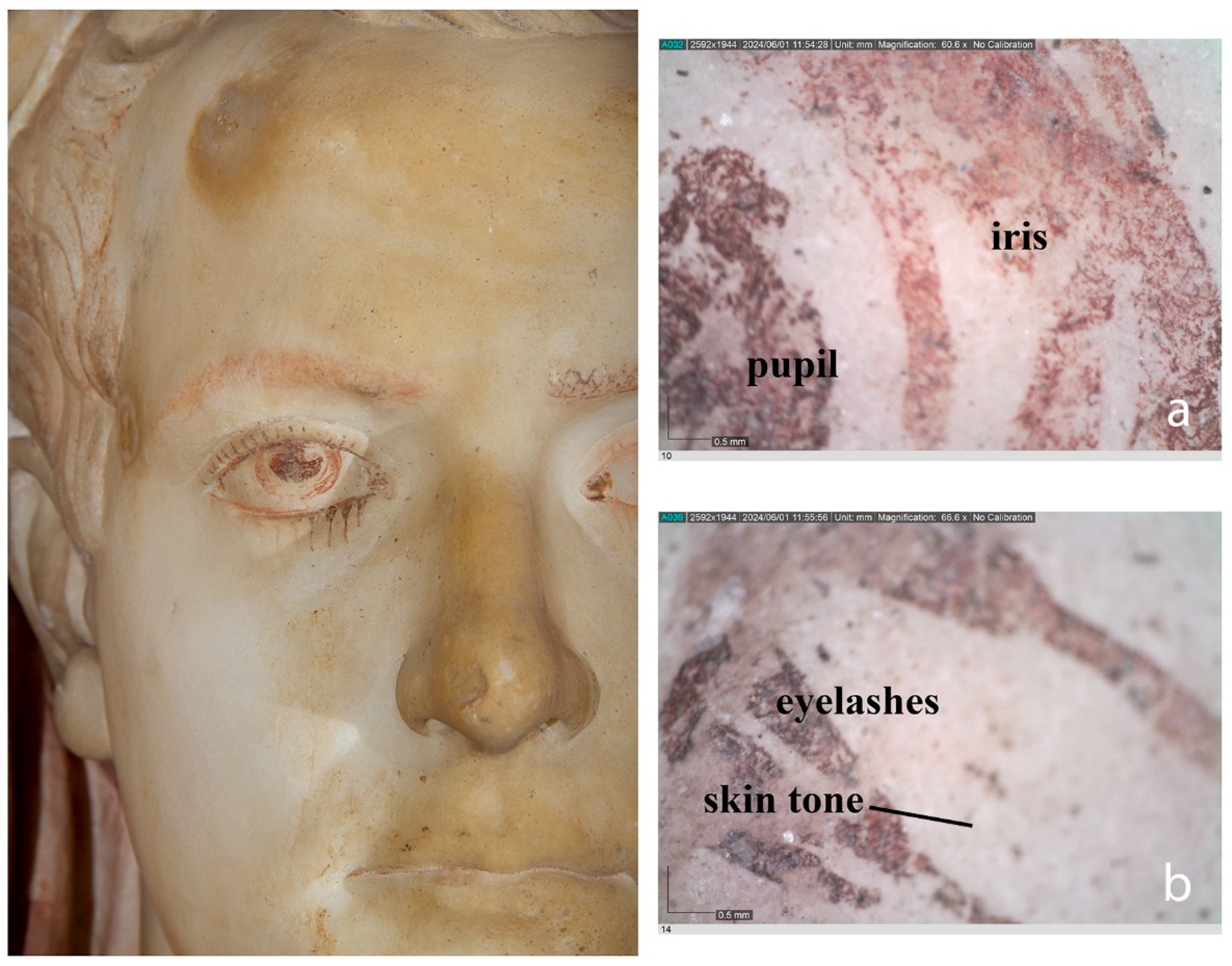
The precise decoration of the facial attributes, particularly the eyes, underscores the exceptional artistic quality of the sculpture. As previously noted, the left eye preserves a remarkably sophisticated stratigraphic sequence, allowing for a detailed reconstruction of the painter’s methodology in depicting irises, pupils and lashes (Figure 13).
Microscopic examination of the head revealed the use of brown pigments within the hair, applied to create depth and texture. The skin tones were rendered through the application of a light yellowish base layer, likely intended to impart a naturalistic warmth to the marble surface. This flesh-tone layer appears to have been applied with varying thickness to modulate light and shadow across the facial contours.
The examination of the eyes revealed a complex paint system. Microscopy images suggested a multi-stage execution process. A preliminary red underdrawing was applied directly onto the marble surface to define the ocular anatomy. This red guide was subsequently overlaid with a lighter flesh-colored pigment, likely to soften the transitions and prepare the area for the paint details. In the final stage, black lines, likely carbon-based, were used with high precision to define the pupils, eyelashes, and eyelids.
Beyond the main garments, additional pigment traces were identified on the right calceus (footwear) and across the upper surface of the statue’s plinth. While these residues indicate that the polychromy once extended to the lowermost details of the monument, their precise chemical composition could not be fully established. These specific regions were subject to limited analytical access, preventing the optimal positioning of the FORS and XRF device heads.

4. Conclusions

The analytical campaign conducted on the two sculptures from Formia provides a rigorous evidence-based reconstruction of the advanced polychromatic systems characteristic of high-status Roman statuary. The synergy between non-invasive imaging, elemental and molecular spectroscopy allowed for the identification of a stratified pictorial scheme that reflects the complexity of ancient polychrome statuary.
The integrated analytical protocol, based primarily on non-invasive methods, allowed the identification of Egyptian blue, iron-based pigments, gilding, and organic red lake pigments. Multiband imaging proved essential for detecting pigment residues and reconstructing decorative patterns that are no longer visible.
The identification of organic red lakes represented a challenge in this research, achieved through the strategic integration of spectroscopic techniques. While FORS established the presence of anthraquinone-based chromophores, the application of SERS was necessary to provide a definitive molecular fingerprint. The analytical data obtained from the female statue definitively confirms the presence of madder lake (Rubia tinctorum), a natural plant-derived dye widely utilized in antiquity for its vibrant pink and red hues. The molecular identification was made possible through the detection of specific alizarin and purpurin markers, which characterize the chemical signature of the Rubia species.
In contrast, the togatus statue exhibits distinct spectral shifts in the reflectance data. While these variations suggest a similarly sophisticated application of natural-based organic colorants, they do not perfectly align with the standard madder profile. These shifts may indicate the use of a different biological source—potentially an insect-derived dye such as kermes—or could be the result of complex interactions between the dye, the metallic mordant, and the inorganic pigments used in the mixture.
Given these nuances, the exact origin of the lake on the togatus remains a hypothesis that requires further analytical confirmation. The ambiguity underscores the necessity of high-resolution molecular diagnostics to differentiate between various anthraquinone-based dyes, as electronic transitions in the visible spectrum can be significantly influenced by the local chemical environment and the specific recipe employed by the Roman workshop.
The exceptional state of pigment conservation on these sculptures underscores the necessity of systematic, high-resolution analytical investigations for the accurate reconstruction of ancient sculptural polychromy. If we focus specifically on sculptures of this type dating to the Julio-Claudian period, several interesting comparisons can be identified, although unfortunately none is supported by analytical data. These include the headless togatus from Tarragona [31,64,65], with traces of red on both the toga and the tunic; a head from Tarragona, possibly representing Augustus [66], covered by a veil bearing traces of red; and, finally, the seated statues of Augustus and Caligula from Torreparedones in Spain, which preserve traces of purple on their togas [35,59].
These findings provide evidence of the sophisticated painting practices inherent in Roman marble statuary, where material selection and stratigraphic complexity were of utmost importance. Ultimately, the successful characterization of this pictorial system validates the efficacy of integrated, multi-technical methodologies in reading the technical and aesthetic evolution of archaeological polychromy.

Author Contributions

Conceptualization, P.L., D.M., G.B., R.I., E.N. and S.L. (Sara Lenzi); methodology, D.M., G.B., R.I. and E.N.; software, D.M., G.B. and R.I.; formal analysis, D.M., G.B., R.I., S.L. (Stefano Legnaioli) and G.L.; investigation, D.M., G.B., R.I. and E.N.; resources, P.L., E.N. and S.L. (Sara Lenzi); data curation, D.M., G.B., R.I., S.L. (Stefano Legnaioli) and G.L.; writing—original draft preparation, D.M., G.B. and R.I.; writing—review and editing, P.L., D.M., G.B., R.I., E.N., S.L. (Sara Lenzi), S.L. (Stefano Legnaioli) and G.L.; supervision, P.L. and D.M. All authors have read and agreed to the published version of the manuscript.

Funding

This research received no external funding.

Institutional Review Board Statement

Not applicable.

Informed Consent Statement

Not applicable.

Data Availability Statement

The original contributions presented in this study are included in the article. Further inquiries can be directed to the corresponding author.

Acknowledgments

The authors wish to thank Cristiana Ruggini and all the staff of the National Archaeological Museum of Formia.

Conflicts of Interest

The authors declare no conflicts of interest.

References and Notes

  1. Liverani, P. Per una ‘Storia del colore’. La scultura policroma romana, un bilancio e qualche prospettiva. In Diversamente Bianco. La Policromia Della Scultura Romana; Liverani, P., Santamaria, U., Eds.; Quasar: Rome, Italy, 2014; pp. 9–32. [Google Scholar]
  2. Østergaard, J.S. The polychromy, sculptural, Greek and Roman. In Oxford Classical Dictionary; Grig, L., Ed.; Oxford Academic: New York, NY, USA, 2015. [Google Scholar] [CrossRef]
  3. Abbe, M. Art. In A Cultural History of Colour in Antiquity; Wharton, D., Ed.; Bloomsbury: London, UK, 2021; pp. 220–248. [Google Scholar]
  4. Sandu, I.C.A. Colour Through Time. In Chromatic Visions; Lecture Notes in Computer Science; Pescarin, S., Barandoni, C., Clay, A., Papadopoulos, G., Sandu, I.C.A., Eds.; Springer: Cham, Switzerland, 2026; Volume 15750. [Google Scholar] [CrossRef]
  5. Rodler, A.; Matthys, S.M.; Brøns, C.; Artioli, G.; Snoeck, C.; Debaille, V.; Goderis, S. Investigating the provenance of Egyptian blue pigments in ancient Roman polychromy. Archeometriai Műhely 2021, 18, 97–108. [Google Scholar] [CrossRef]
  6. Frantzi, G.G.; Mastrotheodoros, G.P.; Theoulakis, P.; Kogou, S.; Psalti, A.; Brecoulaki, H. Unveiling the Original Polychromy of Archaic Architecture: The Gigantomachy on the West Pediment of the Temple of Apollo at Delphi (6th c. B.C.). Heritage 2025, 8, 17. [Google Scholar] [CrossRef]
  7. Ferdani, D.; Barandoni, C.; Bonifazi, F.; Iannaccone, R.; Magrini, D. Multiband photogrammetry for multispectral 3D reconstruction of statuary. J. Cult. Herit. 2025, 76, 74–85. [Google Scholar] [CrossRef]
  8. Aggelakopoulou, E.; Sotiropoulou, S.; Karagiannis, G. Architectural Polychromy on the Athenian Acropolis: An In Situ Non-Invasive Analytical Investigation of the Colour Remains. Heritage 2022, 5, 756–787. [Google Scholar] [CrossRef]
  9. Verri, G.; Granger-Taylor, H.; Jenkins, I.; Sweek, T.; Weglowska, K.; Wootton, W.T. The goddess’ new clothes: The carving and polychromy of the Parthenon sculptures. Antiquity 2023, 97, 1173–1192. [Google Scholar] [CrossRef]
  10. Burgio, L.; Clark, R.J.H. Library of FT-Raman spectra of pigments, minerals, pigment media and varnishes, and supplement to existing library of Raman spectra of pigments with visible excitation. Spectrochim. Acta—Part A Mol. Biomol. Spectrosc. 2001, 57, 1491–1521. [Google Scholar] [CrossRef] [PubMed]
  11. Pozzi, F.; van den Berg, K.J.; Fiedler, I.; Casadio, F. A systematic analysis of red lake pigments in French Impressionist and Post-Impressionist paintings by surface-enhanced Raman spectroscopy (SERS). J. Raman Spectrosc. 2014, 45, 1119–1126. [Google Scholar] [CrossRef]
  12. Campanella, B.; Palleschi, V.; Legnaioli, S. Introduction to vibrational spectroscopies. ChemTexts 2021, 7, 5. [Google Scholar] [CrossRef]
  13. Whitney, A.V.; Van Duyne, R.P.; Casadio, F. An innovative surface-enhanced Raman spectroscopy (SERS) method for the identification of six historical red lakes and dyestuffs. J. Raman Spectrosc. 2006, 37, 993–1002. [Google Scholar] [CrossRef]
  14. Aurigemma, S. Ritratto femminile dell’età di Augusto e statue muliebri iconiche scoperte a Formia. Boll. d’Arte 1930, 10, 216–236. [Google Scholar]
  15. Cassieri, N. Museo Archeologico Formia; Marina di Minturno: Latina, Italy, 2001. [Google Scholar]
  16. Cassieri, N. Formia. Museo Archeologico Nazionale e Monumenti; Istituto poligrafico e Zecca dello Stato, Libreria dello Stato: Roma, Italy, 2013.
  17. Schmidt, E. Römische Frauenstatuen. Ph.D. Thesis, Berlin, Germany, 1967. [Google Scholar]
  18. Kruse, H.J. Römische Weibliche Gewandstatuen des zweiten Jahrhunderts n. Chr. Ph.D. Thesis, Georg-August Universität zu Göttingen, Göttingen, Germany, 1975. [Google Scholar]
  19. Bol, R. Das Statuenprogramm des Herodes-Atticus-Nymphäums; Hoffmann, A., Schumacher, L.-H., Eds.; Walter de Gruyter: Berlin, Germany, 1984; Volume 1. [Google Scholar]
  20. Filges, A. Standbilder Jugendlicher Göttinnen. Klassische und Frühhellenistische Gewandstatuen mit Brustwulst und ihre Kaiserzeitliche Rezeption; Böhlau: Köln, Germany, 1997. [Google Scholar]
  21. Tuccinardi, S. Sculture romane da Formia. Una proposta di lettura in contesto. In Complessi Monumentali e Arredo Scultoreo Nella Regio I: Latium et Campania; Naus: Napoli, Italy, 2017; pp. 49–68. [Google Scholar]
  22. Aurigemma, S. Recenti scoperte in Formia. Boll. d’Arte 1921, 1, 309–335. [Google Scholar]
  23. Aurigemma, S. Scoperte d’arte in Formia e Venafro. L’Illustrazione Ital. 1922, 9, 420–421. [Google Scholar]
  24. Strong, E. Art in Ancient Rome I; C. Scribner’s Sons: New York, NY, USA, 1929. [Google Scholar]
  25. Strong, E. La Scultura Romana da Augusto a Costantino; Fratelli Alinari: Florence, Italy, 1923. [Google Scholar]
  26. Deubner, L. Die Tracht des Römischen Triumphators. Hermes 1934, 69, 316–323. [Google Scholar]
  27. Goethert, F.W. Studien zur Kopienforschung. Mitt. d. Dtsch. Archäol. Inst. Röm. Abt. 1939, 54, 176–219. [Google Scholar]
  28. Zanker, P. Zur Bildrepräsentation führender Männer in mittelitalischen und campanischen Städten zur Zeit der späten Republik und der julisch-claudischen Kaiser. In Les “Bourgeoisies” Municipales Italiennes aux IIe et Ier Siècles av. J.-C.; Centre Jean Bérard: Naples, Italy, 1983; pp. 251–266. [Google Scholar]
  29. Döhl, H.; Zanker, P. La scultura. In Pompei 79. Raccolta di Studi per il Decimonono Centenario Dell’eruzione Vesuviana; Zevi, F., Ed.; Macchiaroli: Naples, Italy, 1984; pp. 177–210. [Google Scholar]
  30. Goette, H.R. Mulleus, Embas, Calceus. Ikonographische Studien zu römischem Schuhwerk. Jahrb. d. Dtsch. Archäol. Inst. 1988, 103, 401–464. [Google Scholar]
  31. Goette, H.R. Studien zu Römischen Togadarstellungen; Philipp von Zabern: Mainz, Germany, 1990; p. 115; Ba 36. [Google Scholar]
  32. Barry, F. A Whiter Shade of Pale: Relative and Absolute White in Roman Sculpture and Architecture. In Revival and Invention: Sculpture and Its Material Histories; Clerbois, S., Droth, M., Eds.; Peter Lang: New York, NY, USA, 2011; pp. 31–62. [Google Scholar]
  33. Mesolella, G. La Decorazione Architettonica di Minturnae, Formiae, Tarracina; L’età Augustea e Giulio-Claudia; L’Erma di Bretschneider: Rome, Italy, 2012. [Google Scholar]
  34. Tuccinardi, S. I ritratti del Museo Archeologico di Formia. In Formiae. Una Città All’inizio Dell’impero; Cassieri, N., Ed.; D’Arco edizioni: Formia, Italy, 2014; pp. 58–66. [Google Scholar]
  35. La Rocca, E. La statua di Augusto di Torreparedones: Un modello per la ricostruzione del divus Augustus pater ad theatrum Marcelli. In Escultura Romana na Hispânia: Atas do X Encontro Internacional de Escultura Romana na Hispânia, Faro-Mértola, Portugal, 27–29 October 2022; Bernardes, J.P., Nogales-Basarrate, T., Gonçalves, L.J., Lopes, V., Lopes, M., Eds.; Universidade do Algarve: Faro, Portugal, 2024; pp. 13–64. [Google Scholar]
  36. Magrini, D.; Bracci, S.; Bartolozzi, G.; Iannaccone, R.; Lenzi, S.; Liverani, P. Revealing Mithras’ color with the ICVBC mobile lab in the museum. Heritage 2019, 2, 2160–2170. [Google Scholar] [CrossRef]
  37. Liverani, P.; Bracci, S.; Iannaccone, R.; Lenzi, S.; Magrini, D.; Mazzei, B. Colours in the dark: New research into catacombs. In Polychromy in Ancient Sculpture and Architecture; Bracci, S., Giachi, G., Paolucci, F., Eds.; Sillabe: Livorno, Italy, 2018; pp. 111–121. [Google Scholar]
  38. Liverani, P.; Bracci, S.; Iannaccone, R.; Lenzi, S.; Magrini, D. New Evidence about the Polychromy of Early Imperial Cycle from the Augusteum of Rusellae (Tuscany). Heritage 2023, 6, 3385–3401. [Google Scholar] [CrossRef]
  39. Accorsi, G.; Verri, G.; Bolognesi, M.; Armaroli, N.; Clementi, C.; Miliani, C.; Romani, A. The Exceptional Near-Infrared Luminescence Properties of Cuprorivaite (Egyptian Blue). Chem. Commun. 2009, 23, 3392–3394. [Google Scholar] [CrossRef] [PubMed]
  40. CNR-IFAC Database. Available online: https://spectradb.ifac.cnr.it/ (accessed on 16 April 2026).
  41. CNR-ISPC (Florence, Italy). Fiber Optics Reflectance Spectra (FORS) Database of Pictorial Materials. Unpublished. [CrossRef]
  42. Lee, P.; Meisel, D. Adsorption and surface-enhanced Raman of dyes on silver and gold sols. J. Phys. Chem. 1982, 86, 3391–3395. [Google Scholar] [CrossRef]
  43. Blume, C. Polychromie Hellenistischer Skulptur: Ausführung, Instandhaltung und Botschaften; Michael Imhof Verlag: Petersberg, Germany, 2015; pp. 1–250. [Google Scholar]
  44. Jeannet, V. Tanagras. Figurines for Life and Eternity: The Musée du Louvre’s Collection of Greek Figurines; The Musee du Louvre: Paris, France, 2010.
  45. Esposito, F.; Falcucci, C.; Ferrara, D. La tecnica esecutiva dei dipinti del Salone 5 della Villa dei Misteri: Cento anni di ipotesi e ricerche. Riv. Studi Pompeiani 2011, 22, 149–158. [Google Scholar]
  46. Clementi, S.; Doherty, B.; Miliani, C.; Romani, A. Spectroscopic characterization of red lake pigments based on alizarin and purpurin. Appl. Phys. A 2008, 92, 25–33. [Google Scholar]
  47. Fonseca, B.; Schmidt Patterson, C.; Ganio, M. Seeing red: Towards an improved protocol for the identification of madder- and cochineal-based pigments by fiber optics reflectance spectroscopy (FORS). Herit. Sci. 2019, 7, 92. [Google Scholar] [CrossRef]
  48. Aceto, L.; Agostino, A.; Fenoglio, E.; Gulmini, M.; Idone, C. Seeing red: Towards an improved protocol for the identification of madder- and cochineal-based pigments by fiber optics reflectance spectroscopy (FORS). npj Herit. Sci. 2019, 2, 32. [Google Scholar]
  49. Bisulca, C.; Picollo, M.; Bacci, M.; Kunzelman, D. UV-Vis-NIR reflectance spectroscopy of red lakes in paintings. In Proceedings of the 9th International Conference on NDT of Art, Jerusalem, Israel, 25–30 May 2008. [Google Scholar]
  50. Van Elslande, E.; Lecomte, S.; Le Hô, A.-S. Micro-Raman spectroscopy (MRS) and surface-enhanced Raman scattering (SERS) on organic colourants in archaeological pigments. J. Raman Spectrosc. 2008, 39, 1001–1006. [Google Scholar] [CrossRef]
  51. Trusso, S.; Saija, F.; Ponterio, R.; Nardo, V.; Aliotta, F.; Mastelloni, M.; Ponterio, R.; Vasi, C. A spectroscopic approach to the study of organic pigments in the field of cultural heritage. Atti Della Accad. Peloritana Dei Pericolanti 2017, 95, A5. [Google Scholar] [CrossRef]
  52. Cañamares, M.V.; Garcia-Ramos, J.; Domingo, C.; Sanchez-Cortes, S. Surface-enhanced Raman scattering study of the adsorption of the anthraquinone pigment alizarin on Ag nanoparticles. J. Raman Spectrosc. 2004, 35, 921–927. [Google Scholar] [CrossRef]
  53. Pozzi, F.; Lombardi, J.; Leona, M. Winsor & Newton original handbooks: A surface-enhanced Raman scattering (SERS) and Raman spectral database of dyes from modern watercolor pigments. Herit. Sci. 2013, 1, 23. [Google Scholar] [CrossRef]
  54. Shadi, I.T.; Chowdhry, B.Z.; Snowden, M.J.; Withnall, R. Semi-quantitative analysis of alizarin and purpurin by surface-enhanced resonance Raman spectroscopy (SERRS) using silver colloids. J. Raman Spectrosc. 2004, 35, 800–807. [Google Scholar] [CrossRef]
  55. Bruni, S.; Guglielmi, V.; Pozzi, F. Historical organic dyes: A surface-enhanced Raman scattering (SERS) spectral database on Ag Lee–Meisel colloids aggregated by NaClO4. J. Raman Spectrosc. 2011, 42, 1267–1281. [Google Scholar] [CrossRef]
  56. Neri, E.; Verità, M. Glass and metal analyses of gold leaf tesserae from 1st to 9th century mosaics. J. Archaeol. Sci. 2013, 40, 4596–4606. [Google Scholar] [CrossRef]
  57. Neri, E.; Verità, M.; Biron, I.; Guerra, M.F. Glass and gold: Analyses of 4th–12th centuries Levantine mosaic tesserae. J. Archaeol. Sci. 2016, 70, 158–171. [Google Scholar] [CrossRef]
  58. Buccolieri, A.; Degl’Innocenti, E.; Cesareo, R.; Castellano, A.; Buccolieri, G. Non-invasive in-situ analysis of a wreath of gold leaves from the National Archaeological Museum of Taranto, Italy. Measurement 2018, 126, 164–167. [Google Scholar] [CrossRef]
  59. Ventura Villanueva, Á.; Fernández Cubero, L. El color de la immagine imperial en Torreparedones: Estudio de la policromía en las estatuas sedentes del foro. In Escultura Romana en Hispania VIII; Baena del Alcázar, L., Márquez, C., Ojeda Nogales, D., Eds.; UCOPress: Córdoba, Spain, 2018; pp. 733–752. [Google Scholar]
  60. Dyer, J.; Tamburini, D.; Sotiropoulou, S. The identification of lac as a pigment in ancient Greek polychromy—The case of a Hellenistic oinochoe from Canosa di Puglia. Dye. Pigm. 2018, 149, 122–132. [Google Scholar] [CrossRef]
  61. Fostiridou, A.; Karapanagiotis, I.; Vivdenko, S.; Lampakis, D.; Mantzouris, D.; Achilara, L.; Manoudis, P. Identification of pigments in Hellenistic and roman funeral figurines. Archaeometry 2016, 58, 453–464. [Google Scholar]
  62. Grifa, C.; Germinario, C.; Pagano, S.; Lepore, A.; De Bonis, A.; Mercurio, M.; Morra, V.; Zuchtriegel, G.; Hay, S.; Esposito, D.; et al. Pompeian pigments. A glimpse into ancient Roman colouring materials. J. Archaeol. Sci. 2025, 177, 106201. [Google Scholar] [CrossRef]
  63. Gliozzo, E.; Pizzo, A.; La Russa, M.F. Mortars, plasters and pigments—Research questions and sampling criteria. Archaeol. Anthropol. Sci. 2021, 13, 193. [Google Scholar] [CrossRef]
  64. Koppel, E.M. Die Römischen Skulpturen von Tarraco; De Gruyter: Berlin, Germany, 1985; pp. 15–16, 28–29, n. 4, pl. 4.1-3. [Google Scholar]
  65. Goette, H.R. Studien zu Römischen Togadarstellungen; Philipp von Zabern: Mainz, Germany, 1990; p. 29, n. 122, f, pl. 5.3. [Google Scholar]
  66. Koppel, E.M. Die Römischen Skulpturen von Tarraco; De Gruyter: Berlin, Germany, 1985; p. 32, n. 44, pl. 12.1-2. [Google Scholar]
Figure 1. Map of Italy highlighting the geographic positions of Rome, the capital city, and Formia.
Applsci 16 07399 g001
Figure 2. Plan of the building with the localization of three statues (redrawn from [14]).
Applsci 16 07399 g002
Figure 3. Roman sculptures from Formia. National Archaeological Museum of Formia, Italy. (a) Female statue (inv. 147680, 166 cm); (b) Togatus (inv. 147614, 204 cm).
Applsci 16 07399 g003
Figure 4. Lower edge of the female figure’s chiton, showing the presence of Egyptian blue (EB). (a) Visible image; (b) VIL image with EB glowing white. @CNR-ISPC.
Applsci 16 07399 g004
Figure 5. Detail of the decoration of the himation on the left side of the female statue (inv. 147680). (a) Visible (VIS) image of the analysed area; (b) VIL image highlighting the presence and spatial distribution of Egyptian blue, glowing white; (c) UVL image showing the spatial localization of red lake and its characteristic reddish luminescence @CNR-ISPC.
Applsci 16 07399 g005
Figure 6. Details of high-magnification images of the polychromy on the female statue (50×). (a) Detail on the himation’s decoration with a mixture of red lake and EB; (b) Detail of the chiton’s decoration obtained with purely EB.
Applsci 16 07399 g006
Figure 7. XRF (a) and FORS (b) spectrum acquired on a pink area of the himation (female statue, inv. 147680, black line), suggesting the presence of a madder-based lake indicated by characteristic absorption features in the FORS spectrum, and Egyptian blue, identified through elemental signals in the XRF analysis through the presence of copper. The reference FORS spectrum of a madder lake [40,41] is shown in red.
Applsci 16 07399 g007
Figure 8. Acquired Raman SERS spectrum showing the characteristic vibrational features used for the identification of a madder lake.
Applsci 16 07399 g008
Figure 9. Details of the XRF spectrum and the high-magnification images (50×) of a golden trace on the chiton of the female statue (inv. 147680).
Applsci 16 07399 g009
Figure 10. Togatus statue. VIL images of two details of the folds of the toga, highlighting the presence and spatial distribution of Egyptian blue, appearing as bright white in gray scale image.
Applsci 16 07399 g010
Figure 11. FORS spectrum acquired on a pink area of the toga (black line), showing characteristic absorption features consistent with the presence of an organic red lake. The reference spectrum of a madder lake [40,41] is shown in red.
Applsci 16 07399 g011
Figure 12. FORS spectrum acquired from a red area of the toga showing the characteristic absorption bands of a hematite-based pigment (black line). The reference spectrum of a red ochre (mainly hematite) [40,41] is shown in red.
Applsci 16 07399 g012
Figure 13. Togatus statue. Optical microscopy details (50×) of (a) the iris and pupil and (b) eyelashes and skin tone.
Applsci 16 07399 g013
Table 1. Cameras and filter setups used for the acquisitions.
TechniqueCameraFilter on LensFilters on Flashes
UVLCANON EOS 7DB + W 468 UV/IR cutB + W 403 UV black
VISCANON EOS 7DB + W 468 UV/IR cutB + W 468 UV/IR cut
VILCANON EOS 7DB + W 093 IRB + W 468 UV/IR cut
Disclaimer/Publisher’s Note: The statements, opinions and data contained in all publications are solely those of the individual author(s) and contributor(s) and not of MDPI and/or the editor(s). MDPI and/or the editor(s) disclaim responsibility for any injury to people or property resulting from any ideas, methods, instructions or products referred to in the content.