1. Introduction
Fermented plant-based beverages have gained attention because they diversify fermented food options and can be formulated as alternatives to conventional dairy products [
1]. Their development requires attention to matrix composition, starter culture performance, product safety, and consumer acceptance. Organic and minimally processed ingredients are also used in this area, although the environmental profile of a specific food cannot be inferred from the ingredient category alone and requires separate assessment across the production chain [
2,
3].
Fermentation with lactic acid bacteria (LAB) is widely used in the production of foods such as yogurts, pickled vegetables, and plant-based fermented beverages. This process can support product preservation by lowering pH and limiting the growth of undesirable microorganisms without intensive processing or synthetic preservatives [
4]. LAB may also affect sensory properties and nutritional value by improving mineral bioavailability and producing bioactive compounds [
5]. One research direction is the use of plant materials or their extracts as natural functional additives in fermentation processes. This approach is consistent with the concept of “food and medicine sharing the same origin”, which is increasingly interpreted in global food science as a basis for modern food–medicine homology, functional food development and preventive nutrition [
6,
7]. Plant materials are considered not only as sources of nutrients, flavor, or color, but also as complex matrices of bioactive compounds that may support the development of functional, preventive, and evidence-based food products. They are sources of polyphenols, flavonoids, and essential oils [
8], many of which show antioxidant or antibacterial properties [
9]. Phenolic compounds may inhibit bacteria by disturbing cell membrane integrity, intracellular pH homeostasis, and enzyme activity, whereas other plant components, including carbohydrates, minerals, or polysaccharides, may support LAB growth and survival [
10,
11]. Moreover, some LAB strains can metabolize plant phenolics through enzymes such as β-glucosidases, esterases, decarboxylases, and reductases, which may modify the antioxidant, antimicrobial, and sensory properties of the fermented product [
12]. Some preparations obtained from mullein flowers, blessed thistle herb, and fumitory herb are also known in traditional phytotherapy.
Mullein flower (
Verbasci flos) is a herbal medicinal substance consisting of the dried flowers, reduced to the corolla and androecium, of three biennial species belonging to the figwort family (
Scrophulariaceae):
Verbascum densiflorum Bertol.,
Verbascum phlomoides L., and
Verbascum thapsus L. In traditional medicine, preparations derived from mullein flowers have been used for various respiratory, gastrointestinal, cardiovascular, and nervous system complaints; however, the officially recognized traditional indication is the relief of sore throat symptoms associated with a dry cough and the common cold [
13]. These properties are associated with bioactive constituents, including mucilage polysaccharides, iridoid glycosides, flavonoids, and saponins.
Cnicus benedictus L., commonly known as blessed thistle, is a plant belonging to the Asteraceae family. The herbal raw material can be the herb, leaves, or the tops of the flowering shoots.
C. benedictus L. can be used to support lactation and for diseases of the digestive system, biliary tract, liver, and pancreas, or in cases of digestive juice deficiency [
14]. It has antimicrobial, antiviral, antioxidant, antinociceptive, and enzyme-inhibiting properties [
15]. Bioactive components contained in it, such as knicin, quercetin, rutin, and chlorogenic acid, work synergistically, supporting their traditional and emerging therapeutic applications, particularly in the treatment of diabetes and as anti-inflammatory agents.
The medicinal raw material of the
Fumaria officinalis L., belonging to the Fumariaceae family, is the fumitory herb (
Fumariae herba). It contains isoquinoline and indenobenzazepine alkaloids, as well as flavonoid compounds (primarily quercetin and kaempferol glycosides), free phenolic acids (ferulic, caffeic, chlorogenic), mucilages, resins and organic acids such as fumaric and malic acid [
16]. The active ingredients in fumitory herb have antispasmodic and choleretic effects, prevent biliary colic and bile duct and gallbladder obstruction, and alleviate indigestion symptoms resulting from liver dysfunction.
F. officinalis is also recommended for lowering blood pressure, treating rheumatic diseases and vegetative neuroses, and for topical use in conjunctivitis, dermatitis, and eczema [
17].
Plant extracts are of interest in fermented food design because they may contribute antioxidant compounds and modify microbial growth, acidification and survival. Their effect on LAB is not uniform. Depending on the extract composition, concentration, bacterial strain and food matrix, plant extracts may be neutral, inhibitory or stimulatory. For this reason, their technological use requires direct testing in the relevant culture and matrix rather than generalization from the botanical raw material alone.
The aim of this study was to evaluate the effect of herbal extracts obtained from V. thapsus, C. benedictus, and F. officinalis on selected parameters of lactic acid bacteria performance under in vitro screening conditions and during the fermentation of organic cow milk and plant-based beverages. The study focused on the growth response of selected LAB strains to herbal extracts, the acidification kinetics of L. acidophilus La-14 in different food matrices, and the viable LAB count after fermentation. In addition, the herbal raw materials and extracts were characterized in terms of microbiological quality of the dried raw material, extraction yield, total phenolic content, and antioxidant activity. The results were intended to assess whether the tested extracts could be considered as functional plant-derived ingredients for fermented milk and plant-based beverages.
2. Materials and Methods
2.1. Materials
The study material consisted of aqueous, ethanol-aqueous (ethanol-water), methanol-aqueous (methanol-water), and acetone-aqueous (acetone-water) extracts obtained from three herbal raw materials, namely the mullein flowers (
Verbascum thapsus L.), blessed thistle herb (
Cnicus benedictus L.), and fumitory herb (
Fumaria officinalis L.), purchased from a herbal shop in Warsaw, Poland. These extracts were prepared according to the protocol previously described by Ziarno et al. [
18], with minor modifications. For this purpose, 10 g of dried herbal raw material and 250 mL of an appropriate 70% aqueous solution of ethanol, methanol, acetone, or water were placed in a flask and stirred in a water bath at 60 °C under a reflux condenser for 3 h. The solid plant residues were then filtered through Whatman No. 1 filter paper, and the appropriate solvents were evaporated under vacuum at 40 °C using a Rotavapor R-200 rotary evaporator (Büchi Labortechnik, Flawil, Switzerland). The resulting extracts were freeze-dried (Alpha 1-4 LSCplus, Osterode am Harz, Germany) and stored tightly closed at −20 °C until further analysis. The extraction yield was assessed using mass balance.
2.2. Microbiological Analysis of Herbal Raw Materials
The microbiological purity of herbal raw materials used for extract preparation was assessed by conventional culture-based methods. The analyses included enumerating total aerobic mesophilic microorganisms,
Enterobacteriaceae bacteria, coliform bacteria, and yeasts and molds. The determinations were performed according to the relevant Polish and international standards: ISO 4833-1:2013 [
19] for total aerobic mesophilic microorganisms, ISO 21528-2:2017 [
20] for
Enterobacteriaceae, ISO 4832:2006 [
21] for coliform bacteria, and ISO 21527-2:2008 [
22] for yeasts and molds.
For each herbal material, 10 g of dried sample was aseptically weighed and transferred into 90 mL of diluent. For enumerating total aerobic mesophilic microorganisms, Enterobacteriaceae, and coliform bacteria, physiological peptone saline was used as the diluent. For the enumeration of yeasts and molds, buffered peptone water supplemented with 30% glycerol was applied. The samples were homogenized for 1 min using a stomacher, and decimal dilutions were prepared in the same diluent. The total aerobic mesophilic count was determined using the pour plate method on Plate Count Agar (PCA) supplemented with yeast extract, glucose, and casein peptone. Aliquots of 1 mL from appropriate decimal dilutions were transferred onto sterile Petri dishes in duplicate, overlaid with molten PCA medium, gently mixed, and incubated at 30 °C for 72 h. The number of Enterobacteriaceae was determined by the pour plate method using Violet Red Bile Glucose Agar (VRBG). Aliquots of 1 mL from appropriate dilutions were transferred onto sterile Petri dishes, overlaid with molten VRBG medium, gently mixed, and incubated at 37 °C for 24 h. Coliform bacteria were enumerated using the pour plate method on Violet Red Bile Lactose Agar (VRBL). Aliquots of 1 mL from appropriate dilutions were transferred onto sterile Petri dishes, overlaid with molten VRBL medium, mixed carefully, and incubated at 37 °C for 24 h. Yeasts and molds were enumerated using the surface plate method on Dichloran Glycerol Agar (DG18). After the medium had solidified and dried, aliquots of the appropriate dilutions were spread over the agar surface using sterile disposable spreaders. The plates were incubated at 25 °C for 7 days.
After incubation, colonies on plates with a countable number were enumerated. The results were calculated in accordance with ISO 7218:2024 [
23] and expressed as colony-forming units per gram of dry herbal material (CFU/g). For data presentation, the results were converted to logarithmic values and expressed as log CFU/g. The detection limit of the plate count methods was 10 CFU/g, corresponding to 1.0 log CFU/g; results below this limit were reported as <1.0 log CFU/g.
2.3. Total Phenolics Content Determination
The total phenolics content of the freeze-dried herbal extracts was determined using the Folin–Ciocalteu reagent according to the method described by Singleton and Rossi [
24], with minor modifications. From the stock solution of each herbal extract, prepared by dissolving 3 mg of the extract in 2 mL of the extraction solvent, 1 mL was taken and diluted to 10 mL with distilled water. Next, 0.5 mL of Folin–Ciocalteu reagent was added, followed by 3 min by 5 mL of saturated sodium carbonate solution, and the volume was made up to 50 mL with distilled water. The solution was left to stand for 1 h at room temperature in the dark. The absorbance at 765 nm was then measured using a Shimadzu UV-1650 PC spectrophotometer (Kyoto, Japan). TPC was expressed as mg gallic acid equivalents per gram of dry extract (mg GAE/g of dry extract) using gallic acid as a reference standard (0.2–5 mg/mL).
2.4. DPPH Radical Scavenging Activity
The DPPH (2,2-diphenyl-1-picrylhydrazyl) radical scavenging activity of the herbal extracts was determined as described by Gow-Chin and Hui-Yin [
25]. For this purpose, 0.5 mL of the stock solution of each herbal extract, prepared by dissolving 3 mg of the extract in 2 mL of solvent, was mixed with 3.5 mL of methanol and 1 mL of a freshly prepared DPPH
· solution in methanol (0.3 mmol/L). The samples were then incubated for 10 min at room temperature in the dark. The absorbance was measured at 517 nm using a Shimadzu UV-1650 PC spectrophotometer (Kyoto, Japan), and the results were expressed as µmol Trolox equivalents (TE) per gram of dry extract using the Trolox standard in the range 8–40 µmol/L.
2.5. ABTS Assay
The ABTS radical-scavenging activity of the herbal extracts was determined according to the procedure described by Re et al. [
26]. First, ABTS radical cations were generated by mixing 5 mL of a 14 mM ABTS solution with 5 mL of a 4.9 mM potassium persulphate solution and leaving the mixture for 12–16 h in the dark at room temperature. Then, 4 mL of the ABTS·+ working solution was mixed with 40 µL of the herbal extract prepared as in the DPPH assay, and after 6 min, the absorbance of the samples was measured at 734 nm using a Shimadzu UV-1650 PC spectrophotometer (Kyoto, Japan). The results were expressed as µmol Trolox equivalents (TE) per gram of dry extract, using Trolox at 0–20 µM.
2.6. Effect of Herbal Extracts on Lactic Acid Bacteria Under In Vitro Conditions
The effect of herbal extracts on the growth of LAB was evaluated using the agar well-diffusion method as a preliminary screening assay. MRS agar (Merck KGaA, Darmstadt, Germany) was used as the culture medium. Stock solutions of the herbal extracts were prepared by dissolving 0.500 g of each freeze-dried extract in 2.0 mL of sterile dimethyl sulfoxide (DMSO), giving a concentration of 250 mg/mL. DMSO was filtered through a 0.22 µm membrane before use and served as the solvent control.
Seven thermophilic LAB monocultures were tested: Lactobacillus acidophilus ATCC 4356, Lactobacillus acidophilus La-14, Lactobacillus acidophilus La-5, Lactobacillus acidophilus NCFM, Lactobacillus delbrueckii subsp. bulgaricus ATCC 11,842, Lactobacillus delbrueckii subsp. lactis ATCC 4797 and Lactobacillus helveticus LH-B01. The monocultures were obtained from the Museum of Clean Cultures of the Division of Milk Technology, Warsaw University of Life Sciences, SGGW-WULS, Warsaw, Poland. Before the assay, each culture was activated overnight in MRS broth (BioMaxima, Lublin, Poland) at 37 °C. For each plate, 2.0 mL of overnight culture was mixed with molten MRS agar. After solidification, 5 mm-diameter wells were cut in the agar and filled with 20 µL of extract solution or DMSO control. Plates were incubated anaerobically at 37 °C for 72 h using Anaerocult™ A (Merck KGaA, Darmstadt, Germany). Each extract-strain combination was tested in two independent repetitions. Inhibition was assessed by the presence of a clear halo around the well. The inhibition zone was measured as the width of the clear halo outside the 5 mm well, so the well diameter was not included in the reported value.
2.7. Effect of Herbal Extracts on Acidification and Viability of Lactic Acid Bacteria During Fermentation of Organic Milk and Plant-Based Beverages
The effects of herbal extracts on LAB acidification and viable cell counts were evaluated under food-matrix fermentation conditions using organic milk and plant-based beverages.
Lactobacillus acidophilus La-14 was selected as a model strain for the fermentation stage due to its high technological relevance and extensive industrial application in the production of functional and probiotic foods. As a well-characterized commercial strain, it exhibits considerable robustness in various food matrices, including dairy and plant-based alternatives, providing a reliable and practical baseline for evaluating fermentation performance in the presence of herbal extracts [
27,
28,
29,
30,
31,
32].
Fermentation trials were carried out in sterile 50 mL portions of organic cow milk, coconut beverage, and soy beverage. Herbal extracts were added at 2 mL per sample, followed by 2 mL of an active L. acidophilus La-14 culture previously propagated in MRS broth. Control samples were prepared in the same way but without herbal extracts. The total volume of each fermentation sample was 54 mL. Samples were mixed and incubated at 45 °C. Fermentation progress was monitored by pH measurement with a benchtop pH meter. In coconut beverage samples, pH was determined after 1.5, 3.0, 4.5, and 6.0 h of incubation. In organic milk and soy beverage samples, pH was additionally measured after 7.5 and 24.0 h. The decrease in pH was used as an indicator of acidifying activity.
After completion of the acidification study, selected samples were analyzed for viable LAB count. Control samples and samples containing extracts that produced the lowest pH values in the fermentation experiment were selected. Viable counts were determined by serial decimal dilutions and the drop plate method described by Herigstad et al. [
33]. MRS agar plates were incubated anaerobically at 37 °C for 72 h. Results were expressed as colony-forming units per milliliter of sample.
2.8. Statistical Analysis
Except for the agar well-diffusion screening assay, which was performed in two independent repetitions and interpreted qualitatively, results were expressed as the arithmetic mean ± standard deviation from three independent replicates. For microbiological analyses, colony counts were converted to log CFU/g or log CFU/mL prior to statistical analysis. Statistical analysis was performed using Statgraphics Plus 4.0 software (Statgraphics Technologies, Inc., The Plains, VA, USA). The data were analyzed by one-way analysis of variance (ANOVA). Significant differences between mean values were determined using Tukey’s post hoc test at p < 0.05.
3. Results and Discussion
3.1. Microbiological Quality of Herbal Raw Materials Used for Extract Preparation
Three dried herbal raw materials used for extract preparation, namely mullein flower, blessed thistle herb, and fumitory herb, were subjected to microbiological evaluation. The analysis covered total microbial count and the presence of Enterobacteriaceae, coliform bacteria, yeasts, and molds. This assessment was performed to document the baseline quality of the plant material entering the extraction process and to support the reproducibility of the experiment. It should not be interpreted as a direct microbiological assessment of the final extracts.
The microbiological quality of the dried herbal materials used for extract preparation varied markedly by botanical raw material. The total microbial count ranged from 4.57 to 6.26 log CFU/g of product, indicating that the tested herbs were not microbiologically uniform before extraction (
Table 1). The highest total microbial count was recorded in fumitory herb, at 6.26 log CFU/g, whereas the lowest was in mullein flower, at 4.57 log CFU/g. Blessed thistle herb showed a slightly higher total microbial count than mullein flower, with 4.86 log CFU/g. More pronounced differences were observed for hygiene indicator microorganisms. Fumitory herb was characterized by the highest counts of
Enterobacteriaceae and coliform bacteria, reaching 4.70 and 4.62 log CFU/g, respectively. These values indicate a substantially higher level of microbial contamination in this material than in the other tested herbs. In the mullein flower,
Enterobacteriaceae and coliform bacteria were detected at lower levels, 2.38 and 2.61 log CFU/g, respectively. In contrast, both microbial groups were below the detection limit in blessed thistle herb, with values reported as <1.00 log CFU/g.
The counts of yeasts and molds were similar in mullein flower and fumitory herb, amounting to 3.53 and 3.51 log CFU/g, respectively. No detectable growth of yeasts and molds was observed in blessed thistle herb, where the value was below 1.00 log CFU/g. This result suggests that, despite its measurable total microbial count, blessed thistle herb showed the most favorable microbiological profile with respect to the tested hygiene indicators and fungal contamination.
These findings are relevant because dried herbs used as starting materials for food ingredients can differ in microbial load before extraction. High counts of Enterobacteriaceae and coliform bacteria in fumitory herb indicate that the microbiological quality of raw plant material should be controlled when herbs are intended for food applications. In the present study, the extracts were prepared with solvent extraction, evaporation, and freeze-drying before being used in fermentation trials. However, the microbiological status of the final extracts was not determined. This is a limitation of the present work and should be included in future studies on herbal extracts intended for fermented foods.
3.2. Extraction Yield
The extraction yields of the herbal materials used in the study are presented in
Table 2. They varied with the type of herbal material and solvent used, as well as the solvents’ polarity and ability to dissolve specific groups of secondary metabolites [
34,
35,
36], ranging from 11.96 to 38.18%. The highest values for this parameter were obtained for mullein flower extracts, whereas extracts obtained from the herb of blessed thistle and fumitory exhibited lower extraction yields. For mullein flowers, the highest extraction yield was obtained with water (38.18%) and 70% acetone (33.32%) as extraction solvents. The remaining extracts obtained from this herbal material were characterized by lower yields exceeding 20%, although these values were higher than those obtained for extracts from the herb of the
C. benedictus and
F. officinalis. This may be due to the presence in the chemical composition of mullein flowers of compounds that have an affinity for water, such as phenolic compounds containing numerous hydroxyl groups capable of binding water molecules, or polysaccharides, including mucilages, which, being a mixture of polysaccharides, absorb water very well, swell easily, and pass into the aqueous phase. In turn, the high extraction yield obtained with the acetone-water system may be due to the complementary effects of both solvents. Water promotes hydration and swelling of the plant matrix, increasing the availability of intracellular compounds, while acetone improves the extraction of less-polar secondary metabolites. A similar importance of the selection of extraction conditions for herbal materials from the
Verbascum species was emphasized by Angeloni et al. [
37] and Babamoradi et al. [
38], indicating that the extraction method and solvent properties influence the resulting chemical profile and the content of extracted components.
For the herb of
C. benedictus, the use of alcoholic solvents, i.e., 70% ethanol and 70% methanol, proved more beneficial, yielding slightly higher extraction yields than other extraction methods, including 70% acetone. This may indicate a higher proportion of compounds with intermediate polarity, which are more soluble in alcohol-water mixtures than in water alone. The use of alcohol-water mixtures is often cited as beneficial for extracting secondary metabolites, as it allows the simultaneous elution of compounds with varying degrees of polarity [
39]. In contrast, for
F. officinalis, the highest extraction yield was obtained with 70% acetone (19.02%), and the lowest was after treating the herbal material with water (14.41%). This result may be related to the specific phytochemical composition of this herbal material and to the presence of compounds that are more soluble in acetone-water systems than in water alone. The importance of optimizing extraction conditions for
F. officinalis was emphasized in the studies by Ahmod et al. [
17], which demonstrated that extraction process parameters influence both the phytochemical profile and the biological potential of the obtained extracts.
The results indicate that extraction efficiency depends not only on solvent polarity but also on plant material properties, including its phytochemical composition and the content of compounds extracted under specific conditions. These differences may result from the varying solubility of secondary metabolites present in the studied materials in aqueous, alcohol-water, or acetone-water systems, as confirmed by the literature on the extraction of bioactive compounds from medicinal plants [
34,
36,
40]. Modern assisted extraction techniques, such as ultrasound-assisted extraction (UAE) and microwave-assisted extraction (MAE), could further improve extraction efficiency by intensifying mass transport and facilitating the release of intracellular compounds from plant tissues. UAE is mainly associated with cavitation effects that promote cell wall disruption and solvent penetration, whereas MAE uses microwave energy to heat polar molecules and accelerate the diffusion of target compounds into the solvent. These techniques are often reported to shorten extraction time, reduce solvent consumption, and improve the recovery of phenolic and other bioactive compounds, although their effectiveness depends strongly on extraction parameters and the type of plant matrix [
41,
42]. It should also be emphasized that extraction yield itself does not necessarily directly reflect the content of specific groups of biologically active compounds and depends not only on the type of plant material used, but also on its origin, place of harvest, drying method, water content, and the presence of many other accompanying substances, which often interact with the extracted components.
3.3. Total Phenolics Content (TPC)
The total polyphenol content in the studied extracts was determined using the Folin–Ciocalteu reagent and is presented in
Table 2. The results indicate that the botanical origin of the herbal material was the primary factor differentiating TPC, while the extraction solvent modified the response within each species. Regardless of the solvent used, fumitory herb was the richest source of compounds reacting with the Folin–Ciocalteu reagent, whereas mullein flower was characterized by the lowest total polyphenol content. The observed differences between herbal materials may be associated with their inherent phytochemical composition, including both the qualitative and quantitative distribution of phenolic compounds. Variability in the structure and localization of these compounds within plant tissues can influence their extractability and, consequently, the measured TPC. Therefore, even when similar extraction conditions are applied, the resulting polyphenol content may differ substantially across plant matrices.
It was also observed that organic-water solvents favored the extraction of Folin-reactive compounds compared with water, although the magnitude of this effect varied with the herbal material used. The most pronounced solvent-related differences were observed for
F. officinalis, whereas in
V. thapsus, the response to different organic-water solvents was less differentiated. These observations suggest that phenolic extraction efficiency depends not only on solvent polarity but also on the phytochemical composition of the plant matrix and the solubility of individual metabolites. It is also worth noting that the interaction between solvent and plant matrix may affect not only extraction efficiency but also the co-extraction of other reducing substances. This may contribute to differences in the measured TPC values, particularly when comparing aqueous and organic-water systems. As a result, the interpretation of solvent effects should consider both extraction selectivity and the potential presence of non-phenolic reducing compounds [
34,
36].
The high total polyphenol content in fumitory extracts is consistent with reports indicating that species of the genus
Fumaria are important sources of polyphenolic compounds and flavonoids, including rutin, isoquercitrin, phenolic acids, quercetin derivatives, and kaempferol [
43,
44]. Previous studies also showed that the composition and activity of
F. officinalis extracts may depend on the extraction technique and temperature, including ultrasound- or microwave-assisted extraction [
45,
46]. However, direct comparison with literature data should be made with caution because published results differ in terms of solvent composition, extraction procedure, plant material basis, and units of expression. Moreover, fumitory herb contains not only phenolic compounds but also other metabolites, including isoquinoline alkaloids such as protopine and chelidonine, which may also contribute to its biological activity [
47].
In contrast, the lower TPC observed for
V. thapsus extracts does not necessarily indicate a lower functional value of this raw material. Grigorov et al. [
48] showed that selected
Verbascum species may differ considerably in chemical composition and biological activity, and the values obtained in the present study may therefore reflect both species-related variability and differences in extraction conditions. This is important because mullein flowers may originate from different pharmacopoeial species, including
V. thapsus,
V. densiflorum, or
V. phlomoides. Amini et al. [
49] also reported marked interspecific differences within the genus
Verbascum, including variation in phenolic acid content, flavonoid profile, and antioxidant activity. Moreover, as indicated by Turker and Gurel [
50] and Gupta et al. [
51], mullein contains other relevant constituents, including verbascoside, aucubin, catalpol, saponins, and polysaccharides. These compounds are not fully reflected in the Folin–Ciocalteu assay, so a lower TPC value should not be interpreted as a direct measure of lower biological potential. Therefore, in the case of
V. thapsus, the total polyphenol content should be considered only as one element of a broader phytochemical profile. Similarly, the phenolic profile of
C. benedictus has been described as complex and dependent on the plant part, origin, and extraction conditions, with chlorogenic acid, rutin, quercetin derivatives, phenolic acids, and flavonoids frequently reported among its important constituents [
52,
53,
54,
55]. In addition, sesquiterpene lactones, lignans, and tannins may contribute to the overall phytochemical and biological profile of this raw material [
15].
3.4. Antioxidant Activity
The antioxidant activity of extracts obtained from mullein flower, blessed thistle herb, and fumitory herb was assessed using DPPH
· radicals and ABTS
·+ cation radicals (
Table 2). It varied significantly depending on the type of herbal material used and the solvent employed for extraction. The highest antioxidant activity, determined by the DPPH method, was observed in fumitory herb extracts, regardless of the organic solvent used, when mixed with water at a 70:30 (solvent:water;
v/
v) ratio. A particularly high capacity to scavenge DPPH· radicals was found in acetone-water extracts (330.89 µmol TE/g of dry extract), methanol-water extracts (324.33 µmol TE/g of dry extract), and ethanol-water extracts (317.48 µmol TE/g of dry extract) of fumitory herb. The lowest antioxidant activity in this group was observed in the aqueous extract of fumitory. However, the obtained value—175.59 µmol TE/g of dry extract—was still higher than that determined for the other herbal extracts studied. It should be emphasized, however, that it was similar to the values obtained for extracts of the herb
C. benedictus, both for those obtained using 70% ethanol (168.83 µmol TE/g of dry extract) and 70% acetone (164.35 µmol TE/g of dry extract). A slightly lower antioxidant activity of the herb
C. benedictus was obtained for the methanol extract (149.03 µmol TE/g of dry extract). In turn, the weakest effect on DPPH
· radicals among the extracts of this herbal material was demonstrated by the aqueous extract (106.11 µmol TE/g of dry extract). These results indicate that, for both herbal materials, despite differences in the numerical values, mixtures of organic solvents with water were more effective than water alone for extracting compounds capable of neutralizing DPPH
· radicals. This phenomenon can be explained by the intermediate polarity of aqueous-organic systems, which enables more efficient extraction of compounds with varying polarities. The presence of water promotes hydration and swelling of the plant material, facilitating the release of components from the cell matrix. At the same time, the organic solvent improves the solubility of less polar secondary metabolites, which may contribute to the antioxidant activity of the extracts studied.
The lowest ability to scavenge DPPH
· radicals was found for mullein flower extracts, particularly the aqueous extract (72.94 µmol TE/g of dry extract) and the acetone-water extract (79.10 µmol TE/g of dry extract). The slight differences in the numerical values obtained for all mullein flower extracts may suggest either a lower content of reducing compounds that react in the DPPH test or a lower susceptibility of these compounds to extraction by the solvents used. In turn, Kolarov et al. [
56] demonstrated that the antioxidant activity of
V. phlomoides extracts significantly depended on the solvent system used. In the DPPH test, the acetone-water extract exhibited the highest activity, while the aqueous extract demonstrated the lowest. Furthermore, mullein flowers were characterized by higher antioxidant activity compared to the leaves, due to their higher flavonoid content. Zhang et al. [
57] demonstrated a lower ability to scavenge DPPH
· radicals by the methanol extract of
V. thapsus compared to standard antioxidants such as BHT or BHA. However, among the aqueous and methanol extracts of three mullein species:
V. nigrum,
V. phlomoides, and
V. thapsus, the methanol extract of
V. nigrum was characterized by the highest antioxidant activity, associated with the highest content of verbascoside, a compound responsible for the medicinal properties of mullein, also having anti-inflammatory and antiviral effects [
58]. Therefore, differences in the results may be due to variations in the plant species and habitat of the plant material, the conditions under which the extract was prepared, and the varying concentrations of individual active compounds.
When antioxidant activity was determined using ABTS
·+ cation radicals, mullein flower extracts also showed the lowest antioxidant activity, ranging from 4.93 to 22.53 µmol TE/g of dry extract. Similar to the DPPH method, the highest antioxidant activity was observed in fumitory herb extracts, namely the acetone-water extract (145.36 µmol TE/g of dry extract), the methanol-water extract (136.76 µmol TE/g of dry extract), and the ethanol-water extract (133.46 µmol TE/g of dry extract). In turn, the antioxidant activity of extracts of the
C. benedictus herb using ABTS
+ cation radicals ranged from 47.03 to 105.60 µmol Trolox/g of dry extract. Aqueous
C. Benedictus extracts, as well as those obtained using the DPPH assay, were characterized by lower numerical values than their counterparts obtained using mixtures of organic solvents with water. Foss et al. [
52] also assessed the antioxidant activity of ten herbal materials, including the whole herb of
C. benedictus, using the ABTS and DPPH assays. Non-hydrolyzed extracts and fractions obtained after alkaline and acid hydrolysis were analyzed, which allowed for the inclusion of phenolic compounds present in both free and bound forms. The
C. benedictus herb exhibited the lowest antioxidant activity among the extracts studied. In the ABTS test, the largest contribution to its antioxidant activity came from phenolic compounds released after alkaline hydrolysis, whereas in the DPPH test, it came from compounds released after acid hydrolysis. In turn, Paun et al. [
59] demonstrated that an extract from the aerial parts of
C. benedictus, enriched in polyphenols and concentrated by ultrafiltration and nanofiltration, exhibited high antioxidant activity, comparable to that of ascorbic acid. These results indicate that the antioxidant potential of the
C. benedictus depends not only on the type of herbal material but also on the extraction method and the degree of polyphenolic compound concentration.
3.5. Growth Response of Lactic Acid Bacteria to Herbal Extracts In Vitro
The growth response of LAB to the herbal extracts was evaluated using the agar well-diffusion method. Seven LAB strains were tested: L. acidophilus ATCC 4356, L. acidophilus La-14, L. acidophilus La-5, L. acidophilus NCFM, L. delbrueckii subsp. bulgaricus ATCC 11,842, L. delbrueckii subsp. lactis ATCC 4797 and L. helveticus LH-B01.
The tested LAB strains showed high tolerance to the herbal extracts. No biologically relevant inhibition zones were observed in any of the 84 extract-strain combinations under the tested conditions. The only borderline halo was observed for L. acidophilus La-14 exposed to the 70% ethanolic extract of C. benedictus L. (DL-E). The halo width was 1.00 to 1.50 mm outside the 5 mm well. Because this response was very small, occurred only in one strain-extract combination and was not confirmed for other L. acidophilus strains, it was not considered convincing evidence of relevant antibacterial activity against LAB under the tested conditions.
Overall, the inhibitory response was not associated with LAB species in general, but rather with a single strain-extract combination. From a technological perspective, the lack of measurable inhibition against nearly all tested strains suggests that the extracts can be further screened using LAB cultures intended for fermented food production. At the same time, the borderline response of L. acidophilus La-14 shows that strain-level testing remains necessary before selecting LAB cultures for products enriched with plant extracts.
The lack of inhibition zones for most combinations is consistent with previous reports showing that selected herbal or plant-derived additives do not necessarily suppress LAB growth. Ziarno et al. [
60] observed that selected herbal extracts did not inhibit the growth of lactic acid bacteria in fermented milk beverages, including yogurts, although higher extract levels could gradually slow fermentation. Similarly, Booyens and Thantsha [
61] reported no visible inhibition zones for
L. acidophilus exposed to garlic extracts. Comparable findings were also reported by Li et al. [
62], who showed that onion juice stimulated the growth and acidification activity of
L. acidophilus NCFM in milk. According to these authors, the stimulatory effect may be associated with the complex composition of onion juice, including polyphenols, sulfur compounds, fructans and minerals.
The present results also agree with the broader view that interactions between LAB and plant phenolics are not uniformly inhibitory. Rodríguez et al. [
63] emphasized that LAB are frequently associated with plant-derived food substrates and may tolerate or metabolize phenolic compounds. Therefore, the absence of inhibition zones in the present experiment may reflect both the intrinsic resistance of the tested LAB strains and the limited antibacterial activity of the extracts under diffusion-test conditions.
These findings are useful for designing fermented foods containing plant extracts, but the agar well-diffusion method provides only preliminary information. The high tolerance observed in this assay should therefore be interpreted not only as the absence of direct growth inhibition, but also as a possible indication of strain-dependent adaptation to phenolic-rich environments [
63]. In food matrices, LAB may respond to phenolic compounds through stress-adaptation mechanisms and enzymatic conversion of selected substrates, including hydrolysis, deglycosylation, decarboxylation or reduction reactions. Such transformations may reduce the inhibitory pressure of some phenolics and generate derivatives with different antimicrobial, antioxidant or sensory properties [
12]. Since phenolic metabolite profiling was not performed in the present study, this mechanism should be considered a possible explanation and a direction for future research rather than a directly confirmed result. It does not fully reflect LAB behavior in food systems, where substrate composition, buffering capacity, sugar availability, oxygen level, extract concentration and phenolic bioavailability may affect acidification and survival. Therefore,
L. acidophilus La-14 was selected for subsequent food-matrix fermentation trials as a primary model strain. The selection of this specific strain was fundamentally driven by its well-documented commercial relevance and widespread industrial application in functional beverage development. This predefined criterion ensures that the fermentation results are technologically applicable, clarifying that the incidental borderline response observed in the screening assay was not the scientific basis for its selection.
3.6. Acidification Kinetics During Fermentation
The acidifying activity of
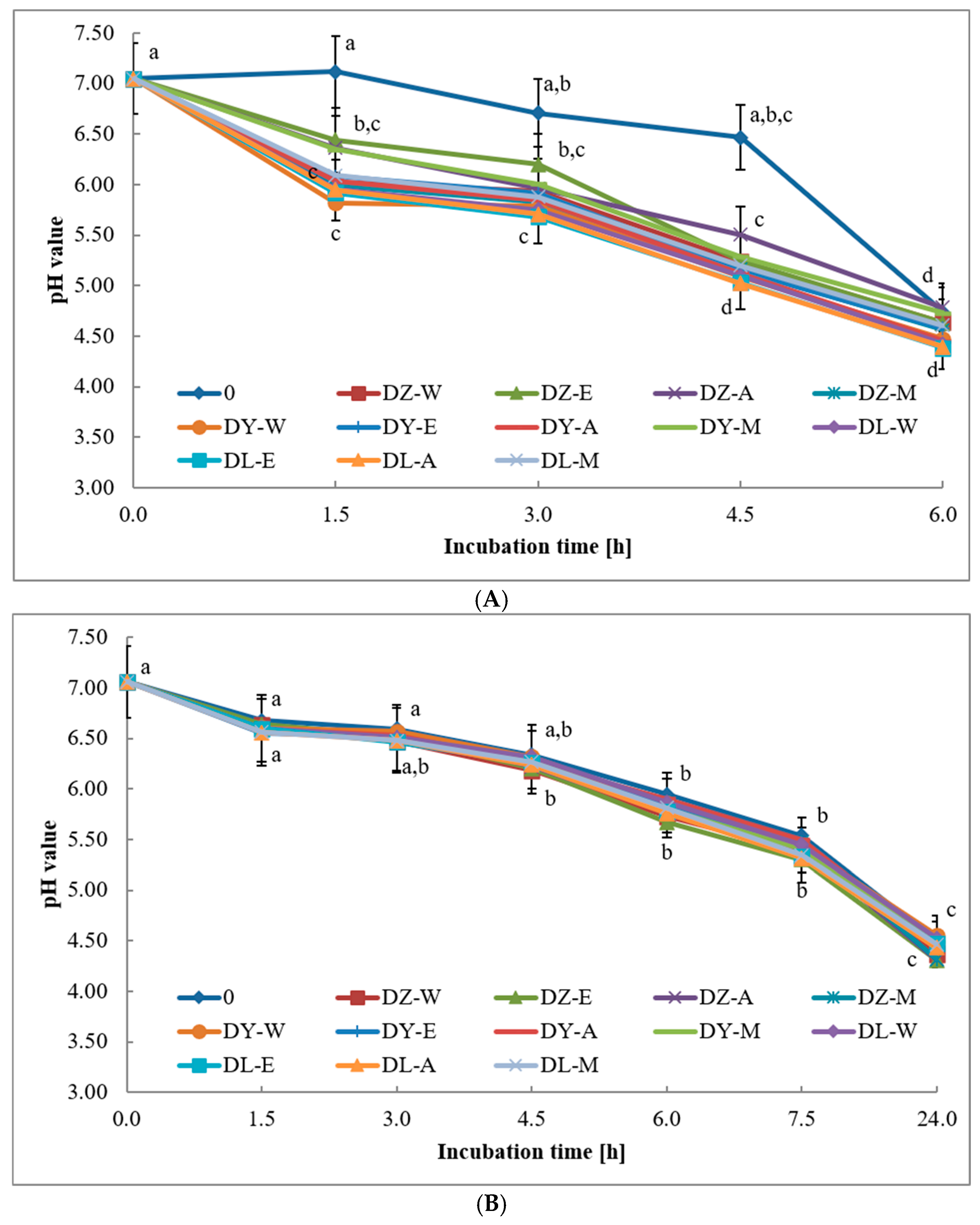
L. acidophilus La-14 depended strongly on the type of fermented matrix. The fastest decrease in pH was observed in organic coconut beverage, whereas the soy beverage and organic milk showed slower acidification and required a longer incubation time to reach the target acidic range. In the coconut beverage, the initial pH was 7.05 and decreased rapidly during the first 1.5 h of fermentation (
Figure 1A). The most pronounced early acidification was observed in the sample supplemented with the aqueous extract of
F. officinalis L., for which the pH decreased to 5.82. After 6 h of incubation, the pH values of the coconut beverage samples ranged from 4.38 to 4.79, indicating that the fermentation process could be stopped at this stage. The control sample acidified more slowly during the first 4.5 h and only then showed a rapid pH decrease, from 6.47 to 4.74. This suggests that selected herbal extracts did not inhibit acidification in the coconut matrix and, in some variants, may have favored faster pH reduction.
The soy beverage showed a more gradual decrease in pH (
Figure 1B). The initial pH was 7.06, and after 1.5 h it remained in a narrow range, from 6.55 to 6.68. After 6 h, the pH dropped below 6.0 in only some samples, with the lowest value, 5.67, recorded in the sample containing the ethanolic extract of
V. thapsus L. After 7.5 h, all samples still had pH values above 5.0. Therefore, fermentation was continued for up to 24 h, when all soy beverage samples reached pH values between 4.31 and 4.56. In contrast to the coconut beverage, the differences between the control sample and the herbal extract samples were small, indicating that the soy matrix had a stronger effect on fermentation kinetics than the extract type.
Organic milk was the slowest acidifying matrix (
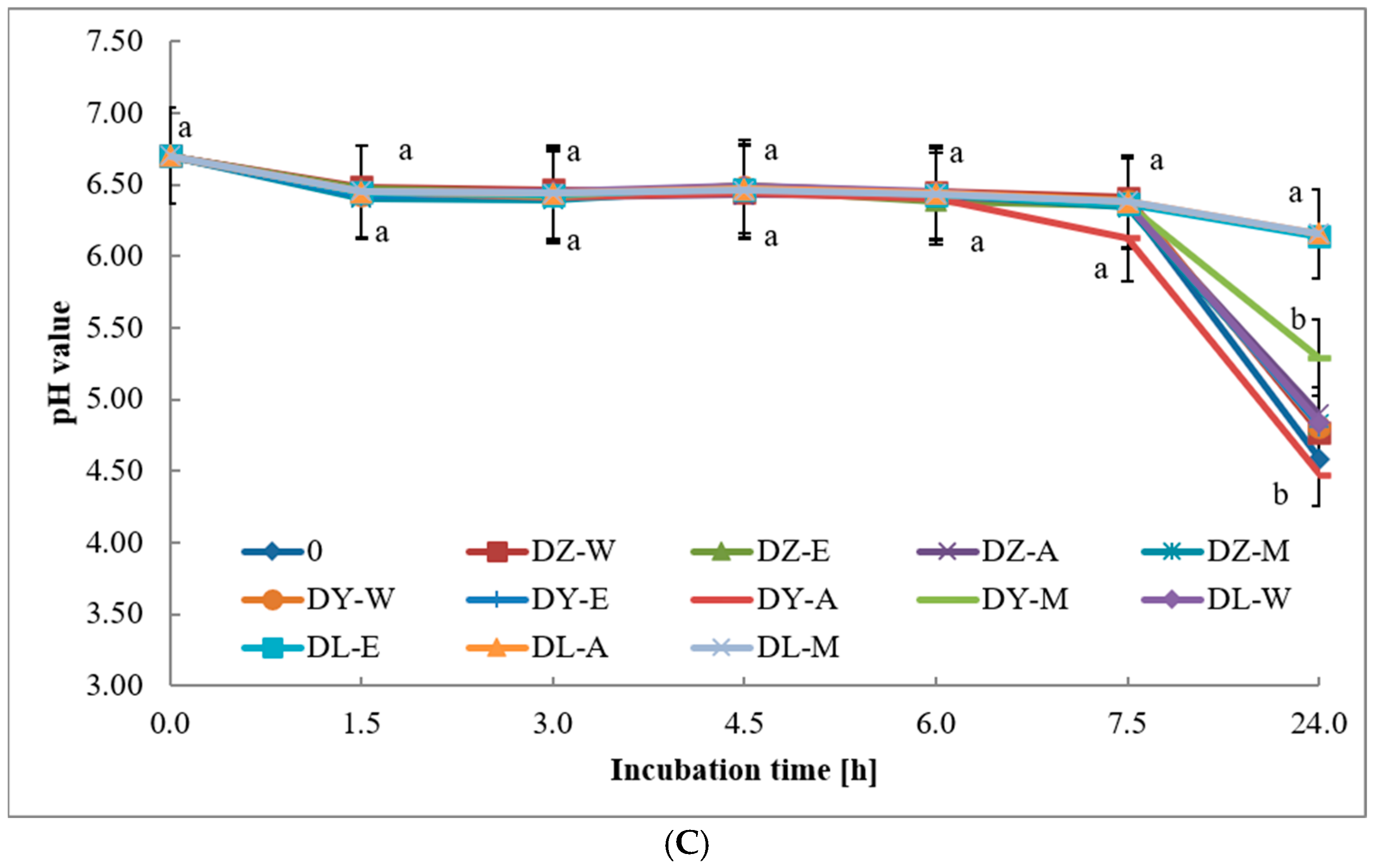
Figure 1C). The initial pH was 6.70, which decreased to approximately 6.40 after 1.5 h across all variants. During the next hours, pH changes remained limited, and after 7.5 h, the pH values were still above 6.0. The lowest value at this stage, 6.13, was recorded for the sample with the aqueous extract of
F. officinalis L. After 24 h, most milk samples reached pH values near 4.0. However, three samples containing extracts of
C. benedictus L. remained above pH 6.0: the ethanolic extract (6.14), the acetonic extract (6.16) and the methanolic extract (6.16). These results indicate that some extracts obtained from
C. benedictus L. in milk may delay acidification.
The rapid acidification of the coconut beverage was unexpected because coconut matrices are usually regarded as less favorable substrates for LAB fermentation due to their relatively low content of fermentable carbohydrates. Soy beverages, in contrast, contain carbohydrates such as glucose, fructose and raffinose, which may support LAB metabolism more effectively [
64]. The present results therefore suggest that the commercial composition of the coconut beverage, including possible naturally occurring or added sugars, should be considered when interpreting fermentation performance. Similar observations concerning matrix-dependent acidification were reported by Ladokun and Oni [
65], who showed differences in pH changes among fermented products prepared from cow milk, soy milk and coconut milk.
3.7. Viability of Lactic Acid Bacteria After Fermentation
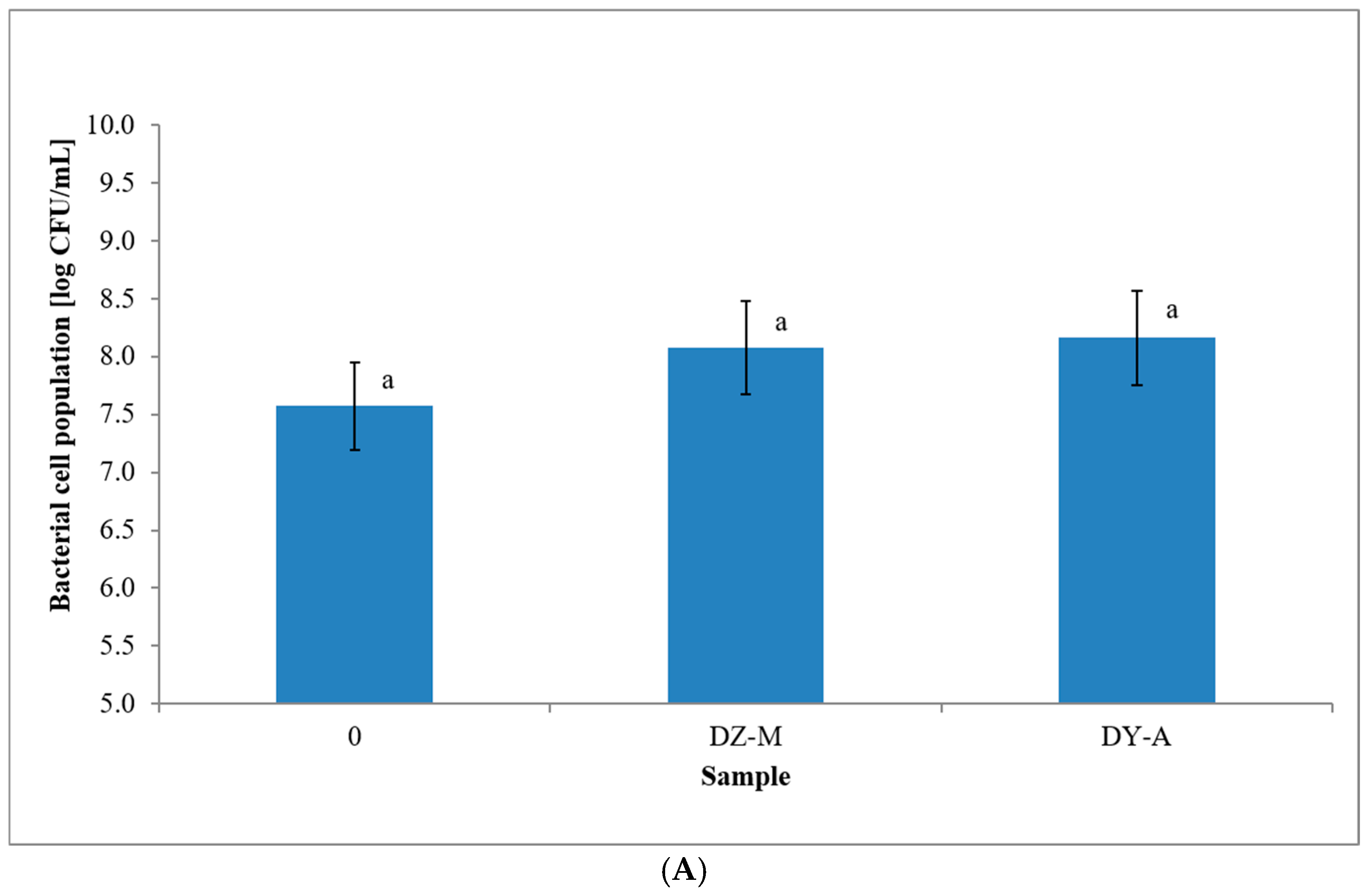
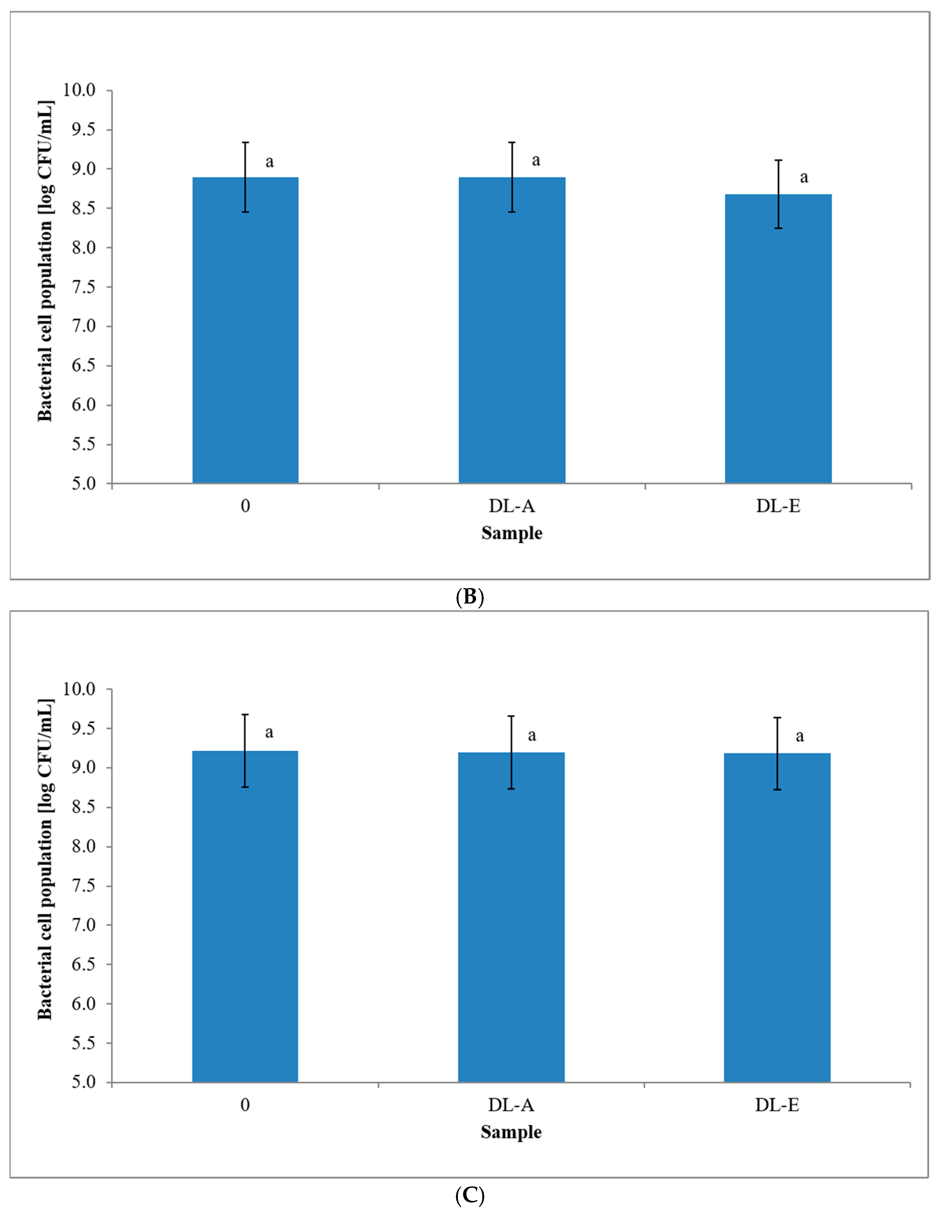
The concentration of viable LAB cells remained high in all analyzed matrices, regardless of the presence or absence of herbal extracts (
Figure 2). All variants within each matrix were assigned to the same statistical grouping. Therefore, the data support the conclusion that supplementation with herbal extract did not significantly affect LAB viability under the tested conditions. In the coconut and soy beverages, viable LAB counts remained near 9 log CFU/mL. In milk, counts were lower but still exceeded 7 log CFU/mL in all analyzed samples.
All analyzed samples contained more than 7 log CFU/mL of viable LAB. This level is relevant for the technological assessment of fermented beverages containing live LAB cultures. Kowalska and Ziarno [
66] reported that fermented cereal and pseudocereal beverages containing LAB and bifidobacteria may maintain viable cell counts above 7 log CFU/mL. The present results are consistent with this observation and indicate that the tested herbal extracts did not compromise LAB survival in coconut, soy, or milk matrices.
LAB survival in fermented products depends on the bacterial genus, species, and strain; the chemical composition of the matrix; acidity; oxygen availability; and storage conditions. Zaręba et al. [
67] showed that the survival of yogurt bacteria and probiotic strains in fermented and non-fermented milk models depended on the type of microorganisms and environmental conditions. In the present study, the high viable counts observed after fermentation indicate that the tested extracts support LAB survival under the applied conditions. They should not be interpreted as a direct measure of overall LAB metabolic activity.
The results show that herbal extracts may be used in the tested fermented beverages without adversely affecting LAB viability. However, their effect on acidification kinetics depended on the matrix. The coconut beverage fermented most rapidly, the soy beverage showed slower acidification, and milk required the longest fermentation time, with extract-dependent delay in samples containing selected C. benedictus extracts. From a technological perspective, the most promising variants were those in which rapid pH reduction was accompanied by high LAB survival. The coconut beverage supplemented with selected extracts met this condition after 6 h of fermentation. The soy beverage also supported high LAB counts, but it required 24 h to reach the target acidity. Milk maintained adequate LAB viability, but selected extracts delayed acidification. These findings are relevant to functional fermented foods and plant-based fermented beverages, in which herbal extracts may be used to add antioxidant compounds while maintaining live LAB counts. Any claims related to gut microbiota modulation, precision nutrition, or health effects would require additional studies, including strain-specific functionality, metabolite formation, phenolic stability during digestion and controlled storage tests.




 ,
,