1. Introduction
On the path to the successful realization of controlled nuclear fusion and the development of next-generation power reactors, such as the International Thermonuclear Experimental Reactor (ITER) and DEMO, one of the key engineering challenges is the selection of plasma-facing materials (PFMs) [
1,
2]. Tungsten (W) is considered one of the leading candidate materials for divertor armour because of its high melting point, excellent thermal conductivity, high sputtering threshold, and very low solubility for hydrogen isotopes [
3,
4].
However, under reactor-relevant operating conditions, tungsten plasma-facing components will be exposed to high heat loads and intense bombardment by low-energy particles, including hydrogen isotopes and helium ash [
5,
6]. Hydrogen plasma exposure can lead to hydrogen trapping in radiation-induced defects, embrittlement, blister formation, and surface erosion [
7,
8,
9]. In contrast, helium irradiation at elevated temperatures promotes the formation of helium-vacancy complexes, nanobubbles, cavities, and, under suitable exposure conditions, nanostructured tungsten fuzz [
10,
11,
12,
13,
14,
15]. It is important to distinguish fully developed W fuzz from more general bubble-related porous surface damage, since these morphologies have different visual features and may form under different combinations of temperature, ion energy, flux, and fluence [
6,
10,
11,
12,
13,
14,
15].
In realistic fusion edge and divertor plasma environments, hydrogen and helium effects occur simultaneously and may interact in a non-additive manner [
16]. Previous experimental studies on linear plasma devices and ion-irradiation facilities have shown that the addition of helium to hydrogen or deuterium exposure can significantly modify the surface morphology of tungsten, the structure of near-surface defects, and hydrogen isotope retention [
17,
18,
19,
20,
21,
22,
23]. Helium-stabilized vacancy complexes and bubbles can act as trapping sites for hydrogen, thereby changing the energetic spectrum of gas retention and the kinetics of thermal desorption [
6,
18,
19,
20,
21,
22,
23]. At the same time, helium-induced porosity and bubble formation may reduce the load-bearing capacity of the near-surface layer and affect the micromechanical response of tungsten, as reported in nanoindentation and mechanical-property studies [
24,
25,
26].
Despite the extensive literature on hydrogen and helium effects in tungsten, additional systematic studies are still needed to clarify how the helium fraction in a steady-state mixed He/H
2 plasma controls the coupled evolution of surface morphology, porosity, mechanical properties, and gas release behavior. Many previous studies focused either on pure hydrogen/deuterium or pure helium exposure, sequential irradiation, or different plasma devices and irradiation conditions. Therefore, a direct comparison under fixed surface temperature, irradiation time, and similar plasma-exposure geometry is useful for identifying the role of plasma composition itself. In the present work, this issue is addressed using the KAZ-PSI linear plasma device, which allows controlled exposure of tungsten samples to steady-state mixed He/H
2 plasma at different helium fractions [
27].
Accordingly, the aim of this work is to systematically investigate the structural and mechanical degradation, as well as gas-retention behavior, of polycrystalline tungsten after prolonged exposure to high-temperature mixed He/H2 plasma. Particular attention is paid to the relationship between helium fraction, surface roughness, SEM-observed morphology, near-surface porosity, nanoindentation response, and thermal desorption behavior.
2. Materials and Methods
The study object was commercially pure, pre-annealed polycrystalline tungsten (W) with a metallic purity of ≥99.97 wt.%, manufactured by Plansee SE (Reutte, Austria). According to the technical specifications, the material density was over 19.1 g/cm
3, which is close to the theoretical value for tungsten (19.3 g/cm
3) and indicates its extremely low initial porosity. The initial average grain size corresponded to grade 6 or finer according to ASTM E112, and the material’s base Vickers hardness was in the range of 450–530 HV. To minimize the influence of impurities on hydrogen isotope capture and radiation-induced defect formation, the material had a strictly controlled chemical composition, which is presented in
Table 1.
For plasma testing, cylindrical samples with a diameter of 6 mm and a height of 5 mm were fabricated from the source material. To minimize the influence of the initial surface topography on the parameters of physical sputtering and ion absorption, the working surface of the samples underwent standardized multi-stage mechanical processing.
Plasma exposure experiments were conducted on the KAZ-PSI (Kazakhstan Plasma Generator for Plasma Surface Interactions) linear plasma system, developed by PlasmaScience LLP for studies of plasma-surface interaction processes in candidate plasma-facing materials [
27]. A detailed schematic diagram of the KAZ-PSI generator was reported in Ref. [
27]. In the present manuscript, the main features of the setup are summarized to clarify the irradiation conditions used in this work.
The KAZ-PSI device consists of a cold-cathode plasma source, a plasma-beam discharge chamber, a gas inlet system, electromagnetic coils for plasma focusing and confinement, a vacuum interaction chamber with a water-cooled target assembly, a biased sample holder, a temperature monitoring system, and a pumping system. The working gas mixture was supplied from external gas cylinders through a controlled gas inlet system and ionized in the discharge region. The system does not produce hydrogen isotopes by nuclear reactions. In this work, molecular hydrogen (H2, protium) was used as a safe non-radioactive laboratory analogue of hydrogen isotopes. Deuterium plasma can be produced on the same system when a D2 cylinder is connected; however, tritium was not used because tritium experiments require specialized nuclear-safety infrastructure. Thus, the present experiments simulate key plasma-surface interaction conditions of mixed hydrogen–helium exposure rather than the exact D–T–He isotope composition of ITER plasma.
Before each experiment, the working chamber was evacuated to a high-vacuum base pressure. Irradiation of the tungsten samples was carried out in steady-state mixed He/H
2 plasma. Four irradiation regimes were used to evaluate the effect of helium fraction while keeping the surface temperature, irradiation time, electron temperature, and electron density within the selected operating range (
Table 2). The total ion flux and the partial H and He fluxes listed in
Table 2 were obtained from plasma diagnostics and are discussed below.
Plasma parameters were determined using electrostatic Langmuir probe measurements and target-current diagnostics. The electron temperature (Te) and plasma density (ne) were measured with a Langmuir probe positioned near the target region. The total ion flux (Γtot) was estimated from the ion saturation current to the target, taking into account the irradiated surface area. The partial hydrogen and helium fluxes were estimated from the measured total ion flux and the nominal He/H2 gas composition, assuming a proportional contribution of ion species under the selected discharge conditions. The degree of ionization was not independently measured in this work; therefore, the reported partial fluxes should be treated as estimated values. The decrease in Γtot with increasing helium fraction is attributed to the higher ionization potential of helium (24.6 eV) compared with hydrogen (13.6 eV for H and 15.4 eV for H2), which reduces the ionization efficiency under fixed discharge conditions. The negative bias applied to the target reduces the contribution of plasma electrons to the measured target current during ion-current measurements. No independent correction for secondary-electron emission was performed in the present work; therefore, the reported total and partial ion fluxes should be considered estimated values derived from target-current diagnostics.
During irradiation, the surface temperature of the samples was maintained at a stable level of 1100 °C. Temperature control of the irradiated zone was performed using a DIKAI infrared pyrometer (model DIT 6 H; DIKAI, Wuhan, China).
The energy of the incident ions (∼115–130 eV) was set by applying a negative bias voltage of −100 V to the target assembly.
Upon completion of the plasma exposure, the samples were removed from the vacuum chamber for comprehensive analysis, the algorithm for which was structured from macroscopic evaluation to microstructural and physical-mechanical testing. First, a quantitative assessment of the surface topography and the degree of surface relief degradation was performed using the contact probe method with an SSR300+ profilometer (Hangzhou Mituolinke Technology Co., Ltd., Hangzhou, China). To ensure high profiling accuracy and to minimize mechanical damage to the forming brittle radiation-induced surface defects (such as helium bubbles, blisters, and nanostructures), scanning was performed at a strictly controlled low probe speed of 0.05 mm/s. The scanning trajectory passed through the central region of the zone of maximum plasma exposure, and the arithmetic mean roughness (Ra) was used as the baseline parameter for comparative erosion analysis.
Surface morphology and transverse fracture cross-sections were examined using an SEM3200 scanning electron microscope (CIQTEK Co., Ltd., Hefei, China) at an accelerating voltage of 15 kV under low-vacuum conditions. SEM analysis was used to study in detail the nature of structural modification at the nanoscale and to assess the depth of radiation defects.
To verify the phase composition of the modified layer and identify possible structural changes within the material, X-ray diffraction analysis (XRD) was employed. Diffractograms were recorded on an X’Pert PRO X-ray diffractometer (Malvern Panalytical, Almelo, The Netherlands) using monochromatic Cu Kα radiation. The mechanical properties of the near-surface layers were evaluated using nanoindentation on a FISCHERSCOPE HM2000 system (Helmut Fischer GmbH, Sindelfingen, Germany) in accordance with DIN EN ISO 14577-1 [
28]. Measurements were performed using a Berkovich indenter at a load of 100 mN with a dwell time of 5 s to reduce creep effects. The main parameters used for degradation analysis were nanohardness (HV) and Young’s modulus (E).
Thermal desorption spectroscopy Thermal desorption spectroscopy (TDS) was used to evaluate gas retention and the capacity of radiation-induced trapping sites.
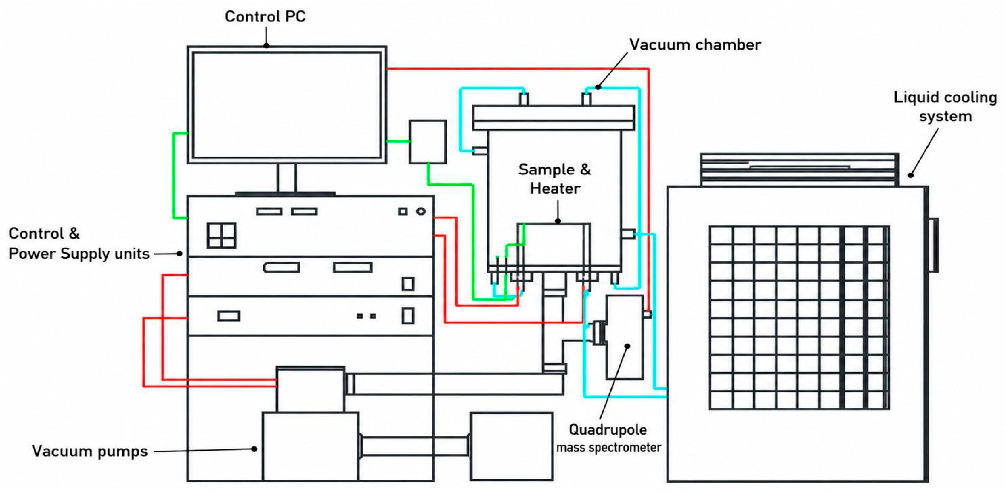
Figure 1 shows the schematic diagram of the experimental setup used for TDS analysis. The TDS facility was designed and assembled at PlasmaScience LLP (Ust-Kamenogorsk, Kazakhstan) and consists of a high-vacuum chamber, a resistive heating system, temperature-control instrumentation, and a quadrupole mass spectrometer for monitoring desorbed gases during programmed heating.
The TDS setup consisted of a high-vacuum chamber equipped with a sample holder, copper current lead, tantalum heater, W-Re thermocouple, pumping system, vacuum gauge, cooling unit, quadrupole mass spectrometer, and control/power-supply units. The sample temperature was controlled using a W–Re thermocouple, while the partial pressure of desorbed hydrogen was recorded using an XT100 quadrupole mass spectrometer (Extorr Inc., New Kensington, PA, USA) tuned to mass 2 (H2). During TDS measurements, the samples were heated to 1000 °C at a rate of 30 °C/min, followed by a 30 min isothermal hold.
3. Results and Discussion
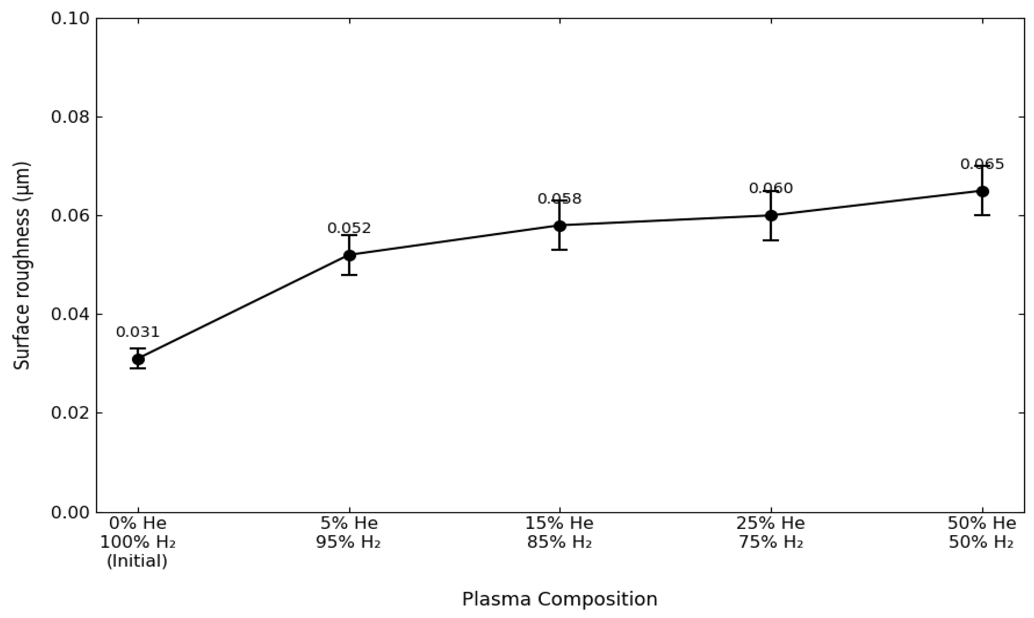
The initial polished surface was characterized by a low roughness value of Ra = 0.031 μm. After He/H
2 plasma exposure, the roughness increased moderately for all irradiated samples (
Figure 2). The changes in Ra among the irradiated samples are small in absolute value and should be interpreted as a supporting indicator of surface modification rather than as the sole evidence of degradation.
Quantitative topography analysis shows that Ra increases from 0.031 μm for the initial polished tungsten to 0.052 μm after exposure to 5% He/95% H2 plasma. With further increase in helium fraction, Ra changes only moderately, reaching 0.058 μm for 15% He/85% H2, 0.060 μm for 25% He/75% H2 and 0.065 μm for 50% He/50% H2. Considering the experimental scatter, this trend indicates a gradual increase in surface roughness with helium fraction, while the more detailed interpretation of surface degradation requires SEM and cross-sectional observations.
SEM images of the exposed surfaces (
Figure 3a–d) show a gradual change in surface morphology with increasing helium fraction in the He/H
2 plasma. At low helium content, the surface contains localized bubble-like and cracked regions. At higher helium fractions, the density of cavities, ruptured features, and open pores increases, indicating more pronounced gas-related surface damage.
As the concentration of helium in the plasma mixture increases (
Figure 3b,c), surface damage becomes more extensive. SEM alone cannot distinguish hydrogen-filled bubbles from helium-filled bubbles; therefore, the observed cavities are described here as gas-related bubbles or pores formed under mixed He/H
2 plasma exposure. Their increased density at higher helium fractions is consistent with the low solubility of helium in tungsten and its tendency to stabilize vacancy-type defects.
For the sample exposed to 50% He/50% H
2 plasma (
Figure 3d), the surface exhibits the highest density of open pores and ruptured bubble-like features among the investigated conditions. The morphology should be interpreted as porous bubble-related surface degradation rather than fully developed tungsten fuzz. The term W fuzz is reserved for characteristic nanofibrous or tendril-like structures reported in the literature.
The apparent surface porosity was estimated from SEM images by image analysis as the ratio of the detected pore area to the total analyzed image area. Representative regions of the plasma-exposed surface were used for each irradiation condition. The calculated porosity increased from 0.2% for the 5% He/95% H2 condition to 0.78% for 15% He/85% H2, 2.703% for 25% He/75% H2 and 3.601% for 50% He/50% H2. These values should be regarded as apparent surface area fractions obtained from the analyzed SEM fields rather than bulk porosity values.
Examination of cross-sections of irradiated samples allowed us to estimate the penetration depth of radiation-induced defects and the thickness of the modified near-surface layer.
Figure 4 shows SEM images of transverse fractures of tungsten after irradiation with He/H
2 plasma at different gas ratios.
At 5% He/95% H2, the modified layer contains mainly small isolated bubble-like defects. With increasing helium fraction, the visible cavities become larger and more numerous. Because the apparent feature size depends on SEM resolution, imaging conditions, and thresholding during image analysis, the sizes are discussed here as approximate ranges rather than exact values with nanometer-level precision.
At 15% He/85% H2, bubble-like features with sizes of the order of several to tens of nanometers are observed. At 25% He/75% H2 and 50% He/50% H2, the cavities become more developed and some features reach several tens of nanometers, with the largest visible pores approaching approximately 80 nm. This trend indicates enhanced coalescence of gas-vacancy complexes at higher helium fractions.
The observed dynamics of subsurface defect formation are attributed to the combined effect of high temperature (1100 °C), continuous ion implantation, and the low solubility of helium in tungsten. At this temperature, migration and coalescence of helium-vacancy complexes can occur, producing larger gas-filled cavities. These cavities generate high internal gas pressure and can locally deform and rupture the near-surface layer.
When gas-filled cavities near the surface grow and coalesce, local deformation of the surrounding tungsten can lead to the opening of pores and the formation of a more damaged near-surface layer. This process explains the increased porosity and the loss of mechanical integrity observed at higher helium fractions.
Cross-sectional SEM observations confirm the formation of a defect-rich layer directly beneath the exposed surface. The depth and density of the cavities increase with helium fraction. The formation of such gas-retaining regions is consistent with the thermal desorption spectroscopy (TDS) results discussed below, which show prolonged high-temperature gas release for samples irradiated at higher helium fractions.
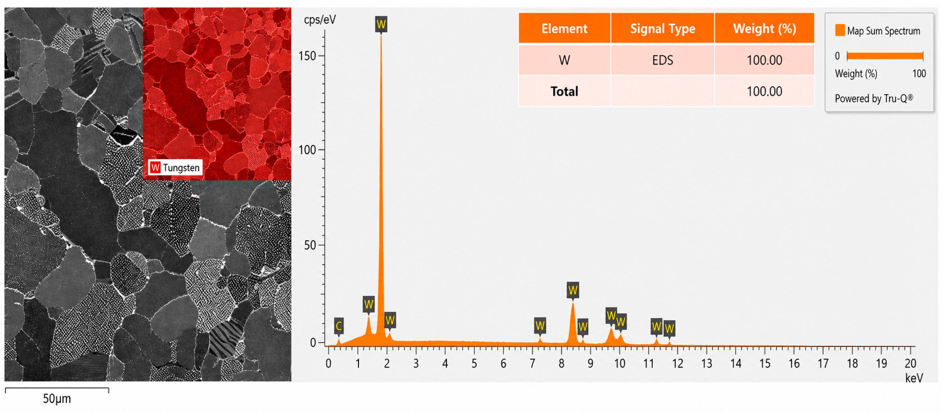
To verify that the observed degradation is not caused by detectable chemical contamination, oxidation, or carburization, local energy-dispersive X-ray spectroscopy (EDS) was performed. EDS analysis was carried out using an XFlash Detector 730M-300 system (Bruker Corporation, Billerica, MA, USA), which enabled additional determination of the elemental composition of the investigated regions.
The EDS elemental mapping and spectrum shown in
Figure 5 confirm that the analyzed near-surface region consists predominantly of tungsten. According to the quantitative EDS results, the W content is 100 wt.% within the detection limits of the method, while no detectable oxygen, carbon, or other impurity elements were observed. This indicates that the surface degradation after He/H
2 plasma exposure is not associated with oxidation, carburization, or chemical corrosion. Therefore, the formation of pores, bubbles, and porous surface morphology should be attributed mainly to plasma-induced radiation damage and gas accumulation in the tungsten matrix.
However, EDS is sensitive only to the near-surface interaction volume and cannot provide complete information about the phase state of the modified layer. Therefore, XRD analysis was additionally performed to verify the phase stability of tungsten and to exclude the formation of secondary crystalline phases, such as tungsten oxides or carbides, throughout the analyzed volume.
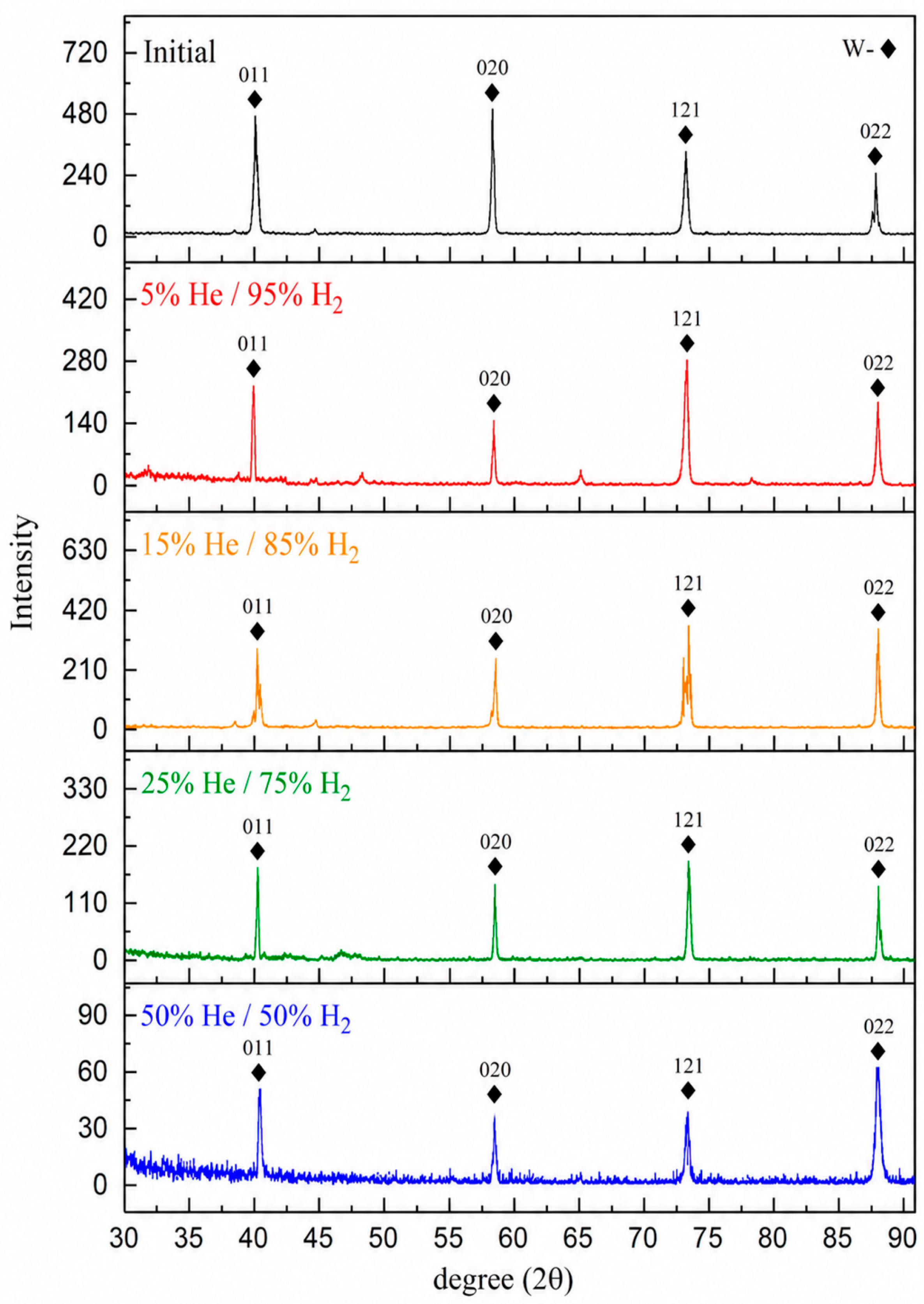
Figure 6 shows comparative XRD patterns of the initial unirradiated tungsten and the samples after exposure to mixed He/H
2 plasma.
The XRD patterns in
Figure 6 show that the phase state of tungsten remains stable after He/H
2 plasma exposure. All spectra contain diffraction peaks corresponding to the body-centered cubic (BCC) structure of metallic tungsten. No additional diffraction peaks attributable to tungsten oxides, carbides, or other secondary crystalline phases were detected within the sensitivity of the method.
In the present study, XRD was used primarily for phase identification and for verifying the absence of detectable secondary crystalline phases rather than for quantitative comparison of peak intensities. Minor variations in relative peak intensity can be influenced by surface roughness, local texture, and the modified near-surface morphology after plasma exposure. Therefore, peak-intensity changes were not used as a quantitative parameter of degradation.
The preservation of the BCC tungsten phase indicates that the observed morphological changes are associated mainly with radiation-induced defects and gas accumulation rather than with bulk phase transformation or chemical reaction. Any possible microstrain effects would require a dedicated line-profile analysis, which is beyond the scope of the present work.
The combination of SEM, EDS, and XRD results therefore supports the interpretation that the plasma-exposed layer undergoes morphological and defect-structure modification while retaining the crystalline phase of metallic tungsten.
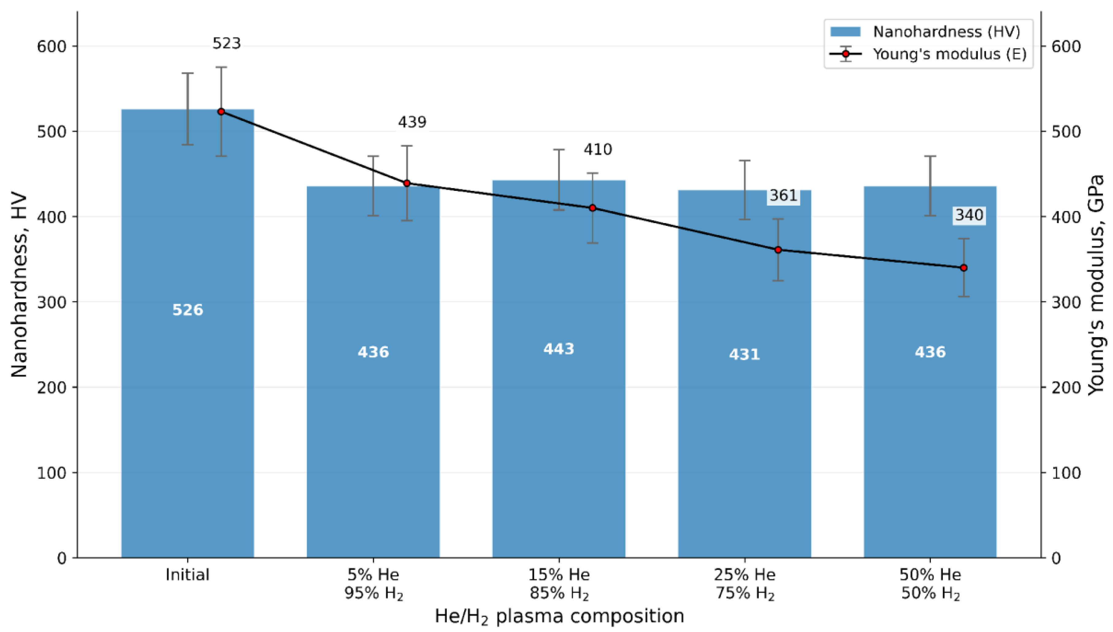
To quantitatively assess the degree of radiation-induced softening and loss of load-bearing capacity in the modified near-surface layer, local physical and mechanical tests were conducted. Changes in the nanohardness (HV) and Young’s modulus (E) of tungsten were evaluated using instrumental nanoindentation on a Fischerscope HM 2000 system. The test results are presented in
Figure 7.
For each irradiation condition, several representative SEM fields from the central plasma-exposed region were analyzed.
After 7 h of He/H2 plasma exposure at 1100 °C, the mechanical response of the irradiated samples differs from that of the initial tungsten. The previous interpretation of a strictly proportional decrease in both nanohardness and Young’s modulus has been corrected. The nanohardness decreases markedly after irradiation compared with the initial state, but the values for the irradiated samples remain relatively close within the experimental scatter. In contrast, Young’s modulus shows a more pronounced decreasing tendency with increasing helium fraction.
This behavior reflects the combined response of a porous, plasma-modified near-surface layer and the underlying tungsten matrix during nanoindentation at 100 mN. The decrease in hardness is associated with plastic deformation and local collapse of the porous surface layer. The decrease in the elastic modulus at higher helium fractions is consistent with the increasing contribution of open porosity and gas-related defects to the measured contact response. Because the indentation response is influenced by both the modified layer and the substrate, the mechanical data are interpreted together with SEM observations rather than as a stand-alone measure of defect depth.
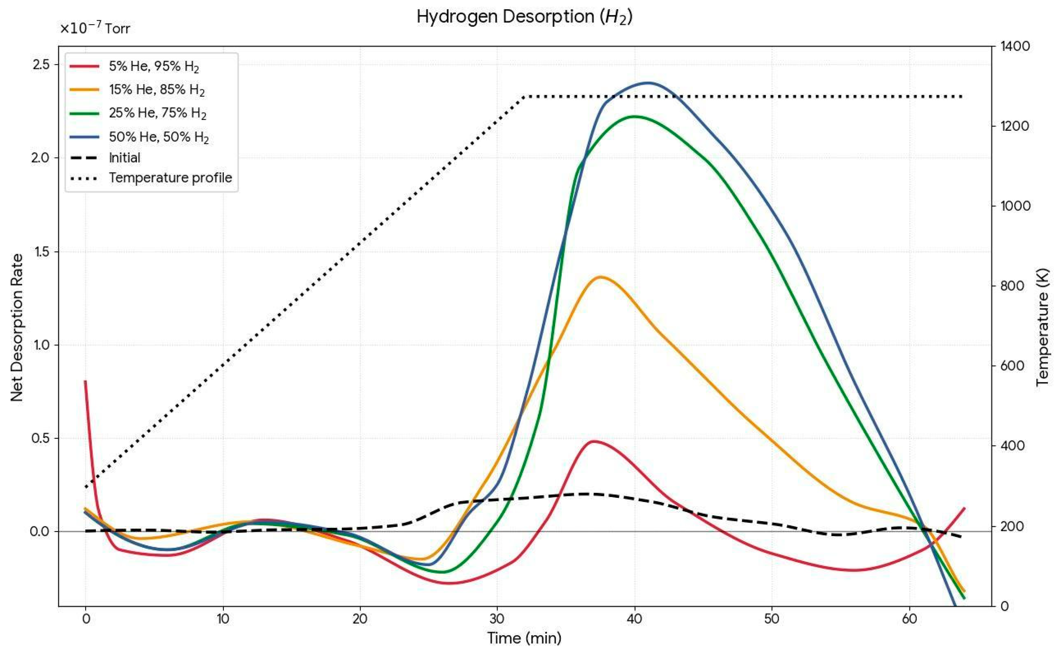
To assess gas retention and the energetic character of trapping sites, TDS analysis was performed. The interpretation of the TDS spectra was revised to avoid overstatement and to clearly distinguish the linear heating stage from the final isothermal hold.
The experimental TDS spectra, showing the kinetics of gas release from tungsten after He/H
2 plasma exposure, are presented in
Figure 8. In this experiment, the samples were heated at 30 °C/min up to 1000 °C (1273 K), followed by a 30 min isothermal hold. Therefore, the time axis is directly related to the temperature program during the heating stage.
For the 5% He/95% H2 condition, the desorption signal is relatively low, indicating limited gas retention under this irradiation regime. Increasing the helium fraction modifies the shape of the desorption curves. The feature previously described as a peak at 1273 K has been reinterpreted: for the samples exposed to 25% He/75% H2 and 50% He/50% H2 the maximum signal near the end of the heating stage corresponds to the transition from linear heating to the isothermal stage rather than to a well-resolved isolated TDS peak at exactly 1273 K.
The prolonged high-temperature tail observed during the isothermal hold at 1000 °C reflects slow gas release from deeper and more stable trapping sites. These traps are associated with vacancy clusters, helium-related cavities, and porous complexes formed at higher helium fractions. The persistence of the desorption signal during the isothermal stage indicates that gas cannot be rapidly released from these traps even at high temperature.
The relationship between helium-induced defects and hydrogen desorption should be considered as a qualitative interpretation supported by SEM and TDS results. Helium has very low solubility in tungsten and preferentially stabilizes vacancy-type defects and bubble-like cavities. These defects can act as trapping sites for hydrogen and can shift gas release toward higher-temperature or prolonged desorption regimes. A direct quantitative correlation between helium and hydrogen desorption would require mass-resolved and depth-resolved analyses, which are beyond the scope of the present work.



 ,
,