1. Introduction
Doxycycline (DOX) is one of the most widely known antibiotics of the tetracycline class, exhibiting broad-spectrum activity against Gram-positive and Gram-negative microorganisms [
1]. It is mainly administered orally and is indicated for respiratory and urinary tract disorders, dermatological infections, as well as specific conditions such as Lyme disease [
2]. In recent years, interest in DOX has been renewed, not only because of its broad antimicrobial spectrum [
3], but also due to the attention it received during the COVID-19 pandemic for its potential anti-inflammatory and antiviral properties [
4].
Absorption occurs mainly within the first 60–90 min of residence in the small intestine (duodenum, proximal jejunum), with significantly reduced rates thereafter (up to 3–5 h). Its bioavailability, however, is often decreased due to interactions with other products (e.g., iron supplements or antacids) or specific dietary habits (dairy products, legumes, spinach) [
5].
Tetracyclines, including doxycycline (DOX), form complexes with multivalent cations such as Fe
3+, Fe
2+, Ca
2+, Mg
2+, and Al
3+ through coordination bonds or hydrogen bonding, and this interaction can markedly influence their solubility and absorption profiles [
6,
7]. A key question, therefore, is the extent to which such ions affect DOX absorption during its residence time in the small intestine. Among these ions, iron is of particular interest, as its coexistence in both the divalent (Fe
2+) and trivalent (Fe
3+) oxidation states within the gastrointestinal environment gives rise to distinct and often opposing pH-dependent complexation behaviors that may critically influence DOX bioavailability. Doxycycline, as a typical member of the tetracycline class, exhibits a strong ability to chelate both divalent and trivalent metal cations, including iron. In the gastrointestinal tract, the redox conversion of ferrous iron (Fe
2+) to ferric iron (Fe
3+) occurs naturally under acidic conditions, particularly in the stomach, thereby increasing the availability of Fe
3+ for complexation [
8]. According to the classical chelation theory for tetracyclines, at low pH, doxycycline exists predominantly in its protonated form (DOX·H
+), which cannot effectively chelate Fe
2+ because the key phenolic and β-diketone groups remain protonated and unavailable for metal binding. As the pH increases toward neutral or alkaline conditions, deprotonation of these groups enhances the chelating capacity of doxycycline, thereby favoring Fe
2+ complexation [
9].
More recent evidence demonstrates that Fe
3+ forms significantly more stable complexes with doxycycline under mildly acidic conditions. The Fe
3+doxycycline complex forms optimally at a pH of approximately 4 with a 1:1 stoichiometry, involving coordination at both the tricarbonylamide group of ring A and the phenolic-diketone oxygens of rings [
10]. These Fe
3+-DOX complexes are stabilized in acidic environments and gradually dissociate as the pH increases toward intestinal values (pH 5–8), without oxidative degradation of doxycycline. For Fe
2+, chelation remains relevant, but the resulting complexes are generally less stable than those of Fe
3+. Their distribution depends on both pH and the Fe
2+/Fe
3+ redox equilibrium. In intermediate to alkaline pH (5–8), deprotonation of doxycycline enhances Fe
2+ binding, although the presence of high concentrations of competing cations (Fe
2+/Fe
3+, Ca
2+, Mg
2+) from food or supplements still promotes the formation of insoluble or poorly soluble complexes [
11].
Clinically, the formation of Fe
2+ and Fe
3+doxycycline complexes leads to a marked reduction in doxycycline bioavailability when co-administered with iron supplements or iron-rich foods, potentially resulting in therapeutic failure and increased risk of antimicrobial resistance [
8,
12,
13,
14].
Taken together, these findings highlight a critical point: Fe
2+ and Fe
3+ exert opposite pH-dependent effects on DOX complexation; Fe
3+ binds strongly under acidic gastric conditions, whereas Fe
2+ binds more effectively under neutral to alkaline intestinal conditions. Because DOX absorption occurs predominantly in the small intestine, where Fe
2+ mediated chelation and the presence of multiple competing cations reduce the fraction of free DOX available for permeation, the net effect is a substantial decrease in systemic exposure. Evidence suggests that, despite the strong Fe
3+ complexation in the stomach, it is the Fe
2+-driven chelation and multivalent–cation interactions in the intestine that ultimately dominate and limit DOX entry into the systemic circulation. Nevertheless, the coexistence of these opposing pH-dependent mechanisms underscores that the overall impact on DOX permeability is not yet fully resolved, and the phenomenon warrants further investigation to clarify which interaction predominates under physiologically relevant conditions. Previous investigations have employed parallel artificial membrane permeation (PAMPA) assays using soybean lecithin-decane and octanol-PAMPA membranes [
15]. However, the static donor-acceptor nature of conventional PAMPA provides only a fixed concentration environment and may not adequately capture the dynamic chelation/dechelation equilibrium between doxycycline and multivalent ions during simultaneous dissolution and permeation. Therefore, the phenomenon could be studied more comprehensively using continuous on-line dissolution/permeability systems that allow a more integrated assessment of potential pharmacokinetic interactions [
16,
17].
In the present study, a combined in vitro protocol was implemented, incorporating an online transition of the drug from the gastric to the intestinal phase dynamically coupled to Franz diffusion cells. This approach aimed to investigate and interpret the reduction in doxycycline concentration during its co-administration with iron or aluminum supplements. Due to the mechanistic complexity and the opposing pH-dependent behaviors of Fe
2+ and Fe
3+, the experimental work primarily focused on elucidating the impact of iron on doxycycline solubility and permeation, while aluminum ions were used as a comparative reference to capture the extent of multivalent–cation interference. Al
3+ is present in aluminum-containing antacids and, owing to its higher trivalent charge, exhibits stronger chelation propensity than the clinically common divalent cations Ca
2+ and Mg
2+, thereby representing an upper limit multivalent cation interference scenario [
18]. In this way, the study seeks to elucidate the mechanisms that influence DOX bioavailability during co-administration, contributing to a more comprehensive interpretation of potential interactions within the ADME framework, while also providing useful insights for the design of future pharmaceutical formulations that may facilitate safer co-administration.
To reliably assess these interactions, accurate quantification of doxycycline throughout the dynamic gastrointestinal to intestinal transition and during permeation experiments is essential. Reliable quantification of doxycycline in the proposed experimental systems requires a robust, flexible, and traceable analytical method. Although various approaches for its determination have been reported (
Table 1), many of them are characterized by long analysis times and high consumption of organic solvents, underscoring the need for an optimized analytical workflow compatible with the demands of the present study. In recent years, the development of simpler, faster, and more environmentally compatible methods has become a central objective in Green Analytical Chemistry, which promotes the reduction in organic solvents, minimization of waste, and optimization of operational cost [
19]. At the same time, White Analytical Chemistry introduces a broader, multicriteria perspective that integrates environmental, economic, and practical considerations in the development of new methods [
20].
Within the HPLC framework, the application of these principles can be achieved through targeted optimization of chromatographic conditions, such as the selection of an appropriate column (shorter lengths or smaller diameters), reduction in analysis time, and replacement of harmful organic solvents [
28].
In this context, a rapid and cost-effective method for the quantitative determination of doxycycline was developed and validated, with emphasis on significantly reducing organic solvent consumption through the use of a short chromatographic column. The method was applied to the assay of DOX in a commercial product and to evaluating the impact of co-administration with iron and aluminum on its dissolution and permeability.
To sum up, the aim of this study was to design a comprehensive methodology that incorporates dissolution rate experiments (in gastric and intestinal media) with doxycycline permeability studies, with the objective of exploring the effect of Fe3+ and Al3+ ions on its biopharmaceutical behavior. For the reliable quantification of the analyte, a validated HPLC-FLD analytical method had to be developed. The method optimization was based on the principles of White Analytical Chemistry and evaluated by the MA tool.
2. Materials and Methods
2.1. Reagents and Solvents
Acetonitrile (ACN, HPLC-gradient grade) was obtained from VWR Chemicals (Radnor, PA, USA). Ultrapure water (18.2 MΩ·cm) was produced using a B30 Water Purification System (Adrona SIA, Riga, Latvia).
Doxycycline, of >97% purity, HPLC (DOX) was purchased from TCI (Tokyo, Japan) and used as the reference standard. Vibramycin® (New York, NY, USA) (100 mg, Pfizer, New York, NY, USA, L2506519, E05-2028) capsules were obtained from a local pharmacy.
Iron (III) chloride hexahydrate (of >98% purity, Sigma Aldrich) was dissolved in the Fe3+ solution.
2.2. Solutions
2.2.1. Phosphate Buffer Solution (20 mM, pH 2)
The buffer was prepared by dissolving 2.8 g of sodium dihydrogen phosphate (Merck, Darmstadt, Germany) in 1 L of ultrapure water. The pH was adjusted to 2.0 using phosphoric acid.
2.2.2. Simulated Gastric Fluid (SGF, pH 1.2)
SGF was prepared by dissolving NaCl (2 g/L; Sigma-Aldrich, Steinheim, Germany) in 1 M (80 mL/L) of HCl (Sigma-Aldrich, Steinheim, Germany). The pH was adjusted to 1.2.
2.2.3. Simulated Intestinal Fluid (SIF, pH 6.8)
SIF was prepared by dissolving KH2PO4 (6.805 g/L) and NaOH (0.896 g/L) in water. The pH was adjusted to 6.8 using a 1 M NaOH solution.
2.2.4. Phosphate-Buffered Saline (PBS, pH 7.4)
PBS was prepared by dissolving 1.44 g Na2HPO4 (Merck, Darmstadt, Germany), 8.0 g NaCl (Sigma-Aldrich, Steinheim, Germany), 0.24 g KH2PO4, and 0.20 g KCl (Chem-Lab NV, Zedelgem, Belgium) in 1 L of distilled water.
2.2.5. Stock Standard Solutions
An accurately weighed amount of 25.0 mg of DOX was dissolved in a 25 mL volumetric flask containing water to obtain a stock solution of 1 mg/mL. Through stepwise serial dilutions, six standard solutions were prepared, with concentrations ranging from 3 to 210 µg/mL.
2.3. Samples Pretreatment
2.3.1. Tablets
For the quantitative and selective extraction of DOX from its pharmaceutical formulation, an accurately weighed amount of powdered tablets equivalent to one Vibarmycin unit was transferred into a volumetric container, and 5 mL of water was added. The mixture was placed in an ultrasonic bath for 15 min and subsequently subjected to magnetic stirring. The resulting dispersion was centrifuged for 10 min at 5000 rpm, and an aliquot of the supernatant was analyzed using the proposed HPLC-FLD method.
2.3.2. Samples from Gastric, Intestinal, and PBS Fluids
Samples (0.7 mL) were withdrawn at predetermined time intervals (30, 60, and 120 min for gastric fluid; 30, 60, 90, 120, 150, and 180 min for intestinal fluid) and analyzed after centrifugation for 10 min at 5000 rpm.
For Franz cell experiments, 0.7 mL was collected from the acceptor chamber at 30, 60, 90, 120, 150, and 180 min, and an equal volume of pre-warmed PBS was added to maintain sink conditions. The collected samples were centrifuged (10 min at 5000 rpm) and analyzed using the validated HPLC method.
2.4. Instruments and Equipment
Chromatographic analyses were performed using an HPLC Shimadzu system (Tokyo, Japan), consisting of two LC-20AD pumps, a DGU-14A degasser, and a SIL-10AD autosampler (injection volume at 30 μL). The CTO-10AS VP column oven was maintained at 40 °C. Detection was carried out using an RF-20A fluorescence detector, operated at a gain setting of ×4 under high-sensitivity conditions. The analytes were monitored with excitation and emission wavelengths of 415 nm and 500 nm, respectively.
Separation was achieved on a reversed-phase Hypersil Gold C18 column (50 mm × 4.6 mm, 5 μm; Thermo, (Waltham, MA, USA)). The analysis was performed under isocratic elution at a flow rate of 1.0 mL/min, using a mobile phase consisting of acetonitrile—20 mM NaH2PO4 buffer (pH 2) 15:85 v/v.
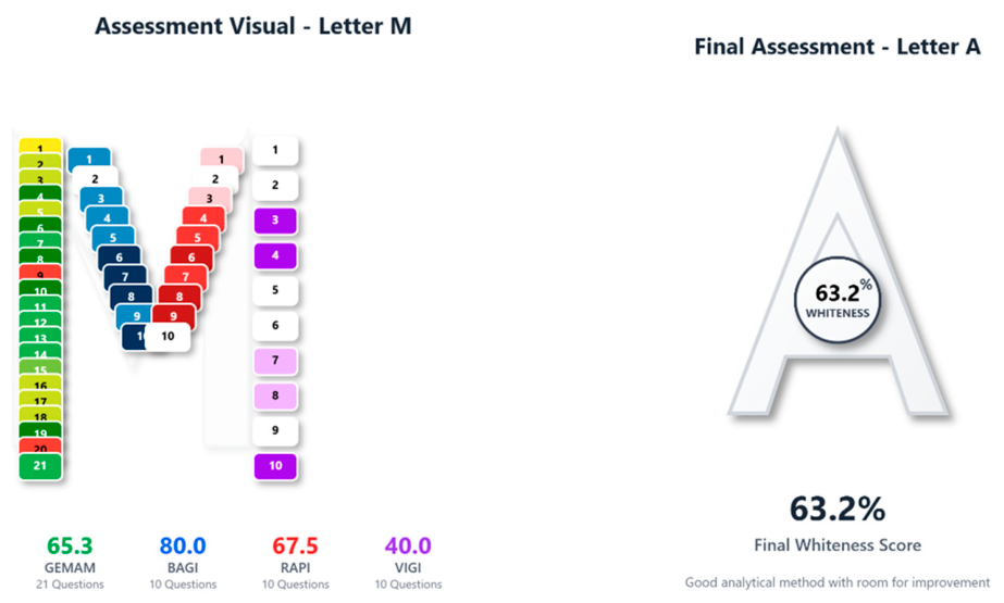
The sustainability of the analytical method was examined using the Multi-Color Assessment (MA) Tool, a platform that integrates four evaluation systems: the Green Evaluation Metric for Analytical Methods (GEMAM), the Blueness Assessment Graphical Index (BAGI), the Redness Analytical Performance Index (RAPI), and the Violet Innovation Grade Index (VIGI). The process followed a structured 51-question protocol covering environmental impact, operational practicality, analytical performance, and the degree of technological innovation. The individual scores were calculated and normalized to produce an overall whiteness Score, reflecting the method’s total sustainability. The platform is freely accessible and automatically generates the corresponding figures.
Franz diffusion cells were used to evaluate the intestinal permeability of both DOX and the DOX complex. A cellulose membrane (12,000–14,000 molecular weight cut-off, Sigma-Aldrich, Darmstadt, Germany) was placed between the donor and recipient chambers (20 mL), providing an effective diffusion area of 4.2 cm2. PBS was used as the acceptor medium, and the temperature was maintained at 37 °C by circulating warm water through the double-walled outer jacket, and gentle agitation was applied at 100 rpm.
The dissolution experiments were carried out using a thermostatic shaking water bath from Witeg (Wertheim, Germany).
2.5. Gastric and Intestinal Protocol
The study was conducted using four different sample compositions: 100 mg DOX as a reference, one Vibramycin® tablet containing 100 mg DOX, 100 mg DOX combined with 28 mg Fe3+, and 100 mg DOX combined with 200 mg Al(OH)3. The amounts of iron and aluminum hydroxide were calculated based on the corresponding ion content of the commercial products SiderAL Forte® and Maalox®. All samples were analyzed in triplicate in both gastric and intestinal media. Incubation was carried out in 40 mL SGF (37 ± 0.5 °C) in a shaking water bath for 2 h, with 0.7 mL aliquots collected at 30 min, 1 h, and 2 h. Following completion of the gastric phase, the required volume of 1 M NaOH was added to neutralize the pH, followed by 80 mL of simulated intestinal fluid. The intestinal phase lasted a total of 3 h, with samples collected at 30 min intervals (V = 0.5 mL).
2.6. In Vitro Permeability Study
A 2 mL volume of intestinal fluid was collected at predetermined time intervals (30, 90, 150 min) and transferred to the Franz cells, replacing an equivalent volume of medium from the donor compartment. Given the sequence of the experiments, the starting point of the permeability study in the Franz cells (0 min) was defined as the time of the first intestinal sampling (30 min). The procedure was carried out under continuous stirring at 100 rpm (37 ± 0.2 °C). In parallel, sampling was also performed from the receptor compartment every 30 min, and the removed volume was replaced with an equal volume of fresh PBS medium. All dissolution and permeation experiments were performed in three independent experimental runs, each including separately prepared donor and receptor systems. Analysis within each run was performed as a single quantitative determination. The collected samples were analyzed using the proposed HPLC-FLD method.
For the permeability assessment, the steady-state flux (Jss) of DOX was determined by plotting the permeated amount per unit area (μg/cm
2) as a function of time (min) and calculating the slope of the linear portion of the curve. Finally, the apparent permeability coefficient (Papp) was calculated using the following equation:
where Cd is the initial concentration of the drug in the donor compartment.
3. Results and Discussion
3.1. Development of the Chromatographic Method
The aim of the present study was to develop a fast and low-cost analytical method, fully compliant with the principles of Green Chemistry and suitable for routine analysis.
Initially, using a mobile phase consisting of a methanol-water system with 0.1% formic acid, the retention mechanism of DOX on various stationary phases was investigated. The proportion of the organic solvent was adjusted in each case so that DOX eluted after the solvent front and within a total analysis time of less than 10 min. Using peak symmetry and base width as evaluation criteria, several 15 cm columns were examined, including C18 Supelco Discovery HS (150 × 4.6 mm, 5 μm), C8 Supelco (150 × 4.6 mm, 5 μm), C4 ACE (150 × 4.6 mm, 5 μm), CN Waters Spherisorb® (150 × 4.6 mm, 5 μm), and phenyl ACE-ACT (150 × 4.6 mm, 5 μm). In all cases, particularly with the phenyl and CN columns, broad peaks were observed, while the C4 column produced a peak with pronounced fronting. The best performance was obtained with the C18 and C8 columns, with the latter exhibiting slightly higher tailing.
Thus, the C18 Supelco Discovery HS (150 × 4.6 mm, 5 μm) was selected as the optimal, and a series of additional experiments was carried out to further improve peak shape. As organic solvents of the mobile phase, both acetonitrile and methanol were evaluated, while the aqueous phase consisted of 0.1% formic acid, NaH2PO4 buffer (20 mM) at pH 2.5, and at pH 6.8. Acetonitrile reduced the peak width of DOX, and lowering the pH caused a slight increase in retention time without significantly affecting peak shape, whereas the use of TFA instead of FA or the addition of EDTA to the mobile phase resulted in even broader peaks. To avoid salt accumulation on the column, water with 0.1% FA and acetonitrile was selected as the optimal mobile phase.
In line with the principles of Green Chemistry and aiming to reduce organic solvent consumption, shorter columns were subsequently tested, including Fast RP (4.6 × 50 mm, 2.5 μm), Silica RP 2.5 μm (4.6 × 50 mm), Silica 100 RP 2.5 μm (4.6 × 50 mm) from Shant Laboratories (Shimadzu, Tokyo, Japan), Supelco Lichrosorb RP-C18 5 μm (100 × 4 mm), Intersil C8 (125 × 4.0 mm, 5 μm), and Thermo Hypersil Gold C18 (50 × 4.6 mm, 5 μm). However, in most cases, DOX either co-eluted with the solvent front or produced broad peaks. The column that provided the best performance and was ultimately selected was the Thermo Hypersil Gold C18 (50 × 4.6 mm, 5 μm).
Further optimization included adjustments to the injection volume (30 μL), mobile phase flow rate (1 mL/min), and column oven temperature (40 °C). Increasing the temperature was deemed necessary, as it improved peak sharpness and reduced the viscosity of the mobile phase.
Finally, to further enhance chromatographic peak quality, a range of solvents was evaluated as potential diluents for doxycycline, including methanol, acetonitrile, water, and mixtures of methanol or acetonitrile with or without the NaH2PO4 buffer (20 mM) at pH 2.5 and 9.2 in a 1:1 v/v ratio. Among all solvents tested, water consistently yielded the most favorable peak characteristics, independent of pH, and was therefore selected as the diluent, in alignment with the principles of Green Chemistry.
3.2. Detector Performance Evaluation and Selection for DOX Quantification
DOX contains multiple ionizable functional groups (alcohol, phenolic, and amino moieties), allowing it to exist as a cation, anion, or zwitterion. Consequently, it exhibits several acid dissociation constants (pKa values). At intermediate pH values, DOX predominantly exists as a zwitterion. When the pH is below 3.02 (pKa
1 = 3.50), DOX is present mainly in its cationic form (DOX
+). Between pH 3.02 and 7.97 (pKa
2 = 7.07), it exists primarily as a zwitterion (DOX
+/−). Between pH 7.97 and 9.15 (pKa
3 = 9.15), it is present as a monoanionic species (DOX
−), while at pH values above 9.15 it forms the dianionic species (DOX
2−) due to deprotonation of the phenolic diketone and tricarbonyl system [
29].
These multiple and pH-dependent ionization states significantly influence its behavior in different environments. Consequently, the key question that arises is what occurs within the gastrointestinal tract and to what extent the presence of cations may affect the permeability of DOX across the intestinal barrier before it reaches systemic circulation. Designing an appropriate experimental procedure requires consideration of the physiological temperature of the human body (37 °C), the pH of the relevant biological fluids, and the residence time of the compound (and/or its complexes) in the stomach (up to 3 h) and the intestine (up to 3 h).
Since the reliable detection and quantification of a chemical compound is crucial for obtaining information related to its structure and its potential degradation by external factors, particular emphasis was placed on selecting an appropriate detector. More specifically, samples of equal concentration (150 μg/mL) DOX, DOX-Fe3+, and DOX-Al(OH)3 were examined in simulated gastric and intestinal fluids at 0 min and 3 h, with or without heating at 37 °C.
The samples were initially analyzed under the proposed chromatographic HPLC conditions using a UV detector and compared with one another. Quantification was performed using a calibration curve (y = (15,368 ± 85)x + 5053 ± 5740, r
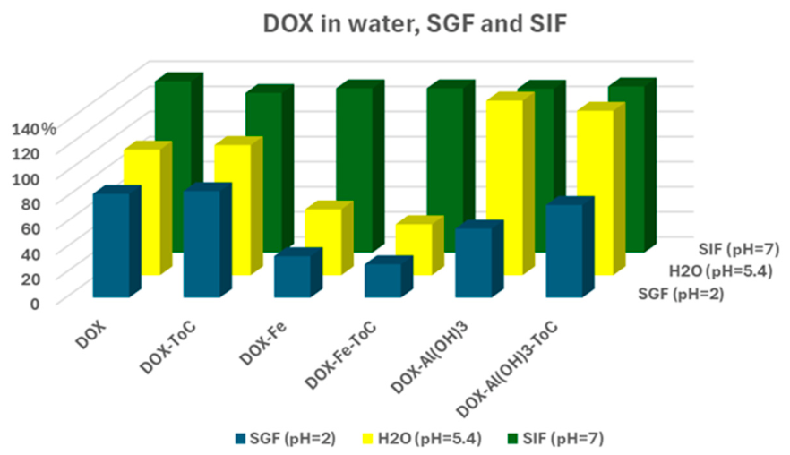
2 = 0.999) constructed at six concentration levels (3–210 μg/mL, n = 3). Based on the UV bar charts (
Figure 1), it was evident that temperature did not substantially affect the signal, except for the DOX-Fe
3+ complex, where a slight decrease in intensity was observed at pH 2 and 5.4. The most notable finding was the strong hypochromic effect of DOX in acidic media, whereas at pH = 7, a clear hyperchromic effect was observed. Furthermore, comparison of pure DOX samples with their corresponding complexes (in the same medium) showed that the Fe
3+ complex exhibited reduced signal intensity at pH 5.4 and 2, while the Al
3+ complex decreased at pH 2 but increased at pH 5.4. In contrast, at pH 7, DOX displayed identical signal intensity whether in free or complex form, with overall higher absorbance (compared to water) due to hyperchromism. The fact that DOX does not form complexes at alkaline pH is confirmed by the stable signal observed across all cases.
In summary, the main conclusion is that during UV detection, variations in signal intensity are primarily determined by hyperchromic and hypochromic effects, and only secondarily by complex formation at specific pH values. Furthermore, at pH < 5, when the compound is present in the same diluent, the signal is altered due to complexation, whereas in alkaline media it remains stable.
The same samples were subsequently examined under identical HPLC conditions using a fluorescence detector (FLD). The results demonstrated a consistent and stable DOX signal across all tested conditions, indicating that neither bathochromic/hypsochromic shifts nor complex formation interfered with the analytical response. This study was also an indirect stability evaluation of the analyte for 3 h at 37 °C, in terms of the dynamic in vitro protocol.
Table 2 further summarizes the matrix effect of DOX in SIF and SGF fluids (37 °C) in the presence of Fe
3+ or Al
3+, with recovery values calculated using a water-based reference standard.
Given this confirmed stability of the fluorescence signal under all matrix conditions, and considering that the aim of the present study was to quantify the total amount of DOX (in complexed or uncomplexed form) permeating through the Franz cell barrier, the fluorescence detector was deemed more appropriate, as it provides reliable and sensitive responses under these conditions.
3.3. Method Validation
Τhe method was validated in accordance with the requirements of ICH Q2(R2) [
30], including the evaluation of system suitability, selectivity, linearity, limits of detection and quantification, precision, accuracy, and robustness.
3.3.1. System Suitability
The performance of the chromatographic system was evaluated through key parameters indicative of its efficiency and reliability. Specifically for DOX, the following values were determined: a retention time of 3.39 min, a tailing factor of 1.29, a theoretical plate number of 1141.92, and a height equivalent to a theoretical plate (HETP, USP) of 44 μm.
3.3.2. Selectivity
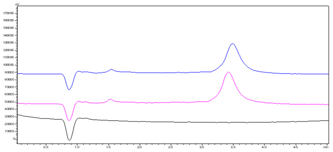
The selectivity of the method was confirmed by comparing chromatograms obtained from a blank diluent and a pretreated tablet sample. The analytical method demonstrated adequate selectivity, as no peaks originating from the matrix were observed at the retention times of the target analytes (
Figure 2). In SGF, SIF, or PBS as diluent in the presence of iron or aluminum, the chromatogram of the blank solution was like water, with no interfering peaks. Carryover was assessed by performing three consecutive injections of the highest calibration level, followed by a blank injection. No additional peaks were detected in the blank chromatogram, indicating the absence of carryover [
31]. Furthermore, the matrix effect of DOX in simulated intestinal (SIF) and gastric (SGF) fluids at 37 °C, in the presence of Fe
3+ or Al
3+, was thoroughly investigated (
Section 2.3,
Table 2) and was found not to affect the outcome.
3.3.3. Linearity, LOD, and LOQ
Linearity was evaluated using six standard solutions prepared in water, each analyzed in triplicate (n = 3). The calibration curve was constructed from the obtained AUC (area under the curve) values and was described by the following equation:
The reliability of the method was further assessed by calculating the % y-intercept (intercept × 100/response at 100%), which was found to be within the acceptable limit, 1.87% (<2%). The limits of detection (LOD) and quantification (LOQ) were calculated using the following equations:
where Sy/x is the residual standard deviation and
Slope is the (x) variable of the calibration curve.
Based on these equations, LOD was found to be 0.8 μg/mL and LOQ 2.7 μg/mL.
3.3.4. Precision and Repeatability
Precision was evaluated through intra-day (repeatability) and inter-day (intermediate precision) analyses at three concentration levels (low, medium, and high). Measurements were performed within a single day and across three consecutive days. The results, expressed as %RSD, are summarized in
Table 3.
3.3.5. Accuracy
Accuracy was evaluated using five DOX samples of known concentration (n = 3) for intra-day accuracy and three samples with different concentrations (low, medium, high) for three consecutive days (n = 3). The measured concentrations, obtained from the calibration curve, were compared with the corresponding theoretical values to calculate the % recovery (
Table 4). The method demonstrated satisfactory accuracy, with a mean % recovery value of 100 ± 2%.
Additionally, the accuracy of the method was assessed with respect to its ability to recover the active substance from a Vibramycin® (100 mg) tablet formulation. Sample preparation was performed according to the extraction procedure on five individual tablets, processed separately. The mean % recovery was calculated to be 104.6% (%RSD = 2.1%).
3.3.6. Robustness
The robustness of the chromatographic system was evaluated by introducing small, deliberate variations in critical operating parameters and assessing their influence on the tailing factor (Tf) and peak area (AUC). The tested parameters included slight modifications in the mobile phase flow rate (±0.05 mL/min), column oven temperature (±2 °C), organic solvent ratio in the mobile phase (±1%), salt concentration (±5 mM), pH (±0.5), and emission wavelength (±2 nm). In all cases, the obtained %RSD values for both Tf and AUC remained within the acceptable limits (
Table 5), indicating that none of the tested variations had a significant impact on chromatographic performance. These findings confirm that the method is robust under the conditions evaluated, with the only exception involving parameters that influence the residence time of the analyte within the detector’s flow cell (primarily flow rate) [
32].
3.4. Dissolution Test and Permeability Study
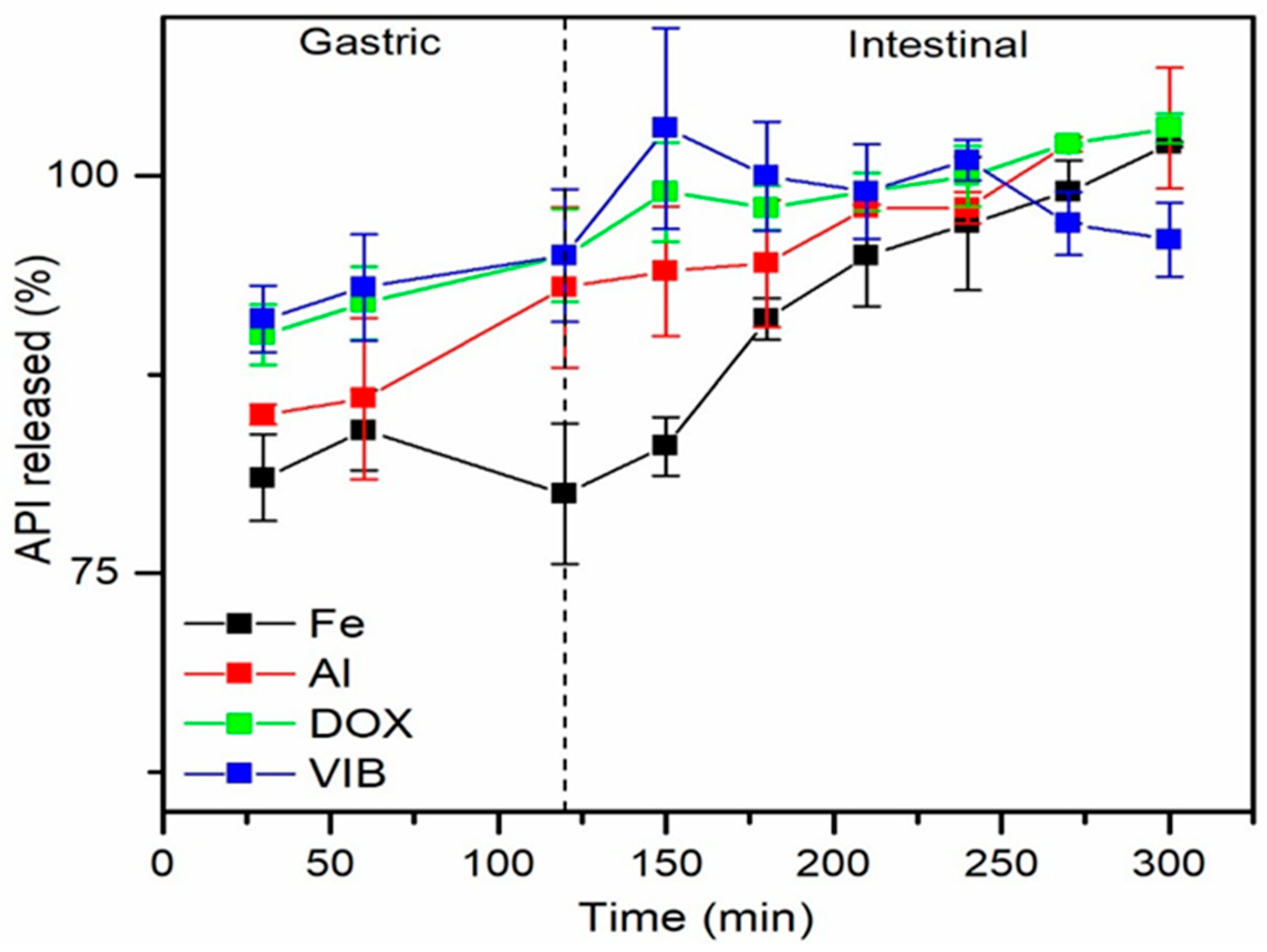
Figure 3 presents the in vitro release profiles in gastric and intestinal fluids of pure DOX, DOX in tablets (VIB), and DOX complexed with Fe
3+/Fe
2+ (Fe) and Al
3+ (Al). According to the results, in simulated gastric fluid, DOX is detected at ~90% within the first 30 min, both in its pure form and in the tablet, while the presence of Al
3+ and Fe
3+ slightly reduces the detected amount to 85% and 81%, respectively. The negative effect of iron becomes more evident at the 2 h sampling point, where DOX reaches approximately 94% in all cases except in the presence of Fe
3+, where only 80% is detected. After the samples transition to alkaline pH, a small corresponding increase is observed during the first 30 min, followed by a gradual rise in DOX concentration that eventually approaches 100% in all samples.
To evaluate whether the dissolution and permeability rates of DOX were affected by its surrounding environment, a paired t-test was performed to determine whether the paired measurements differed significantly. The analysis was conducted at a significance level of α = 0.05. The test returned p-values < 0.05 for both the DOX–Fe3+ vs. DOX–Al3+ pair (p = 0.020) and the DOX vs. DOX-Fe3+ pair (p = 0.008), indicating that these paired datasets differed significantly from each other. In contrast, no statistically significant difference was observed between DOX and DOX in tablets (p = 0.97).
The distinct behavior of DOX in the presence of Fe
3+ can be interpreted by considering both the oxidation state of iron (Fe
2+/Fe
3+) at different pH values and its solubility, which under certain conditions leads to precipitation. Equally important is the protonation state of DOX, which determines its ability to form chelate complexes. DOX, whose solubility is favored at pH 1–6 [
33], interacts with metal ions primarily through coordination bonds involving its hydroxyl and amine donor groups, as well as through hydrogen bonding [
7].
Table 6 sums up the main bibliographic facts in which the pharmacokinetic behavior of DOX can be interpreted in the presence of iron. It should be noted that no direct monitoring of iron speciation or Fe
2+/Fe
3+ interconversion was performed during the present experiments. Τherefore, the proposed mechanistic interpretation should be considered as a proof of concept framework supported by bibliographic evidence rather than a direct verified experimental confirmation of the predominant iron species under each condition.
At very low gastric pH (1–2), DOX is highly protonated, limiting the availability of donor groups for chelation. Although Fe3+ is fully soluble at this pH, the strong competition from H+ reduces the efficiency of complex formation. In the intermediate pH range (3–6), partial deprotonation of the phenolic/enolic groups enhances the donor capacity of DOX, while Fe3+ remains soluble, leading to the highest levels of chelation. At neutral to slightly alkaline pH (7–8), DOX becomes more negatively charged and capable of strong chelation with Fe 3+, given that the Fe2+-DOX complexes are not stable. However, Fe3+ undergoes hydrolysis and precipitates as Fe(OH)3, reducing the amount of iron available for complexation. Thus, the experimentally observed decrease in DOX-Fe chelation at this pH is mainly due to the reduced availability of Fe3+ rather than a diminished ability of DOX to form complexes.
When Fe3+ is replaced by Al3+, the behavior changes markedly. Al3+ hydrolyzes and precipitates at much lower pH values (around 4–5) as Al(OH)3(s), and it forms significantly weaker complexes with DOX compared to Fe3+. As a result, Al3+ only remains soluble under strongly acidic conditions, and its ability to form chelates is limited; at neutral or slightly alkaline pH, Al3+ is essentially absent from the solution due to precipitation. Moreover, Al3+ does not participate in redox reactions, meaning that the Al-DOX system is governed exclusively by hydrolysis and solubility, in contrast to Fe3+, which remains available for chelation across a broader pH range.
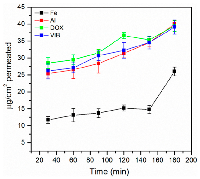
Following the completion of the DOX dissolution study in gastric and intestinal media, its permeation across cellulose membranes was evaluated using Franz diffusion cells. In this experiment, pure DOX, DOX from the tablet formulation (VIB), and DOX in the presence of Al
3+ exhibited comparable Jss and Papp values, whereas the presence of Fe
3+ resulted in approximately a two-fold reduction in the amount of DOX reaching the receptor compartment (
Table 7,
Figure 4). Since the analytical method quantified equal concentrations of free DOX in the donor phase under all conditions, the reduced permeation observed with Fe
3+ cannot be attributed to differences in the available DOX in solution. A reasonable explanation is that at pH 7, Fe
3+ undergoes hydrolysis and forms hydroxide or colloidal species that interact with the membrane surface, generating an additional diffusional barrier (fouling layer) or a region of local DOX adsorption at the membrane interface. This interfacial effect increases the resistance to diffusion and decreases the effective flux of DOX, despite identical donor concentrations. In contrast, the hydroxide species formed by Al
3+ under the same conditions appear to exert minimal influence on the membrane, which is consistent with the similar permeation profiles observed for pure DOX, VIB, and DOX in the presence of Al
3+. However, no direct membrane characterization, precipitate quantification, or adsorption control experiments were performed in the present study. Therefore, this interpretation should be regarded as a mechanistically plausible hypothesis rather than a directly demonstrated membrane fouling phenomenon.
Similarly, it should be noted that the Franz diffusion cells used in the present study model only passive diffusion across an inert membrane and do not reproduce key physiological characteristics of the intestinal environment. In vivo, doxycycline absorption may be influenced by active transport systems, dependent ion interactions, the unstirred water layer, and the presence of mucus and enzymes. Cellulose was selected as a simplified and low-cost option with hydrophilic, inert, porous diffusional support that allows a controlled passive transport assessment while minimizing additional chemical interactions with the tested drug. Therefore, the values reported in this study should be interpreted as mechanistic indicators of how metals affect the passive component of doxycycline transport rather than as predictors of absolute intestinal bioavailability.
Furthermore, the present study is a simplified dynamic protocol that provides mechanistic insights that are consistent with and supported by in vivo protocols. It is known that
per-os iron can reduce the concentration of DOX and decrease its t
1/2 according to in vivo studies in people [
14]. In addition, in vivo experiments in chickens have also shown a decrease in tetracycline in the blood in the presence of iron [
34]. Further studies have suggested a decrease in absorption [
5,
35]. In this context, in vitro studies like this can provide relevant mechanistic evidence without replacing the in vivo experiments on drug interactions.
3.5. Evaluation Using Unified MA Tool
The environmental sustainability of the proposed analytical method was assessed via a unified MA tool, an online platform that integrates four existing assessment frameworks into a single evaluation system for analytical methods [
36]. It combines GEMAM (greenness), BAGI (practicality), RAPI (performance), and VIGI (innovation) through a 51-question evaluation protocol. In addition, the tool calculates individual scores for each dimension and computes an overall whiteness Score that reflects the method’s total sustainability. Scores below 39% are considered poor, 40–59% moderate, 60–79% good, and ≥80% excellent.
According to the results (
Figure 5), the proposed HPLC-FLD method demonstrated a strong environmental sustainability profile (65.3%), primarily due to its low organic solvent consumption during both the sample pretreatment and chromatographic analysis.
The method also exhibited excellent practical applicability, achieving an 80% BAGI score, which reflects highly accessible instrumentation, low operational cost, and simple procedures that support routine laboratory implementation. Its outstanding practical feasibility makes it well-suited for widespread adoption with minimal operational barriers and efficient resource utilization.
The RAPI score of 67% reflects the comprehensive ICH Q2(R2) validation performed, including specificity, accuracy, precision, robustness, and sensitivity parameters that ensure regulatory compliance. This relatively high score is expected, as the method adheres to established ICH guidelines that systematically address the core validation elements evaluated by RAPI.
The VIGI assessment indicates a moderate level of innovation, supported by the method’s alignment with White Chemistry principles and its regulatory integration. The conceptual framework of the study also contributes to the overall score.
Overall, the analytical method demonstrates good sustainability, with a 63.2% whiteness Score, indicating a well-balanced performance across the four critical dimensions of environmental impact, practical applicability, analytical performance, and technological innovation. It represents a solid and comprehensive analytical solution that effectively integrates sustainability principles with practical implementation requirements, making it a reliable choice for routine analytical applications.
4. Conclusions
The present study developed a combined methodology integrating dissolution-rate experiments (in gastric and intestinal media) with doxycycline permeation studies, aiming to investigate the influence of Fe3+ and Al3+ ions on its biopharmaceutical behavior. The results demonstrated rapid and nearly complete release of DOX under gastric conditions for all tested forms, with only minor reductions observed in the presence of Fe3+. Following the transition to intestinal pH, DOX achieved complete solubilization in all cases, indicating that neither metal ion substantially alters its overall dissolution profile.
In contrast, the permeation experiments revealed a selective effect of Fe3+. Despite identical concentrations of free DOX in the donor compartment of the Franz cells, the presence of Fe3+ resulted in significantly lower flux and apparent permeability compared with pure DOX, the tablet formulation, and the Al3+ condition. This reduction is likely associated with interfacial phenomena at the membrane surface, such as the formation of Fe-derived hydroxide or colloidal species at pH 7, which increase diffusional resistance without affecting the drug concentration in solution. Al3+ did not induce a comparable effect, consistent with the similar permeation profiles observed across the corresponding samples.
This work represents an initial in vitro evaluation of doxycycline permeation through a cellulose membrane using Franz diffusion cells. A key limitation of the study is that the proposed Fe3+-related interfacial mechanism is supported only indirectly by transport data, as no direct membrane characterization was performed. Future research should therefore include physicochemical or spectroscopic analysis of the membrane to confirm the hypothesized interactions, as well as experiments conducted in biorelevant media to better simulate physiological conditions. Additionally, ex vivo permeation studies using intestinal tissue would provide a more physiologically relevant validation of the proposed mechanism and further complement the in vitro findings.
Finally, the validated HPLC-FLD method proved to be rapid, economical, and environmentally compatible, offering high sensitivity and operational flexibility while minimizing solvent consumption. Overall, the combined methodology established in this work provides an efficient and practical framework for evaluating factors that influence the bioavailability of doxycycline.



 ,
,