Article

Astrocytes in the CA1 Field of the Hippocampus as Targets of Magnoflorine Action: The Relevance to Astrogial Structural and Functional Modulation After Acute and Chronic Administration—A Preliminary Study


by
Aleksandra Krawczyk
1,†,
Radosław Szalak
1,*,†,
Małgorzata Komar
1,*,
Dorota Nieoczym
2,
Wirginia Kukula-Koch
3,
Wojciech Koch
4,
Ömer Gürkan Dilek
5 and
Marcin B. Arciszewski
1
1
Faculty of Veterinary Medicine, Department of Animal Anatomy and Histology, University of Life Sciences, Akademicka Str. 12, 20-033 Lublin, Poland
2
Faculty of Biology and Biotechnology, Institute of Biological Sciences, Department of Functional Anatomy and Cytobiology, Maria Curie-Skłodowska University, Akademicka Str. 19, 20-033 Lublin, Poland
3
Department of Pharmacognosy with Medicinal Plants Garden, Medical University of Lublin, Chodźki Str. 1, 20-093 Lublin, Poland
4
Department of Food and Nutrition, Medical University of Lublin, Chodźki Str. 4a, 20-093 Lublin, Poland
5
Faculty of Veterinary Medicine, Department of Anatomy, Mehmet Akif Ersoy University, Cevat Sayili Boulevard 118, Degirmenler District, 15-030 Burdur, Turkey
*
Authors to whom correspondence should be addressed.
These authors contributed equally to this work.
Appl. Sci. 2026, 16(10), 4960; https://doi.org/10.3390/app16104960
Submission received: 1 April 2026 / Revised: 8 May 2026 / Accepted: 12 May 2026 / Published: 15 May 2026
(This article belongs to the Special Issue Dietary Bioactive Compounds and Their Neuroprotective Potential)

Featured Application

The demonstrated dose-, time-, and layer-dependent modulation of astrocyte morphology and reactivity in the CA1 field of the hippocampus in mice by magnoflorine (MGN) suggests that this compound may affect astroglia-mediated mechanisms involved in neurotransmission and regulation of the neuronal microenvironment. Through increased arborisation of astrocytic processes, magnoflorine may contribute to changes in synaptic organisation under both physiological and neuronal stress conditions.

Abstract

Astrocytes play a crucial role in maintaining neuronal microenvironment homeostasis and regulating synaptic plasticity within the hippocampus. Magnoflorine (MGN), a naturally occurring isoquinoline alkaloid, has demonstrated biological activity in the central nervous system. However, its effects on astroglial cells remain poorly understood. The present study aimed to evaluate the impact of acute and chronic administration of MGN (10 and 20 mg/kg body weight) on the morphology and morphometric parameters of GFAP-positive astrocytes in the CA1 field of the mouse hippocampus. Immunohistochemical and morphometric analyses were performed in the oriens layer (SO), pyramidal layer (SP), radiate layer (SR), and lacunose-molecular layer (SLM). MGN significantly modulated astrocyte density, cell size, and the number of processes in a dose-, time-, and layer-dependent manner. A heterogeneous and layer-specific astroglial response was particularly evident following chronic administration of the tested compound. Together with the observed lack of significant differences in analysed parameters, decreases were mainly detected after administration of the low MGN dose, whereas the 20 mg/kg dose induced primarily increased structural complexity. Thus, the direction of changes was not uniform across all layers. The most prominent changes were detected in the SLM layer. Overall, MGN modulated astrocyte morphology and reactivity in a context-dependent manner. These findings indicate a modulatory influence of MGN on astroglial structural plasticity rather than a uniform directional effect. Although the observed changes may be associated with alterations in astroglia-mediated mechanisms involved in maintaining neuronal homeostasis and responses to stress, their functional significance requires further investigation.

1. Introduction

Astrocytes are one of the primary types of glial cells in the central nervous system (CNS) and play a crucial role in maintaining the homeostasis of the neuronal microenvironment. The star-shaped cells have numerous processes that repeatedly divide into small branches. Their endings reach, among others, synapses and Ranvier nodes of nerve fibres and, in the form of end feet, cover the surfaces of blood vessels [1,2,3,4]. Astrocyte processes exhibit the activity of numerous ion channels, receptors for various neurotransmitters, cytokines, chemokines, growth factors, and transporters [5,6]. This enables astrocytes to contribute to maintaining ionic homeostasis, be involved in the metabolism of many neurotransmitters, including glutamate (Glu), and ensure the integrity and proper permeability of the blood–brain barrier (BBB). Additionally, astrocytes support synaptogenesis, which is essential for learning and memory. Furthermore, they can be a source of many active substances, including those with neuroprotective and synaptic properties, as well as growth factors that positively impact neuronal survival and function [1,2,3,4,7,8,9,10,11]. Astroglia also play an important role in the pathogenesis of many acute and chronic CNS diseases, such as epilepsy, stroke, traumatic brain injury, as well as Alzheimer’s disease, Parkinson’s disease and amyotrophic lateral sclerosis [12,13]. Astrocytes respond dynamically to any changes in the neuronal microenvironment. This can be observed by overgrowth of their bodies and processes (hypertrophy) and increased synthesis of various proteins, including glial fibrillary acidic protein (GFAP). In mature astrocytes, GFAP builds the cell’s cytoskeleton, influencing its shape, structural stability, and mechanical strength. This protein also controls astrocyte motility and, consequently, synaptic activity. GFAP plays a crucial role in stiffening astrocytic processes and in the permeability and maintenance of the BBB. It interacts with many other intracellular proteins and adhesion molecules, including specific Glu transporters. Maintaining the homeostasis of this neurotransmitter is crucial, as its excess in the extracellular space can lead to excitotoxicity and neuronal and glial death. Astroglia take up Glu from the extracellular space, thereby clearing excess Glu from the synaptic cleft, which represents one of the key neuroprotective mechanisms [7,11,14,15,16,17,18].
The hippocampus is a part of the olfactory system, playing a significant role in memory and learning processes [19]. The CA1 field of the hippocampus is composed of the oriens layer (SO), the pyramidal layer (SP), the radiate layer (SR), and the lacunose-molecular layer (SLM). The SO layer includes the axons of pyramidal neurons, whose neuron bodies form the SP layer. The SR layer contains its dendrites, whereas the SLM layer features terminal dendritic branches and a large number of synaptic connections [20,21]. Glutamatergic fibres of the perforant pathway, connecting the temporal lobe with the hippocampus, and glutamatergic collaterals of Schaffer pyramidal cells in the CA3 field of the hippocampus, terminate within the SLM layer [22].
Magnoflorine (MGN) is a naturally occurring isoquinoline alkaloid that belongs to the aporphine subclass and is widely distributed among various plant families. One of its richest natural sources is the root of Berberis vulgaris, a plant long used in traditional medicine for its health-promoting properties. The compound can be effectively isolated from plant material using modern chromatographic techniques, such as centrifugal partition chromatography, which ensures the high purity and reproducibility of the extract. Numerous studies have confirmed the broad, multi-system activity of MGN, demonstrating its antioxidant, anti-inflammatory, immunomodulatory, antimicrobial, and anticancer properties. It has also been associated with anxiolytic and potential neuroprotective effects, making it an object of growing scientific interest [23,24,25]. Previous research, including recent experimental studies, has shown that MGN may exert beneficial effects on the CNS by influencing neuronal calcium homeostasis, supporting hippocampal neuron function, and modulating parvalbumin expression—factors essential for maintaining memory processes. The compound has also been observed to enhance learning and memory performance in animal models, suggesting its potential role in improving cognitive functions and protecting against neurodegenerative changes associated with aging and diseases such as dementia or Alzheimer’s disease [26,27].
Considering that normal neuronal activity depends closely on metabolic and trophic support provided by astroglial cells, it can be hypothesised that MGN may also induce structural alterations in astrocytes. To our knowledge, the effects of MGN on hippocampal astrocytes have not been investigated. Therefore, preliminary studies were undertaken to evaluate the effects of different doses of MGN, administered either once or for seven consecutive days, on astroglial morphology and reactivity in the CA1 field of the hippocampus in mice.

2. Materials and Methods

2.1. Animals

The experiments were carried out on 48 (n = 48) naïve male Swiss mice (Experimental Medicine Center, Lublin, Poland) weighing 20–30 g, and 4 weeks of age. Mice were housed in groups of 8 per home cage (38 × 22 × 18 cm3), made of white Plexiglas. The animals were maintained under standard laboratory conditions (12 h light/dark cycle, room temperature 21+/−1 °C) with free access to tap water and laboratory chow (Agropol, Motycz, Poland) in their home cages and adapted to the laboratory conditions for 7 days. Subsequently, the animals were randomly assigned to six experimental groups (n = 8): two control groups corresponding to the acute and chronic treatment regimens; two groups receiving MGN at a dose of 10 mg/kg body weight (b.w.) under acute or chronic administration; and two groups receiving MGN at a dose of 20 mg/kg b.w. under acute or chronic administration. The number of animals was selected to sustain a proper statistical analysis of results. All experiments were performed between 8:00 and 15:00 on animals assessed visually to be in good health and putting on weight correctly, similar to other animals.
All studies were carried out according to the ARRIVE guidelines to improve the reporting of animal research and the quality of the studies and were conducted in accordance with the National Institute of Health Guidelines for the Care and Use of Laboratory Animals, and with the European Community Council Directive for the Care and Use of Laboratory Animals of 22 September 2010 (2010/63/EU). Furthermore, on 19 June 2015, we obtained the agreement to conduct the aforementioned studies (no. 33/2015) from the Local Ethical Committee for Animal Experiments in Lublin. All gathered animals were used for this study. No randomisation technique or blinding of the study was implemented. The experiments were performed in accordance with the principles of substitution, limitations, and improvements (3R).

2.2. Drugs

Magnoflorine (MGN) (96.2%) was isolated from the methanolic extract of Berberis vulgaris root according to a previously described protocol [26] using centrifugal partition chromatography (CPC), using a bisphatic solvent system composed of chloroform/methanol/water (4:3:3 v/v) with 20 mM of hydrochloric acid and triethylamine, as reported in the authors’ earlier work [26]. Prior to administration, MGN was freshly prepared on the day of the experiment by dissolving the compound in saline in a mortar. The solution was administered intraperitoneally (i.p.) at a volume of 10 mL/kg. Fresh MGN solutions at doses of 10 and 20 mg/kg b.w. were used. The selected doses (10 and 20 mg/kg b.w.) were based on our previous behavioural study [26], in which 20 mg/kg significantly improved memory performance, whereas 10 mg/kg produced a borderline effect without altering locomotor activity, indicating that both doses were behaviourally well tolerated. Control animals received injections of saline at the same volume and via the same route of administration.
In the acute treatment regimen, MGN (10 or 20 mg/kg) or saline was administered intraperitoneally as a single dose. In the chronic treatment regimen, MGN (10 or 20 mg/kg) or saline was administered intraperitoneally once daily for 7 consecutive days.

2.3. Immunohistochemical Analyses

The animals were euthanised by decapitation, and the collected brains were immediately fixed in 10% buffered formalin (pH 7.0). After 12 h of fixation at 4 °C, the tissues were gradually dehydrated in increasing concentrations of ethanol. As described previously [26], the brains were subsequently embedded in paraffin blocks and sectioned into 5 µm thick slices using a microtome (Microm HM 360, Microm, Walldorf, Germany). The obtained sections were mounted on adhesive glass slides (Superfrost Plus, Thermo Scientific, Braunschweig, Germany).
Immunohistochemical staining was performed on frontal mouse brain sections containing the hippocampus using the indirect peroxidase–antiperoxidase (PAP) method with antibodies obtained from Sigma-Aldrich (St. Louis, MO, USA). All reagents were diluted according to the manufacturer’s instructions in 0.5 M Tris-buffered saline (TBS; pH 7.6; Sigma-Aldrich St. Louis, MO, USA), which was also used for washing steps. Deparaffinized and hydrated sections from each animal were incubated with 3% H2O2 for 30 min to block endogenous peroxidase activity, followed by incubation with goat serum (G9023, 1:10; Sigma-Aldrich St. Louis, MO, USA) for 20 min at room temperature (RT).
Sections were then incubated with a primary rabbit anti-GFAP antibody (G9269, 1:80; Sigma-Aldrich St. Louis, MO, USA) for 16 h at 4 °C. After washing, the sections were incubated at RT for 1 h with a secondary goat anti-rabbit IgG antibody conjugated to peroxidase (A9169, 1:400; Sigma-Aldrich St. Louis, MO, USA). Immunoreactivity was visualised using 3,3′-diaminobenzidine tetrachloride (DAB; 32750; Sigma-Aldrich St. Louis, MO, USA) as the chromogen. Sections were subsequently rinsed in distilled water and counterstained with Mayer’s haematoxylin (MHS80; Sigma-Aldrich St. Louis, MO, USA) according to standard histological procedures. Negative control sections were processed in parallel, omitting the primary antibody. The specificity of the anti-GFAP antibody had been previously confirmed using rat brain sections [28].

2.4. Morphological and Morphometric Analyses

Morphological analyses of GFAP-positive (GFAP+) cells were performed in individual layers of the CA1 field of the mouse hippocampus using an Olympus BX 51 light microscope coupled with an Olympus Colour View III digital camera (Olympus, Tokyo, Japan). Morphometric assessment of the studied glia was performed using Cell^D software version 5.1. GFAP-positive cells were identified based on clearly visible brown cytoplasmic immunoreactivity around the cell nucleus, exceeding the background staining level and showing morphology consistent with astrocytic cell bodies and processes. The same microscope settings, illumination, image acquisition parameters, and Cell^D software version 5.1 measurement criteria were applied to all sections to ensure consistency of thresholding and morphometric analysis across all experimental groups.
The density of GFAP+ cells was analysed in 120 microscopic fields (15 per animal) covering distinct layers of the CA1 hippocampal region. Each field had an area of 2.50 × 10⁻³ mm2 and was overlaid with a grid. Randomly selected fields that completely covered the hippocampal layer under study were analysed. The results are presented as the mean number of GFAP+ astrocytes ± standard deviation in the examined surface area. The size of GFAP+ astrocytes in the CA1 hippocampal layers was assessed by analysing at least 100 randomly selected astrocytes. Measurements of astrocytes were performed along three axes (vertical, horizontal, and oblique), and the mean astrocyte size was calculated. In addition, the mean number of GFAP+ astrocyte processes was determined in Cell^D software by counting extensions at the point of their departure from the cell body in at least 100 randomly selected GFAP+ astrocytes per animal in individual CA1 layers of the hippocampus in both the control and experimental groups, using the same predefined measurement criteria for all groups.

2.5. Statistical Analysis

Statistical analyses were performed using Graphpad Prism software version 8.0 (San Diego, CA, USA).
The results concerning the morphometric parameters of GFAP+ astrocytes, i.e., their density, number of their processes and mean size, were verified for normality of distribution using the Shapiro–Wilk test. The effect of dose was analysed separately for the single-administration and chronic-administration conditions using the Kruskal–Wallis test followed by Dunn’s post hoc test. For these analyses, differences were considered statistically significant if p ≤ 0.05.
The effect of treatment duration on the evaluated morphological parameters of astrocytes was assessed separately within each dose level using the Mann–Whitney test. Because three Mann–Whitney tests were performed in this family of comparisons, a Bonferroni correction was applied, and the threshold for statistical significance was set at raw p ≤ 0.0167 (equivalently, Bonferroni-adjusted p ≤ 0.05).

3. Results

3.1. Morphological Analyses

In all animals studied, astrocytes were found to be immunoreactive for GFAP in all layers of the CA1 field of the hippocampus. This protein was localised in the cytoplasm of the bodies, particularly at the origin of the processes, as well as in the processes themselves.
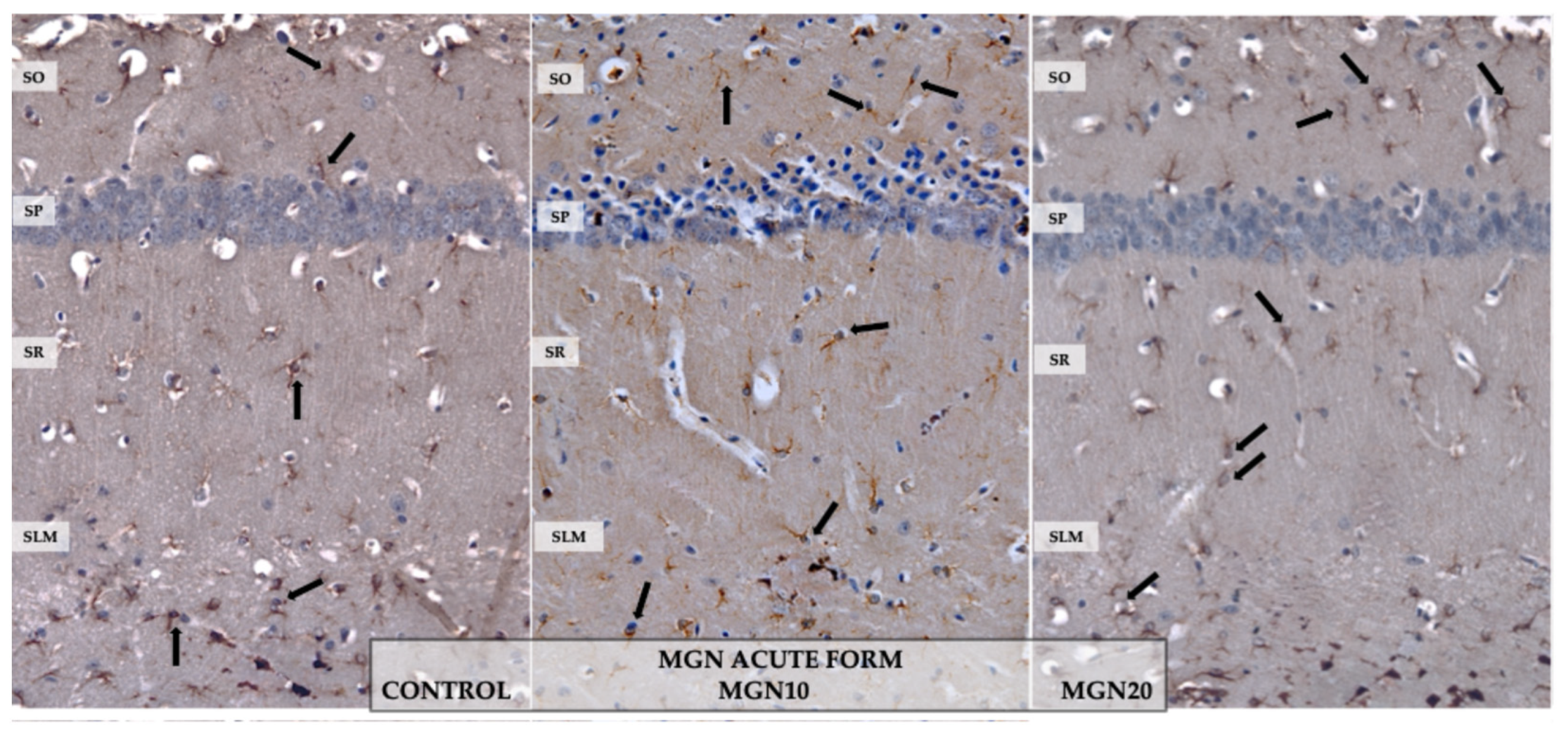
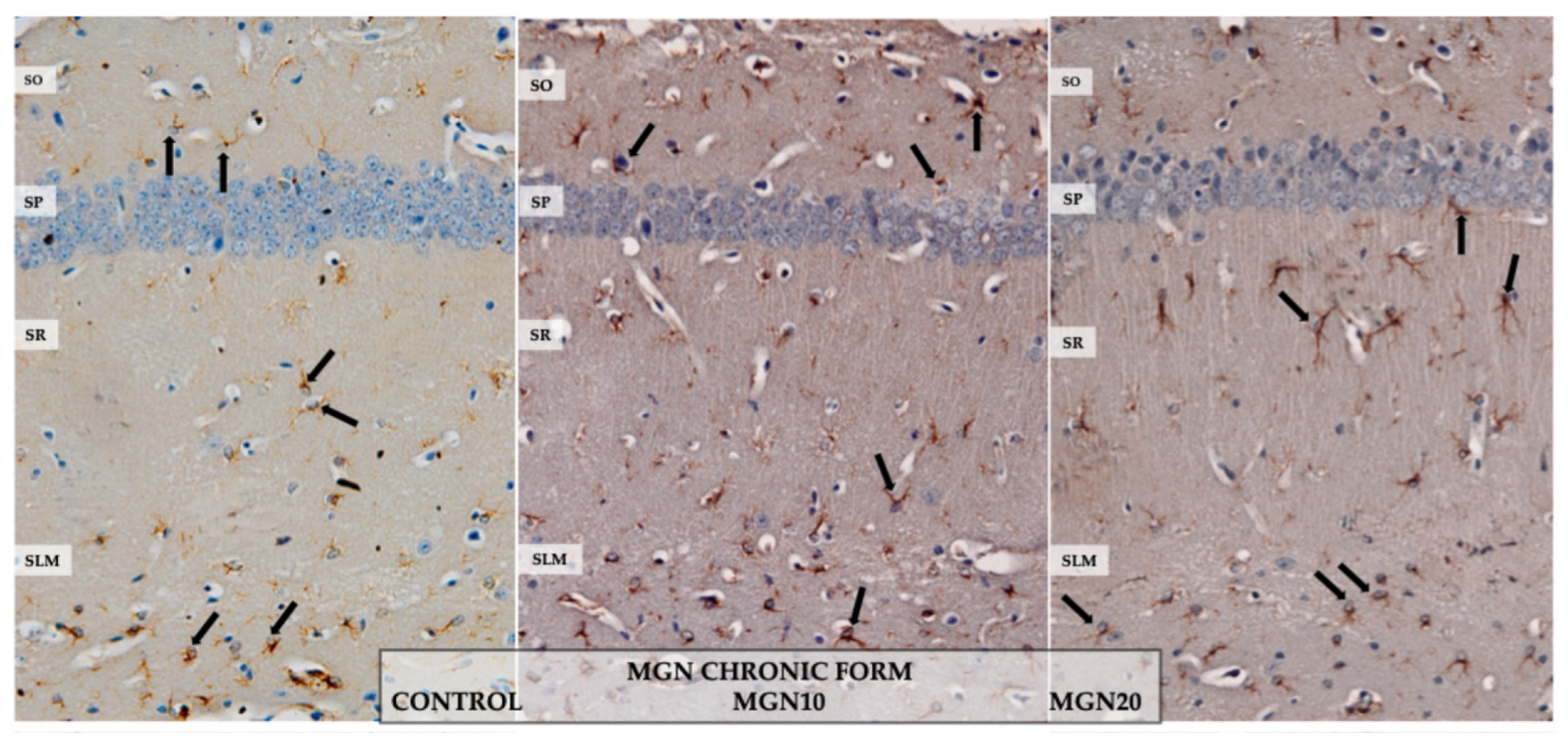
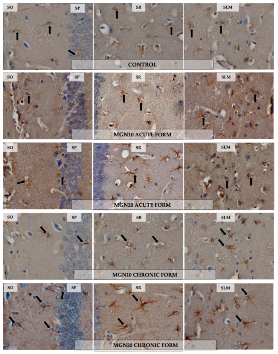
In the SO layer, astrocytes in the control group, in animals receiving a single administration of MGN at both doses and in those treated for 7 days with MGN at 10 mg/kg showed GFAP+ cytoplasm at the point of departure from the body of long, thin, and weakly branched immunostained primary processes. In contrast, in animals chronically administered MGN 20 mg/kg, the GFAP+ astrocytic processes were thicker and exhibited more secondary branches (Figure 1, Figure 2 and Figure 3).
In the SP layer, in animals from the control group and those receiving acute MGN administration at both doses, GFAP+ cells were characterised by a sparse amount of brown cytoplasm, limited mainly to thin and sparsely branched processes and their origins (Figure 1 and Figure 3). In mice treated with MGN for 7 days, especially at the higher dose, most astrocyte cell bodies exhibited GFAP+ cytoplasm, which particularly accumulated at the origins of long, thicker primary processes immunopositive for the tested protein (Figure 2 and Figure 3).
In the SR layer, in the control animals and in the MGN 10 mg/kg and 20 mg/kg groups after acute administration, most astrocytes exhibited GFAP immunoreactivity in short, thin, and sparsely branched processes, as well as at their origin. After a single administration of MGN at a dose of 20 mg/kg, cells with dark brown, longer, thick, and sparsely branched processes were also observed (Figure 1 and Figure 3). In animals receiving MGN chronically at both doses, astrocytes were characterised by a greater amount of immunopositive cytoplasm, particularly at the origins of primary processes, some of which were thicker and highly branched (Figure 2 and Figure 3).
In the SLM layer, in the control group and in the groups that received a single administration of MGN at both doses, astrocytes were characterised mainly by a small amount of cytoplasm immunoreactive for the tested protein within the cell body, and by a brown reaction product located predominantly in short, thicker, and sparsely branched processes. Some cells showed GFAP+ primary processes that underwent repeated branching, resulting in the formation of smaller and thinner branches (Figure 1 and Figure 3). In mice receiving chronic MGN, especially at the higher dose, astrocytes exhibited increased cytoplasmic immunopositivity for the tested protein within the cell body and in thicker, secondarily branched primary processes (Figure 2 and Figure 3).

3.2. Morphometric Analyses

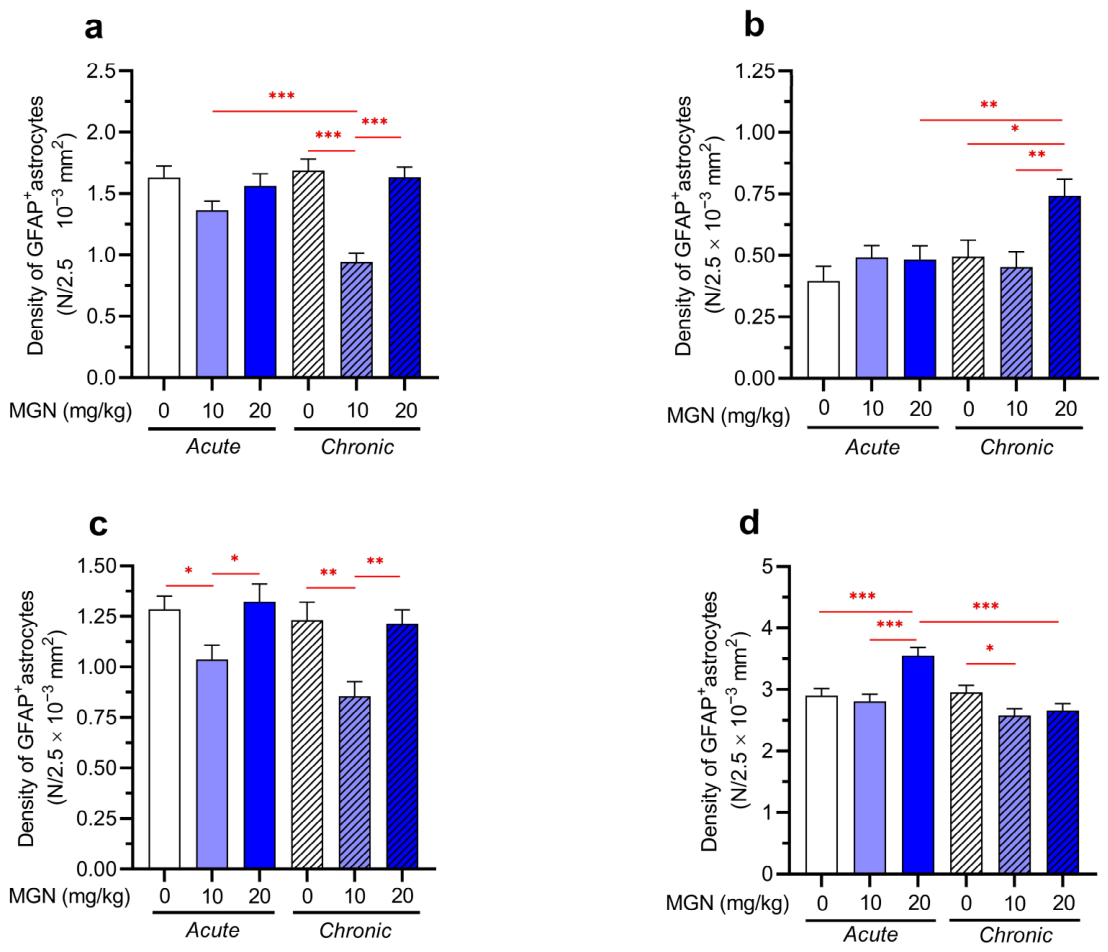
In the acute treatment model, MGN (10 and 20 mg/kg) did not induce any statistically significant changes in the density of GFAP+ astrocytes in the SO layer (KW = 4.462, p = 0.1074, Figure 4a). In contrast to acute administration, chronic exposure revealed pronounced differences between the groups (KW = 41.34, p < 0.001). Chronic treatment with MGN at a dose of 10 mg/kg significantly reduced the density of GFAP+ astrocytes in the SO layer compared with the control group (p < 0.001). Conversely, MGN at a dose of 20 mg/kg administered chronically resulted in a significant increase in GFAP+ astrocyte density relative to the 10 mg/kg dose (p < 0.001), with values comparable to those observed in the control group. A significant difference in the density of GFAP+ astrocytes was detected between groups of animals after acute and chronic treatment with MGN at a dose of 10 mg/kg (p < 0.0002), indicating a marked effect of exposure duration on astroglial density.
In the SP layer (Figure 4b), acute treatment with MGN did not change GFAP+ astrocyte density (KW = 2.842, p = 0.2415), but statistically significant changes were observed in groups exposed to chronic treatment (KW = 12.99, p = 0.0015). Chronic administration of MGN at a dose of 20 mg/kg resulted in a significant increase in GFAP+ astrocyte density compared with the respective control group (p < 0.05) as well as the group treated with MGN at a dose of 10 mg/kg (p < 0.01). These findings indicate that, in the SP layer, the effects of MGN become evident only after prolonged exposure.
In the SR layer (Figure 4c), significant changes in GFAP+ astrocyte density were observed following both acute (KW = 8.342, p = 0.0154) and chronic (KW = 15.63, p = 0.0004) MGN administration. In the acute administration regimen, the 10 mg/kg dose caused a significant reduction in GFAP+ astrocyte density compared with the control group (p < 0.05). MGN at a dose of 20 mg/kg did not change the density of GFAP+ astrocytes significantly compared to the control group (p > 0.05), but their density was statistically higher than in the group treated with a dose of 10 mg/kg (p < 0.05). Similar changes in the density of GFAP+ astrocytes were observed in groups treated chronically. Chronic administration of MGN at 10 mg/kg led to a significant decrease in astrocyte density relative to the control group (p < 0.01). A dose of 20 mg/kg did not change the density of the astrocytes compared to the control group and increased it in comparison to the group administered with MGN at a dose of 10 mg/kg (p < 0.01). There were no statistically significant changes in the astrocyte density between the respective acutely and chronically treated groups.
The most pronounced and heterogeneous changes in GFAP+ astrocyte density in the analysed CA1 layers of the hippocampus were observed in the SLM layer (Figure 4d). In the acute treatment regimen (KW = 20.22, p < 0.0001), MGN administered at a dose of 20 mg/kg induced a significant increase in GFAP+ astrocyte density compared with both the control group (p < 0.001) andthe group treated with a dose of 10 mg/kg (p < 0.001). In contrast, a different pattern was observed under chronic conditions (KW = 6.02, p = 0.0493). Chronic administration of MGN at 10 mg/kg resulted in a significant reduction in GFAP+ astrocyte density compared with the control group (p < 0.05), whereas a dose of 20 mg/kg did not produce significant differences relative to the control. Additionally, a significant difference in the density of GFAP+ astrocytes was noted between groups subjected to acute and chronic administration of MGN at a dose of 20 mg/kg (p < 0.0003), indicating an attenuation of the effect observed following acute exposure.
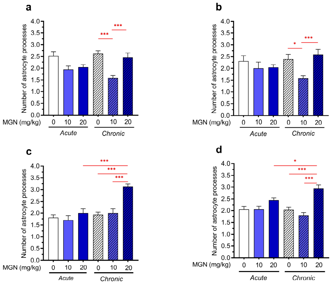
Although the results of the Kruskal–Wallis test indicated the presence of statistically significant changes (KW = 6.323, p = 0.0424), the post hoc analysis did not reveal any significant differences between the number of astrocytic processes in the SO layer in groups subjected to the acute administration regimen (Figure 5a). In contrast, statistically significant changes were noted between groups treated chronically (KW = 25.41, p < 0.0001). Chronic administration of MGN at a dose of 10 mg/kg significantly reduced the number of astrocytic processes compared with the control group (p < 0.001). A dose of 20 mg/kg resulted in a significant increase relative to a dose of 10 mg/kg (p < 0.001), with values comparable to those observed in the control group (p > 0.05).
In the SP layer (Figure 5b), pronounced differences between the groups were observed only after the chronic treatment regimen (acute treatment: KW = 1.167, p = 0.558; chronic treatment: KW = 17.0, p = 0.0002). Chronic administration of MGN at a dose of 10 mg/kg significantly reduced the number of astrocytic processes compared with the respective control group (p < 0.05). The number of astrocyte processes in the group treated chronically with MGN at a dose of 20 mg/kg was significantly higher than in the group treated with a dose of 10 mg/kg (p < 0.001), but it did not differ from the control group (p > 0.05).
The acute treatment regimen did not affect the number of astrocyte processes in the SR layer (KW = 1.449, p = 0.4845; Figure 5c). Statistically significant changes in this parameter were noted in groups treated chronically (KW = 42.85, p < 0.0001). Chronic administration of MGN at a dose of 20 mg/kg resulted in a significant increase in the number of astrocyte processes compared with both the chronic control group (p < 0.001) and relative to the group treated with the 10 mg/kg dose (p < 0.001). Moreover, the number of astrocyte processes following chronic administration at a dose of 20 mg/kg was significantly higher than that observed after acute administration of the same dose (p < 0.0003).
Although the Kruskal–Wallis test suggested the presence of statistically significant differences in the number of astrocyte processes in the SLM layer between groups treated acutely (KW = 7.12, p = 0.0284), post hoc analysis did not show such differences (Figure 5d). Pronounced differences in this layer were observed after the chronic treatment regimen (KW = 29.76, p < 0.0001). Administration of MGN at a dose of 10 mg/kg did not result in significant changes compared with the chronic control group (p > 0.05). In contrast, a dose of 20 mg/kg induced a significant increase in the number of astrocyte processes relative to the chronic control group (p < 0.001) and to the group treated with a dose of 10 mg/kg (p < 0.001). Additionally, a significant difference between groups exposed to acute and chronic administration at a dose of 20 mg/kg was detected (p < 0.0167).
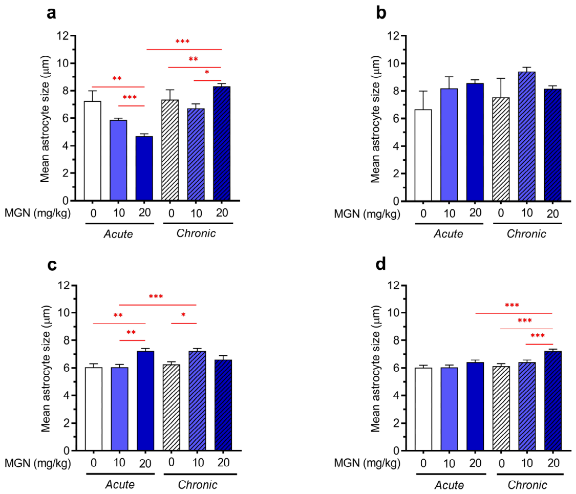
In the SO layer (Figure 6a), statistically significant changes in the mean astrocyte size were noted following both acute (KW = 18.33, p < 0.0001) and chronic treatment (KW = 9.738, p = 0077) with MGN. Under acute treatment conditions, a significant reduction in astrocyte size was observed between the control group and the group treated with MGN at a dose of 20 mg/kg (p < 0.01), as well as between groups treated with doses of 10 and 20 mg/kg (p < 0.001). Chronic treatment with MGN at a dose of 20 mg/kg significantly increased mean astrocyte size in comparison to the control group (p < 0.01). The group treated with a dose of 10 mg/kg showed a significant reduction in the cell size compared with the group administered a 20 mg/kg dose (p < 0.05). Statistical analysis of the results also showed the impact of dosage time on the average size of astrocytes in the group treated with a dose of 20 mg/kg (p < 0.0003).
In the SR layer (Figure 6c), statistically significant differences were noted both between groups administered with MGN acutely (KW = 14.71, p = 0.0006) and chronically (KW = 8.645, p = 0.0133). Single (acute) administration of MGN at a dose of 20 mg/kg resulted in a significant increase in mean astrocyte size compared to both the respective control group (p < 0.01) and the group treated with a dose of 10 mg/kg (p < 0.01). In the chronic model, a significant increase in mean astrocyte size was detected following administration of a dose of 10 mg/kg (p > 0.05), but not a dose of 20 mg/kg (p > 0.05).
In the SLM layer (Figure 6d), statistically significant differences were observed between groups treated with MGN chronically (KW = 19.45, p < 0.0001). Administration of MGN at a dose of 20 mg/kg resulted in a significant increase in mean astrocyte size compared with both the control group (p < 0.001) and the group treated with a dose of 10 mg/kg (p < 0.001). Moreover, prolonged treatment with MGN at a dose of 20 mg/kg significantly increased mean astrocyte size in comparison to the group treated with this dose acutely (p < 0.0003).

4. Discussion

GFAP+ astrocytes were observed in the CA1 field of the hippocampus in all studied animals. These cells were most numerous in the SLM layer, which is consistent with the findings of other authors [22]. The present study demonstrates that MGN administration modulates both morphological and quantitative parameters of astrocytes; however, these effects are strongly dependent on dose, duration of exposure, and hippocampal layer. Importantly, the observed changes were more pronounced following chronic administration compared with the acute paradigm.
Immunohistochemical analysis showed that prolonged administration of MGN, particularly at the higher dose of 20 mg/kg, increased GFAP immunoexpression in the cytoplasm of astrocytic cell bodies and in their thicker, more extensively branched processes. Morphometric analyses confirmed a clear dose-, time-, and layer-dependent effect of MGN. Acute administration of MGN significantly affected only the density and size of GFAP+ astrocytes. Following administration of the low dose (10 mg/kg), a decrease in the density of the studied glia was observed in the SR layer. In contrast, the higher dose of MGN (20 mg/kg) significantly increased the density of GFAP+ astrocytes in the SLM layer, as well as their size in the SR layer, in contrast to the SO layer. Chronic administration of MGN at the lower dose (10 mg/kg) resulted in a significant decrease in GFAP+ astrocyte density in the SO, SR, and SLM layers, as well as a reduction in the number of astrocytic processes in the SO layer. At the same time, an increase in the size of the studied glial cells was observed in the SR layer. In contrast, the higher dose of MGN (20 mg/kg) significantly increased the density of GFAP+ astrocytes in the SP layer, the number of astrocytic processes in the SR and SLM layers, and the cell size in the SO and SLM layers.
The heterogeneous effects observed across hippocampal layers, doses, and treatment durations suggest that MGN does not exert a uniform action on astroglial cells. Instead, the present findings indicate a region-specific and context-dependent astroglial response. The hippocampus is a structurally and functionally heterogeneous formation, and the CA1 layers (SO, SP, SR, SLM) differ in synaptic organization, metabolic demand, and susceptibility to excitatory and oxidative stress [20,21]. These differences may underlie the variable sensitivity of astrocytes to MGN observed in the present study. One plausible explanation is that MGN acts as a modulator of cellular homeostasis, whose effects depend on the baseline physiological state of the local microenvironment. In regions with higher synaptic activity or metabolic load, MGN may exert stabilising effects, whereas in less affected regions, it may induce adaptive or compensatory structural remodelling. Importantly, the lack of consistent directionality across all parameters suggests that MGN does not act as a classical neuroprotective agent, but rather as a biologically active compound influencing astroglial plasticity and neuron–glial interactions.
Morphologically altered astrocytes characterised by numerous secondary branching processes and cytoplasm immunopositive for the examined protein are classified as hypertrophic. Under physiological conditions, such changes in astrocytic morphology have been described, among others, in response to physical exercise, exposure to enriched environments, and the use of various diets [12]. Hypertrophic astrocytes have also been observed in the cerebral cortex of rodents as they acquire new motor skills, and these alterations have been found to correlate with synaptogenesis. However, mice that repeated previously learned skills did not exhibit changes in astroglial morphology nor in synapse number [29]. These findings suggest that astrocytic hypertrophy may be induced by synaptic plasticity, a phenomenon involving structural and functional changes in interneuronal connections and, consequently, neural networks [30,31,32]. In this context, the hypertrophic changes observed in the present study may reflect alterations in astrocyte–synapse interactions rather than a purely protective response. Such structural remodelling is consistent with adaptive plasticity processes but cannot be directly interpreted as beneficial without functional validation. The glial responses revealed in our study may be related to the beneficial effect of MGN on memory in mice, especially at a dose of 20 mg/kg, as demonstrated in a previous report [23]. The glial responses revealed in our study may be related to previously reported effects of MGN on memory. However, in the absence of behavioural or functional data, such associations should be interpreted with caution. In the hippocampus, learning and memory formation rely on the mechanism of long-term synaptic potentiation (LTP) [33,34]. In adult individuals, under conditions of in vitro-induced LTP, significant structural changes within synapses are observed within tens of seconds to minutes, which is referred to as structural plasticity [35]. This phenomenon is related to the variability in the shape of synapses, their number, the size of the postsynaptic density and the shape of the dendritic spines that form the synapses [36].
Thin, perisynaptic astrocyte processes (PAPs) play an important role in synaptic transmission. They contact and envelop the pre- and postsynaptic poles, constituting a crucial element of the so-called tripartite synapse. In this way, astrocytes can simultaneously contact multiple synapses, enabling their close interaction with neurons [37]. The efficacy of synaptic transmission and synaptic stability depend on the degree of synapse coverage by PAPs and their mobility [38]. A loss of synaptic stability may lead to the development of neurodegenerative disorders, thereby resulting in impairment of memory, learning, and cognitive functions [39,40]. Under physiological conditions, hippocampal astrocytes tightly ensheathe approximately 60% of synaptic connections with their processes [41,42,43]. In the rat hippocampal dentate gyrus, astrocytes significantly increase their branching and synaptic coverage following LTP induction, with these changes being particularly prominent within 8 h after stimulation. LTP thus exerts a substantial influence on the spatial relationship between astrocyte processes and potentiated synapses in the dentate gyrus neuropil. Simultaneously, the extension of astrocytic processes and the increase in their branching may contribute to the stabilisation of neuronal networks, providing structural support to surrounding neurons [44,45]. PAPs detect neurotransmitters within the synaptic cleft and surrounding extracellular space, enabling them to regulate and modulate synaptic strength and activity, as well as neuronal excitability [46,47,48,49]. Therefore, MGN-induced changes in astrocytic morphology may influence synaptic coverage and signalling efficiency. However, without direct electrophysiological or functional data, these effects remain speculative.
The morphological features observed in the present study, including increased branching and hypertrophy of astrocytic processes, may reflect alterations in neuron–glia interactions. However, without direct assessment of synaptic function or plasticity, such interpretations remain speculative. Similarly, although previous studies suggest a relationship between astrocytic structural changes and synaptic plasticity mechanisms underlying learning and memory [30,31,32], the present study does not provide direct functional evidence linking MGN-induced astroglial alterations with cognitive outcomes and should therefore be interpreted with caution.
Astrocytes support and protect neurons during neurotransmission. These cells play a crucial role in Glu metabolism, which is particularly important for plasticity and learning. This compound is an agonist of NMDA receptors, whose activation underlies LTP in the CA1 field of the hippocampus [50,51,52]. PAP membranes contain transporters specific for this neurotransmitter, such as glutamate–aspartate transporter (GLAST) and glutamate transporter 1 (GLT-1). However, it must be clearly stated that the present study did not assess glutamate transporter expression or their activity. These transporters enable astroglia to take up Glu from the extracellular space, thereby clearing excess Glu from the synaptic cleft [53]. This process protects neurons from prolonged stimulation and prevents the development of excitotoxicity. In this situation, the excessive and rapid increase in intracellular calcium ion levels in stimulated cells activates numerous mechanisms that lead to damage and even death of neurons and glia cells [10,54,55,56,57]. Under conditions of neuronal hyperexcitation, an increase in the activity of these transporters has been observed, and enhanced GLT-1 expression immediately results in a decrease in extracellular Glu levels [58]. In adult mice, in a model of epilepsy, a significant increase in GLT-1 and GLAST immunoreactivity was observed in the first 3 days after intrahippocampal injection of kainic acid (an NMDA receptor agonist). However, after a week, a significant decrease was observed, which correlated with the occurrence of epileptic seizures in the animals [59]. The GLT-1 transporter colocalises with glial intermediate filaments in astrocytes, and the cytoskeleton is involved in the trafficking and assembly of this transporter along astrocytic processes. Furthermore, GFAP is involved in the anchoring of the GLAST transporter to the plasma membrane, which is upregulated in cultured astrocytes maintained under conditions of elevated Glu concentrations [60,61,62,63]. These results suggest that GFAP plays a significant role in enhancing the intracellular transport of Glu from the synaptic cleft. The increased GFAP immunoreactivity in astroglial cells, as well as the density of GFAP+ astrocytes in the CA1 field of the hippocampus of mice in a chronic MGN model, especially at a high dose, may therefore result from increased formation of intermediate glial filaments. These structures not only stabilise perisynaptic astrocyte projections, strengthening neuronal connections, but also participate in maintaining normal neuronal excitability and neurotransmission. GFAP is a widely used indicator of astrocyte morphology, but not of all their subpopulations. It is a marker for reactive astrocytes. In non-reactive astrocytes, the level of this protein is undetectable by immunohistochemical methods [64]. Increased GFAP immunoreactivity in astroglial cells is one of the features of reactive gliosis in response to various changes in the perineuronal microenvironment. In this case, astrocytes that are immunonegative for the studied protein may become immunopositive without changes in their overall number. However, glial proliferation and migration cannot be excluded and may significantly contribute to an apparent increase in astroglial cell numbers [11,12,15,16]. Although these mechanisms provide a plausible biological framework, the present study did not assess glutamate transporter expression or function. Therefore, any interpretation linking MGN-induced astroglial changes with glutamatergic regulation remains hypothetical.
Reactive glial response is observed in many acute and chronic CNS diseases, such as epilepsy, stroke, traumatic brain injury, Alzheimer’s disease, and Parkinson’s disease. The pathogenesis of these disorders involves glutamate-induced excitotoxicity, in the metabolism of which astrocytes play a key role [11,12,13,54,55,56,58,65,66,67,68].
Numerous studies have shown that high levels of this neurotransmitter can lead to cell death due to the development of oxidative stress. This toxicity mechanism was confirmed in cultured astrocytes from the cerebral cortex of neonatal rats exposed to Glu, followed by vitamin E treatment, which completely blocked this process [69]. Similarly, a significant increase in GFAP immunoreactivity was observed in the cerebellum of adult rats that received both monosodium glutamate and ascorbic acid (vitamin C), which has antioxidant properties. The authors indicate a likely beneficial effect of vitamin C on astrocytes, as it prevents oxidative damage and may ultimately increase their survival [70].
The above observations may suggest that, due to its antioxidant properties, MGN may enhance the proper functioning of astrocytes, making them more resistant to oxidative stress. As a result, it may help better protect neurons from damage by providing a stable and safe microenvironment for synaptic transmission. However, the proposed involvement of oxidative stress pathways cannot be confirmed within the scope of the present study, as no biochemical markers of oxidative stress were analysed.
At the same time, our studies demonstrated that chronic administration of a lower dose of MGN to the selected layers of the CA1 field of the hippocampus in mice resulted in a reduction in the density of GFAP+ cells and their processes. This distinct response can be interpreted as a mechanism that enables better adaptation of astrocytes to the chemically altered microenvironment. The low dose may inhibit unnecessary astroglial activation, which is optimal for physiological glutamatergic transmission. Furthermore, GFAP, as a structural protein, limits the mobility of PAPs, whereas its decrease increases their mobility, allowing for better alignment of astrocytic processes with active synapses [30,71]. Taken together, the findings of the present study indicate that MGN induces complex, non-uniform changes in astroglial morphology that depend on dose, the duration of exposure, and hippocampal layer. This pattern suggests a context-dependent modulation of astrocyte structure rather than a uniform or directional biological effect.
The present study is preliminary and has several limitations that should be acknowledged. First, the analysis was based exclusively on GFAP immunoreactivity, which reflects only a subset of astrocytes and primarily indicates changes in morphology of reactive astroglial. The lack of additional astrocytic markers, such as S100β or Aldh1L1, limits the ability to fully characterise astrocyte populations. Second, female mice were not included in the present study in order to minimize biological variability related to fluctuations in sex steroid hormones, which are known to affect astrocyte morphology and GFAP expression [72]. Nevertheless, this represents an important limitation, and future studies should determine whether the astroglial response to magnoflorine is sex-dependent. Third, this study did not include functional, molecular, or biochemical analyses. In particular, no assessment of neuronal condition, synaptic plasticity, glutamate transporter expression, or oxidative stress markers was performed. Therefore, the functional significance of the observed astroglial changes remains unclear. Fourth, behavioural and electrophysiological assessments were not performed, precluding direct evaluation of potential effects on cognitive function or synaptic activity. Last is the lack of formal blinding during the morphometric evaluation and the absence of a defined randomization procedure. Although all experimental steps, including tissue processing, immunohistochemical staining, image acquisition, and quantitative analyses, were performed according to a strictly standardized protocol, the possibility of observer-related bias cannot be completely excluded. Therefore, the obtained morphometric differences should be interpreted with appropriate caution and confirmed in future studies using blinded assessment procedures.

5. Conclusions

In summary, MGN modulates astrocyte morphology and reactivity in the CA1 field of the male mice hippocampus in adose-, time-, and hippocampal layer-dependent manner. A complex and layer-specific astroglial response was particularly evident following chronic administration of the tested compound. Together with the observed lack of significant differences in morphology and analysed parameters in some layers, decreases were mainly detected after administration of the low MGN dose (10 mg/kg), whereas increases were observed primarily after the high dose (20 mg/kg). Thus, the direction of changes was not uniform across all layers, and the observed differences may reflect local microenvironmental specificity as well as differences in astrocyte sensitivity. Notably, the most substantial changes were detected in the SLM layer, suggesting increased sensitivity of astroglia located in this CA1 layer to MGN-induced modulation. Importantly, the observed heterogeneity of responses indicates that MGN does not exert a uniform effect on astroglial cells but rather induces region- and context-dependent structural alterations.
The obtained results suggest that MGN may induce adaptive morphological remodelling of astrocytes in a context-dependent manner. These effects may be associated with alterations in astroglia-mediated mechanisms involved in maintaining neuronal homeostasis and responses to stress. However, given the exclusively morphological nature of the present study, these interpretations should be treated with caution. The present findings do not support a clear or uniform neuroprotective action of MGN but rather point toward a modulatory influence on astroglial structure and plasticity. Therefore, their functional significance and potential biological relevance remain to be elucidated in future studies incorporating molecular, biochemical, and functional approaches.
These findings provide preliminary evidence that MGN may influence astroglial structural plasticity in the hippocampus and may serve as a basis for future studies investigating its functional and therapeutic relevance in neurodegenerative and cognitive disorders.

Author Contributions

Conceptualization, R.S., M.K. and A.K.; methodology, R.S., A.K., W.K.-K. and D.N.; software, R.S. and Ö.G.D.; validation, R.S., A.K. and M.K.; formal analysis, R.S., D.N., W.K. and Ö.G.D.; investigation, R.S., W.K.-K. and A.K.; resources, R.S., A.K. and M.B.A.; data curation, R.S. and M.K.; writing—original draft preparation, R.S., A.K. and M.K.; writing—review and editing, R.S., A.K. and M.K.; visualization, R.S., A.K., D.N. and M.K.; supervision, M.K. and M.B.A.; project administration, R.S. and W.K.; funding acquisition, M.K., R.S. and M.B.A. All authors have read and agreed to the published version of the manuscript.

Funding

This research received no external funding.

Institutional Review Board Statement

The described studies were performed in accordance with the guidelines set to improve the quality of the animal research and reporting of the animal studies, following the European Community Council Directive for the Care and Use of Laboratory Animals of 22 September 2010 (2010/63/EU), and the National Institute of Health Guidelines for the Care and Use of Laboratory Animals. Furthermore, on 19 June 2015, we obtained the agreement to conduct the aforementioned studies (number: 33/2015) from the Local Ethical Committee for Animal Experiments in Lublin.

Informed Consent Statement

Not applicable.

Data Availability Statement

The data that support the findings of this study are available from the corresponding author upon reasonable request.

Conflicts of Interest

The authors declare no conflicts of interest.

References

  1. Vasile, F.; Dossi, E.; Rouach, N. Human Astrocytes: Structure and Functions in the Healthy Brain. Brain Struct. Funct. 2017, 222, 2017–2029. [Google Scholar] [CrossRef] [PubMed]
  2. Li, B.; Han, L.; Cao, B.; Yang, X.; Zhu, X.; Yang, B.; Zhao, H.; Qiao, W. Use of Magnoflorine-Phospholipid Complex to Permeate Blood-Brain Barrier and Treat Depression in the CUMS Animal Model. Drug Deliv. 2019, 26, 566–574. [Google Scholar] [CrossRef]
  3. Chiareli, R.A.; Carvalho, G.A.; Marques, B.L.; Mota, L.S.; Oliveira-Lima, O.C.; Gomes, R.M.; Birbrair, A.; Gomez, R.S.; Simão, F.; Klempin, F.; et al. The Role of Astrocytes in the Neurorepair Process. Front. Cell Dev. Biol. 2021, 9, 665795. [Google Scholar] [CrossRef]
  4. Gradisnik, L.; Velnar, T. Astrocytes in the Central Nervous System and Their Functions in Health and Disease: A Review. World J. Clin. Cases 2023, 11, 3385–3394. [Google Scholar] [CrossRef]
  5. Gabryel, B.; Kasprowska, D.; Kost, A.; Łabuzek, K.; Urbanek, T. Astrocytes in Ischemic Stroke—A Potential Target for Neuroprotective Strategies. Postępy Hig. Med. Dośw. 2015, 69, 384–397. [Google Scholar] [CrossRef]
  6. Valles, S.L.; Singh, S.K.; Campos-Campos, J.; Colmena, C.; Campo-Palacio, I.; Alvarez-Gamez, K.; Caballero, O.; Jorda, A. Functions of Astrocytes under Normal Conditions and after a Brain Disease. Int. J. Mol. Sci. 2023, 24, 8434. [Google Scholar] [CrossRef] [PubMed]
  7. Sofroniew, M.V.; Vinters, H.V. Astrocytes: Biology and Pathology. Acta Neuropathol. 2010, 119, 7–35. [Google Scholar] [CrossRef]
  8. Verkhratsky, A.; Nedergaard, M. Physiology of Astroglia. Physiol. Rev. 2018, 98, 239–389. [Google Scholar] [CrossRef] [PubMed]
  9. Pinky, N.F.; Wilkie, C.M.; Barnes, J.R.; Parsons, M.P. Region- and Activity-Dependent Regulation of Extracellular Glutamate. J. Neurosci. 2018, 38, 5351–5366. [Google Scholar] [CrossRef] [PubMed]
  10. Mahmoud, S.; Gharagozloo, M.; Simard, C.; Gris, D. Astrocytes Maintain Glutamate Homeostasis in the CNS by Controlling the Balance between Glutamate Uptake and Release. Cells 2019, 8, 184. [Google Scholar] [CrossRef]
  11. Hol, E.M.; Pekny, M. Glial Fibrillary Acidic Protein (GFAP) and the Astrocyte Intermediate Filament System in Diseases of the Central Nervous System. Curr. Opin. Cell Biol. 2015, 32, 121–130. [Google Scholar] [CrossRef]
  12. Verkhratsky, A.; Butt, A.; Li, B.; Illes, P.; Zorec, R.; Semyanov, A.; Tang, Y.; Sofroniew, M.V. Astrocytes in Human Central Nervous System Diseases: A Frontier for New Therapies. Signal Transduct. Target. Ther. 2023, 8, 396. [Google Scholar] [CrossRef]
  13. Lee, H.-G.; Wheeler, M.A.; Quintana, F.J. Function and Therapeutic Value of Astrocytes in Neurological Diseases. Nat. Rev. Drug Discov. 2022, 21, 339–358. [Google Scholar] [CrossRef] [PubMed]
  14. Potokar, M.; Morita, M.; Wiche, G.; Jorgačevski, J. The Diversity of Intermediate Filaments in Astrocytes. Cells 2020, 9, 1604. [Google Scholar] [CrossRef]
  15. Middeldorp, J.; Hol, E.M. GFAP in Health and Disease. Prog. Neurobiol. 2011, 93, 421–443. [Google Scholar] [CrossRef] [PubMed]
  16. McKeon, A.; Benarroch, E.E. Glial Fibrillary Acid Protein: Functions and Involvement in Disease. Neurology 2018, 90, 925–930. [Google Scholar] [CrossRef] [PubMed]
  17. Pekny, M.; Pekna, M. Astrocyte Intermediate Filaments in CNS Pathologies and Regeneration. J. Pathol. 2004, 204, 428–437. [Google Scholar] [CrossRef] [PubMed]
  18. Matias, I.; Morgado, J.; Gomes, F.C.A. Astrocyte Heterogeneity: Impact to Brain Aging and Disease. Front. Aging Neurosci. 2019, 11, 59. [Google Scholar] [CrossRef]
  19. Andersen, P.; Morris, R.; Amaral, D.; Bliss, T.; O’Keefe, J. (Eds.) The Hippocampus Book; Oxford University Press: Oxford, UK, 2006. [Google Scholar]
  20. El Falougy, H.; Kubikova, E.; Benuska, J. The Microscopical Structure of the Hippocampus in the Rat. Bratisl. Lek. Listy 2008, 109, 106–110. [Google Scholar] [PubMed]
  21. Romani, A.; Schürmann, F.; Markram, H.; Migliore, M. Reconstruction of the Hippocampus. In Computational Modelling of the Brain; Giugliano, M., Negrello, M., Linaro, D., Eds.; Advances in Experimental Medicine and Biology; Springer International Publishing: Cham, Germany, 2022; Volume 1359, pp. 261–283. [Google Scholar]
  22. Korzhevskii, D.É.; Otellin, V.A.; Grigor’ev, I.P.; Kostkin, V.B.; Polenov, S.A.; Lentsman, M.V.; Balestrino, M. Structural Organization of Astrocytes in the Rat Hippocampus in the Post-Ischemic Period. Neurosci. Behav. Physiol. 2005, 35, 389–392. [Google Scholar] [CrossRef]
  23. Kukula-Koch, W. The Elevation of LC-ESI-Q-TOF-MS Response in the Analysis of Isoquinoline Alkaloids from Some Papaveraceae and Berberidaceae Representatives. J. Anal. Methods Chem. 2017, 2017, 8384107. [Google Scholar] [CrossRef]
  24. Okon, E.; Kukula-Koch, W.; Jarzab, A.; Halasa, M.; Stepulak, A.; Wawruszak, A. Advances in Chemistry and Bioactivity of Magnoflorine and Magnoflorine-Containing Extracts. Int. J. Mol. Sci. 2020, 21, 1330. [Google Scholar] [CrossRef]
  25. Xu, L.; Yang, W.; Hu, J.; Han, C.-M.; Li, P.-F. Three New Isoquinoline Alkaloids from the Whole Plants of Thalictrum Tenue with Cytotoxic Activities. J. Asian Nat. Prod. Res. 2020, 22, 618–625. [Google Scholar] [CrossRef]
  26. Szalak, R.; Matysek, M.; Koval, M.; Dziedzic, M.; Kowalczuk-Vasilev, E.; Kruk-Slomka, M.; Koch, W.; Arciszewski, M.B.; Kukula-Koch, W. Magnoflorine from Berberis Vulgaris Roots—Impact on Hippocampal Neurons in Mice after Short-Term Exposure. Int. J. Mol. Sci. 2023, 24, 7166. [Google Scholar] [CrossRef] [PubMed]
  27. Szalak, R.; Komar, M.; Kowalczuk-Vasilev, E.; Kruk-Slomka, M.; Zagórska, J.; Arciszewski, M.B.; Dziedzic, M.; Koch, W.; Kukula-Koch, W. Can Magnoflorine Improve Memory? Immunohistochemical Studies on Parvalbumin Immunoreactive Neurons and Fibers of Mice Hippocampus. Nutrients 2024, 17, 137. [Google Scholar] [CrossRef]
  28. Krawczyk, A.; Jadwiga, J.-A. Evaluation of Immunoreactive for Glial Fibrillary Acidic Protein Astrocytes in the Periaqueductal Gray Matter of Rats Treated with Monosodium Glutamate. Med. Weter. 2023, 79, 22–27. [Google Scholar] [CrossRef]
  29. Kleim, J.A.; Markham, J.A.; Vij, K.; Freese, J.L.; Ballard, D.H.; Greenough, W.T. Motor Learning Induces Astrocytic Hypertrophy in the Cerebellar Cortex. Behav. Brain Res. 2007, 178, 244–249. [Google Scholar] [CrossRef] [PubMed]
  30. Lawal, O.; Ulloa Severino, F.P.; Eroglu, C. The Role of Astrocyte Structural Plasticity in Regulating Neural Circuit Function and Behavior. Glia 2022, 70, 1467–1483. [Google Scholar] [CrossRef]
  31. Abbott, L.F.; Nelson, S.B. Synaptic Plasticity: Taming the Beast. Nat. Neurosci. 2000, 3, 1178–1183. [Google Scholar] [CrossRef]
  32. Lee, H.-K. Synaptic Plasticity and Phosphorylation. Pharmacol. Ther. 2006, 112, 810–832. [Google Scholar] [CrossRef] [PubMed]
  33. Ota, Y.; Zanetti, A.T.; Hallock, R.M. The Role of Astrocytes in the Regulation of Synaptic Plasticity and Memory Formation. Neural Plast. 2013, 2013, 185463. [Google Scholar] [CrossRef]
  34. Kandel, E.R.; Dudai, Y.; Mayford, M.R. The Molecular and Systems Biology of Memory. Cell 2014, 157, 163–186. [Google Scholar] [CrossRef]
  35. Nakahata, Y.; Yasuda, R. Plasticity of Spine Structure: Local Signaling, Translation and Cytoskeletal Reorganization. Front. Synaptic Neurosci. 2018, 10, 29. [Google Scholar] [CrossRef] [PubMed]
  36. Matsuzaki, M.; Honkura, N.; Ellis-Davies, G.C.R.; Kasai, H. Structural Basis of Long-Term Potentiation in Single Dendritic Spines. Nature 2004, 429, 761–766. [Google Scholar] [CrossRef]
  37. Araque, A.; Parpura, V.; Sanzgiri, R.P.; Haydon, P.G. Tripartite Synapses: Glia, the Unacknowledged Partner. Trends Neurosci. 1999, 22, 208–215. [Google Scholar] [CrossRef] [PubMed]
  38. Bernardinelli, Y.; Randall, J.; Janett, E.; Nikonenko, I.; König, S.; Jones, E.V.; Flores, C.E.; Murai, K.K.; Bochet, C.G.; Holtmaat, A.; et al. Activity-Dependent Structural Plasticity of Perisynaptic Astrocytic Domains Promotes Excitatory Synapse Stability. Curr. Biol. 2014, 24, 1679–1688. [Google Scholar] [CrossRef]
  39. Kasai, H.; Fukuda, M.; Watanabe, S.; Hayashi-Takagi, A.; Noguchi, J. Structural Dynamics of Dendritic Spines in Memory and Cognition. Trends Neurosci. 2010, 33, 121–129. [Google Scholar] [CrossRef]
  40. Koleske, A.J. Molecular Mechanisms of Dendrite Stability. Nat. Rev. Neurosci. 2013, 14, 536–550. [Google Scholar] [CrossRef]
  41. Bushong, E.A.; Martone, M.E.; Jones, Y.Z.; Ellisman, M.H. Protoplasmic Astrocytes in CA1 Stratum Radiatum Occupy Separate Anatomical Domains. J. Neurosci. 2002, 22, 183–192. [Google Scholar] [CrossRef] [PubMed]
  42. Ventura, R.; Harris, K.M. Three-Dimensional Relationships between Hippocampal Synapses and Astrocytes. J. Neurosci. 1999, 19, 6897–6906. [Google Scholar] [CrossRef] [PubMed]
  43. Witcher, M.R.; Kirov, S.A.; Harris, K.M. Plasticity of Perisynaptic Astroglia during Synaptogenesis in the Mature Rat Hippocampus. Glia 2007, 55, 13–23. [Google Scholar] [CrossRef] [PubMed]
  44. Araque, A.; Perea, G. Glial Modulation of Synaptic Transmission in Culture. Glia 2004, 47, 241–248. [Google Scholar] [CrossRef]
  45. Yang, G.; Pan, F.; Gan, W.-B. Stably Maintained Dendritic Spines Are Associated with Lifelong Memories. Nature 2009, 462, 920–924. [Google Scholar] [CrossRef]
  46. Bains, J.S.; Oliet, S.H.R. Glia: They Make Your Memories Stick! Trends Neurosci. 2007, 30, 417–424. [Google Scholar] [CrossRef]
  47. Derouiche, A.; Anlauf, E.; Aumann, G.; Mühlstädt, B.; Lavialle, M. Anatomical Aspects of Glia–Synapse Interaction: The Perisynaptic Glial Sheath Consists of a Specialized Astrocyte Compartment. J. Physiol.-Paris 2002, 96, 177–182. [Google Scholar] [CrossRef]
  48. Reichenbach, A.; Derouiche, A.; Kirchhoff, F. Morphology and Dynamics of Perisynaptic Glia. Brain Res. Rev. 2010, 63, 11–25. [Google Scholar] [CrossRef]
  49. Matyash, V.; Kettenmann, H. Heterogeneity in Astrocyte Morphology and Physiology. Brain Res. Rev. 2010, 63, 2–10. [Google Scholar] [CrossRef] [PubMed]
  50. Allen, N.J. Astrocyte Regulation of Synaptic Behavior. Annu. Rev. Cell Dev. Biol. 2014, 30, 439–463. [Google Scholar] [CrossRef] [PubMed]
  51. Chung, W.-S.; Allen, N.J.; Eroglu, C. Astrocytes Control Synapse Formation, Function, and Elimination. Cold Spring Harb. Perspect. Biol. 2015, 7, a020370. [Google Scholar] [CrossRef]
  52. Lushnikova, I.; Skibo, G.; Muller, D.; Nikonenko, I. Synaptic Potentiation Induces Increased Glial Coverage of Excitatory Synapses in CA1 Hippocampus. Hippocampus 2009, 19, 753–762. [Google Scholar] [CrossRef]
  53. Jurga, A.M.; Paleczna, M.; Kadluczka, J.; Kuter, K.Z. Beyond the GFAP-Astrocyte Protein Markers in the Brain. Biomolecules 2021, 11, 1361. [Google Scholar] [CrossRef]
  54. Belov Kirdajova, D.; Kriska, J.; Tureckova, J.; Anderova, M. Ischemia-Triggered Glutamate Excitotoxicity from the Perspective of Glial Cells. Front. Cell. Neurosci. 2020, 14, 51. [Google Scholar] [CrossRef] [PubMed]
  55. Lai, T.W.; Zhang, S.; Wang, Y.T. Excitotoxicity and Stroke: Identifying Novel Targets for Neuroprotection. Prog. Neurobiol. 2014, 115, 157–188. [Google Scholar] [CrossRef]
  56. Neves, D.; Salazar, I.L.; Almeida, R.D.; Silva, R.M. Molecular Mechanisms of Ischemia and Glutamate Excitotoxicity. Life Sci. 2023, 328, 121814. [Google Scholar] [CrossRef]
  57. Plotegher, N.; Filadi, R.; Pizzo, P.; Duchen, M.R. Excitotoxicity Revisited: Mitochondria on the Verge of a Nervous Breakdown. Trends Neurosci. 2021, 44, 342–351. [Google Scholar] [CrossRef]
  58. Wang, J.; Wang, F.; Mai, D.; Qu, S. Molecular Mechanisms of Glutamate Toxicity in Parkinson’s Disease. Front. Neurosci. 2020, 14, 585584. [Google Scholar] [CrossRef] [PubMed]
  59. Peterson, A.R.; Binder, D.K. Regulation of Synaptosomal GLT-1 and GLAST during Epileptogenesis. Neuroscience 2019, 411, 185–201. [Google Scholar] [CrossRef]
  60. Zhou, J.; Sutherland, M.L. Glutamate Transporter Cluster Formation in Astrocytic Processes Regulates Glutamate Uptake Activity. J. Neurosci. 2004, 24, 6301–6306. [Google Scholar] [CrossRef]
  61. Sullivan, S.M.; Lee, A.; Björkman, S.T.; Miller, S.M.; Sullivan, R.K.P.; Poronnik, P.; Colditz, P.B.; Pow, D.V. Cytoskeletal Anchoring of GLAST Determines Susceptibility to Brain Damage. J. Biol. Chem. 2007, 282, 29414–29423. [Google Scholar] [CrossRef] [PubMed]
  62. Sullivan, S.M.; Björkman, S.T.; Miller, S.M.; Colditz, P.B.; Pow, D.V. Structural Remodeling of Gray Matter Astrocytes in the Neonatal Pig Brain after Hypoxia/Ischemia. Glia 2010, 58, 181–194. [Google Scholar] [CrossRef]
  63. Gegelashvili, G.; Civenni, G.; Racagni, G.; Danbolt, N.C.; Schousboe, I.; Schousboe, A. Glutamate Receptor Agonists Up-Regulate Glutamate Transporter GLAST in Astrocytes. NeuroReport 1996, 8, 261–265. [Google Scholar] [CrossRef] [PubMed]
  64. Kimelberg, H.K. The Problem of Astrocyte Identity. Neurochem. Int. 2004, 45, 191–202. [Google Scholar] [CrossRef] [PubMed]
  65. Burda, J.E.; Sofroniew, M.V. Reactive Gliosis and the Multicellular Response to CNS Damage and Disease. Neuron 2014, 81, 229–248. [Google Scholar] [CrossRef]
  66. Li, K.; Li, J.; Zheng, J.; Qin, S. Reactive Astrocytes in Neurodegenerative Diseases. Aging Dis. 2019, 10, 664. [Google Scholar] [CrossRef] [PubMed]
  67. Barker-Haliski, M.; White, H.S. Glutamatergic Mechanisms Associated with Seizures and Epilepsy. Cold Spring Harb. Perspect. Med. 2015, 5, a022863. [Google Scholar] [CrossRef]
  68. Tannenberg, R.; Scott, H.; Westphalen, R.; Dodd, P. The Identification and Characterization of Excitotoxic Nerve-Endings in Alzheimer Disease. Curr. Alzheimer Res. 2004, 1, 11–25. [Google Scholar] [CrossRef] [PubMed]
  69. Chen, C.; Liao, S.; Kuo, J. Gliotoxic Action of Glutamate on Cultured Astrocytes. J. Neurochem. 2000, 75, 1557–1565. [Google Scholar] [CrossRef]
  70. Hashem, H.E.; El-Din Safwat, M.D.; Algaidi, S. The Effect of Monosodium Glutamate on the Cerebellar Cortex of Male Albino Rats and the Protective Role of Vitamin C (Histological and Immunohistochemical Study). J. Mol. Histol. 2012, 43, 179–186. [Google Scholar] [CrossRef]
  71. Han, Y.; Huang, L.; Wu, J.; Wan, G.; Mo, L.; Liu, A. The Role of Astrocytes in Poststroke Rehabilitation. Brain Behav. 2025, 15, e70551. [Google Scholar] [CrossRef]
  72. Suárez, I.; Bodega, G.; Rubio, M.; Fernández, B. Sexual Dimorphism in the Hamster Cerebellum Demonstrated by Glial Fibrillary Acidic Protein (GFAP) and Vimentin Immunoreactivity. Glia 1992, 5, 10–16. [Google Scholar] [CrossRef] [PubMed]
Figure 1. GFAP-immunoreactive cells in the CA1 field of the mouse hippocampus from the control group and groups treated with 10 mg/kg and 20 mg/kg of MGN in the acute form. The oriens layer (SO), the pyramidal layer (SP), the radiate layer (SR), and the lacunose-molecular layer (SLM). The arrows indicate the GFAP+ astrocytes. Magnification ×20.
Applsci 16 04960 g001
Figure 2. GFAP-immunoreactive cells in the CA1 field of the mouse hippocampus from the control group and groups treated with 10 mg/kg and 20 mg/kg of MGN in the chronic form. The oriens layer (SO), the pyramidal layer (SP), the radiate layer (SR), and the lacunose-molecular layer (SLM). The arrows indicate the GFAP+ astrocytes. Magnification ×20.
Applsci 16 04960 g002
Figure 3. GFAP-immunoreactive cells in the CA1 field of the mouse hippocampus from the control group and groups treated with 10 mg/kg and 20 mg/kg of MGN in the acute and chronic form. The oriens layer (SO), the pyramidal layer (SP), the radiate layer (SR), and the lacunose-molecular layer (SLM). The arrows indicate the GFAP+ astrocytes. Magnification ×60.
Applsci 16 04960 g003
Figure 4. The effect of acute and chronic magnoflorine (MGN; 10 and 20 mg/kg) administration on the density of GFAP+ astrocytes in the CA1 layers of the mouse hippocampus. Quantitative analysis was performed in the oriens layer—SO (a), the pyramidal layer—SP (b), the radiate layer—SR (c), and the lacunose-molecular layer—SLM (d). Resulted obtained in groups subjected to acute or chronic administration were analysed separately using the Kruskal–Wallis test with Dunn’s post hoc test ((a) acute: KW = 4.462, p = 0.1074, chronic: KW = 41.34, p < 0.001; (b) acute: KW = 2.842, p = 0.2415, chronic: KW = 12.99, p = 0.0015; (c) acute: KW = 8.342, p = 0.0154, chronic: KW = 15.63, p = 0.0004; (d) acute: KW = 20.22, p < 0.0001, chronic: KW = 6.02, p = 0.0493)). Additionally, two respective groups that received the same treatment acutely or chronically were compared using the Mann–Whitney test with a Bonferroni correction. Data are presented as the mean ± SEM (N/2.5 × 10−3 mm2). Statistical significance is indicated as follows: Dunn’s post hoc test, * p < 0.05, ** p < 0.01, and *** p < 0.001; Mann–Whitney test with a Bonferroni correction, * p < 0.0167, ** p < 0.0033, and *** p < 0.0003. Red asterisks denote significant differences between the indicated groups.
Applsci 16 04960 g004
Figure 5. The effect of acute and chronic magnoflorine (MGN; 10 and 20 mg/kg) administration on the number of astrocytic processes in the CA1 layers of the mouse hippocampus. Quantitative analysis was performed on the oriens layer—SO (a), the pyramidal layer—SP (b), the radiate layer—SR (c), and the lacunose-molecular layer—SLM (d). The results obtained in groups subjected to acute or chronic administration were analysed using the Kruskal–Wallis test with Dunn’s post hoc test ((a) acute: KW = 6.323, p = 0.0424, chronic: KW = 25.41, p < 0.0001; (b) acute: KW = 1.167, p = 0.558, chronic: KW = 17.0, p = 0.0002; (c) acute: KW = 1.449, p = 0.4845, chronic: KW = 42.85, p = 0.0001; (d) acute: KW = 7.12, p = 0.0284, chronic: KW = 29.76, p < 0.0001)). Additionally, two respective groups that received the same treatment acutely or chronically were compared using the Mann–Whitney test with a Bonferroni correction. Data are presented as the mean ± SEM. Statistical significance is indicated as follows: Dunn’s post hoc test, * p < 0.05, and *** p < 0.001; Mann–Whitney test with a Bonferroni correction, * p < 0.0167, and *** p < 0.0003. Red asterisks denote significant differences between the indicated groups.
Applsci 16 04960 g005
Figure 6. The effect of acute and chronic magnoflorine (MGN; 10 and 20 mg/kg) administration on the mean astrocyte size in the CA1 layers of the mouse hippocampus. Quantitative analysis was performed in the oriens layer—SO (a), the pyramidal layer—SP (b), the radiate layer—SR (c), and the lacunose-molecular layer—SLM (d). The results obtained in groups subjected to acute or chronic administration were analysed using the Kruskal–Wallis test with Dunn’s post hoc test ((a) acute: KW = 18.33 p = 0.0001, chronic: KW = 9.738, p < 0.0077; (b) acute: KW = 2.092, p = 0.3709, chronic: KW = 5.36, p = 0.0685; (c) acute: KW = 14.71, p = 0.0006, chronic: KW = 8.645, p = 0.0133; (d) acute: KW = 4.493, p = 0.1057, chronic: KW = 19.45, p < 0.0001)). Additionally, two respective groups that received the same treatment acutely or chronically were compared using the Mann–Whitney test with a Bonferroni correction. Data are presented as the mean ± SEM. Statistical significance is indicated as follows: Dunn’s post hoc test, * p < 0.05, ** p < 0.01, and *** p < 0.001; Mann–Whitney test with a Bonferroni correction, * p < 0.0167, ** p < 0.0033, and *** p < 0.0003. Red asterisks denote significant differences between the indicated groups.
Applsci 16 04960 g006
Disclaimer/Publisher’s Note: The statements, opinions and data contained in all publications are solely those of the individual author(s) and contributor(s) and not of MDPI and/or the editor(s). MDPI and/or the editor(s) disclaim responsibility for any injury to people or property resulting from any ideas, methods, instructions or products referred to in the content.