Article

Plant Iridoids Affect Intraocular Pressure and Vascular Flow in the Rabbit Eye

1
Department of Pharmacology, Wroclaw Medical University, Mikulicza-Radeckiego 2, 50-345 Wroclaw, Poland
2
Ophthalmology Clinic, Uniwersytecki Szpital Kliniczny, Borowska 213, 50-556 Wrocław, Poland
3
Department of Preclinical Sciences, Pharmacology and Medical Diagnostics, Faculty of Medicine, Wroclaw University of Science and Technology, Wybrzeże Wyspiańskiego 27, 50-370 Wrocław, Poland
4
Department of Food Chemistry and Biocatalysis, Wrocław University of Environmental and Life Sciences, Norwida 25, 50-375 Wrocław, Poland
*
Author to whom correspondence should be addressed.
Appl. Sci. 2025, 15(9), 5055; https://doi.org/10.3390/app15095055
Submission received: 5 April 2025 / Revised: 27 April 2025 / Accepted: 28 April 2025 / Published: 1 May 2025
(This article belongs to the Special Issue Natural Products and Bioactive Compounds)

Abstract

:
For plant-derived raw materials, there are very few studies regarding the effect of intraocular administration on intraocular pressure (IOP) and associated blood flow. Traditional folk medicine uses many natural resources for eye disorders. However, in the main, these exhibit an anti-inflammatory and moisturizing effect. The intraocular pressure reduction and neuroprotective effects are known, but only for orally administered products. In the work presented here, the effect of eight natural iridoids in concentrations of 0.1 and 0.5% in saline on IOP and blood flow in iris vessels was studied in white New Zealand rabbits. No ocular adverse effects were observed during the whole experiment. We demonstrated, for the first time, significant reductions in IOP for five of the eight iridoids tested at a concentration of 0.5%. These were verbenalin, aucubin, oleuropein, gentiopicroside, and secologanin. The highest effect of IOP lowering, a nearly 1.5 mmHg difference from baseline, was observed for verbenalin 2 h after administration. An increase in vascular inflow was observed only with the administration of aucubin, catalpol, and gentiopicroside at 2 and 3 h after administration of the 0.5% solution. This effect was contrary to the result for the reference—timolol—which significantly reduced flow by more than 100 flux during the first hours of the experiment. In summary, selected iridoids could be considered, after further investigation, as natural components for ophthalmic formulation in the prevention of eye diseases.

1. Introduction

Iridoids are secondary terpenoid plant metabolites, structurally distinguished by the presence of cyclopentane-pyrans fused with bicyclic ten-carbon cis. A slit in any of the rings builds the subgroup called the seco-iridoids group (from Latin seco, “to cut”). About 800 iridoids that include 60 non-glycosidic and 30 seco-iridoids are identified [1]. They have been found in plants from many families, mainly Apocynaceae, Lamiaceae, Loganiaceae, Rubiaceae, Scrophulariaceae, Verbenaceae, and Caprifoliaceae. Many of the herbs used as the main bioactive compounds in well-established traditional medicines possess iridoids, such as Nepeta cataria (nepetalactone), Olea europea (ligstroside and oleuropein), Harpagophytum procumbens, known as the devil’s claw (harpagoside), as well as Valleriana officinalis (valeopotriatres). Iridoids, both as constituents of plant extracts and as pure isolated substances, exhibit a wide range of pharmacological activities, such as cardiovascular, hepatoprotective, hypoglycemic, antimutagenic, antispasmodic, anticancer, antiviral, immunomodulatory, laxative, and anti-inflammatory effects [2]. Infusions and extracts obtained from plants such as chamomile (Matricaria chamomilla), ribwort plantain (Plantago lanceolata), common mallow (Malva sylvestris), witch hazel (Hamamelis virginiana), and eyebright (Euphrasia officinalis) have all been used to treat eye diseases [3]. Eyebright extracts have been used for hundreds of years in traditional folk medicine to treat inflammation, as well as dry eye syndrome [4], conjunctivitis [5], hordeolum, and ocular allergy symptoms [6]. Well-established compounds of eyebright belong to the iridoid group, with examples including aucubin, catalpol, euphroside, vernicoside, and acteoside. Aucubin, the main component isolated from Aucuba japonica extracts, exhibits well-documented activity in the treatment of dry eye disease. Apoptotic aucubin and extracts rich in iridoid were found to decrease apoptotic cells in the cornea [7]. Iridoid catalpol (Catal) is also known to have pleiotropic protective effects in the treatment of neurodegenerative diseases, ischemic diseases, ischemic stroke, metabolic disorders, and others.
Glaucoma affects more than 70 million people worldwide, of whom approximately 10% are bilaterally blind. Glaucoma is a group of diseases with different causes of onset, characterized by neuropathy of the optic nerve caused by progressive damage to the retinal ganglion cells (RGCs). Degeneration of these cells leads to a characteristic appearance of the optic nerve disc, with corresponding loss of the visual field, often accompanied by elevated intraocular pressure. Advanced disease results in permanent, irreversible loss of vision [8]. The most common form of glaucoma in the Western world is primary open-angle glaucoma (POAG) [9,10].
The pharmacological treatment of glaucoma involves lowering intraocular pressure, improving vascular flow, and neuroprotection. The mainstay of treatment is the reduction of intraocular pressure, most commonly with topical eye drop medications, whose mechanism is to reduce the formation of aqueous fluid, increasing choroidal outflow or conventional outflow through the angle of glaucoma [8].
Glaucoma is not the only eye disease that can be caused by vascular disorders. Among the most serious and common are central retinal vein thrombosis or, more rarely, central retinal artery occlusion [11,12]. Retinal vein obstruction (RVO) is the second most common retinal vascular condition, affecting 16.4 million people worldwide in 2008 [13]. Risk factors for RVO include hypertension, myocardial infarction, stroke, and higher total cholesterol and creatinine levels [13].
Iridoids as a group of compounds where chosen in our preliminary study, where iridoid- (loganin, loganic acid, secologanin) rich extracts where administrated orally to rabbits. We found decreasing levels of IOP. Due to this fact, we decided to investigate pure commercially available compounds to check their activity in a rabbit model.
Currently, there is a lack of studies involving the effects of pure natural iridoid structured substances on intraocular pressure (IOP), as well as vascular flow, in the eye. The aim of this study is the evaluation of the effect of 0.5 and 0.1% solutions of eight pure, naturally occurring sources of iridoids and seco-iridoids on IOP and vascular flow in the eyes of New Zealand rabbits [14].

2. Materials and Methods

The approval for the study was obtained from the local ethics committee (No. 21/2015 dated 18 March 2015).
The iridoids used in the experiment where purchased from Sigma Aldrich, St. Louis, MO, USA. The purity was above 98%, according to HPLC (High-Performance Liquid Chromatography).
For the evaluation of the effect of iridoids on intraocular pressure and blood flow in the blood vessels of the iris, 14 young, sexually mature New Zealand white rabbits, aged between 6 months and 1 year, were used: 7 males and 7 females. The average female mass was 4.5 kg, whereas for male rabbits, it was 4.1 kg. Each compound was tested in 7 rabbits, of mixed sex. The time interval between the experiments (use of new compounds) was 3–4 days. In all experiments, control was the untreated eye. During the experiment, the rabbits were kept in individual cages at 21–23 °C. The animals were fed a full-meal blend feed for rabbits (a special mix for the time of biological testing LSK, Agropol S.J., Motycz, Poland), ad libitum, with unlimited access to drinking water.
Before the experiment itself, the animals were under 4-week quarantine. Their behavior and physical condition were observed during that time. The measurement of data throughout the experiment was carried out by one person. This course of action was aimed at domestication, as well as stress reduction, as stress could interfere with the results of the experiment. The experiment was performed within two months (1 day of experimentation, followed by a 3–4 day interval).
A solution of iridoids was administered in an aqueous solution of artificial tears containing 0.15% sodium hyaluronate (vehicle). The iridoids that were administered topically included verbenalin (VERB, 1), loganin (LOGA, 2), aucubin (AUCU, 3), catalpol (CATAL, 4), harpagoside (HARP, 5), oleuropein (OLEU, 6), gentiopicroside (GENCJ, 7), and secologanin (SECO, 8), as presented on Figure 1. In addition, a 0.5% timolol solution was also administered as a positive control.
Experimental drops where prepared by dissolving the corresponding iridoids in Kropia eyedrops, S-lab, Mirków, Poland (0.3% hyaluronic acid, without preservatives). A 0.15% solution was obtained by dilution in distilled water. In the first part of the study, each animal was administered 0.5% iridoids in a 0.9% NaCl (saline) fraction intraconjunctivally, in a volume of 1 drop corresponding to the volume of 50 µL to the right eye (study group), and to the left eye, saline in the volume of 1 drop (50 µL) was administered as a placebo (control group). Then, the intraocular pressure was measured in both eyes at six time points: before (time 0) and 1, 2, 3, and 6 h after iridoid and vehicle administration. An induction Icare VET rebound tonometer (Icare Finland Oy, Vantaa, Finland) was used for the measurement of intraocular pressure, without anesthesia, before measurement. We repeated the experiment with the 0.1% iridoids in the same way.
In the second part of the study, each animal was administered iridoids intraconjunctivally, in a volume of 1 drop (50 µL) to the right eye, and to the left eye, saline was once again administered in the volume of 1 drop (50 µL) as the control. Blood flow in the iris was measured before (time 0) and 1, 2, 3, and 6 h after administration. The measurement was completed using a laser Doppler flow meter—Laser Blood Flow Monitor MBD3 (Moor Instruments, Axminster, GB). Following local anesthesia of the cornea (Alcaine, Alcon Laboratories, Inc., Fort Worth, TX, USA), the laser probe was placed in direct contact with the cornea (approximately 2 mm from the corneal limbus). The laser beam was directed perpendicularly to the surface of the iris. The measurement time was 50 s. During that time, the apparatus performed 500 measurements of momentary capillary flow.
The interval between the first and second part of the described experiment was 2 days.

Statistical Analysis

The following tests were used for the determination of differences between groups: the nonparametric Wilcoxon test was used for comparison of intraocular pressure values, and the Student’s t-test was used for the comparison of iris blood flow values. Statistical analysis was carried out using Statistica, version 13.1, Dell Inc., Tulsa, OK, USA.

3. Results

In the course of the experiment conducted after intravitreal administration of the aforementioned iridoids, no conjunctival irritation nor other disorderly effects were observed in the anterior segment of the eye in any of the variants. No adverse effects on the eye were noticed on the days following the administration of the substances.

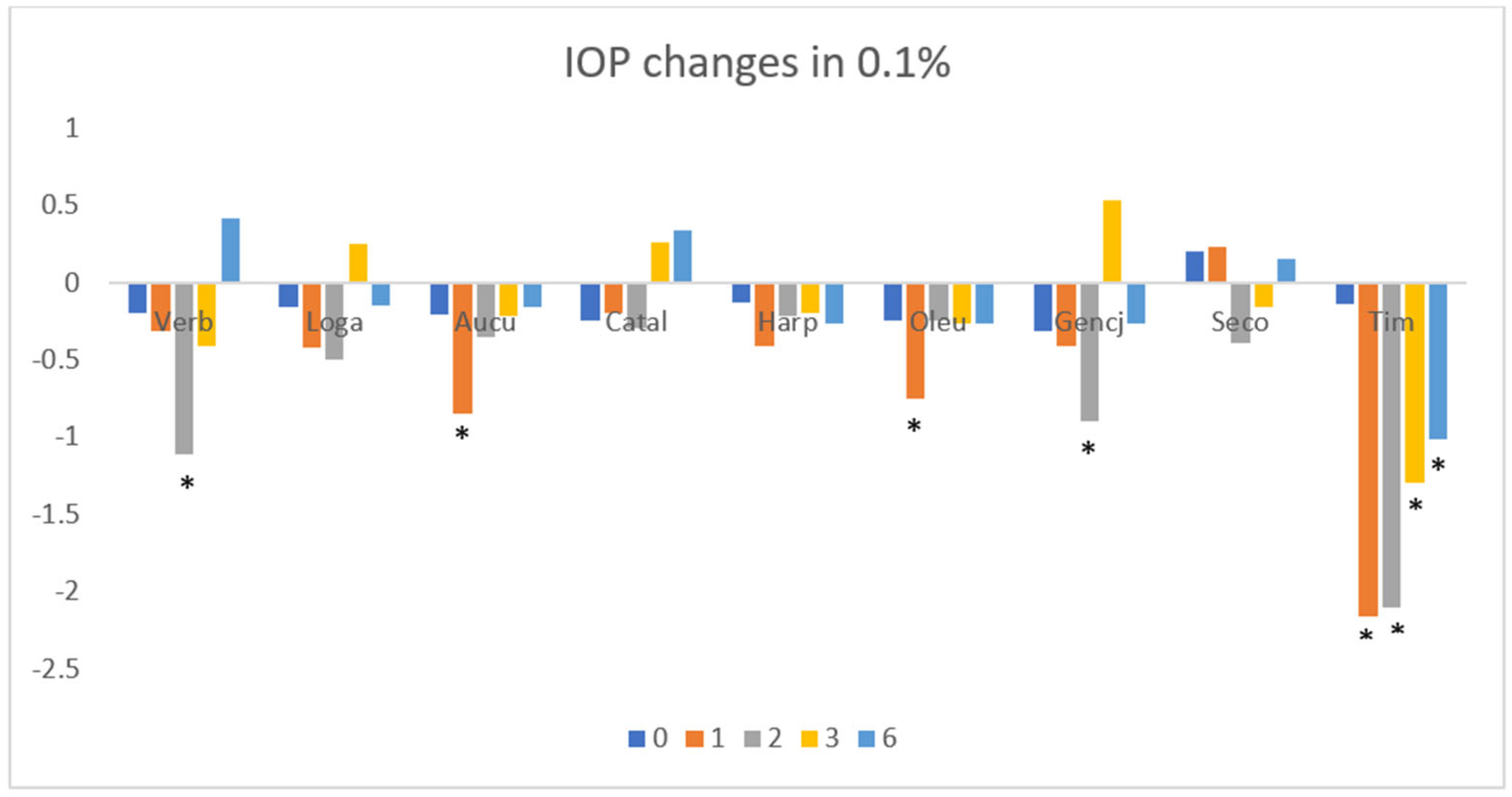
3.1. Intraocular Pressure

Intraocular pressure was found to be significantly reduced with the administration of 0.5% verbenalin, aucubin, oleuropein, gentiopicroside, and secologanin; however, pressure reductions were lower (by more than 1 mmHg) than those noted for the 0.5% timolol solution (Figure 2). In the case of loganin and harpagoside, a trend towards lower pressure was evident. A lack of effect was noted for catalpol. The greatest reduction in pressure was noted at 1 or 2 h after administration.
After administration of the 0.1% solution, statistically significant reductions in intraocular pressure were noted for verbenaline, aucubine, oleuropein, and gentiopicroside; the pressure reductions were lower than those noted after the administration of the 0.5% concentration and the 0.5% timolol solution (Figure 3). A slight reduction was noted for loganin, with no effect observed after the administration of catalpol, harpagoside, and secologanin.

3.2. Blood Flow

Of all the iridoids tested, statistically significant changes in flows were observed only for aucubin, catalpol, and gentiopicroside at concentrations of 0.5%. Changes were measured at different times after intraconjunctival administration. Two hours following the administration of gentiopicroside and aucubin, an increase blood flow of 38 and 40 flux, respectively, was observed (Figure 4). The highest effect of blood flow was observed for catapol, which increased the flow during the second hour of the experiment, yielding a value 48 flux. None of the compounds tested showed a longer effect on blood flow. As a control, the 0.5% concentration of timolol reduced iris blood flow for a value of 97 flux after the first hour up to 56 flux at 6 h after administration. In all variants, no effect was observed for the 0.1% concentration (Figure 5).

4. Discussion

To date, there are remarkably few reports that describe the direct effects of applying both plant extracts and isolated pure substances to the eye. Forskolin, a natural compound from the labdane diterpene group isolated from Plectranthus barbatus, was tested in experimental therapy in an open-label study [14] in patients with open-angle glaucoma. Research conducted in 90 adult patients has proven a decrease in IOP after 4 weeks of ocular administration by 5.4 mm Hg. Forskolin, by increasing cAMP, could help improve fluid drainage through the trabecular meshwork, reducing pressure inside the eye [15,16]. Cannabinoids (cannabidiol and tetrahydrocannabinol) in nanoemulsions containing Carbopol® 940 NF at a concentration of 1% significantly reduced IOP by 4.5 mm Hg in eyes treated for up to 360 min in a Dutch Belted male rabbit model [17]. A possible mechanism of cannabinoids is an interaction with the CB1 and CB2 receptors in ocular tissues, which affects fluid outflow and reduces IOP. Contrary to the above-mentioned results, 0.4% and 1.6% nanoemulsions of cannabinodiol in polysorbate 80 and medium-chain triglyceride exhibited no effect on IOP in a mouse model.
Experimental eye drops containing cannabinoids have been sold on Jamaica, but no extensive results have yet been delivered [18]. The results of the topical ophthalmic administration of pure tetrahydrocannabinol are not clear at this time. Jay et al. found no effect of 1% THC on changes in IOP pressure [19]. The lack of THC activity was also repeated in other experiments by Green [20]. In contrast to these results, the formulation in tocrisolve (a mixture of phospholipids and ethanol in water) with Pluronic-F68 stabilized commercial soybean oil emulsion presented a 28% reduction in IOP in the eighth hour after administration in a mouse model [21]. Interestingly, the effect of cannabinoids was sex-dependent. The authors concluded that THC lowers IOP by activating two receptors, CB1 and GPR18. Adelli et al. found reduced the IOP peak by after 2 h and 47% after 4 h of administration of THC eye formulations containing mono- and di-valine esters as auxiliary substances [22].
Razali et al. proved that topical administration of drops containing 0.2% trans-resveratrol (bioactive compound present in red grapes, red wine, Fallopia japonica) in 3% polyvinylpyrolidone decreased the IOP by 30% in rats with steroid-induced ocular hypertension [23]. In the experiment, resveratrol enhanced ocular blood flow and reduced oxidative stress in the trabecular meshwork, improving fluid outflow and lowering pressure. Davis investigated eyedrops containing 0.43% curcumin (isolated from Curcuma longa) in a nanoemulsion with Pluronic-F127 stabilized tocopherol polyethene glycol 1000 succinate nanoparticles, reducing hypertension in cobalt chloride-induced hypoxia in rats [24].
More reports can be found regarding experiments in which natural substances were supplemented orally. Hirooka et al. found that within 5 months, Ginkgo biloba extract administered orally decreased IOP by 29.8% in rats with chronic elevated intraocular pressure [25]. The authors also observed positive changes in retinal ganglion cell loss in the investigated group. Dorairaj et al. noticed that patients with primary open-angle glaucoma and IOPs ranging between 15 and 23 mmHg who were treated with 120 mg of ginkgo biloba extract at 30 months achieved a significant decrease in this value [26]. On the other hand, it was shown that oral Gingko biloba extract has no impact on IOP in consecutive patients with newly-diagnosed NTG [27]. Orally administered extract of Cordyceps cicadae, an entomogenous fungus, containing N6-(2 hydroxyethyl)-adenosine as a bioactive compound, significantly (60.5%) reduced IOP at a dose of 0.2 mg/kg b.w, after 56 days [28]. High extracts of pine bark and bilberry fruits decreased IOP in Japanese subjects with primary open-angle glaucoma after 4 weeks of administration at 130 mg per day [29]. Catalpol has a positive effect on atherosclerotic lesions in rabbits with alloxan-induced diabetes. The possible mechanisms may be related to the inhibition of the inflammatory response after oxidative stress and antifibrosis, as well as to the extracellular matrix. Catalpol administration alleviated diabetic atherosclerosis in diabetic rabbits, as demonstrated by the significant inhibition of neointima hypertrophy and macrophage recruitment. Therapy increased the superoxide dismutase activity, glutathione peroxidase, and plasma levels of total antioxidant status, while reducing the levels of malondialdehyde, protein carbonyl groups, and the advanced glycation end product. Cat also reduced the levels of tumor necrosis factor-α in the circulation, monocyte chemotactic protein-1, and vascular cell adhesion molecule-1a, and it also reduced the expression of mRNA and transforming growth factor-β1 and collagen IV in the vessels [30] Catalpol was able to reduce LPS (Escherichia coli serotype O55:B5)-induced microvascular barrier damage and hemorrhage in the mesenteric circulation by targeting both TLR-4 and Src, thus attenuating the phosphorylation of Src kinase, phosphatidylinositol 3-phosphatidyl kinase, and focal kinase, as well as cathepsin B activation [31]. Catalpol and puerarin are two monomers of Rehmannia glutinosa and lobed kudzuvine root, which are herbs commonly used together in ancient prescriptions of traditional Chinese medicine for cerebral ischemia [32]. Liu et al. investigated the effects of freeze-dried Catal powder and puerarin on cerebral ischemia in rats. They found an increase in regional cerebral blood flow, a reduced infarct volume, protection of vascular integrity, and inhibition of endothelial cell apoptosis in vivo. In a rat model of stroke after occlusion of the right middle cerebral artery, catalpol was found to affect neuroprotection and angiogenesis through the JAK2/STAT3 signaling pathway, mediated by STAT3 activation and VEGF expression. Catalpol may be used as a potential treatment for stroke [33]. Catalpol exhibited potent neuroprotective effects in mice after experimental autoimmune encephalomyelitis. Cat also plays a role in remyelination by promoting the expression of the transcription factors Olig1 and Olig2 [34]. Catalpol and geniposide, as two types of iridoid glycosides with high activity, are the main bioactive components of Rehmannia glutinosa and Gardenia jasminoides Ellis, respectively. Preclinical experiments have shown that they have significant neuroprotective effects against Alzheimer’s disease, Parkinson’s disease, stroke, and depression [35]. Catalpol, the main active component of the roots of Rehmannia glutinosa, can reduce liver damage in a mouse model of cholestasis by inhibiting oxidative stress and enhancing the potential of the mitochondrial membrane. This compound, derived from the roots of Rehmannia glutinosa, displays antioxidant and anti-inflammatory effects, reduces apoptotic protein expression, improves mitochondrial membrane potential, increases ATP and glutathione content, and inhibits lipid peroxidation [36]. The administration of iridoid glycosides from Morinda citrifolia fruit extract improves blood flow and has a fibrinolytic effect, which is attributed to the iridoid glycoside asperulosic acid [37,38]. In a mouse model of hindlimb ischemia, there was a significant acceleration in perfusion recovery and a reduction in muscle tissue damage in the group receiving auxubine (Au). Au was found to promote angiogenesis in peripheral ischemia through the vascular endothelial cell growth factor (VEGF)/Akt/endothelial nitric oxide synthase (eNOS) signaling pathway. The authors qualify Au as a potent new compound with potential use in the treatment of peripheral vascular disease [39]. Aucubin can protect RGC retinal ganglion cells in diabetic rats, inhibit RGC apoptosis, and reduce oxidative stress and inflammatory response, and 10 mg/kg of aucubin showed optimal efficacy. This mechanism may be related to inhibition of the p38MAPK signaling pathway [40]. Topical administration of aucubin eye drops has shown beneficial effects on abnormal ocular changes caused by urban particular matter (UPM), such as excessive tear secretion and lacrimal gland damage. The results of the study demonstrate the beneficial effects of aucubin in dry eye disease [41]. Picroside II protects the blood–brain barrier by inhibiting the oxidative signaling pathway in cerebral ischemia-reperfusion injury [42]. It also inhibits neuronal apoptosis and improves the morphology and structure after cerebral ischemia in rats [43]. Ginsenoside, a non-peptide angiogenic agent, administered in a rat model of myocardial infarction effectively improved myocardial perfusion and preserved left ventricular function during infarction [44]. Combination therapies have been used for more than 2500 years in traditional Chinese medicine. A preparation of three components, ginsenosides, berberine, and jasminoide, acting synergistically, was employed in the treatment of rats affected by obstruction of the middle cerebral artery caused by focal cerebral ischemia. The respective proportions of the substances displayed a good pharmacological effect on acute ischemic stroke and allowed the strongest synergistic effect. The researchers pointed out that in addition to restoring blood supply and protecting easily damaged cells in the ischemic area as early as possible, attention should be also paid to the removal of toxic metabolites [45].
Other iridoids also have effects on microcirculation. Oleuropein and pinorezinol were able to protect rats from pial microcirculation in reperfusion injury, increase nitric oxide release, and reduce oxidative stress, maintaining the distribution of pial blood flow. Control rats showed reduced arteriolar diameter, microvascular leakage, leukocyte adhesion in the venules, and reduced capillary perfusion. Pretreatment with oleuropein or pinorezinol determined dilatation in all rows of arterioles, decreasing microvascular leakage and leukocyte adhesion via protection of capillary perfusion. Test substances, administered together, prevented microvascular damage to a greater extent [46]. In a study of the effects of gentiopicroside on diabetic peripheral neuropathy in rats, gentiopicroside was found to exert a protective effect on nerves and attenuated experimental neuropathy by improving nerve blood flow through regulation of the PPAR-γ/AMPK/ACC signaling pathway. Motor and sensory nerve conduction velocity was also improved [47]. A neuroprotective role for morroniside was observed in spinal cord injury in rats, and treatment improved functional recovery. Behavioral improvements were associated with a higher survival of neuronal- and oligodendrocytes, which increased the ability of the injured spinal cord to form myelin and repair tissue, ultimately contributing to improved neurological outcomes. Morroniside treatment decreased oxygen free radical levels and increased antioxidant enzyme activity [48]. In rat studies, oleuropein, the main component of olive leaves, reduces oxidative stress, displaying antiapoptotic and anti-inflammatory effects in a model of kidney damage caused by ureteral obstruction [37]. Rehmanniae Radix and Cornus officinalis are a typical pair of Chinese medicinal herbs used in clinical practice to treat diabetic nephropathy. The main active constituents are catapol and loganin, respectively. In a diabetic nephropathy model, it was found that LOGA and CATAL alone or in combination protected renal function from damage, prevented extracellular matrix hyperplasia and glycogen deposition, and alleviated podocyte loss detected by histological and immunohistochemical tests. LOGA and CATAL alone or in combination attenuated AGEs-induced podocyte apoptosis in vitro. LOGA and CATAL alone or in combination inhibited the activation of the RAGE/p38 MAPK/p65 NF-κB and RAGE/Nox4/p65 NF-κB pathways in podocytes. The inhibitory effects of the drug combination were more pronounced than the effects of individual treatment. Both compounds inhibited apoptosis, oxidative stress, and inflammation [49]. Also, Hwang et al. have proven the interaction of loganin with muscarine receptors present in the eyes [50]. Stimulation of M3 receptors by loganin in the ciliary muscle contractions as a result of the stretching of the trabeculae facilitated the outflow of aqueous fluid through Schlemm’s canal, lowering of intraocular pressure. This mechanism is similar to pilocarpine, a known muscarine agonist, used in glaucoma treatment.
In the treatment of glaucoma, strategies that delay or arrest the loss of RGC have been recognized as potentially beneficial for vision preservation, but their success depends on a thorough understanding of the mechanisms that lead to RGC dysfunction and death. The main pharmacological treatment strategies include reducing intraocular pressure, improving vascular flow, and neuroprotection. The mainstay of glaucoma treatment is the reduction in intraocular pressure, most commonly via topical eye drop medications and less commonly, using laser or surgical procedures. Most drug therapies focus on reducing the formation of aqueous fluid and increasing choroidal outflow or conventional outflow through the angle of glaucoma [8].
Known pharmaceuticals currently used in ophthalmological practice administered intraconjunctivally could act on different mechanisms. Prostaglandins bind to specific F2α receptors in the cells of the ciliary body and the trabecular meshwork, causing an improvement in aqueous humor through the uveoscleral pathway, which is a significant route for fluid drainage from the eye. As a consequence, the reduction in fluid volume lowers IOP. The β-blockers, such as timolol, inhibit the production of aqueous humor by the ciliary body and also alter the vascular dynamics in the eye, potentially reducing blood flow to the ciliary body and further decreasing aqueous humor production. Finaly, β-blockers could act indirectly by reducing the pressure gradient across the trabecular meshwork, which can enhance aqueous humor drainage. α-2 adrenergic agonists such as brimonidine bind to α-2 adrenergic receptors in the ciliary body, which leads to reduced production of aqueous humor. Additionally, it enhances the flow of aqueous humor through the uveoscleral pathway, further contributing to a decrease in IOP. Carbonic anhydrase inhibitors such as brinzolamide inhibit anhydrase in the ciliary body, leading to decreased bicarbonate production. This results in reduced fluid transport and lower aqueous humor production, thus decreasing IOP. Muscarinic agonists such as pilocarpine, bind to muscarinic receptors on the ciliary muscle, causing it to contract, increasing the outflow of aqueous humor through the trabecular meshwork.
Some forms of glaucoma present with completely normal intraocular pressure and abnormalities in vascular blood flow, an example of which is normal pressure glaucoma (NTG). Previous studies have shown that NTG is associated with various systemic diseases, including migraines, Alzheimer’s disease, primary vascular dysregulation, and Flammer syndrome. Vascular factors are involved in these diseases. The mechanisms underlying the abnormal ocular blood flow in NTG are still unclear, but the risk factors for glaucomatous optic neuropathy are probably oxidative stress, vasospasm, and endothelial dysfunction [50]. The USG-Doppler study of extraocular flow in glaucoma patients showed lower extraocular velocities, higher retinal venous saturation, and increased choroidal thickness asymmetry compared to the results for the healthy group [51]. Glaucoma, as a neurodegenerative disease, is also of interest for researchers in terms of identifying various neuroprotection strategies as a method of treatment [52]. It is thought that RGC death may display a variety of molecular bases. These include impaired axonal transport, deprivation of neurotrophic factors, toxic pro-neurotrophins, activation of intrinsic and extrinsic apoptotic signals, mitochondrial dysfunction, excitotoxic damage, oxidative stress, abnormal reactive glial behavior, and loss of synaptic connectivity. The above observations could form the basis for new potential targets for neuroprotection [53].
Unfortunately, neuroprotective treatment is long-lasting and currently, there are no suitable markers to assess its efficacy shortly after the drug is administered, as can be measured after administering eye drops delivering hypotensive drugs, where the effect of lowering intraocular pressure is seen shortly after the drug is administered. A comparison of the results of the clinical trials of neuroprotective drugs with conventional treatment does not provide conclusive results [54,55,56,57,58].

5. Conclusions

Some iridoids, such as verbenalin, aucubin, oleuropein, gentiopicroside, and secologanin, have the potential to lower intraocular pressure. However, they display a weaker effect than that of the standard 0.5% timolol treatment. We found that a 0.5% concentration of verbenalin, administered intraconjunctivally, significantly decreases IOP after 3 and 4 h. In terms of aucubin, oleuropein, gentiopicroside, and secologanin, this effect was observed only in the first or second hour. Only verbenalin, aucubin, oleuropein and gentiopicroside show a decreasing IOP effect in concentrations of 0.1%, but the effect was less observed than that for the 0.5% concentrations. In comparison to the timolol values of IOP, these changes where significantly weaker. In terms of iris blood flow, increases were only observed for aucubin, catalpol, and gentiopicroside. contrary to the results for timolol, which increased for all time points via intraconjunctival administration. Catalpol, gentiopicroside, and aucubin increased statistically significant blood flow 2 and 3 h, respectively, after administration only at a concentration of 0.5% In conclusion, applied iridoids could be considered for further investigation as promising agents in neurodegenerative diseases and in patients with vascular flow disorders in the eyeball. Although there is considerable evidence linking iridoids to benefits in maintaining ocular and other organ health, more in-depth research is needed to determine whether this promising compound is suitable for use as an evidence-based complementary and alternative medicine in the treatment of ocular diseases presenting with elevated intraocular pressure and vascular blood flow disorders.

Author Contributions

D.S.: investigation, visualization, project administration, writing of manuscript, formal analysis; T.S.: conceptualization; A.S. (Adam Szeląg): funding acquisition; A.S. (Antoni Szumny): writing of manuscript, conceptualization, and investigation. All authors have read and agreed to the published version of the manuscript.

Funding

This work was supported by the statutory budget of Wroclaw Medical University (SUB.A080.19.024).

Institutional Review Board Statement

The animal study protocol was approved by the Lokalna Komisja Bioetyczna, Wrocław Poland. The approval for the study was obtained from the committee through approval No. 21/2015, dated 18 March 2015.

Informed Consent Statement

Not applicable.

Data Availability Statement

Available on demand.

Conflicts of Interest

The authors declare no conflict of interest.

Abbreviations

The following abbreviations are used in this manuscript:
IOPintraocular pressure
VERBverbenalin
LOGAloganin
AUCUaucubin
CATALcatalpol
HARPharpagoside
OLEUoleuropein
GENCJgentiopicroside
SECOsecologanin
RGCsretinal ganglion cells
POAGprimary open-angle glaucoma
RVORetinal vein obstruction
HPLCHigh-Performance Liquid Chromatography
THCTetrahydrocannabinol
NTGnormal tension glaucoma
VEGFvascular endothelial cell growth factor

References

  1. Wang, L.; Meng, X.; Zhou, H.; Liu, Y.; Zhang, Y.; Liang, H.; Hou, G.; Kang, W.; Liu, Z. Iridoids and active ones in patrinia: A review. Heliyon 2023, 9, e16518. [Google Scholar] [CrossRef]
  2. Kouda, R.; Yakushiji, F. Recent advances in Iridoid chemistry: Biosynthesis and chemical synthesis. Chem.–Asian J. 2020, 15, 3771–3783. [Google Scholar] [CrossRef]
  3. Amle, V.S.; Rathod, D.A.; Keshamma, E.; Kumar, V.; Kumar, R.; Saha, P. Bioactive Herbal Medicine Use for Eye Sight: A Meta Analysis. J. Res. Appl. Sci. Biotechnol. 2022, 1, 42–50. [Google Scholar] [CrossRef]
  4. Paduch, R.; Woźniak, A.; Niedziela, P.; Rejdak, R. Assessment of eyebright (Euphrasia officinalis L.) extract activity in relation to human corneal cells using in vitro tests. Balk. Med. J. 2014, 31, 29–36. [Google Scholar] [CrossRef]
  5. Sharma, P.; Singh, G. A review of plant species used to treat conjunctivitis. Phytother. Res. 2002, 16, 1–22. [Google Scholar] [CrossRef]
  6. Bielory, L.; Heimall, J. Review of complementary and alternative medicine in treatment of ocular allergies. Curr. Opin. Allergy Clin. Immunol. 2003, 3, 395–399. [Google Scholar] [CrossRef]
  7. Kang, W.S.; Jung, E.; Kim, J. Aucuba japonica Extract and Aucubin Prevent Desiccating Stress-Induced Corneal Epithelial Cell Injury and Improve Tear Secretion in a Mouse Model of Dry Eye Disease. Molecules 2018, 23, 2599. [Google Scholar] [CrossRef]
  8. Weinreb, R.N.; Aung, T.; Medeiros, F.A. The Pathophysiology and Treatment of Glaucoma. JAMA 2014, 311, 1901. [Google Scholar] [CrossRef]
  9. Quigley, H.A.; Broman, A.T. The number of people with glaucoma worldwide in 2010 and 2020. Br. J. Ophthalmol. 2006, 90, 262–267. [Google Scholar] [CrossRef]
  10. He, S.; Stankowska, D.L.; Ellis, D.Z.; Krishnamoorthy, R.R.; Yorio, T. Targets of Neuroprotection in Glaucoma. J. Ocul. Pharmacol. Ther. 2018, 34, 85–106. [Google Scholar] [CrossRef]
  11. Schorr, E.M.; Rossi, K.C.; Stein, L.K.; Park, B.L.; Tuhrim, S.; Dhamoon, M.S. Characteristics and Outcomes of Retinal Artery Occlusion: Nationally Representative Data. Stroke 2020, 51, 800–807. [Google Scholar] [CrossRef]
  12. Youn, T.S.; Lavin, P.; Patrylo, M.; Schindler, J.; Kirshner, H.; Greer, D.M.; Schrag, M. Current treatment of central retinal artery occlusion: A national survey. J. Neurol. 2018, 265, 330–335. [Google Scholar] [CrossRef]
  13. Song, P.; Xu, Y.; Zha, M.; Zhang, Y.; Rudan, I. Global epidemiology of retinal vein occlusion: A systematic review and meta-analysis of prevalence, incidence, and risk factors. J. Glob. Health 2019, 9, 010427. [Google Scholar] [CrossRef]
  14. Majeed, M.; Nagabhushanam, K.; Natarajan, S.; Vaidyanathan, P.; Karri, S.K.; Jose, J.A. Efficacy and safety of 1% forskolin eye drops in open angle glaucoma–An open label study. Saudi J. Ophthalmol. 2015, 29, 197–200. [Google Scholar] [CrossRef]
  15. Szumny, D.; Sozański, T.; Kucharska, A.Z.; Dziewiszek, W.; Piórecki, N.; Magdalan, J.; Chlebda-Sieragowska, E.; Kupczynski, R.; Szeląg, A.; Szumny, A. Application of cornelian cherry iridoid-polyphenolic fraction and Loganic acid to reduce intraocular pressure. Evid.-Based Complement. Altern. Med. 2015, 1, 939402. [Google Scholar] [CrossRef]
  16. Alasbahi, R.; Melzig, M. Forskolin and derivatives as tools for studying the role of cAMP. Die Pharm.-Int. J. Pharm. Sci. 2012, 67, 5–13. [Google Scholar]
  17. Wagh, V.; Patil, P.; Surana, S.; Wagh, K. Forskolin: Upcoming antiglaucoma molecule. J. Postgrad. Med. 2012, 58, 199–202. [Google Scholar] [CrossRef]
  18. Senapati, S.; Youssef, A.A.A.; Sweeney, C.; Cai, C.; Dudhipala, N.; Majumdar, S. Cannabidiol loaded topical ophthalmic nanoemulsion lowers intraocular pressure in normotensive Dutch-belted rabbits. Pharmaceutics 2022, 14, 2585. [Google Scholar] [CrossRef]
  19. Pinar-Sueiro, S.; Rodríguez-Puertas, R.; Vecino, E. Cannabinoid applications in glaucoma. Arch. Soc. Española Oftalmol. Engl. Ed. 2011, 86, 16–23. [Google Scholar] [CrossRef]
  20. Jay, W.; Green, K. Multiple-drop study of topically applied 1% delta 9-tetrahydrocannabinol in human eyes. Arch. Ophthalmol. 1983, 101, 591–593. [Google Scholar] [CrossRef]
  21. Green, K.; Symonds, C.M.; Oliver, N.W.; Elijah, R.D. Intraocular pressure following systemic administration of cannabinoids. Curr. Eye Res. 1982, 2, 247–253. [Google Scholar] [CrossRef]
  22. Miller, S.; Daily, L.; Leishman, E.; Bradshaw, H.; Straiker, A. Δ9-Tetrahydrocannabinol and cannabidiol differentially regulate intraocular pressure. Investig. Ophthalmol. Vis. Sci. 2018, 59, 5904–5911. [Google Scholar] [CrossRef]
  23. Adelli, G.R.; Bhagav, P.; Taskar, P.; Hingorani, T.; Pettaway, S.; Gul, W.; ElSohly, M.A.; Repka, M.A.; Majumdar, S. Development of a Δ9-tetrahydrocannabinol amino acid-dicarboxylate prodrug with improved ocular bioavailability. Investig. Ophthalmol. Vis. Sci. 2017, 58, 2167–2179. [Google Scholar] [CrossRef]
  24. Razali, N.; Agarwal, R.; Agarwal, P.; Tripathy, M.; Kapitonova, M.Y.; Kutty, M.K.; Smirnov, A.; Khalid, Z.; Ismail, N.M. Topical trans-resveratrol ameliorates steroid-induced anterior and posterior segment changes in rats. Exp. Eye Res. 2016, 143, 9–16. [Google Scholar] [CrossRef]
  25. Davis, B.M.; Pahlitzsch, M.; Guo, L.; Balendra, S.; Shah, P.; Ravindran, N.; Malaguarnera, G.; Sisa, C.; Shamsher, E.; Hamze, H. Topical curcumin nanocarriers are neuroprotective in eye disease. Sci. Rep. 2018, 8, 11066. [Google Scholar] [CrossRef]
  26. Hirooka, K.; Tokuda, M.; Miyamoto, O.; Itano, T.; Baba, T.; Shiraga, F. The Ginkgo biloba extract (EGb 761) provides a neuroprotective effect on retinal ganglion cells in a rat model of chronic glaucoma. Curr. Eye Res. 2004, 28, 153–157. [Google Scholar] [CrossRef]
  27. Dorairaj, S.; Ritch, R.; Liebmann, J.M. Visual improvement in a patient taking ginkgo biloba extract: A case study. Explore 2007, 3, 391–395. [Google Scholar] [CrossRef]
  28. Guo, X.; Kong, X.; Huang, R.; Jin, L.; Ding, X.; He, M.; Liu, X.; Patel, M.C.; Congdon, N.G. Effect of Ginkgo biloba on visual field and contrast sensitivity in Chinese patients with normal tension glaucoma: A randomized, crossover clinical trial. Investig. Ophthalmol. Vis. Sci. 2014, 55, 110–116. [Google Scholar] [CrossRef]
  29. Lee, L.-Y.; Hsu, J.-H.; Fu, H.-I.; Chen, C.-C.; Tung, K.-C. Lowering the intraocular pressure in rats and rabbits by cordyceps cicadae extract and its active compounds. Molecules 2022, 27, 707. [Google Scholar] [CrossRef]
  30. Manabe, K.; Kaidzu, S.; Tsutsui, A.; Mochiji, M.; Matsuoka, Y.; Takagi, Y.; Miyamoto, E.; Tanito, M. Effects of French maritime pine bark/bilberry fruit extracts on intraocular pressure for primary open-angle glaucoma. J. Clin. Biochem. Nutr. 2021, 68, 67–72. [Google Scholar] [CrossRef]
  31. Liu, J.Y.; Zheng, C.Z.; Hao, X.P.; Zhang, D.J.; Mao, A.W.; Yuan, P. Catalpol ameliorates diabetic atherosclerosis in diabetic rabbits. Am. J. Transl. Res. 2016, 8, 4278–4288. [Google Scholar]
  32. Zhang, Y.P.; Pan, C.S.; Yan, L.; Liu, Y.Y.; Hu, B.H.; Chang, X.; Li, Q.; Huang, D.D.; Sun, H.Y.; Fu, G.; et al. Catalpol restores LPS-elicited rat microcirculation disorder by regulation of a network of signaling involving inhibition of TLR-4 and SRC. Am. J. Physiol. Gastrointest. Liver Physiol. 2016, 311, G1091–G1104. [Google Scholar] [CrossRef]
  33. Liu, Y.; Tang, Q.; Shao, S.; Chen, Y.; Chen, W.; Xu, X. Lyophilized Powder of Catalpol and Puerarin Protected Cerebral Vessels from Ischemia by Its Anti-apoptosis on Endothelial Cells. Int. J. Biol. Sci. 2017, 13, 327–338. [Google Scholar] [CrossRef]
  34. Dong, W.; Xian, Y.; Yuan, W.; Huifeng, Z.; Tao, W.; Zhiqiang, L.; Shan, F.; Ya, F.; Hongli, W.; Jinghuan, W.; et al. Catalpol stimulates VEGF production via the JAK2/STAT3 pathway to improve angiogenesis in rats’ stroke model. J. Ethnopharmacol. 2016, 191, 169–179. [Google Scholar] [CrossRef]
  35. Sun, Y.; Ji, J.; Zha, Z.; Zhao, H.; Xue, B.; Jin, L.; Wang, L. Effect and Mechanism of Catalpol on Remyelination via Regulation of the NOTCH1 Signaling Pathway. Front. Pharmacol. 2021, 12, 628209. [Google Scholar] [CrossRef]
  36. Zhang, X.; Liu, K.; Shi, M.; Xie, L.; Deng, M.; Chen, H.; Li, X. Therapeutic potential of catalpol and geniposide in Alzheimer’s and Parkinson’s diseases: A snapshot of their underlying mechanisms. Brain Res. Bull. 2021, 174, 281–295. [Google Scholar] [CrossRef]
  37. Gao, X.; Xu, J.; Liu, H. Protective effects of catalpol on mitochondria of hepatocytes in cholestatic liver injury. Mol. Med. Rep. 2020, 22, 2424–2432. [Google Scholar] [CrossRef]
  38. Kaeidi, A.; Sahamsizadeh, A.; Allahtavakoli, M.; Fatemi, I.; Rahmani, M.; Hakimizadeh, E.; Hassanshahi, J. The effect of oleuropein on unilateral ureteral obstruction induced-kidney injury in rats: The role of oxidative stress, inflammation and apoptosis. Mol. Biol. Rep. 2020, 47, 1371–1379. [Google Scholar] [CrossRef]
  39. Murata, K.; Abe, Y.; Futamura-Masuda, M.; Uwaya, A.; Isami, F.; Deng, S.; Matsuda, H. Effect of Morinda citrifolia fruit extract and its iridoid glycosides on blood fluidity. J. Nat. Med. 2014, 68, 498–504. [Google Scholar] [CrossRef]
  40. Chen, L.; Yang, Y.; Zhang, L.; Li, C.; Coffie, J.W.; Geng, X.; Qiu, L.; You, X.; Fang, Z.; Song, M.; et al. Aucubin promotes angiogenesis via estrogen receptor beta in a mouse model of hindlimb ischemia. J. Steroid Biochem. Mol. Biol. 2017, 172, 149–159. [Google Scholar] [CrossRef]
  41. Feng, M.; Jiang, X.; Zhang, Q.; Wang, Q.; She, C.; Li, Z. Aucubin protects against retinal ganglion cell injury in diabetic rats via inhibition of the p38MAPK pathway. Am. J. Transl. Res. 2023, 15, 1007–1016. [Google Scholar]
  42. Park, S.-B.; Jung, W.K.; Yu, H.-Y.; Kim, Y.H.; Kim, J. Effect of Aucubin-Containing Eye Drops on Tear Hyposecretion and Lacrimal Gland Damage Induced by Urban Particulate Matter in Rats. Molecules 2022, 27, 2926. [Google Scholar] [CrossRef]
  43. Zhai, L.; Liu, M.; Wang, T.; Zhang, H.; Li, S.; Guo, Y. Picroside II protects the blood-brain barrier by inhibiting the oxidative signaling pathway in cerebral ischemia-reperfusion injury. PLoS ONE 2017, 12, e0174414. [Google Scholar] [CrossRef]
  44. Wang, T.; Zhao, L.; Guo, Y.; Zhang, M.; Pei, H. Picroside II Inhibits Neuronal Apoptosis and Improves the Morphology and Structure of Brain Tissue following Cerebral Ischemic Injury in Rats. PLoS ONE 2015, 10, e0124099. [Google Scholar] [CrossRef]
  45. Wei, H.J.; Yang, H.H.; Chen, C.H.; Lin, W.W.; Chen, S.C.; Lai, P.H.; Chang, Y.; Sung, H.W. Gelatin microspheres encapsulated with a nonpeptide angiogenic agent, ginsenoside Rg1, for intramyocardial injection in a rat model with infarcted myocardium. J. Control. Release Off. J. Control. Release Soc. 2007, 120, 27–34. [Google Scholar] [CrossRef]
  46. Li, S.; Wu, C.; Chen, J.; Lu, P.; Chen, C.; Fu, M.; Fang, J.; Gao, J.; Zhu, L.; Liang, R.; et al. An effective solution to discover synergistic drugs for anti-cerebral ischemia from traditional Chinese medicinal formulae. PLoS ONE 2013, 8, e78902. [Google Scholar] [CrossRef]
  47. Lapi, D.; Di Maro, M.; Mastantuono, T.; Battiloro, L.; Sabatino, L.; Muscariello, E.; Colantuoni, A. Effects of oleuropein and pinoresinol on microvascular damage induced by hypoperfusion and reperfusion in rat pial circulation. Microcirculation 2015, 22, 79–90. [Google Scholar] [CrossRef]
  48. Lu, Y.; Yao, J.; Gong, C.; Wang, B.; Zhou, P.; Zhou, S.; Yao, X. Gentiopicroside Ameliorates Diabetic Peripheral Neuropathy by Modulating PPAR-Γ/AMPK/ACC Signaling Pathway. Cell. Physiol. Biochem. 2018, 50, 585–596. [Google Scholar] [CrossRef]
  49. Duan, F.X.; Shi, Y.J.; Chen, J.; Song, X.; Shen, L.; Qi, Q.; Ding, S.Q.; Wang, Q.Y.; Wang, R.; Lü, H.Z.; et al. The neuroprotective role of morroniside against spinal cord injury in female rats. Neurochem. Int. 2021, 148, 105105. [Google Scholar] [CrossRef]
  50. Chen, Y.; Chen, J.; Jiang, M.; Fu, Y.; Zhu, Y.; Jiao, N.; Liu, L.; Du, Q.; Wu, H.; Xu, H.; et al. Loganin and catalpol exert cooperative ameliorating effects on podocyte apoptosis upon diabetic nephropathy by targeting AGEs-RAGE signaling. Life Sci. 2020, 252, 117653. [Google Scholar] [CrossRef]
  51. Hwang, E.S.; Kim, H.B.; Lee, S.; Kim, M.J.; Lee, S.O.; Han, S.M.; Maeng, S.; Park, J.H. Loganin enhances long-term potentiation and recovers scopolamine-induced learning and memory impairments. Physiol. Behav. 2017, 17, 243–248. [Google Scholar] [CrossRef]
  52. Fan, N.; Wang, P.; Tang, L.; Liu, X. Ocular Blood Flow and Normal Tension Glaucoma. BioMed Res. Int. 2015, 2015, 308505. [Google Scholar] [CrossRef]
  53. Abegão Pinto, L.; Willekens, K.; Van Keer, K.; Shibesh, A. Ocular blood flow in glaucoma—The Leuven Eye Study. Acta Ophthalmol. 2016, 94, 592–598. [Google Scholar] [CrossRef]
  54. Naik, S.; Pandey, A.; Lewis, S.A.; Rao, B.S.S.; Mutalik, S. Neuroprotection: A versatile approach to combat glaucoma. Eur. J. Pharmacol. 2020, 881, 173208. [Google Scholar] [CrossRef]
  55. Almasieh, M.; Wilson, A.M.; Morquette, B.; Cueva Vargas, J.L.; Di Polo, A. The molecular basis of retinal ganglion cell death in glaucoma. Prog. Retin. Eye Res. 2012, 31, 152–181. [Google Scholar] [CrossRef]
  56. Sena, D.F.; Lindsley, K. Neuroprotection for treatment of glaucoma in adults. Cochrane Database Syst. Rev. 2017, 1, Cd006539. [Google Scholar] [CrossRef]
  57. Ganguly, S.; Wulff, D.; Phan, C.-M.; Jones, L.W.; Tang, X.S. Injectable and 3D Extrusion Printable Hydrophilic Silicone-Based Hydrogels for Controlled Ocular Delivery of Ophthalmic Drugs. ACS Appl. Bio Mater. 2024, 7, 6286–6296. [Google Scholar] [CrossRef]
  58. Su, W.; Wang, R.; Qian, C.; Li, X.; Tong, Q.; Jiao, T. Research progress review of preparation and applications of fluorescent hydrogels. J. Chem. 2020, 2020, 8246429. [Google Scholar] [CrossRef]
Figure 1. Chemical structures of investigated iridoids: (1) verbenalin (VERB), (2) loganin (LOGA), (3) aucubin (AUCU), (4) catalpol (CATAL), (5) harpagoside (HARP), (6) oleuropein (OLEU), (7) gentiopicroside (GENCJ) and (8) secologanin (SECO).
Applsci 15 05055 g001
Figure 2. Intraocular pressure results before and 1, 2, 3, and 6 h following administration of 0.5% iridoid solution. The graph shows the difference in pressure values compared to the first baseline measurement. Negative values indicate a decrease in IOP. An asterisk indicates a statistically significant effect. Verb: verbenalin; Loga: loganin, Aucu: aucubin; Catal: catalpol; Harp: harpagoside; Oleu: oleuropein; Gencj: gentiopicroside; Seco: secologanin; Tim: timolol.
Applsci 15 05055 g002
Figure 3. Intraocular pressure (mm Hg) results before and 1, 2, 3, and 6 h after administration of 0.1% iridoid solution. The graph shows the difference in pressure values compared to the first baseline measurement. Negative values indicate a decrease in IOP. An asterisk indicates a statistically significant effect. Verb: verbenalin; Loga: loganin, Aucu: aucubin; Catal: catalpol; Harp: harpagoside; Oleu: oleuropein; Gencj: gentiopicroside; Seco: secologanin; Tim: timolol.
Applsci 15 05055 g003
Figure 4. Iris blood flow (flux, a.u.) before and 1, 2, 3 and 6 h after administration of a 0.5% iridoid solution. The graph shows the difference in flow values compared to the first baseline measurement. Timolol was administered as a reference at 0.5%. Positive values indicate an increase in iris blood flow. An asterisk indicates a statistically significant effect. Verb: verbenalin; Loga: loganin, Aucu: aucubin; Catal: catalpol; Harp: harpagoside; Oleu: oleuropein; Gencj: gentiopicroside; Seco: secologanin; Tim: timolol.
Applsci 15 05055 g004
Figure 5. Iris blood flow (flux, a.u.) before and 1, 2, 3 and 6 h following administration of a 0.1% iridoid solution. The graph shows the difference in flow values compared to the first baseline measurement. Timolol was administered as reference at 0.5%. Positive values indicate an increase in iris blood flow. An asterisk indicates a statistically significant effect. Verb: verbenalin; Loga: loganin, Aucu: aucubin; Catal: catalpol; Harp: harpagoside; Oleu: oleuropein; Gencj: gentiopicroside; Seco: secologanin; Tim: timolol.
Applsci 15 05055 g005
Disclaimer/Publisher’s Note: The statements, opinions and data contained in all publications are solely those of the individual author(s) and contributor(s) and not of MDPI and/or the editor(s). MDPI and/or the editor(s) disclaim responsibility for any injury to people or property resulting from any ideas, methods, instructions or products referred to in the content.

