Article

In Vitro Wear Properties of a New CAD/CAM Dental Resin Composite in a Chewing Simulation Test Opposing Itself

by
Camillo D’Arcangelo
1,†,
Mirco Vadini
1,†,
Lorenzo Vanini
2,
Giuseppe Daniele Rondoni
3,
Edoardo Sorrentino
1,‡ and
Francesco De Angelis
1,*,‡
1
Unit of Restorative Dentistry and Endodontics, Department of Medical, Oral and Biotechnological Science, School of Dentistry, “G. D’Annunzio” University of Chieti, 66100 Chieti, Italy
2
Private Practice, Corso S. Gottardo 25, 6830 Chiasso, Switzerland
3
Private Dental Technician, 17100 Savona, Italy
*
Author to whom correspondence should be addressed.
These authors contributed equally to this work.
Appl. Sci. 2025, 15(9), 5023; https://doi.org/10.3390/app15095023
Submission received: 28 March 2025 / Revised: 22 April 2025 / Accepted: 25 April 2025 / Published: 30 April 2025

Abstract

:
Wear resistance is of paramount importance for the long-term success of dental materials, especially when they are used for extensive indirect restorations in full-mouth rehabilitations. The present in vitro study aimed to evaluate the two-body wear resistance of a new and recently introduced CAD/CAM resin composite disc (Ena Cad, Micerium S.pA.), to compare it to the wear resistance of other two well-known and already marketed CAD/CAM composites (Brilliant Crios, Coltene/Whaledent AG; Grandio disc, Voco Gmbh) and to a conventional type 3 gold alloy (Aurocast 8, Nobil-Metal). Ten cylindrical specimens (diameter 8 mm, height 6 mm) were manufactured with each material and subjected to a two-body wear test in a dual-axis chewing simulator, performing 120,000 chewing cycles opposing antagonists (2-mm-diameter round tip) made out of the same corresponding materials. The total vertical wear (mm) and the total volumetric loss (mm3) for each sample/antagonist pair were calculated. Representative scanning electron microscope images were also taken. Data were statistically analyzed using one-way analysis of variance tests. No statistically significant differences were recorded among the wear properties of the restorative materials under investigation. The Ena Cad disc showed a wear resistance comparable to the type 3 gold alloy and to the already marketed Brilliant Crios and Grandio disc.

1. Introduction

Wear is defined as the net material loss from its surface, occurring as a result of the following three different processes: abrasion, attrition and erosion. The physiologic average enamel wear has been estimated to be around 30–35 µm per year [1] and it is generally related to phenomena such as microplowing, microcutting, microcracking and microfatigue [1,2]. An “ideal” dental restorative material should adequately simulate the mechanical properties of the natural tooth hard tissues, including wear resistance. Indeed, a reduced wear resistance can quickly compromise both the function and aesthetics [3,4,5,6,7], while an excessively high wear resistance can jeopardize an advantageous intraoral self-functionalization process of the restoration [8]. Gold-based dental alloys would be ideally suitable for posterior restorations because of their wear behavior that is similar to the natural enamel [9,10], their marginal precision [9,11] and the reduced abrasiveness towards the antagonistic dentition [12]. Unfortunately, in recent decades, the intrinsically unpleasing aesthetic of gold alloys has drastically limited their use, even for the posterior teeth occlusal surfaces. Nevertheless, the selection of alternative materials with a wear resistance that is analogous to natural enamel is still essential, especially when dealing with extensive full-mouth rehabilitations.
Nowadays, resin composites are amongst the most employed restorative materials. Composite resins are made of an organic matrix, inorganic fillers, bonding agents, pigments, catalysts and inhibitors [13]. In terms of organic matrix composition, urethane dimethacrylate (UDMA) showed higher mechanical properties compared to triethylene glycol dimethacrylate (TEGDMA), ethoxylated bisphenol A-dimethacrylate (Bis-EMA) and bisphenol A-glycidyl methacrylate (Bis-GMA) [14]. For small- and medium-sized cavities, composites can be applied with a direct technique by the clinician, while for large-sized cavities, indirect restorations are suggested [15]. During the curing step, resin composite properties are affected by the double carbon bonds conversion degree to single ones [16]. Light-curing of direct restorations does not guarantee a complete monomer conversion [17], due to approximately up to 45% of unreacted monomers and double bonds [18]. Indirect restorations can receive further extra oral polymerization steps, subjecting them to additional heat-curing cycles [19], finally enhancing their mechanical properties [20,21,22,23].
Concerning indirect dental materials, the recent implementation of industrially pre-polymerized resin-based composites, made available as CAD/CAM resin-based blocks or discs, has further enhanced their indications for a widespread and increasing number of different clinical applications. In comparison to light-cured composites, CAD/CAM materials show superior mechanical and surface properties [24,25,26], color stability [27,28,29], reduced bacterial adhesion [30,31] and a decreased release of monomers [32]. Furthermore, the CAD/CAM workflow seems to promote patient satisfaction [33], reducing the clinical chairside time and laboratory costs [34]. At the same time, the CAD/CAM restorations’ intaglio surface fit [35,36,37,38] appears at least similar to what is achievable using more traditional analogical laboratory workflows.
Among the different CAD/CAM manufacturing methods, the subtractive systems use milling machines to cut prefabricated and industrially polymerized discs or blocks into the desired shape [39]. The pre-polymerized resins are manufactured under elevated temperature (>100 °C) and pressure conditions (>150 MPa) to obtain highly dense material [40,41]. This allows us to significantly increase the cross-linking degree (up to 90–95%) and achieve a more uniform polymerization, as high temperature increases the chains’ mobility and the monomer conversion, while high pressure balances the shrinkage and reduces the number of defects and their size [41,42]. The isotropic mixture compaction increases its density, promoting the reduction of defects in number and size [41], which may further improve the material mechanical properties [43,44]. Polymerization with high temperature and high pressure limits the shrinkage because of monomers getting closer together [41]. High pressure can promote a more homogeneous in-mass polymerization than light-curing, as this reaction requires a light-source and some distance from the composite [41]. All of the above-mentioned potential benefits have led to a strong and increasing interest towards CAD/CAM resin-based composites lately, raising the need for carefully investigating the laboratory performances of any new material that is made available on the market, in order to independently confirm the presence of adequate mechanical properties. Then, this in vitro study aimed to investigate the two-body wear resistance of a newly introduced CAD/CAM resin-based composite disc (Ena Cad, Micerium S.p.A), comparing it to a conventional type 3 gold alloy (Aurocast 8, Nobil-Metal) and to two other well-known and already marketed CAD/CAM resin-based composites (Brilliant Crios, Coltene/Whaledent AG; Grandio disc, VOCO GmbH). The null hypothesis was that no significant wear resistance differences could be observed.

2. Materials and Methods

A complete list of the materials employed is given in (Table 1).

2.1. Samples Fabrication

The sample size for the present in vitro study was established based on similar previous research [45,46,47,48]. Ten (n = 10) cylindrical specimens, 8 mm in diameter and 6 mm high, were shaped by milling the three pre-polymerized CAD/CAM materials investigated (Ena Cad, Brilliant Crios, Grandio disc).
Ten (n = 10) gold alloy (Aurocast 8) cylindrical specimens, having the same dimensions as above, were manufactured using the traditional lost-wax technique. Briefly, cylindrical wax models were realized and, after wax sprues application, they were put in a silicone melting ring. A gypsum-based investment material was poured and left to harden. The wax was burned out in a furnace (Magma No. 2300-0500; Renfert GmbH, Hilzingen, Germany) and the ring was subsequently placed in a casting machine (ASM 30; Tecno-Gaz S.p.A, Sala Baganza, Italy). After gold alloy melting, the centrifuge was activated. The investment was finally removed and the samples were obtained.

2.2. Antagonist Abraders Fabrication

One set of ten (n = 10) standard antagonists was manufactured for each restorative material. A stainless-steel cone with a 2-mm-diameter round tip was employed as template abrader. For CAD/CAM materials, the antagonist milling was carried out after scanning the stainless-steel template (Figure 1A). For Aurocast 8, wax replicas of the stainless-steel abrader were made by taking and pouring polyvinylsiloxane impressions. Then, gold alloy antagonist abraders were cast using the traditional lost-wax technique, as described above.

2.3. Polishing Procedures

For all specimens and antagonists, the polishing step was carried out using an electric handpiece at about 15,000 rpm and manual pressure for 60 s. All CAD/CAM resin composite samples and antagonist abraders have been polished with a goat bristle brush using 3-μm (Shiny A, Micerium) and 1-μm (Shiny B, Micerium) diamond pastes. Then, a felt wheel (Shiny F, Micerium) was used for the aluminum oxide paste (Shiny C, Micerium).
Gold alloy cylindrical specimens and antagonists were polished with silicone rubbers (Blue Fine and Pink Extra-Fine Midgets, HP #15, Dedeco International (Long Eddy, NY, USA)) and felt wheels using a dedicated diamond paste (Dia Past, NobilMetal S.p.A., Villafranca d’Asti, Italy).

2.4. Wear Testing

Each cylinder was placed in a two-axis chewing simulator (CS-4.2, SD Mechatronik GmbH, Feldkirchen-Westerham, Germany), after a storage of 24 h at 37 °C, and tested for 120,000 chewing cycles against an abrader manufactured from the same restorative material with a vertical force of 50 N. Both cylindrical specimens and antagonist abraders were fixed in the respective holders with acrylic resin (VariDur 200, Buehler, IL, USA). The chewing simulation parameters were set according to the Ivoclar method [48,49,50,51]. During wear testing, specimens were kept submerged in artificial saliva (Glandosane, Stadapharm GmbH, Bad Vilbel, Germany).

2.5. Data Analysis

After the chewing simulation test, a three-dimensional surface of the cylindrical samples was obtained. A CAD/CAM scanner (inEos X5, Dentsply Sirona, Charlotte, NC, USA) was employed to digitally acquire the worn surfaces. To calculate the sample vertical wear (mm) and volumetric loss (mm3), the STL (standard triangulation language) file obtained was converted into a DXF (drawing interchange file) file, in order to upload it into a computer-aided design software (AutoCAD 2009, Autodesk Inc., San Rafael, CA, USA) [48].
The antagonist vertical wear (mm) was measured as the difference among the height of each antagonist abrader before and after the chewing simulator test. Accordingly, the antagonist volumetric loss (mm3) was evaluated as the abraded spherical cap volume given by the following formula:
antagonist volumetric loss = π·h2·(3R − h)/3
where the spherical cap height (h) (Figure 1B) corresponds to the antagonist vertical wear (mm), while the radius of spherical cap (R) is 1 mm, because of the intrinsic geometry of all of the abraders. Total vertical wear (mm) was quantified as the sum of the sample and abrader vertical wear. Likewise, the total volumetric loss (mm3) was quantified as the sum of the sample and antagonist volumetric loss. Means (and standard deviations) of total vertical wear (mm) and total volumetric loss (mm3) were both calculated and compared to one-way analysis of variance (ANOVA) tests and Tukey’s honestly significant difference tests (α = 0.05).

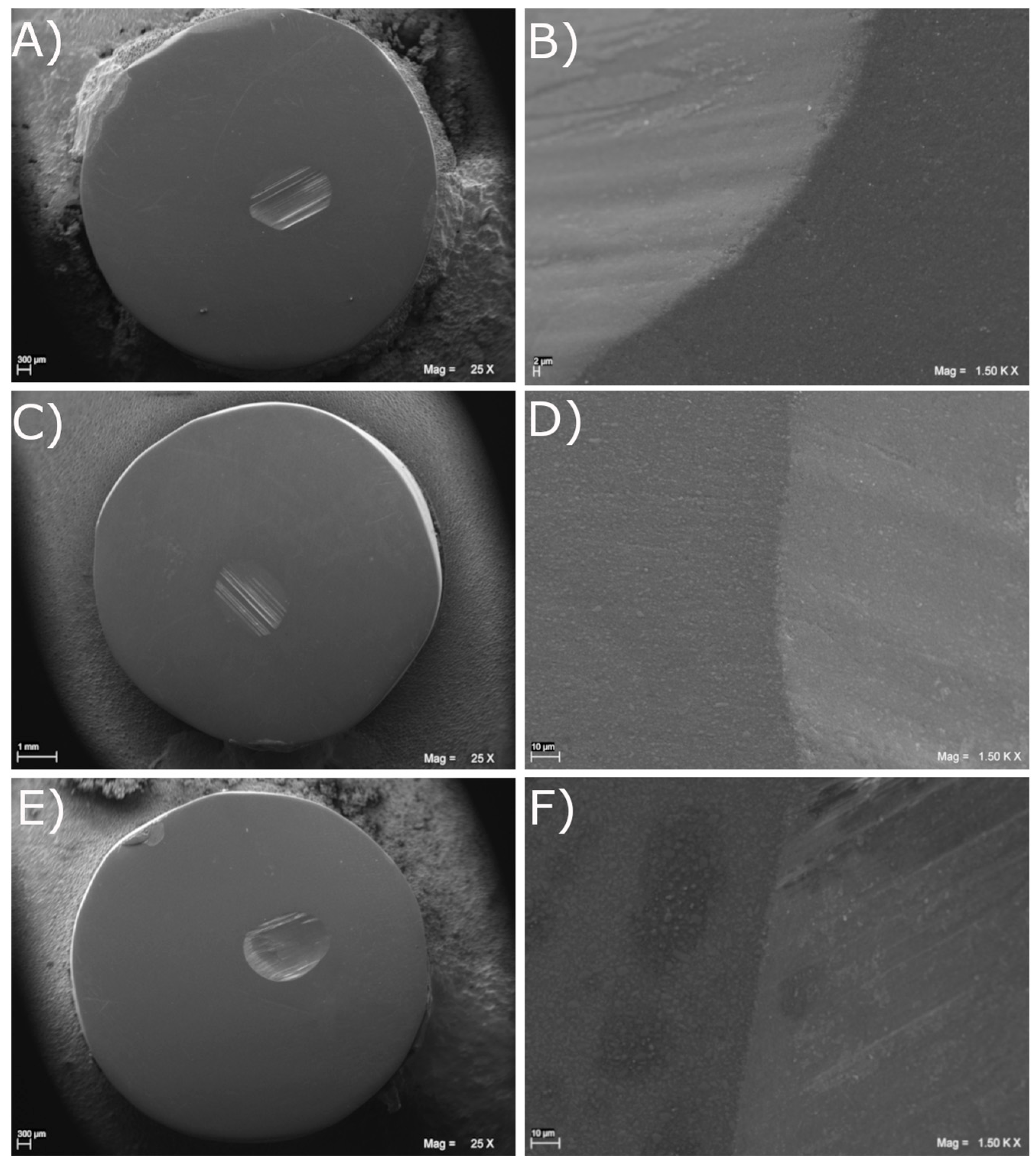
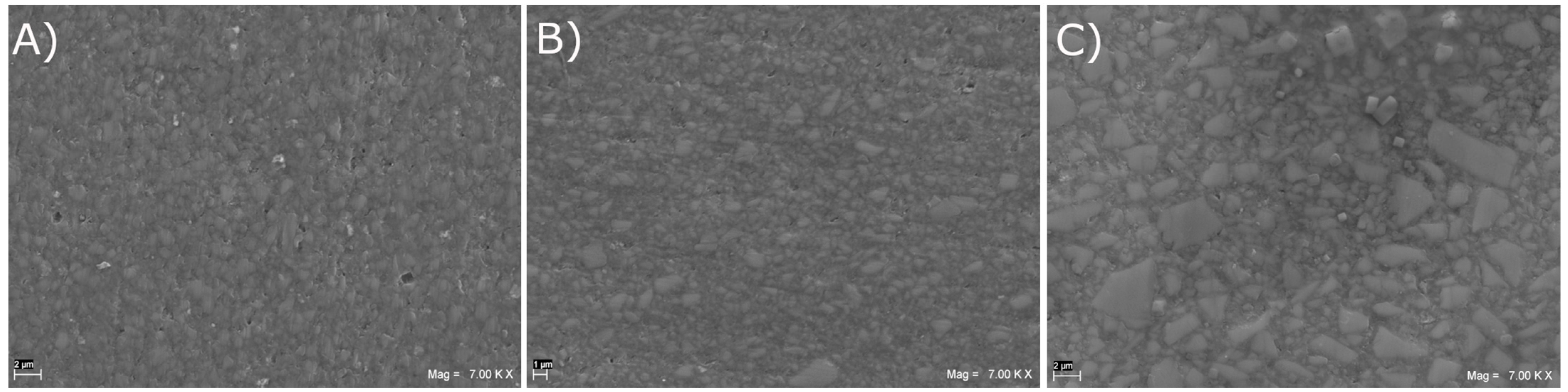
2.6. Scanning Electron Microscopy Analysis

A scanning electron microscope (SEM) (EVO 50 XVP LaB6, Carl Zeiss SMT Ltd., Cambridge, UK) evaluation was performed after the sputter-coating of cylindrical specimens (except for Aurocast 8), which were observed at 25×, 1500× and 7000× magnifications to analyze the wear facets and the material surface features. SEM conditions were set as follows: high vacuum (2 × 10⁻⁷ Torr), emission current 10 pA, accelerating voltage 10 kV and working distance around 6 mm.

3. Results

Mean values (and standard deviations [SD]) for the vertical wear and the volumetric loss obtained on each material are shown in Table 2 and Table 3. According to the one-way ANOVA tests, no significant differences (p > 0.05) were observed among the experimental groups concerning both total vertical wear and total volumetric loss.
Compared to Aurocast 8, modestly increased, but not statistically significant, differences were recorded on total vertical wear mean values for Brilliant Crios and Grandio disc. Slightly lower, but still not significant, differences were recorded for the total vertical wear of Ena Cad discs compared to all of the other tested materials. As for the total volumetric loss, slightly reduced, but not significant, differences were recorded for the total volumetric loss of the Ena Cad, Brilliant Crios and Grandio disc compared to Aurocast 8. SEM images of the wear facets observed on the cylindrical specimens are shown in Figure 2. The Ena Cad, Brilliant Crios and Grandio discs were quite similar in dimension for the worn areas, without important signs of chipping over the transition between worn and unworn surfaces. At 7000× magnification (Figure 3), Ena Cad showed filler particle sizes ranging quite uniformly between 0.2 µm and 3 µm. Brilliant Crios showed a less homogeneous filler size range, with particles ranging between 0.1 µm and 4 µm. Finally, the Grandio disc showed the highest filler size, with filler particles up to 7 µm.

4. Discussion

In full-mouth rehabilitations, the selection of an adequate restorative material is of paramount importance. The choice of restoring worn teeth with additive materials seems to be an ideal option, as this allows for the remaining tooth structure to be maximally preserved [52]. Full-mouth rehabilitations often lead to prosthetic artificial occlusion, with the upper arch opposing the same material on the lower arch [53]. In order to in vitro mimic such a clinical scenario, in this study the restorative materials were tested in a chewing simulator opposing antagonist abraders manufactured from the same corresponding materials. The tested materials were compared in terms of total vertical wear (i.e., the sum of sample vertical wear and antagonist vertical wear) and the total volumetric loss (i.e., the sum of sample volumetric loss and antagonist volumetric loss). The null hypothesis investigated had to be accepted. No statistically significant differences were recorded in terms of total wear among all restorative materials under investigation. The total wear depth and total volumetric loss mean values after 120,000 chewing simulation cycles opposing abraders manufactured from the same material had statistically similar results. On the basis of the obtained findings, all of the CAD/CAM resin-based materials exhibited adequate wear resistance, since their total wear depth and total volume loss results were statistically comparable to the type 3 gold alloy. In previous in vitro studies, Aurocast 8 showed human enamel-like wear rates [47,48]. An excessive tooth wear can compromise the occlusion and tooth–jaw relationships, and this may lead to muscle fatigue and impaired aesthetics and function [3,4,5,6,7]. In particular, parafunctional patients are those at higher risk of pathologically increased tooth wear (over 140 μm per year) [54].
Many attempts have been made to correlate the materials’ wear resistance to other material properties [55,56,57]. The gold wear behavior is provided by its metal bonds [5] and, as for other metals, seems proportional to its hardness [58]. As far as composites are concerned, hardness seems more related to the material abrasiveness [56,57], as the antagonist wear is produced by hard filler particles emerging from the worn resin matrix [56,57]. The size of fillers, the inter-particle distance, the matrix formulation and the silane agent can also affect composite wear properties [59,60,61].
The Ena Cad disc is a CAD/CAM resin-based radiopaque composite material with a high cross-linked polymeric matrix, in which a filler percentage of approximately 70% is embedded. Concerning the total vertical wear and total volumetric loss, as a result of 120,000 chewing simulation cycles against abraders manufactured from the same material, Ena Cad showed a wear resistance close to the type 3 gold alloy. Ena Cad also performed quite similarly to the other two CAD/CAM composite resins investigated herein. Ena Cad was proposed by the manufacturer to fabricate fully anatomical monoliths, inlays, crowns and superstructures for functional and aesthetic rehabilitation, also on implants. The chemical composition and the optimized high-density filler technology might explain the promising wear behavior observed herein.
Brilliant Crios is a CAD/CAM resin-based material whose fillers are embedded in a cross-linked methacrylate matrix. According to the manufacturer, the small filler particles of Brilliant Crios can be strongly organized, consequently reducing the polymer matrix exposition during the wear process [62,63]. However, in the present study, SEM images revealed that the fillers’ size (ranging between 0.1 µm and 4 µm) was slightly larger than what was claimed for Brilliant Crios by the manufacturer (below 0.1 µm). In a two-body wear in vitro study, several CAD/CAM materials (Brilliant Crios, Lava Ultimate, Vita Enamic, Vita Suprinity), have been tested against different antagonist abraders (human tooth enamel, composite resin, feldspathic porcelain). Brilliant Crios showed maximum depth and mean maximum depth values compared to the other tested CAD/CAM materials when the antagonist abrader was enamel, while against porcelain its wear depth was slightly lower than Vita Enamic and similar to Lava Ultimate [64]. In our in vitro study, the choice of using the same restorative material as an antagonist plays a crucial role, because it mimics the clinical issues that are faced in extensive full-mouth rehabilitations involving both the upper and lower jaws, or when selecting a suitable restorative material to be placed opposing already restored antagonist teeth [46].
The Grandio disc is a nano-hybrid CAD/CAM composite, consisting of 86% inorganic fillers embedded in a polymeric matrix. According to Lukomska-Szymanska [65], Grandio disc showed an adequate flexural and tensile strength. The use of different sizes of fillers enabled us to load higher amounts of strong filler particles [14]. This could also explain its positive wear resistance results. Specifically, the filler size particle reduction, as well as the high filler load, can effectively optimize the resin composite wear resistance [66,67,68,69]. The SEM analysis found, for the Grandio disc, the largest particle size among the three materials tested (up to 7 µm).
The surface roughness may determine early wear in a two-body simulation [70]. Indeed, the polishing degree can affect the specimens’ wear resistance, and a reduced wear is expected with smooth surfaces [71,72], which may ultimately extend the longevity of a restoration. Polishing after milling or after intraoral adjustments is mandatory, to guarantee adequate clinical performance [73]. An adequate polishing should keep a surface roughness (Ra) value below 0.2 μm, as it reduces bacterial adhesion [74], which is more easily achieved if the composite resin particle size decreases [75]. In this study, the roughness of each tested material surface was carefully standardized, subjecting all specimens and abraders to the well-known and clinically accepted polishing procedures under highly controlled laboratory conditions. Following the SEM analysis, Ena Cad had the smallest and the most homogeneous filler particle size range compared to the rest of the CAD/CAM dental resin composites tested, which suggests potential aesthetic advantages and better polishing.
The wear resistance was the only mechanical property analyzed in this in vitro study. Additional tests, including Vickers microhardness, a flexural strength test or compressive strength test, might provide further interesting information, which is useful to better characterize the mechanical behaviors of the examined materials. The complexity of the oral cavity concerning temperature, humidity and pH changes is hardly reproduced by an in vitro test [76,77]. The present results showed sample vertical wear mean values ranging between 0.061–0.086 mm, which is approximately twice the intraoral physiological enamel wear per year (0.035 mm) reported in previous papers [1]. However, it must be considered that in vivo wear is a complex mechanism that may strongly vary from patient to patient [78]. According to previous research [49,50,51,79,80], in the present study the Ivoclar two-body wear test method was used, and this is based on the simulation of 120,000 chewing cycles. Some authors stated that, in clinical conditions, approximately 330,000 chewing cycles are registered in one year of mastication [51]. This would mean that 120,000 cycles should correspond to 132.7 days (approximately 4 months) of clinical chewing strokes [45]. Yilmaz and Sadeler [81], on the other hand, reported that the number of in vitro mastication cycles (120,000, 240,000 or 480,000) might approximately correspond to 6 months, 1 year or 2 years in vivo, respectively. Thus, it still seems rather uncertain whether and to what extent in vitro results can perfectly match the clinical situations [49]. It must be also underlined that the specimens of the in vitro model used herein cannot rely on the periodontal ligament and alveolar bone elasticity that support the physiological masticatory loads of the human enamel in vivo [76,82]. Despite all of the above limitations, in vitro models still play a crucial role to obtain predictive pre-clinical information which may help to foresee the performance and the possible clinical use of any new material that is placed on the market, in a relatively short amount of time.

5. Conclusions

The Ena Cad disc showed a wear resistance comparable to the type 3 gold alloy and to the already marketed Brilliant Crios and Grandio disc, suggesting it as a suitable indirect restorative material, even in the case of full-mouth rehabilitations including the upper and lower arch.

Author Contributions

Conceptualization, C.D., L.V., G.D.R. and F.D.A.; methodology, M.V., E.S. and F.D.A.; software, M.V., E.S. and F.D.A.; validation, C.D. and F.D.A.; formal analysis, C.D., M.V., E.S. and F.D.A.; investigation, M.V., G.D.R. and E.S.; resources, C.D., L.V., G.D.R. and F.D.A.; data curation, L.V., E.S. and F.D.A.; writing—original draft preparation, E.S. and F.D.A.; writing—review and editing, C.D., M.V., L.V., G.D.R., E.S. and F.D.A.; visualization, M.V., E.S. and F.D.A.; supervision, C.D. and F.D.A.; project administration, C.D.; funding acquisition, C.D. All authors have read and agreed to the published version of the manuscript.

Funding

This research received no external funding.

Institutional Review Board Statement

Not applicable.

Informed Consent Statement

Not applicable.

Data Availability Statement

The data presented in this study are available on request from the corresponding author.

Conflicts of Interest

M.V., E.S. and F.D.A. declare that they have no conflicts of interest. C.D., L.V. and G.D.R. declare that they have received support from commercial sources and received honoraria for speaking at symposia by companies that sell products used in this study.

References

  1. Lambrechts, P.; Braem, M.; Vuylsteke-Wauters, M.; Vanherle, G. Quantitative in vivo wear of human enamel. J. Dent. Res. 1989, 68, 1752–1754. [Google Scholar] [CrossRef]
  2. Seghi, R.R.; Rosenstiel, S.F.; Bauer, P. Abrasion of human enamel by different dental ceramics in vitro. J. Dent. Res. 1991, 70, 221–225. [Google Scholar] [CrossRef] [PubMed]
  3. Yip, K.H.; Smales, R.J.; Kaidonis, J.A. Differential wear of teeth and restorative materials: Clinical implications. Int. J. Prosthodont. 2004, 17, 350–356. [Google Scholar]
  4. Dahl, B.L.; Oilo, G. In vivo wear ranking of some restorative materials. Quintessence Int. 1994, 25, 561–565. [Google Scholar] [PubMed]
  5. Ramp, M.H.; Suzuki, S.; Cox, C.F.; Lacefield, W.R.; Koth, D.L. Evaluation of wear: Enamel opposing three ceramic materials and a gold alloy. J. Prosthet. Dent. 1997, 77, 523–530. [Google Scholar] [CrossRef] [PubMed]
  6. Zeng, J.; Sato, Y.; Ohkubo, C.; Hosoi, T. In vitro wear resistance of three types of composite resin denture teeth. J. Prosthet. Dent. 2005, 94, 453–457. [Google Scholar] [CrossRef]
  7. Ogle, R.E.; Davis, E.L. Clinical wear study of three commercially available artificial tooth materials: Thirty-six month results. J. Prosthet. Dent. 1998, 79, 145–151. [Google Scholar] [CrossRef]
  8. De Angelis, F.; Buonvivere, M.; Sorrentino, E.; Rondoni, G.D.; D’Arcangelo, C. Wear Properties of Conventional and High-Translucent Zirconia-Based Materials. Materials 2022, 15, 7324. [Google Scholar] [CrossRef]
  9. Johnson, R.; Verrett, R.; Haney, S.; Mansueto, M.; Challa, S. Marginal Gap of Milled versus Cast Gold Restorations. J. Prosthodont. Off. J. Am. Coll. Prosthodont. 2017, 26, 56–63. [Google Scholar] [CrossRef]
  10. Christensen, G.J. The most conservative and longest lasting restorative service. Dent. Clin. N. Am. 1976, 20, 351–364. [Google Scholar] [CrossRef]
  11. Park, J.M.; Hong, Y.S.; Park, E.J.; Heo, S.J.; Oh, N. Clinical evaluations of cast gold alloy, machinable zirconia, and semiprecious alloy crowns: A multicenter study. J. Prosthet. Dent. 2016, 115, 684–691. [Google Scholar] [CrossRef]
  12. Barco, M.T., Jr.; Synnott, S.A. Precision metal occlusal surfaces for removable partial dentures. Int. J. Prosthodont. 1989, 2, 365–367. [Google Scholar] [PubMed]
  13. Fong, H.; Dickens, S.H.; Flaim, G.M. Evaluation of dental restorative composites containing polyhedral oligomeric silsesquioxane methacrylate. Dent. Mater. Off. Publ. Acad. Dent. Mater. 2005, 21, 520–529. [Google Scholar] [CrossRef] [PubMed]
  14. Mokhtar, M.M.; Farahat, D.S.; Eldars, W.; Osman, M.F. Physico-mechanical properties and bacterial adhesion of resin composite CAD/CAM blocks: An in-vitro study. J. Clin. Exp. Dent. 2022, 14, e413–e419. [Google Scholar] [CrossRef] [PubMed]
  15. D’Arcangelo, C.; Vadini, M.; Buonvivere, M.; De Angelis, F. Safe clinical technique for increasing the occlusal vertical dimension in case of erosive wear and missing teeth. Clin. Case Rep. 2021, 9, e04747. [Google Scholar] [CrossRef]
  16. Daronch, M.; Rueggeberg, F.A.; De Goes, M.F. Monomer conversion of pre-heated composite. J. Dent. Res. 2005, 84, 663–667. [Google Scholar] [CrossRef]
  17. Peutzfeldt, A. Resin composites in dentistry: The monomer systems. Eur. J. Oral Sci. 1997, 105, 97–116. [Google Scholar] [CrossRef]
  18. Amirouche-Korichi, A.; Mouzali, M.; Watts, D.C. Effects of monomer ratios and highly radiopaque fillers on degree of conversion and shrinkage-strain of dental resin composites. Dent. Mater. Off. Publ. Acad. Dent. Mater. 2009, 25, 1411–1418. [Google Scholar] [CrossRef]
  19. Vadini, M.; D’Amario, M.; De Angelis, F.; Falco, A.; D’Arcangelo, C. No-Prep Rehabilitation of Fractured Maxillary Incisors with Partial Veneers. J. Esthet. Restor. Dent. Off. Publ. Am. Acad. Esthet. Dent. 2016, 28, 351–358. [Google Scholar] [CrossRef]
  20. Grazioli, G.; Francia, A.; Cuevas-Suárez, C.E.; Zanchi, C.H.; Moraes, R.R. Simple and Low-Cost Thermal Treatments on Direct Resin Composites for Indirect Use. Braz. Dent. J. 2019, 30, 279–284. [Google Scholar] [CrossRef]
  21. Lepesqueur, M.; Surriaga, P.; Masache, M.E.; Vasquez, B.; Pena, M.; Gomes, O.M.M.; Dominguez, J.A. Effects on Micro-Hardness and Conversion Level of Two Types of Resins Undergoing Post-Polymerization Treatments/Efectos sobre microdureza y grado de conversion de dos tipos de resinas sometidas a tratamientos de pospolimerizacion/Efeitos sobre microdureza e grau de conversao de dois tipos de resinas submetidas a tratamentos de pos-polimerizacao. Rev. Nac. De. Odontol. 2015, 11, 49–57. [Google Scholar]
  22. Cruz, F.L.G.; Carvalho, R.F.; Batista, C.H.T.; Siqueira-Júnior, H.M.; Queiroz, J.R.C.; Leite, F.P.P. Efecto del tratamiento térmico y de fibras de polietileno en la resistencia a la flexión de resinas compuestas. Acta Odontol. Venez. 2014, 52, 3–4. [Google Scholar]
  23. Zamalloa-Quintana, M.; López-Gurreonero, C.; Santander-Rengifo, F.M.; Ladera-Castañeda, M.; Castro-Pérez Vargas, A.; Cornejo-Pinto, A.; Cervantes-Ganoza, L.; Cayo-Rojas, C. Effect of additional dry heat curing on microflexural strength in three types of resin composite: An in vitro study. Crystals 2022, 12, 1045. [Google Scholar] [CrossRef]
  24. Aguirre, B.C.; Chen, J.H.; Kontogiorgos, E.D.; Murchison, D.F.; Nagy, W.W. Flexural strength of denture base acrylic resins processed by conventional and CAD-CAM methods. J. Prosthet. Dent. 2020, 123, 641–646. [Google Scholar] [CrossRef]
  25. Al-Dwairi, Z.N.; Tahboub, K.Y.; Baba, N.Z.; Goodacre, C.J. A Comparison of the Flexural and Impact Strengths and Flexural Modulus of CAD/CAM and Conventional Heat-Cured Polymethyl Methacrylate (PMMA). J. Prosthodont. Off. J. Am. Coll. Prosthodont. 2020, 29, 341–349. [Google Scholar] [CrossRef] [PubMed]
  26. Al-Dwairi, Z.N.; Tahboub, K.Y.; Baba, N.Z.; Goodacre, C.J.; Özcan, M. A Comparison of the Surface Properties of CAD/CAM and Conventional Polymethylmethacrylate (PMMA). J. Prosthodont. Off. J. Am. Coll. Prosthodont. 2019, 28, 452–457. [Google Scholar] [CrossRef]
  27. Al-Qarni, F.D.; Goodacre, C.J.; Kattadiyil, M.T.; Baba, N.Z.; Paravina, R.D. Stainability of acrylic resin materials used in CAD-CAM and conventional complete dentures. J. Prosthet. Dent. 2020, 123, 880–887. [Google Scholar] [CrossRef]
  28. Gruber, S.; Kamnoedboon, P.; Özcan, M.; Srinivasan, M. CAD/CAM Complete Denture Resins: An In Vitro Evaluation of Color Stability. J. Prosthodont. Off. J. Am. Coll. Prosthodont. 2021, 30, 430–439. [Google Scholar] [CrossRef]
  29. Alp, G.; Johnston, W.M.; Yilmaz, B. Optical properties and surface roughness of prepolymerized poly(methyl methacrylate) denture base materials. J. Prosthet. Dent. 2019, 121, 347–352. [Google Scholar] [CrossRef]
  30. Murat, S.; Alp, G.; Alatalı, C.; Uzun, M. In Vitro Evaluation of Adhesion of Candida albicans on CAD/CAM PMMA-Based Polymers. J. Prosthodont. Off. J. Am. Coll. Prosthodont. 2019, 28, e873–e879. [Google Scholar] [CrossRef]
  31. Al-Fouzan, A.F.; Al-Mejrad, L.A.; Albarrag, A.M. Adherence of Candida to complete denture surfaces in vitro: A comparison of conventional and CAD/CAM complete dentures. J. Adv. Prosthodont. 2017, 9, 402–408. [Google Scholar] [CrossRef] [PubMed]
  32. Ayman, A.D. The residual monomer content and mechanical properties of CAD\CAM resins used in the fabrication of complete dentures as compared to heat cured resins. Electron. Physician 2017, 9, 4766–4772. [Google Scholar] [CrossRef]
  33. Kattadiyil, M.T.; Jekki, R.; Goodacre, C.J.; Baba, N.Z. Comparison of treatment outcomes in digital and conventional complete removable dental prosthesis fabrications in a predoctoral setting. J. Prosthet. Dent. 2015, 114, 818–825. [Google Scholar] [CrossRef]
  34. Srinivasan, M.; Schimmel, M.; Naharro, M.; Neill, C.O.; McKenna, G.; Müller, F. CAD/CAM milled removable complete dentures: Time and cost estimation study. J. Dent. 2019, 80, 75–79. [Google Scholar] [CrossRef] [PubMed]
  35. Hsu, C.Y.; Yang, T.C.; Wang, T.M.; Lin, L.D. Effects of fabrication techniques on denture base adaptation: An in vitro study. J. Prosthet. Dent. 2020, 124, 740–747. [Google Scholar] [CrossRef]
  36. McLaughlin, J.B.; Ramos, V., Jr.; Dickinson, D.P. Comparison of Fit of Dentures Fabricated by Traditional Techniques Versus CAD/CAM Technology. J. Prosthodont. Off. J. Am. Coll. Prosthodont. 2019, 28, 428–435. [Google Scholar] [CrossRef] [PubMed]
  37. Srinivasan, M.; Cantin, Y.; Mehl, A.; Gjengedal, H.; Müller, F.; Schimmel, M. CAD/CAM milled removable complete dentures: An in vitro evaluation of trueness. Clin. Oral. Investig. 2017, 21, 2007–2019. [Google Scholar] [CrossRef] [PubMed]
  38. Goodacre, B.J.; Goodacre, C.J.; Baba, N.Z.; Kattadiyil, M.T. Comparison of denture base adaptation between CAD-CAM and conventional fabrication techniques. J. Prosthet. Dent. 2016, 116, 249–256. [Google Scholar] [CrossRef]
  39. da Costa, L.P.G.; Zamalloa, S.I.D.; Alves, F.A.M.; Spigolon, R.; Mano, L.Y.; Costa, C.; Mazzo, A. 3D printers in dentistry: A review of additive manufacturing techniques and materials. Clin. Lab. Res. Dent. 2021, 1–10. [Google Scholar] [CrossRef]
  40. Abualsaud, R.; Gad, M.M. Flexural Strength of CAD/CAM Denture Base Materials: Systematic Review and Meta-analysis of In-vitro Studies. J. Int. Soc. Prev. Community Dent. 2022, 12, 160–170. [Google Scholar] [CrossRef]
  41. Nguyen, J.F.; Migonney, V.; Ruse, N.D.; Sadoun, M. Resin composite blocks via high-pressure high-temperature polymerization. Dent. Mater. Off. Publ. Acad. Dent. Mater. 2012, 28, 529–534. [Google Scholar] [CrossRef] [PubMed]
  42. Lambert, H.; Durand, J.C.; Jacquot, B.; Fages, M. Dental biomaterials for chairside CAD/CAM: State of the art. J. Adv. Prosthodont. 2017, 9, 486–495. [Google Scholar] [CrossRef]
  43. McCabe, J.F.; Ogden, A.R. The relationship between porosity, compressive fatigue limit and wear in composite resin restorative materials. Dent. Mater. Off. Publ. Acad. Dent. Mater. 1987, 3, 9–12. [Google Scholar] [CrossRef] [PubMed]
  44. Brosh, T.; Ferstand, N.; Cardash, H.; Baharav, H. Effect of polymerization under pressure on indirect tensile mechanical properties of light-polymerized composites. J. Prosthet. Dent. 2002, 88, 381–387. [Google Scholar] [CrossRef]
  45. De Angelis, F.; D’Arcangelo, C.; Malíšková, N.; Vanini, L.; Vadini, M. Wear Properties of Different Additive Restorative Materials Used for Onlay/Overlay Posterior Restorations. Oper. Dent. 2020, 45, E156–E166. [Google Scholar] [CrossRef] [PubMed]
  46. D’Arcangelo, C.; Vanini, L.; Rondoni, G.D.; Vadini, M.; De Angelis, F. Wear Evaluation of Prosthetic Materials Opposing Themselves. Oper. Dent. 2018, 43, 38–50. [Google Scholar] [CrossRef]
  47. D’Arcangelo, C.; Vanini, L.; Rondoni, G.D.; De Angelis, F. Wear properties of dental ceramics and porcelains compared with human enamel. J. Prosthet. Dent. 2016, 115, 350–355. [Google Scholar] [CrossRef]
  48. D’Arcangelo, C.; Vanini, L.; Rondoni, G.D.; Pirani, M.; Vadini, M.; Gattone, M.; De Angelis, F. Wear properties of a novel resin composite compared to human enamel and other restorative materials. Oper. Dent. 2014, 39, 612–618. [Google Scholar] [CrossRef]
  49. Heintze, S.D. How to qualify and validate wear simulation devices and methods. Dent. Mater. Off. Publ. Acad. Dent. Mater. 2006, 22, 712–734. [Google Scholar] [CrossRef]
  50. Heintze, S.D.; Faouzi, M.; Rousson, V.; Ozcan, M. Correlation of wear in vivo and six laboratory wear methods. Dent. Mater. Off. Publ. Acad. Dent. Mater. 2012, 28, 961–973. [Google Scholar] [CrossRef]
  51. Heintze, S.D.; Reichl, F.X.; Hickel, R. Wear of dental materials: Clinical significance and laboratory wear simulation methods -A review. Dent. Mater. J. 2019, 38, 343–353. [Google Scholar] [CrossRef]
  52. Grütter, L.; Vailati, F. Full-mouth adhesive rehabilitation in case of severe dental erosion, a minimally invasive approach following the 3-step technique. Eur. J. Esthet. Dent. Off. J. Eur. Acad. Esthet. Dent. 2013, 8, 358–375. [Google Scholar]
  53. Bencharit, S.; Schardt-Sacco, D.; Border, M.B.; Barbaro, C.P. Full mouth rehabilitation with implant-supported prostheses for severe periodontitis: A case report. Open Dent. J. 2010, 4, 165–171. [Google Scholar] [CrossRef] [PubMed]
  54. Ahmed, K.E.; Whitters, J.; Ju, X.; Pierce, S.G.; MacLeod, C.N.; Murray, C.A. Clinical Monitoring of Tooth Wear Progression in Patients over a Period of One Year Using CAD/CAM. Int. J. Prosthodont. 2017, 30, 153–155. [Google Scholar] [CrossRef] [PubMed]
  55. Anusavice, K.J.; Shen, C.; Rawls, H.R. Phillips’ Science of Dental Materials; Elsevier Health Sciences: Philadelphia, PA, USA, 2012. [Google Scholar]
  56. Suzuki, S.; Leinfelder, K.F. Wear of enamel cusps opposed by posterior composite resin. Quintessence Int. 1993, 24, 885–890. [Google Scholar]
  57. Shimane, T.; Endo, K.; Zheng, J.H.; Yanagi, T.; Ohno, H. Wear of opposing teeth by posterior composite resins--evaluation of newly developed wear test methods. Dent. Mater. J. 2010, 29, 713–720. [Google Scholar] [CrossRef]
  58. Fischer, T.E.; Anderson, M.P.; Jahanmir, S. Influence of fracture toughness on the wear resistance of yttria-doped zirconium oxide. J. Am. Ceram. Soc. 1989, 72, 252–257. [Google Scholar] [CrossRef]
  59. Garoushi, S.; Lassila, L.; Vallittu, P.K. Impact of Fast High-Intensity versus Conventional Light-Curing Protocol on Selected Properties of Dental Composites. Materials 2021, 14, 1381. [Google Scholar] [CrossRef]
  60. Cha, H.S.; Lee, Y.K.; Lim, B.S.; Rhee, S.H.; Yang, H.C. Evaluation of wear resistance of dental resin composites with a 3D profilometer. J. Biomed. Mater. Res. Part. B Appl. Biomater. 2004, 71, 414–420. [Google Scholar] [CrossRef]
  61. Lim, B.S.; Ferracane, J.L.; Condon, J.R.; Adey, J.D. Effect of filler fraction and filler surface treatment on wear of microfilled composites. Dent. Mater. Off. Publ. Acad. Dent. Mater. 2002, 18, 1–11. [Google Scholar] [CrossRef]
  62. Bayne, S.C.; Taylor, D.F.; Heymann, H.O. Protection hypothesis for composite wear. Dent. Mater. Off. Publ. Acad. Dent. Mater. 1992, 8, 305–309. [Google Scholar] [CrossRef] [PubMed]
  63. Sumino, N.; Tsubota, K.; Takamizawa, T.; Shiratsuchi, K.; Miyazaki, M.; Latta, M.A. Comparison of the wear and flexural characteristics of flowable resin composites for posterior lesions. Acta Odontol. Scand. 2013, 71, 820–827. [Google Scholar] [CrossRef]
  64. Zaim, B.; Serin Kalay, T.; Purcek, G. Friction and wear behavior of chairside CAD-CAM materials against different types of antagonists: An in vitro study. J. Prosthet. Dent. 2022, 128, 803–813. [Google Scholar] [CrossRef] [PubMed]
  65. Lukomska-Szymanska, M.; Radwanski, M.; Kharouf, N.; Mancino, D.; Tassery, H.; Caporossi, C.; Inchingolo, F.; de Almeida Neves, A.; Chou, Y.F.; Sauro, S. Evaluation of Physical-Chemical Properties of Contemporary CAD/CAM Materials with Chromatic Transition “Multicolor”. Materials 2023, 16, 4189. [Google Scholar] [CrossRef] [PubMed]
  66. Tanaka, K.; Someya, T.; Kawada, E.; Ohyama, T.; Yoshinari, M.; Takemoto, S.; Hattori, M. In vitro wear behavior of restorative resin composites against bovine enamel. Dent. Mater. J. 2020, 39, 915–923. [Google Scholar] [CrossRef]
  67. Elfakhri, F.; Alkahtani, R.; Li, C.; Khaliq, J. Influence of filler characteristics on the performance of dental composites: A comprehensive review. Ceram. Int. 2022, 48, 27280–27294. [Google Scholar] [CrossRef]
  68. Blackham, J.T.; Vandewalle, K.S.; Lien, W. Properties of hybrid resin composite systems containing prepolymerized filler particles. Oper. Dent. 2009, 34, 697–702. [Google Scholar] [CrossRef]
  69. Koottathape, N.; Takahashi, H.; Iwasaki, N.; Kanehira, M.; Finger, W.J. Two- and three-body wear of composite resins. Dent. Mater. Off. Publ. Acad. Dent. Mater. 2012, 28, 1261–1270. [Google Scholar] [CrossRef]
  70. Turssi, C.P.; De Moraes Purquerio, B.; Serra, M.C. Wear of dental resin composites: Insights into underlying processes and assessment methods--a review. J. Biomed. Mater. Res. Part. B Appl. Biomater. 2003, 65, 280–285. [Google Scholar] [CrossRef]
  71. Preis, V.; Behr, M.; Handel, G.; Schneider-Feyrer, S.; Hahnel, S.; Rosentritt, M. Wear performance of dental ceramics after grinding and polishing treatments. J. Mech. Behav. Biomed. Mater. 2012, 10, 13–22. [Google Scholar] [CrossRef]
  72. Heintze, S.D.; Cavalleri, A.; Forjanic, M.; Zellweger, G.; Rousson, V. Wear of ceramic and antagonist--a systematic evaluation of influencing factors in vitro. Dent. Mater. Off. Publ. Acad. Dent. Mater. 2008, 24, 433–449. [Google Scholar] [CrossRef] [PubMed]
  73. Matzinger, M.; Hahnel, S.; Preis, V.; Rosentritt, M. Polishing effects and wear performance of chairside CAD/CAM materials. Clin. Oral Investig. 2019, 23, 725–737. [Google Scholar] [CrossRef] [PubMed]
  74. Bollen, C.M.; Lambrechts, P.; Quirynen, M. Comparison of surface roughness of oral hard materials to the threshold surface roughness for bacterial plaque retention: A review of the literature. Dent. Mater. Off. Publ. Acad. Dent. Mater. 1997, 13, 258–269. [Google Scholar]
  75. Aydın, N.; Topçu, F.T.; Karaoğlanoğlu, S.; Oktay, E.A.; Erdemir, U. Effect of finishing and polishing systems on the surface roughness and color change of composite resins. J. Clin. Exp. Dent. 2021, 13, e446–e454. [Google Scholar] [CrossRef]
  76. Soriano-Valero, S.; Román-Rodriguez, J.L.; Agustín-Panadero, R.; Bellot-Arcís, C.; Fons-Font, A.; Fernández-Estevan, L. Systematic review of chewing simulators: Reality and reproducibility of in vitro studies. J. Clin. Exp. Dent. 2020, 12, e1189–e1195. [Google Scholar] [CrossRef]
  77. Condon, J.R.; Ferracane, J.L. Evaluation of composite wear with a new multi-mode oral wear simulator. Dent. Mater. Off. Publ. Acad. Dent. Mater. 1996, 12, 218–226. [Google Scholar] [CrossRef]
  78. Heintze, S.D.; Barkmeier, W.W.; Latta, M.A.; Rousson, V. Round robin test: Wear of nine dental restorative materials in six different wear simulators—Supplement to the round robin test of 2005. Dent. Mater. Off. Publ. Acad. Dent. Mater. 2011, 27, e1–e9. [Google Scholar] [CrossRef]
  79. Heintze, S.D.; Zellweger, G.; Cavalleri, A.; Ferracane, J. Influence of the antagonist material on the wear of different composites using two different wear simulation methods. Dent. Mater. Off. Publ. Acad. Dent. Mater. 2006, 22, 166–175. [Google Scholar] [CrossRef]
  80. Hu, X.; Zhang, Q.; Ning, J.; Wu, W.; Li, C. Study of Two-Body Wear Performance of Dental Materials. J. Natl. Med. Assoc. 2018, 110, 250–255. [Google Scholar] [CrossRef]
  81. Yılmaz, E.C.; Sadeler, R. Investigation of three-body wear of dental materials under different chewing cycles. Sci. Eng. Compos. Mater. 2018, 25, 781–787. [Google Scholar] [CrossRef]
  82. Aksel, H.; Askerbeyli-Örs, S.; Deniz-Sungur, D. Vertical root fracture resistance of simulated immature permanent teeth filled with MTA using different vehicles. J. Clin. Exp. Dent. 2017, 9, e178–e181. [Google Scholar] [CrossRef] [PubMed]
Figure 1. (A) Image of pre-test antagonist abrader, showing its conic shape with a 2-mm-diameter round tip. (B) Image of post-test worn antagonist abrader: the height (h) of the worn spherical cap was used within the formula for the calculation of the antagonist volumetric loss.
Applsci 15 05023 g001
Figure 2. Scanning electron microphotographs at two different magnifications of the wear facets observed on representative specimens from the CAD/CAM composite materials tested: Ena Cad [(A), 25×; (B), 1500×], Brilliant Crios [(C), 25×; (D), 1500×] and Grandio disc [(E), 25×; (F), 1500×].
Applsci 15 05023 g002
Figure 3. Scanning electron microphotographs (original magnification 7000×) showing different sizes of the filler particles embedded within the polymeric matrix of the CAD/CAM composite materials tested: Ena Cad (A), Brilliant Crios (B) and Grandio disc (C).
Applsci 15 05023 g003
Table 1. Tested materials and their technical data.
MaterialLot NumberShadeManufacturerTechnical Data
Ena Cad030724UD 3.5Micerium S.p.A (Genova, Italy)Highly cross-linked polymer blends (UDMA, BDDMA), in which inorganic silicate glass filling material are embedded to 71.56% by weight. Average particle size: 0.80 µm and a variation range of 0.20 µm to 3.0 µm. Stabilizers, light stabilizers and pigments are included
Brilliant CriosM85399A2Coltene/Whaledent AG (Altstätten, Switzerland)Resin matrix: Cross-linked methacrylates; Filler: SiO2 (size < 20 nm), Barium glass (size < 1.0 µm); Inorganic pigments
Grandio disc2336410A2VOCO GmbH (Cuxhaven, Germany)Nano-hybrid composite. Grandio disc contains 86% w/w inorganic fillers embedded within a polymer matrix
Aurocast 815L 02 55-Nobil-Metal S.p.A. (Villafranca d’Asti, Italy)Type 3 high-gold dental alloy. Composition (w/w): Au = 85.4%, Ag = 9.0%, Cu = 5.0%, Pd = 1.0%, Ir = 1.0%
Table 2. Sample vertical wear (mm), antagonist vertical wear (mm) and total vertical wear (mm) mean (and standard deviations [SD]) values are summarized in the table.
MaterialSample Vertical Wear (SD)
A
Antagonist Vertical Wear (SD)
B
Total Vertical Wear (SD)
A + B
Ena Cad 0.061 (0.044) a0.140 (0.037) a0.202 (0.045) a
Brilliant Crios0.067 (0.036) a0.212 (0.107) a0.279 (0.093) a
Grandio disc0.086 (0.050) a0.179 (0.079) a0.265 (0.097) a
Aurocast 80.076 (0.017) a0.141 (0.050) a0.217 (0.045) a
Same superscript letters denote a non-statistically significant difference (p > 0.05).
Table 3. Sample volumetric loss (mm3), antagonist volumetric loss (mm3) and total volumetric loss (mm3) mean (and standard deviations [SD]) values are summarized in the table.
MaterialSample Volumetric Loss (SD)
C
Antagonist Volumetric Loss (SD)
D
Total Volumetric Loss (SD)
C + D
Ena Cad 0.055 (0.042) b0.062 (0.029) a0.117 (0.043) a
Brilliant Crios0.049 (0.030) b0.156 (0.128) a0.206 (0.118) a
Grandio disc0.078 (0.066) b0.109 (0.092) a0.186 (0.125) a
Aurocast 80.148 (0.065) a0.066 (0.036) a0.213 (0.069) a
Same superscript letters denote a non-statistically significant difference (p > 0.05).
Disclaimer/Publisher’s Note: The statements, opinions and data contained in all publications are solely those of the individual author(s) and contributor(s) and not of MDPI and/or the editor(s). MDPI and/or the editor(s) disclaim responsibility for any injury to people or property resulting from any ideas, methods, instructions or products referred to in the content.

