
Evaluation of the Impact of Various Functional Fillers on Key Properties of Dental Composites

 , ,
, ,  ,
,  and
and 
Abstract
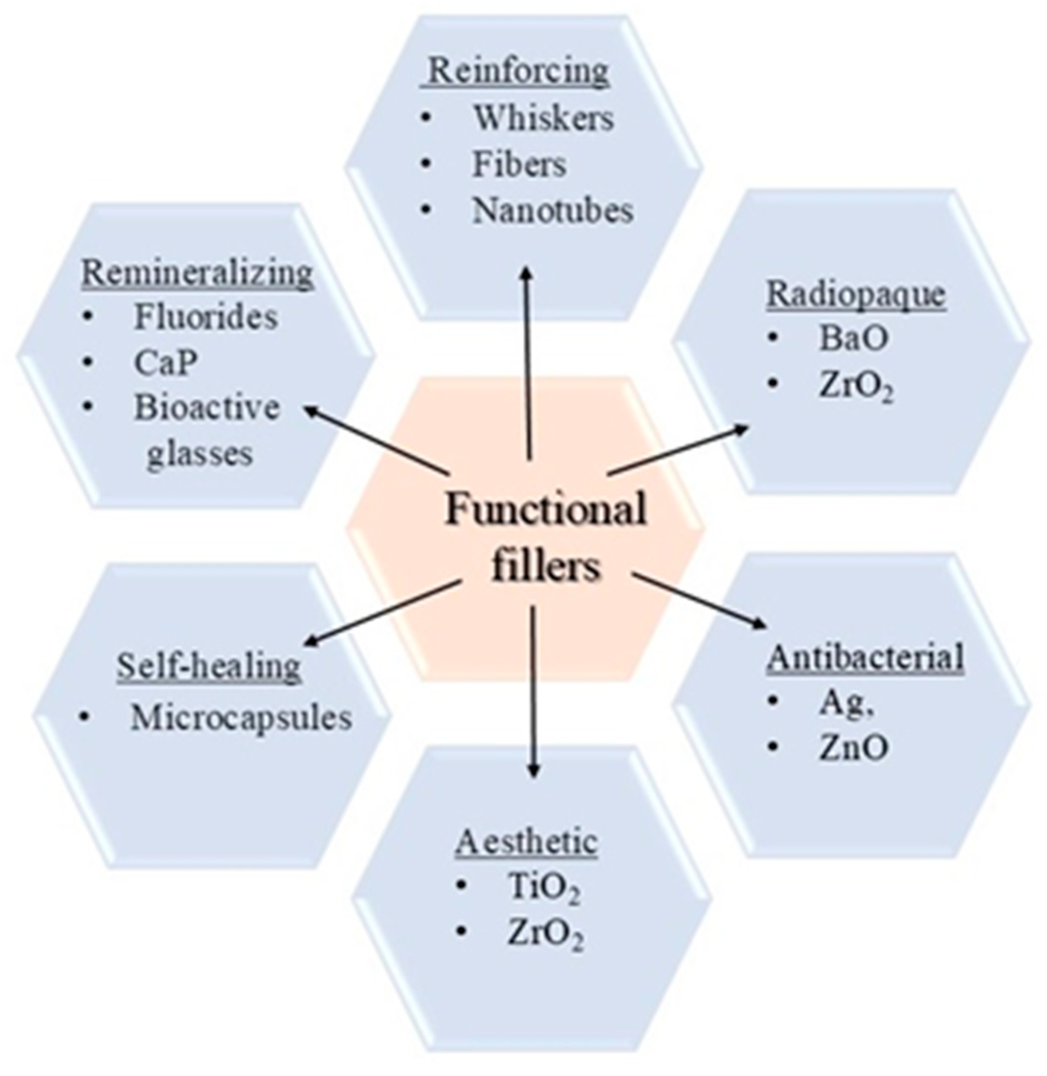
1. Introduction
2. Materials and Methods
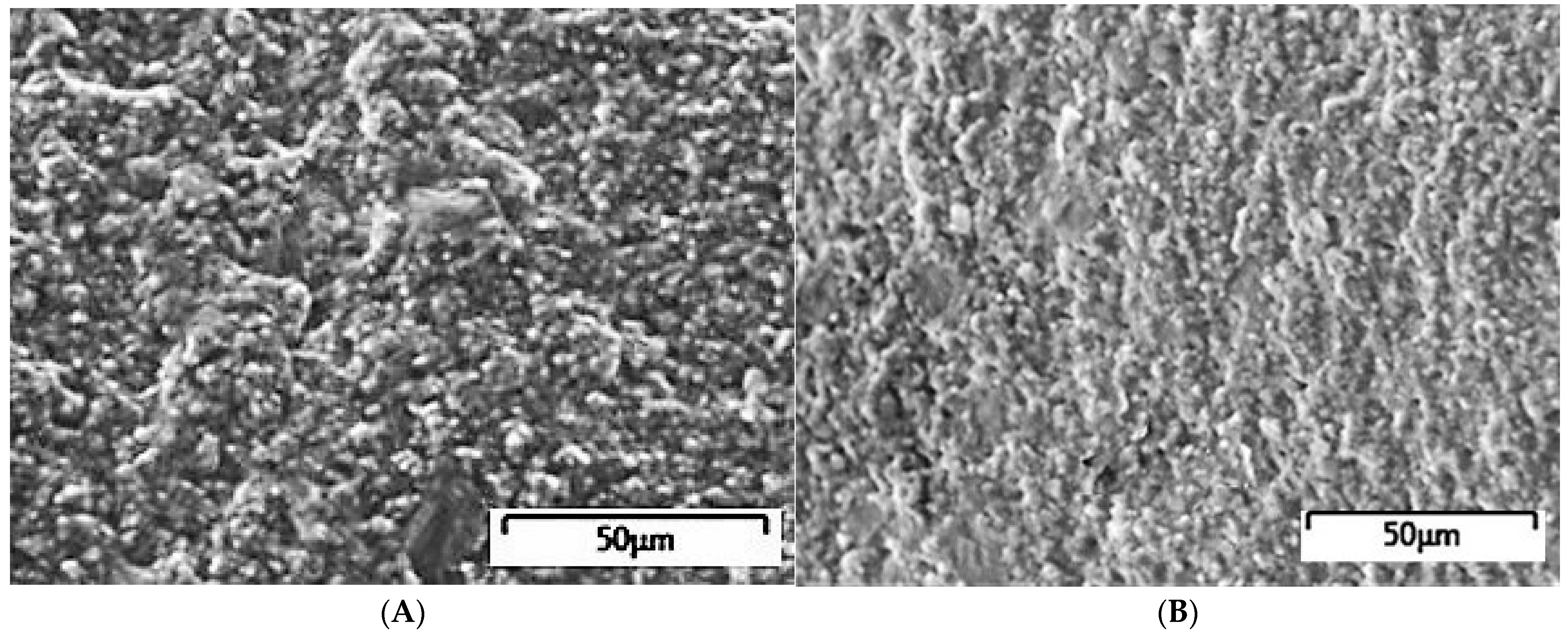
2.1. SEM Analysis
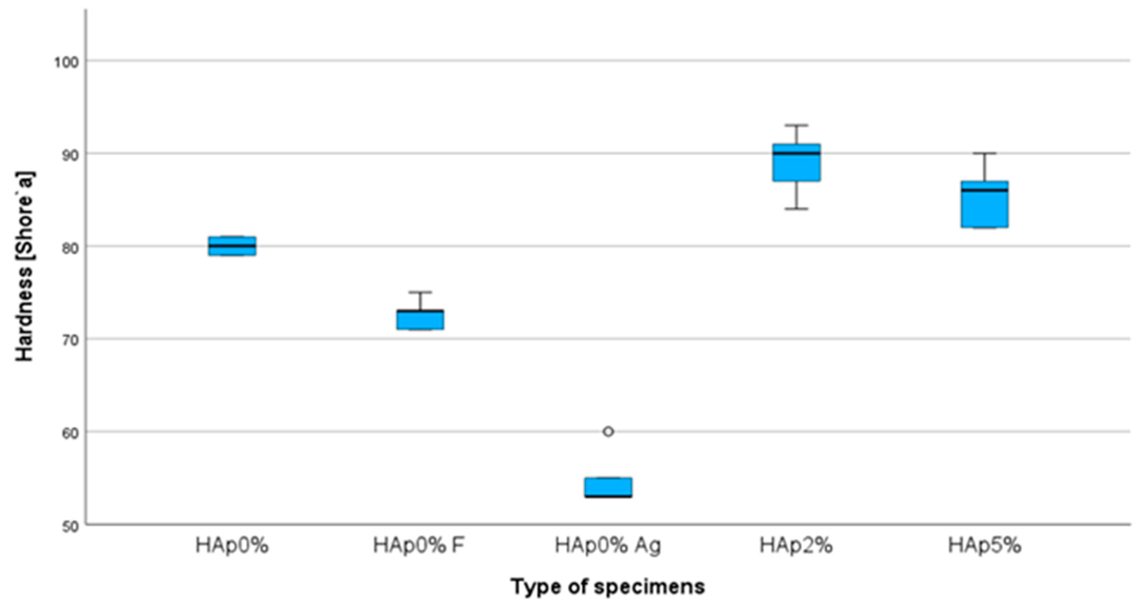
2.2. Hardness Measurements
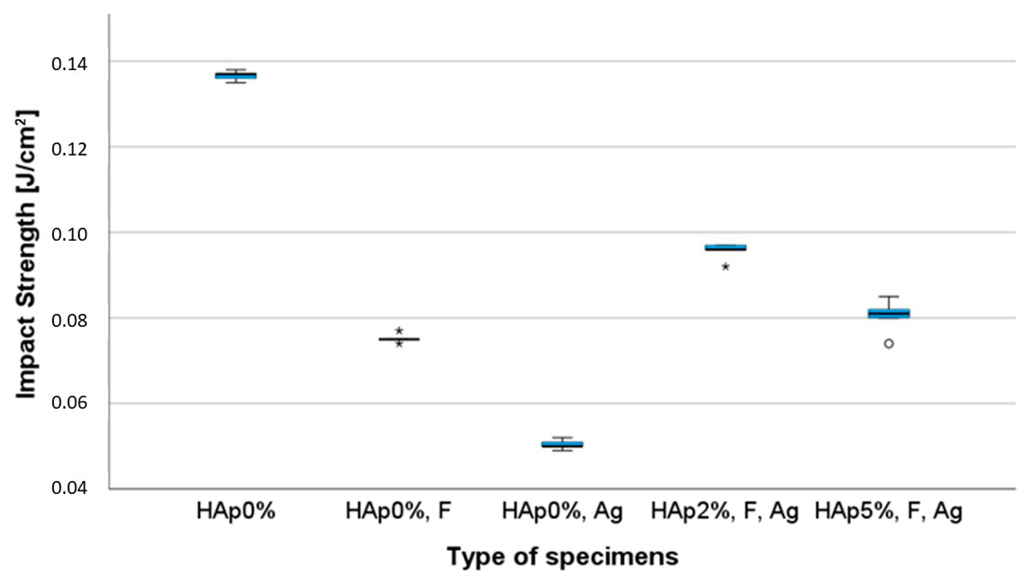
2.3. Impact Strength Test
2.4. Compressive Strength Test
2.5. Bending Strength Test
2.6. Tribological Wear Resistance Test
2.7. Statistical Analysis of Measured Data
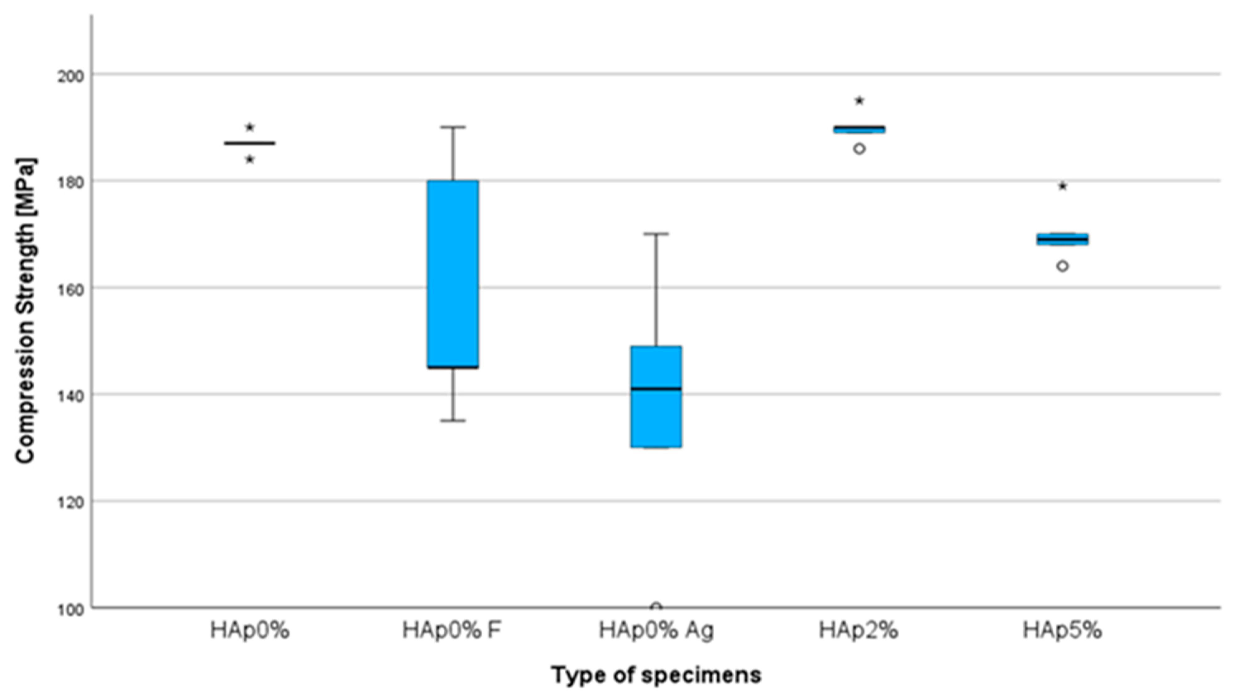
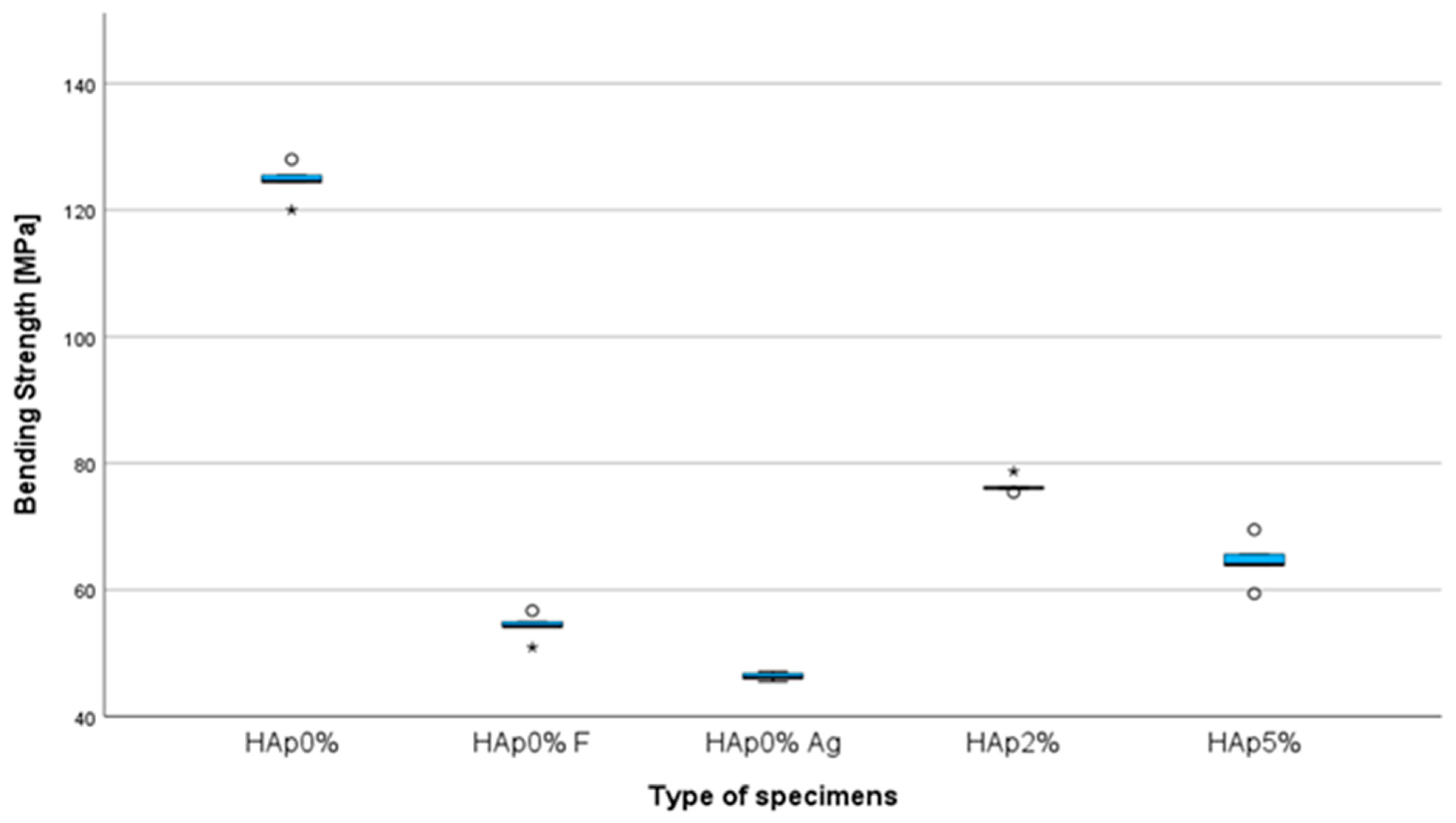
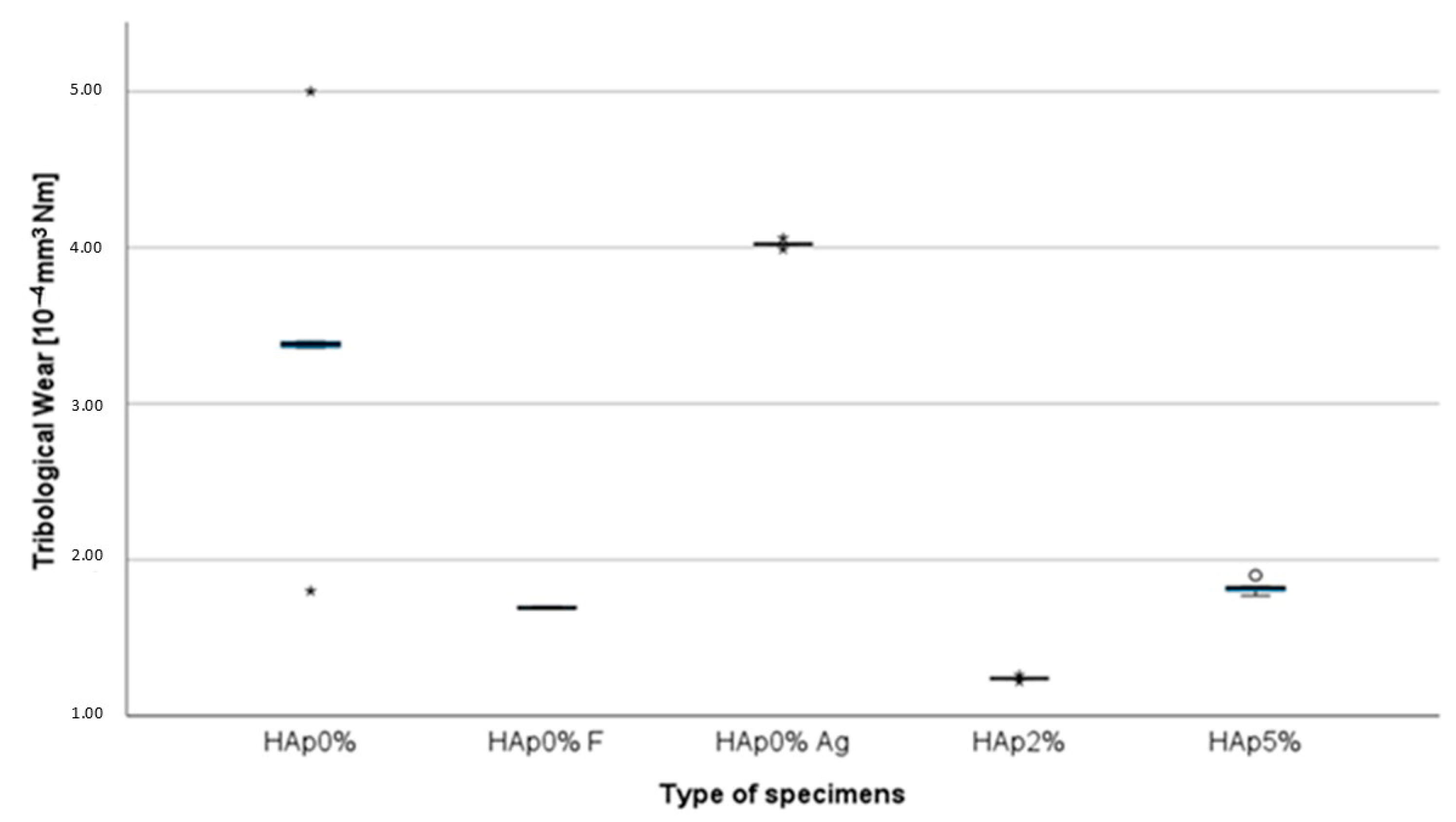
3. Results
4. Discussion
5. Conclusions
6. Patents
Author Contributions
Funding
Institutional Review Board Statement
Informed Consent Statement
Data Availability Statement
Conflicts of Interest
References
- Kreth, J.; Merritt, J.; Qi, F. Bacterial and host interactions of oral streptococci. DNA Cell Biol. 2009, 28, 397–403. [Google Scholar] [CrossRef] [PubMed]
- Zhang, J.S.; Chu, C.-H.; Yu, O.Y. Oral Microbiome and Dental Caries Development. Dent. J. 2022, 10, 184. [Google Scholar] [CrossRef] [PubMed]
- De Soet, J.J.; Van Gemert-Schriks, M.C. Host and microbiological factors related to dental caries development. Caries Res. 2008, 42, 340–347. [Google Scholar] [CrossRef] [PubMed]
- Chen, X.; Daliri, E.B.-M.; Kim, N.; Kim, J.-R.; Yoo, D.; Oh, D.-H. Microbial Etiology and Prevention of Dental Caries: Exploiting Natural Products to Inhibit Cariogenic Biofilms. Pathogens 2020, 9, 569. [Google Scholar] [CrossRef]
- Do Nascimento, C.; Pita, M.S.; de Souza Santos, E.; Monesi, N.; Pedrazzi, V.; de Albuquerque Junior, R.F.; Ribeiro, R.F. Microbiome of titanium and zirconia dental implants abutments. Dent. Mater. 2016, 32, 93–101. [Google Scholar] [CrossRef]
- Paolone, G.; Scolavino, S.; Gherlone, E.; Spagnuolo, G. Direct Esthetic Composite Restorations in Anterior Teeth: Managing Symmetry Strategies. Symmetry 2021, 13, 797. [Google Scholar] [CrossRef]
- Aminoroaya, A.; Esmaeely, N.R.; Nouri, K.S.; Panahi, P.; Das, O.; Ramakrishna, S. A Review of Dental Composites: Methods of Characterizations. ACS Biomater. Sci. Eng. 2020, 13, 3713–3744. [Google Scholar] [CrossRef]
- Yadav, R.; Lee, H.; Lee, J.H.; Singh, J.K.; Lee, H.H. A comprehensive review: Physical, mechanical, and tribological characterization of dental resin composite materials. Tribol. Int. 2023, 179, 108102. [Google Scholar] [CrossRef]
- Wang, X.; Cai, Q.; Zhang, X.; Wei, Y.; Xu, M.; Yang, X.; Ma, Q.; Cheng, Y.; Deng, X. Improved performance of Bis-GMA/TEGDMA dental composites by net-like structures formed from SiO2 nanofiber fillers. Mater. Sci. Eng. 2016, 59, 464–470. [Google Scholar] [CrossRef]
- Tian, M.; Gao, Y.; Liu, Y.; Liao, Y.; Hedin, N.E.; Fong, H. Fabrication and evaluation of Bis-GMA/TEGDMA dental resins/composites containing nano fibrillar silicate. Dent. Mater. 2008, 24, 235–243. [Google Scholar] [CrossRef]
- De Santis, R.; Gloria, A.; Maietta, S.; Martorelli, M.; De Luca, A.; Spagnuolo, G.; Riccitiello, F.; Rengo, S. Mechanical and Thermal Properties of Dental Composites Cured with CAD/CAM Assisted Solid-State Laser. Materials 2018, 11, 504. [Google Scholar] [CrossRef] [PubMed]
- Sila, P.C.; Porto-Neto, S.; Lizarelli, R.F.; Bagnato, V.S. Orthodontic brackets removal under shear and tensile bond strength resistance tests—A comparative test between light sources. Laser Phys Lett. 2007, 3, 220–226. [Google Scholar]
- Silva, E.H.; Albuquerque, R.C.; Lanza, L.D.; Vieira, G.C.; Peixoto, R.; Alvim, H.; Yoshida, M. Influence of different light sources on the conversion of composite resins. Indian J. Dent. Res. 2011, 22, 790–794. [Google Scholar] [CrossRef] [PubMed]
- García, A.H.; Lozano, M.A.M.; Vila, J.C.; Escribano, A.B.; Galve, P.F. Composite resins. A review of the materials and clinical indications. Med. Oral Patol. Oral Cir. Bucal. 2006, 11, 215–220. [Google Scholar]
- Van Landuyt, K.L.; Snauwaert, J.; De Muncka, J.; Peumansa, M.; Yoshidac, Y.; Poitevin, A.; Coutinho, E.; Suzuki, K.; Lambrechts, P.; Van Meerbeeka, B. Systematic review of the chemical composition of contemporary dental adhesives. Biomaterials 2007, 28, 3757–3785. [Google Scholar] [CrossRef] [PubMed]
- Rüttermann, S.; Dluzhevskaya, I.; Großsteinbeck, C.; Raab, W.H.M.; Janda, R. Impact of replacing Bis-GMA and TEGDMA by other commercially available monomers on the properties of resin-based composites. Dent. Mater. 2010, 26, 353–359. [Google Scholar] [CrossRef]
- Domingo, C.; Arcís, R.W.; Osorio, E.; Osorio, R.; Fanovich, M.A.; Rodríguez-Clemente, R.; Toledano, M. Hydrolytic stability of experimental hydroxyapatite-filled dental composite materials. Dent. Mater. 2003, 19, 478–486. [Google Scholar] [CrossRef]
- Mirică, I.-C.; Furtos, G.; Bâldea, B.; Lucaciu, O.; Ilea, A.; Moldovan, M.; Câmpian, R.-S. Influence of Filler Loading on the Mechanical Properties of Flowable Resin Composites. Materials 2020, 13, 1477. [Google Scholar] [CrossRef]
- Chistyakov, E.M.; Kolpinskaya, N.; Posokhova, V.; Chuev, V. Dental Composition Modified with Aryloxyphosphazene Containing Carboxyl Groups. Polymers 2020, 12, 1176. [Google Scholar] [CrossRef]
- Karabela, M.M.; Sideridou, I.D. Synthesis and study of properties of dental resin composites with different nanosilica particles size. Dent. Mater. 2011, 27, 825–835. [Google Scholar] [CrossRef]
- Azmy, E.; Al-Kholy, M.R.Z.; Fattouh, M.; Kenawi, L.M.M.; Helal, M.A. Impact of nanoparticles additions on the strength of dental composite resin. Int. J. Biomater. 2022, 2022, 1165431. [Google Scholar] [CrossRef]
- Ghada, H.; Naguib, T.B.; Jumana, M.; Alaa, T.; Abdulghani, M.; Rabab, A.; Hamed, M. Noninvasive assessment of novel nanohybrid resin cement adaptation using cross-polarization optical coherence tomography. Dent. Mater. 2024, 40, 643–652. [Google Scholar]
- Hameed, H.K.; Abdul Rahman, H. The effect of addition nano particle ZrO2 on some properties of Autoclave Processed Heat cure acrylic denture base material. J. Baghdad Coll. Dent. 2015, 27, 32–39. [Google Scholar] [CrossRef]
- Yazi, W.; Meifang, Z. Functional fillers for dental resin composites. Acta Biomater. 2022, 122, 50–65. [Google Scholar] [CrossRef]
- Mirhashemi, A.; Bahador, A.; Sodagar, A.; Pourhajibagher, M.; Amiri, A.; Gholamrezayi, E. Evaluation of antimicrobial properties of nano-silver particles used in orthodontics fixed retainer composites: An experimental in-vitro study. J. Dent. Res. Dent. Clin. Dent. Prospect. 2021, 15, 87–93. [Google Scholar] [CrossRef] [PubMed] [PubMed Central]
- Hanif, A.; Ghani, F. Mechanical properties of an experimental resin based composite containing silver nanoparticles and bioactive glass. Pak. J. Med. Sci. 2020, 36, 776–781. [Google Scholar] [CrossRef] [PubMed] [PubMed Central]
- Dan-Lei, Y.; Ya-Nan, C.; Qian, S.; Mei, L.; Hao, N.; Jie-Xin, W. Antibacterial activity and reinforcing effect of SiO2–ZnO complex cluster fillers for dental resin composites. Biomater. Sci. 2021, 9, 1795–1804. [Google Scholar] [CrossRef]
- Şomoghi, R.; Semenescu, A.; Pasăre, V.; Chivu, O.R.; Nițoi, D.F.; Marcu, D.F.; Florea, B. The Impact of ZnO Nanofillers on the Mechanical and Anti-Corrosion Performances of Epoxy Composites. Polymers 2024, 16, 2054. [Google Scholar] [CrossRef]
- Demarco, F.F.; Correa, M.B.; Cenci, M.S.; Moraes, R.R.; Opdam, N.J. Longevity of posterior composite restorations: Not only a matter of materials. Dent. Mater. 2012, 28, 87–101. [Google Scholar] [CrossRef]
- Astvaldsdottir, A.; Dagerhamn, J.; van Dijken, J.W.; Naimi-Akbar, A.; Sandborgh-Englund, G.; Tranaeus, S.; Nilsson, M. Longevity of posterior resin composite restorations in adults—A systematic review. J. Dent. 2015, 43, 934–954. [Google Scholar] [CrossRef]
- Liu, Z.; Liang, H.; Shi, T.; Xie, D.; Chen, R.; Han, X.; Shen, L.; Wang, C.; Tian, Z. Additive manufacturing of hydroxyapatite bone scaffolds via digital light processing and in vitro compatibility. Ceram. Int. 2019, 45, 11079–11086. [Google Scholar] [CrossRef]
- Sousa, A.C.; Biscaia, S.; Alvites, R.; Branquinho, M.; Lopes, B.; Sousa, P.; Valente, J.; Franco, M.; Santos, J.D.; Mendonça, C.; et al. Assessment of 3D-Printed Polycaprolactone, Hydroxyapatite Nanoparticles and Diacrylate Poly(ethylene glycol) Scaffolds for Bone Regeneration. Pharmaceutics 2022, 14, 2643. [Google Scholar] [CrossRef] [PubMed]
- Klimek, L.; Kopacz, K.; Śmielak, B.; Kula, Z. An Evaluation of the Mechanical Properties of a Hybrid Composite Containing Hydroxyapatite. Materials 2023, 16, 4548. [Google Scholar] [CrossRef] [PubMed]
- Taheri, M.M. Fluoridated hydroxyapatite nanorods as novel fillers for improving mechanical properties of dental composite: Synthesis and application. Mater. Desing 2015, 82, 119–125. [Google Scholar] [CrossRef]
- EN ISO 4049; Dentistry–Polymer-Based Restorative Materials. ISO: Geneva, Switzerland, 2009.
- ASTM G133-05; Standard Test Method for Linearly Reciprocating Ball-on-Flat Sliding Wear. ISO: Geneva, Switzerland, 2010.
- PN-EN ISO 868; Plastics and Ebonite—Determination of Indentation Hardness by Means of a Durometer (Shore Hardness). ISO: Geneva, Switzerland, 2003.
- PN-EN ISO 604; Plastics—Determination of Compressive Properties 2002. ISO: Geneva, Switzerland, 2002.
- PN-EN ISO 179-2:2020-12; Plastics—Determination of Charpy Impact Properties. ISO: Geneva, Switzerland, 2020.
- ASTM D2240; Standard Test Method for Rubber Property—Durometer Hardness. ISO: Geneva, Switzerland, 2017.
- Banaszek, K.; Klimek, L. Ti(C, N) as Barrier. Coatings 2019, 9, 432. [Google Scholar] [CrossRef]
- Dziedzic, K.; Zubrzycka-Wróbel, J. Research on tribological properties of dental composite materials. Adv. Sci. Technol. Res. J. 2016, 10, 144–149. [Google Scholar] [CrossRef]
- Kula, Z.; Klimek, L.; Dąbrowska, K.; Neves, C.B.; Roque, J.C. Selected Mechanical Properties of Dental Hybrid Composite with Fluorine, Hydroxyapatite and Silver Fillers. J. Compos. Sci. 2024, 8, 232. [Google Scholar] [CrossRef]
- Kutbay, I.; Yilmaz, B.; Evis, Z.; Usta, M. Effect of calcium fluoride on mechanical behavior and sinterability of nano-hydroxyapatite and titania composites. Ceram. Int. 2014, 40, 14817–14826. [Google Scholar] [CrossRef]
- Seyedmajidi, S.; Seyedmajidi, M. Fluorapatite: A Review of Synthesis, Properties and Medical Applications vs. Hydroxyapatite. Iran. J. Mater. Sci. Eng. 2022, 19, 1–8. [Google Scholar]
- Tredwin, C.H.J.; Young, A.M.; Neel, E.A.A. Hydroxyapatite, fluor-hydroxyapatite and fluorapatite produced via the sol–gel method: Dissolution behaviour and biological properties after crystallization. J. Mater. Sci. Mater. Med. 2014, 25, 47–53. [Google Scholar] [CrossRef]
- Durner, J.; Stojanovic, M.; Urcan, E.; Hickel, R.; Reichl, F.X. Influence of silver nano-particles on monomer elution from light-cured composites. Dent. Mater. 2011, 27, 631–636. [Google Scholar] [CrossRef]
- Gryczynski, Z.; Lukomska, J.; Lakowicz, J.R.; Matveeva, E.G.; Gryczynski, I. Depolarized light scattering from silver nanoparticles. Chem. Phys. Lett. 2006, 421, 189–192. [Google Scholar] [CrossRef] [PubMed]
- Chadda, H.; Satapathy, B.K.; Patnaik, A.; Ray, A.R. Mechanistic interpretations of fracture toughness and correlations to wear behavior of hydroxyapatite and silica/hydroxyapatite filled bis-GMA/TEGDMA micro/hybrid dental restorative composites. Compos. Part B Eng. 2017, 130, 132–146. [Google Scholar] [CrossRef]
- Kula, Z.; Klimek, L.; Kopacz, K.; Śmielak, B. Evaluation of the Effect of the Addition of Hydroxyapatite on Selected Mechanical and Tribological Properties of a Flow-Type Composite. Materials 2022, 15, 9016. [Google Scholar] [CrossRef] [PubMed]
- Sirait, M.; Sinulingga, K.; Siregar, N.; Doloksaribu, M.E. Characterization of hydroxyapatite by cytotoxicity test and bending test. J. Phys. Conf. Ser. 2022, 2193, 012039. [Google Scholar] [CrossRef]
- Annur, H.; Kaelani, Y. Pengujian Bending Biomaterial Hidroksiapatit dari Tulang Sapi sebagai Prosthesis Sendi Rahang (TMJ) pada Manusia. J. Tek. ITS 2015, 4, 37–40. [Google Scholar]
- Neves, C.B.; Costa, J.; Nepomuceno, L.; Madeira, A.; Portugal, J.; Bettencourt, A. Microhardness and Flexural Strength after Chemical Aging of chlorhexidine delivery systems based on acrylic resin. Rev. Port. Estomatol. Med. Dentária Cir. Maxilofac. 2019, 60, 104–110. [Google Scholar] [CrossRef]
- Costa, J.; Bettencourt, A.; Madeira, A.; Nepomuceno, L.; Portugal, J.; Neves, C.B. Surface Properties after Chemical Aging of chlorhexidine delivery systems based on acrylic resin. Rev. Port. Estomatologi. Med. Dentária Cir. Maxilofac. 2019, 60, 155–162. [Google Scholar] [CrossRef]
- Sajewicz, E. On evaluation of wear resistance of tooth enamel and dental materials. Wear 2006, 260, 1256. [Google Scholar] [CrossRef]

| HAp 0% | Composite Bis-GMA, Hydroxyapatite-free |
| HAp 0%, F | Composite Bis-GMA, Hydroxyapatite-free, contains fluoride |
| HAp 0%, Ag | Composite Bis-GMA, Hydroxyapatite-free, contains nanosilver |
| HAp 2%, F, Ag | Composite Bis-GMA, contains Hydroxyapatite, fluoride, and nanosilver |
| HAp 5%, F, Ag | Composite Bis-GMA, contains Hydroxyapatite, fluoride, and nanosilver |
| Sample Symbol | Composite Type | Resin Type | Filler Content HAp [wt%] | Filler Size HAp [µm] | Filler Content F [wt%] | Filler Content Ag [wt%] | Filler Size Ag [nm] |
|---|---|---|---|---|---|---|---|
| HAp 0% | light-cured | Bis-GMA | - | - | - | - | - |
| HAp 0%, F | light-cured | Bis-GMA | - | - | 0.2 | - | - |
| HAp 0%, Ag | light-cured | Bis-GMA | - | - | - | 1 | <100 |
| HAp 2%, F, Ag | light-cured | Bis-GMA | 2 | 30 | 0.2 | 1 | <100 |
| HAp 5%, F, Ag | light-cured | Bis-GMA | 5 | 30 | 0.2 | 1 | <100 |
Disclaimer/Publisher’s Note: The statements, opinions and data contained in all publications are solely those of the individual author(s) and contributor(s) and not of MDPI and/or the editor(s). MDPI and/or the editor(s) disclaim responsibility for any injury to people or property resulting from any ideas, methods, instructions or products referred to in the content. |
© 2025 by the authors. Licensee MDPI, Basel, Switzerland. This article is an open access article distributed under the terms and conditions of the Creative Commons Attribution (CC BY) license (https://creativecommons.org/licenses/by/4.0/).
Share and Cite
Kula, Z.; Neves, C.B.; Dąbrowska, K.; Roque, J.C.; Klimek, L. Evaluation of the Impact of Various Functional Fillers on Key Properties of Dental Composites. Appl. Sci. 2025, 15, 4961. https://doi.org/10.3390/app15094961
Kula Z, Neves CB, Dąbrowska K, Roque JC, Klimek L. Evaluation of the Impact of Various Functional Fillers on Key Properties of Dental Composites. Applied Sciences. 2025; 15(9):4961. https://doi.org/10.3390/app15094961
Chicago/Turabian StyleKula, Zofia, Cristina Bettencourt Neves, Katarzyna Dąbrowska, João Carlos Roque, and Leszek Klimek. 2025. "Evaluation of the Impact of Various Functional Fillers on Key Properties of Dental Composites" Applied Sciences 15, no. 9: 4961. https://doi.org/10.3390/app15094961
APA StyleKula, Z., Neves, C. B., Dąbrowska, K., Roque, J. C., & Klimek, L. (2025). Evaluation of the Impact of Various Functional Fillers on Key Properties of Dental Composites. Applied Sciences, 15(9), 4961. https://doi.org/10.3390/app15094961