Review

A Review of Transcranial Electrical and Magnetic Stimulation Usefulness in Major Depression Disorder—Lessons from Animal Models and Patient Studies

by
Florin Zamfirache
1,
Cristina Dumitru
2,
Deborah-Maria Trandafir
3,
Andrei Bratu
1 and
Beatrice Mihaela Radu
1,*
1
Department of Anatomy, Animal Physiology and Biophysics, Faculty of Biology, University of Bucharest, Splaiul Independentei 91-95, 050095 Bucharest, Romania
2
Department of Educational Sciences, Faculty of Educational Sciences, Social Sciences and Psychology, The National University of Science and Technology POLITEHNICA Bucharest, Pitești University Centre, Targul din Vale, 1, 110040 Pitesti, Romania
3
“Prof. Dr. Alexandru Obregia” Clinical Psychiatry Hospital, Șoseaua Berceni 10, 041914 Bucharest, Romania
*
Author to whom correspondence should be addressed.
Appl. Sci. 2025, 15(7), 4020; https://doi.org/10.3390/app15074020
Submission received: 9 March 2025 / Revised: 24 March 2025 / Accepted: 2 April 2025 / Published: 5 April 2025
(This article belongs to the Special Issue Biosignal and Motion Measurements)

Abstract

:
Chronic depression causes long-term structural and functional brain damage, making new effective therapies for depressed patients essential. Up to 30% of patients with depression are resistant to treatment and experience adverse effects. Alternative therapies may help achieve remission when used separately or with traditional therapies. Transcranial direct current stimulation (tDCS) and transcranial magnetic stimulation (tMS) are helpful therapeutic interventions for major depression (MMD). tDCS and tMS are noninvasive techniques that modulate the excitability of different brain regions. It has been shown to be safe and effective as monotherapy or in combination with other therapeutic interventions, such as antidepressants or psychotherapy. This review analyzes the current knowledge of using tDCS and tMS in animal models and clinical studies, both as monotherapy and/or combined with other therapeutic approaches.

1. Introduction

Major depressive disorder (MDD) is known as the most debilitating mental health issue with significant morbidity and mortality [1]. Depression is a mental disorder affecting both mental and physical health. It is a heterogeneous group of pathological conditions with different etiologies that, according to DSM-5-TR, might include symptoms such as depressed mood, loss of enjoyment, difficulty concentrating, lack of energy, sleep and appetite disturbances, feelings of worthlessness, guilty feelings, and suicidal thoughts [2]. MDD is diagnosed when an individual exhibits a minimum of five symptoms over the course of two consecutive weeks, denoting a substantial deterioration in functioning. At least one of these symptoms must be a depressed mood or an absence of interest or pleasure in activities. The prevalence of major depression worldwide has been estimated at over 300 million patients, representing 4.4% of the world’s population. Globally, as populations grow and more people reach the age when depression and anxiety are most common, the number of people affected is increasing, particularly in low-income countries [3]. According to Liu et al. [4], there was a 49.86% increase in the number of cases of depression worldwide between 1990 and 2017. Major depression is considered a syndrome that results from psychological, environmental, and genetic factors. Often, a family history of it, major changes in lifestyle, chronic conditions, various medications, or substance abuse may cause it. Despite the growing number of studies, the exact mechanisms underlying MDD remain unclear [5].
Current psychiatric guidelines recommend several antidepressants and cognitive-behavioral psychotherapy as first-line treatments for MDD. In addition, even new medications produce side effects that reduce tolerability and increase the risk for patients despite advances in psychopharmacology. Psychotherapy, in turn, is expensive, time-consuming, unsuitable for all patients, and unavailable in underdeveloped parts of the world [6]. Alternative noninvasive therapies such as electrical and magnetic brain stimulation are increasingly being researched, but their usefulness is still debated.
The present study has been designed to evaluate the therapeutic potential of two noninvasive interventions for MDD. The interventions under scrutiny are transcranial direct current stimulation (tDCS) and transcranial magnetic stimulation (tMS). A scoping review of the extant evidence from both animal models and clinical studies has been undertaken to determine the efficacy of these interventions as standalone treatments and in combination with conventional therapies such as antidepressants and psychotherapy.

1.1. Cognitive Decline in Depression

Although MDD is the most common condition and often has a high recurrence rate, currently used therapeutic approaches do not provide an effective treatment for all those who suffer from it. Depression is a syndrome because it implies a cluster of symptoms and signs that tend to occur together. The two “basic” symptoms of depression are a low mood and a loss of interest in activities. In addition, people may experience changes in appetite, sleep problems, fatigue, guilt, difficulty concentrating, thoughts of death, or impaired memory and cognitive function. MDD is associated with structural and functional brain abnormalities that contribute to its debilitating effects, including significant cognitive impairment.
MDD includes cognitive dysfunction as a diagnostic criterion, described as a reduced ability to think or concentrate. Other cognitive problems may consist of deficiencies in executive function, problems concentrating and paying attention, learning and memory, and processing speed. Objective assessments of cognitive performance suggest that patients with melancholic depression have a significantly greater degree of impairment in memory and executive function compared with patients with non-melancholic depression. Furthermore, the severity of symptoms, the cumulative duration of depressive episodes, and the presence of comorbidities were all independently and negatively correlated with cognitive function [7].
Research suggests that cognitive deficits are more pronounced in melancholic depression compared to non-melancholic subtypes. Additionally, factors such as symptom severity, cumulative duration of depressive episodes, and comorbid conditions can further exacerbate cognitive decline. The most complex cases require differential diagnosis techniques to distinguish between reversible cognitive impairment related to depression and neurodegenerative conditions such as dementia. However, despite advances in neuroscience, no definitive biomarkers exist for diagnosing MDD or guiding individualized treatment selection. In contrast, despite significant advances in neuroscience, no biomarkers are available to diagnose or identify preferred treatments for people with MDD [8].
Neuroimaging has played a crucial role in enhancing our understanding of the potential neural mechanisms involved in depression, as well as the cognitive impairments observed in depressed individuals. Various morphological, functional, and molecular approaches have been employed to explore these mechanisms. Diffusion Tensor Imaging (DTI) studies using voxel-based morphometry and diffusion tensor imaging showed changes in the anterior cingulate cortex, frontal cortex, island, thalamus, and left longitudinal fasciculus. Abnormal functional connectivity was observed in several brain regions, including the hippocampus, prefrontal white matter, right solitary bundle, corpus callosum, and inferior fronto-occipital bundle. These regions showed altered connectivity within the evidence network (mainly involving the anterior insular cortex and anterior dorsal cingulate cortex) and the cognitive control network (involving the dorsolateral prefrontal cortex and pregenual anterior cingulate cortex). Additionally, there was network hyperactivity observed in the default network (comprising the posterior cingulate, medial prefrontal cortex, precuneus, and temporo-parietal cortex) as well as in the affective network (involving the amygdala, subgenual cingulate, and pregenual cingulate), all of which are associated with cognitive deficits in MDD [8].
Cognitive dysfunction is a key pathological characteristic of MDD, but it is frequently neglected and underappreciated in the diagnosis and treatment of the condition. It significantly influences psychosocial outcomes, including an individual’s mental and social well-being and ability to function, which directly impact productivity and the chances of returning to work. As a result, it is crucial to assess both subjective and objective cognitive function measures in order to enhance functional outcomes for those with MDD. Conventional therapeutic approaches to MDD are insufficient, including antidepressant medications and psychotherapy, and remain the primary treatment options for MDD. However, these interventions are not universally effective, with up to 30% of patients experiencing treatment resistance. Pharmacological treatments primarily target mood symptom recovery, but evidence suggests that patients in remission continue to experience clinically significant cognitive deficits that impair their functional capacity. This gap between mood remission and functional recovery underscores the need for treatments that address functionally relevant areas, such as cognitive domains. It is crucial to investigate further existing clinical approaches for their independent, direct, and meaningful impacts on cognition. Currently, vortioxetine is among the approved pharmacological treatments for MDD with demonstrated direct and independent pro-cognitive benefits [9].
Barriers to effective mental health care include a lack of resources, side effects that cause many people to drop out of treatment, and the social stigmatization of mental disorders. Antidepressants and various psychotherapeutic approaches can be beneficial in the fight against major depression. Still, a significant number of people do not have access to them and struggle with this disorder. New treatment approaches, such as tDCS, could considerably impact their lives as it does not come with this kind of barrier. tDCS, together with Cognitive control therapy, has been shown to ameliorate depressive symptoms by a response rate of about 25%. It might be specifically beneficial to older patients suffering from depression and especially to those more at risk of cognitive decline [10]. tDCS and tMS are promising neuromodulatory interventions that modulate cortical excitability and functional connectivity in key brain regions implicated in MDD.

1.2. Structural Brain Abnormalities in Major Depressive Disorder

Long-term MDD has been found to induce brain changes, with reduced volume or functionality observed in regions responsible for emotional processing and cognitive functions. Neuroimaging studies have shown that individuals with MDD exhibit decreased gray matter volumes in the prefrontal cortex, amygdala, hippocampus, and thalamus. These structural changes are thought to underline key symptoms of MDD, such as negative affect and difficulties in decision-making, as these regions play a critical role in mood regulation and the processing of emotional stimuli.
In individuals with depression, functional and metabolic brain irregularities have been identified, and these changes often respond to treatment. Resting-state studies using positron emission tomography (PET) have revealed decreased cerebral blood flow and metabolism in the left dorsolateral prefrontal cortex (DLPFC), alongside increased metabolism in the right DLPFC in cases of MDD [11]. The hippocampus, a region commonly known to be involved in memory formation, is found to be affected by MDD, which can cause a reduction in total volume, which may lead to impairments in memory and cognitive functions of individuals [12].
Surface-based morphometry (SBM) highlights other structural nuances, such as cortical thickness and surface area changes. Studies have noted reduced cortical thickness in MDD, particularly in the ACC, superior temporal gyrus, and orbitofrontal cortex. Thinning in these areas may reflect neurodegenerative processes tied to depressive symptomatology, with implications for emotion regulation and social cognition [12].
There is increasing evidence of impaired neuroplasticity in MDD, such as the observation of widespread functional and structural abnormalities [13]. Brain plasticity has also been observed to be dysfunctional in terms of long-term potentiation and depression modification [9]. Compared to healthy individuals, depressed ones have lower levels of glutamate/glutamine (Glx) in the anterior cingulate compared to the same age control group [14]. Glx deficiency in patients with depression is probably an essential metabolic marker underlying hypometabolism. It reflects functional changes due to illness, most likely a reduced efficiency of the glutamatergic system and the associated occurrence of specific symptoms related to the affected brain areas. For example, psychomotor retardation and depressive mood may be symptoms associated with left DLPFC dysfunction [15,16]. Low glutamate/glutamine levels in unipolar depressive patients are successfully restored after electroconvulsive therapy [17]. Research has shown that stimulating the left dorsolateral prefrontal cortex (DLPFC) using transcranial magnetic stimulation (tMS) can trigger increased dopamine release in the striatum [18]. The study by Dubin [19] demonstrates that rhythmic stimulation of the left DLPFC at 10 Hz facilitates the release of serotonin in limbic structures.
Recent neuroimaging research has linked the left DLPFC to emotional judgment, while the right DLPFC is associated with anticipation and attention related to emotional judgment. The valence-lateralization theory, which suggests that the left prefrontal cortex is dominant for positive emotions and the right for negative emotions, is supported by fMRI studies in healthy individuals. These studies demonstrate a linear or parametric relationship between negative and positive emotional judgment and neural activity in the left and right DLPFC. Specifically, reduced activity in the left DLPFC is associated with impaired emotional judgment and abnormal modulation by positive and negative emotional valence. Conversely, increased activity in the right DLPFC appears to correlate with the attentional regulation of emotional judgment. Most tDCS studies reveal the anode targeting at left DLPFC and cathode targeting at right DLPFC, modulating the neurotransmitters released that regulate synaptic plasticity of this region frequently affected in MDD. Anodal stimulation was found to reduce Gamma-Aminobutyric Acid (GABA), and Cathodal stimulation has been shown to reduce glutamatergic and increase GABA concentrations [20].

1.3. EEG/qEEG Recordings in Depression

Various studies are investigating the use of Electroencephalography (EEG) and Quantitative Electroencephalography (qEEG) in treating depression by detecting hemispheric asymmetry in brain electrical activity in people with MDD compared to healthy controls. These may serve as potential risk indicators for depression and other emotional disorders, as well as in tracking the effectiveness of various therapeutic approaches.
Even if more and more studies are performed in this area, using qEEG as a predictor of the efficacy of antidepressant treatment remains a challenging task for clinicians. The primary benefit that qEEG can bring in the treatment of major depression is the possible diminution of the number of typical unsuccessful treatment options explored by a psychiatrist before the right antidepressant is found.
In the qEEG analysis, an increase in alpha power in the left hemisphere and a decrease in beta values in the left parietal-occipital cortex, along with hypercoherence in the right anterior region, were identified in individuals with unipolar depression.
Clinicians can use the coronary artery disease (CAD) system as a nonlinear method of depression diagnosis to detect and confirm the disorder early [21].
In addition to the traditional depression scales used to assess a patient’s level of depression subjectively, qEEG is an emerging technique that can objectively identify changes in brain abnormalities when compared to usual population standards and can be used to monitor treatment effects.
The potential biomarker capabilities of EEG used to increase the chances of antidepressant efficacy [22] have been identified as a method that can influence treatment outcomes and could significantly increase the remission rate in the treatment of depression.
qEEG is still not considered a clinically reliable predictor of a specific antidepressive treatment, as current studies still lack scientific validity and insufficiently replicate previous findings. Future studies will need to remedy the current limitations before this method can be proposed as a guideline for psychiatrists [23].

2. Methods

This study employed a scoping review methodology to evaluate the effectiveness of tDCS and tMS as alternative or adjunctive treatments for MDD, particularly in patients resistant to conventional therapies. By analyzing evidence from both clinical studies and animal models, the study seeks to assess the potential of these neuromodulation techniques in improving treatment outcomes, cognitive function, and neuroplasticity in individuals with MDD.
Research Objectives:
  • To investigate the neurobiological mechanisms through which tDCS and tMS modulate brain activity in the treatment of MDD.
  • To explore findings from animal models and their translational relevance to clinical applications for MDD treatment.
  • To provide recommendations for integrating tDCS and tMS into personalized treatment approaches for patients with treatment-resistant depression.
Our review was guided by the following research question:
RQ. How do tDCS and tMS modulate brain activity and improve treatment outcomes in patients with MDD, particularly those resistant to conventional therapies?
The scoping review methodology was chosen to provide a broad synthesis of existing research, particularly in both animal models and human clinical studies, based on the criteria outlined in the Preferred Reporting Items for Systematic Reviews and Meta-Analyses (PRISMA) guidelines for scoping reviews.
For the paper extracted for this review, we considered the following criteria: (1) Our study includes preclinical animal models and human patients diagnosed with MDD. The clinical studies evaluated include participants with treatment-resistant depression and those undergoing tDCS/tMS as monotherapy or combined with antidepressants or psychotherapy. (2) We considered peer-reviewed journal articles, clinical trials, and preclinical studies investigating the therapeutic use of tDCS and tMS in MDD. Both standalone and combination interventions were included. (3) The primary outcomes analyzed include efficacy in symptom reduction (measured through depression rating scales such as HDRS, MADRS, and BDI), cognitive function improvements, and neuroplasticity-related biomarkers (e.g., neuroimaging and neurophysiological data).
Inclusion criteria. Studies published in peer-reviewed journals that assess the effectiveness of tDCS or tMS in MDD, including clinical trials, systematic reviews, meta-analyses, and animal model research.
Exclusion criteria. Studies lacking primary data, case reports, studies on neuromodulation methods other than tDCS/tMS, and non-English publications.
Search strategy and replicability. While this study follows a scoping review approach, we adhered to a structured literature search strategy using PubMed, Scopus, and Web of Science databases. The search terms included “transcranial direct current stimulation”, “transcranial magnetic stimulation”, “Major Depressive Disorder”, and “neuromodulation”. No restrictions were placed on the publication year to capture the full scope of available literature.

3. Brain Stimulation Techniques

Noninvasive brain stimulation (tDCS) has been studied extensively in recent years, aiming to improve neural plasticity, and is, therefore, a technique used in the treatment of depression. Along with transcranial magnetic stimulation (rTMS), it is being studied and used as a treatment for major depression, especially for patients resistant to conventional treatment methods [24].
Noninvasive electrical brain stimulation therapy is increasingly considered to be safe, tolerable, and practical, either as monotherapy or in combination with other interventions, such as medication and psychotherapy. Moreover, because stimulation parameters can be targeted to specific brain areas, the noninvasive neurostimulation technology is also in the process of shifting the paradigm towards a precision-oriented framework that takes into account “knowledge of the underlying brain circuits. at the base of complex cognitive, emotional and self-reflexive functions”, to guide individualized patient-oriented treatments [6]. HD-tDC consists of four small disk electrodes arranged around a central electrode that helps to determine the direction of the unifocal modulation. Thus, greater individualization proves helpful in adapting to the possible perturbations offered by the assembly, and by using MRI techniques, there can be pre-existing mapping for the optimal placement of the electrodes.
In this review, we are particularly interested in the use of tDCS for depression. Regulatory authorities have already approved other therapies, such as rtMS treatment, for clinical use in many countries, including the United States, Israel, Canada, and Brazil [25]. Still, access to such equipment is limited, resulting in several limitations for patients who can benefit from it. On the other hand, tDCS devices are more accessible to the general public due to their low cost and portability.
In principle, tDCS is a noninvasive form of transcranial stimulation (Figure 1) that alters the excitability of cortical tissue by applying a weak direct current (0.5–2 mA) through electrodes placed on the scalp. Several tDCS protocols have been developed and tested in clinics, varying the number of sessions, the current intensity, the stimulating electrode size, or the electrodes’ localization. In the treatment of depression, the most common tDCS protocols [26,27,28] imply the positioning of the anodal electrode on F3 (left dorsolateral prefrontal cortex), while the reference electrode is placed on F4 (right dorsolateral prefrontal cortex), Fp2 (right orbitofrontal cortex), or F8 (right inferior frontal gyrus) (Figure 1).
The therapeutic application of electrical stimulation dates to the first century, when placing a live electric fish on the scalp was used to alleviate headaches through its strong electric current. Antal et al. [29] documented that applying electric currents to the scalp could alleviate symptoms of melancholy. The beneficial effects of tDCS have been reported in the treatment of psychiatric (especially depression and schizophrenia) and neurological disorders, as well as in the rehabilitation of cognitive, motor, and sensory functions after a stroke [27].
In 1804, Aldini first documented that applying electric currents to the scalp could help alleviate symptoms of melancholy [30]. Noninvasive brain stimulation (NIBS) refers to techniques that stimulate the brain without the need for any implants. One such method, tDCS, involves applying a weak electric current through electrodes placed on the scalp. This modulates the resting potential of neuronal membranes but does not directly induce neuronal firing [31].
Similarly, transcranial alternating current stimulation (tACS) uses a sinusoidal alternating current that is delivered at a specific frequency to influence the brain’s natural oscillatory patterns [32]. Another form of NIBS, transcranial random noise stimulation (tRNS), involves an alternating current with random frequencies and amplitudes [33].
tMS is another noninvasive brain stimulation technique that uses a magnetic coil to generate a local electric current by electromagnetic induction. This is applied to a focal region of the brain, resulting in depolarization or hyperpolarization, neuronal excitation, or inhibition [34]. When the motor cortex is stimulated, the electric current travels to the descending fibers of the spinal cord and causes muscle contraction. tMS depolarizes myelinated axons in the stimulated cortex, initiating a cascade of neural events propagating locally and to distant brain regions via cortico-cortical and cortico-subcortical connections. The brain’s current state influences the extent and duration of both local and remote tMS effects and involves a balance of excitatory (glutamatergic) and inhibitory (GABA-ergic) neurotransmission [35]. In clinical practice, high-frequency or low-frequency rTMS is most used, while tMS-theta-burst (TBS) treats depression [36].
Other invasive brain stimulation methods are Deep brain stimulation (DBS) and Vagus nerve stimulation (VNS). VNS involves delivering electrical signals to the vagus nerve using a device implanted under the skin, while DBS involves placing electrodes in targeted regions of the brain.
Electroconvulsive therapy (ECT) involves placing electrodes on the scalp to induce generalized convulsive activity, resulting in a seizure, and is performed under general anesthesia. While it could be considered a noninvasive form of brain stimulation, ECT is generally categorized as more invasive than methods like tDCS and TMS, as it induces a seizure and requires anesthesia, even though it does not involve surgery (see Figure 2). All these techniques fall under the broader category of transcranial electrical stimulation (TES). tDCS offers several advantages, including cost-effectiveness, portability, and a relatively low risk of side effects. In terms of efficacy, ECT remains highly effective for severe, treatment-resistant depression (TRD) and is often the “gold standard”, showing quicker and more robust effects than other neuromodulation therapies. Research results reveal more significant results in reducing depressive symptoms, as measured by tests such as the Hamilton Depression Scale (HDRS) or Beck Depression Inventory (BDI). However, effects on anxiety symptoms or suicidal ideation are more pronounced with ECT compared to tDCS and rTMS. Magnetic stimulation is effective, especially in resistant depression, but the effect is smaller compared to ECT. Despite this, it remains more tolerable for patients and is beneficial for those with mild-to-moderate MDD. Recent evidence suggests that rTMS may be well-tolerated among those who have experienced adverse reactions to medication. In patients with mild to moderate MDD, studies have shown encouraging results with tDCS, and several studies show it to be beneficial when integrated alongside medication-based treatments. However, the evidence for its benefit in treatment-resistant depression is still inconclusive. The variability in findings across different studies underscores the importance of establishing standardized protocols to ensure more reliable outcomes [37].
ECT is by far the most effective and rapid treatment for major depressive disorder (MDD). It has the best results in reducing suicidality and alleviating severe MDD, making it particularly valuable when other treatments have failed. However, rTMS is noninvasive and does not require anesthesia, making it a safer, easier-to-administer alternative with fewer cognitive side effects. It is thus widely accepted and more convenient for outpatient settings, especially for patients averse to ECT’s invasiveness. tDCS is the least invasive and least costly of the three, making it accessible in outpatient and even home-based treatments. This portability and its mild side effect profile make tDCS favorable for prolonged treatment regimens and combination therapies [37].
As a limitation, ECT is associated with cognitive side effects, particularly memory loss, which can be both short-term and longer-lasting. Stigma and the need for anesthesia further limit its acceptability despite its efficacy. rTMS requires daily sessions over several weeks to achieve a clinical response, making it logistically demanding. While it is well-tolerated, its efficacy can be limited, particularly in patients with severe depression. tDCS efficacy in TRD is not consistently established, and results are typically less robust than ECT or rTMS. Its lower intensity of stimulation means slower response rates and its clinical effectiveness is highly dependent on proper application techniques, which vary across studies [37].
Transcranial DCS (tDCS) as a practical depression intervention is mainly due to its portable design, accessibility, and easy-to-use configuration. The method has shown promise in improving neurological conditions, including depression, migraines, and motor deficits, by delivering low-level electrical currents to specific brain regions. Achieving consistent results depends on carefully calibrated factors—electrode placement, current intensity, and session duration—that researchers meticulously manage in clinical studies but can be harder to regulate outside the lab. Despite the popularity of tDCS for both therapeutic and cognitive enhancement purposes, its efficacy outside controlled environments can be inconsistent due to these varying parameters and individual differences in responsiveness to stimulation [38].
The real-world application of tDCS introduces practical challenges, notably patient adherence and the requirement for trained professionals to supervise sessions. Although some patients can independently use home-based tDCS devices, adherence to treatment schedules may decrease without the structured oversight typical of clinical settings. Electrode placement significantly impacts the results of tDCS as a method of treating depression. Inappropriate placement, even with relatively minor differences, can significantly influence the results, decreasing the effectiveness of the intervention or causing patient dissatisfaction. For individuals who do not have regular access to trained professionals, maintaining consistent, high-quality tDCS treatments can be a challenge [39].
The widespread use of tDCS is influenced by safety concerns. Although it is generally considered a safe method, especially compared to more invasive neuromodulation methods, the efficacy and safety of tDCS depend on compliance with specific parameters that may not always be controllable. Scientific research precisely controls various parameters to avoid adverse effects, but in a non-clinical setting, there is a risk of misuse. Not respecting the guidelines regarding session duration, electrode placement, or current intensity can lead to ineffective or even unsafe results, especially if the patient is to use tDCS outside of clinical supervision [38].
Despite these limitations of tDCS, there is a steady improvement in methods to make it more adaptable for unsupervised use. Devices are now easier to use, and misuse can be prevented through features such as preset stimulation parameters and fixed or guided electrode placement. This reduces risks and improves accessibility for non-professional users. However, even with these efforts, some details remain, such as individual variability in brain anatomy and response to tDCS, that complicate efforts to standardize home use. Continuous monitoring and possibly periodic adjustments based on feedback remain essential to ensure effectiveness for diverse users and applications [39].

3.1. The Effect of tDCS and tMS on Neuroplasticity

Neuroplasticity is a broad term that describes the brain’s ability to adapt and learn through cellular and molecular changes in neurons, resulting in alterations to both the activity and structure of neural networks. Synaptic plasticity involves synaptogenesis, the formation and organization of new synapses, while non-synaptic plasticity refers to processes like neuronal migration and neurogenesis. These changes can be detected through brain imaging, which reveals lasting alterations in regional neural function or neuroanatomy [40]. Luque-Casado et al. [41] reported that tDCS could also be used as a tool for neuroenhancement in terms of cognitive functions such as working memory. This is a highly relevant aspect as studies have reported that cognitive reserves play the role of an important neuroprotective factor in mood disorders such as depression [42]. Additionally, Brunoni et al. [43] found that cognitive dysfunction is a core symptom of MDD, so the use of tDCS as a neuroenhancer could potentiate the internal physiological mechanisms as a symptom-oriented therapy.
The neurophysiological effects of tDCS typically extend beyond the immediate stimulation period [31]. Long-term potentiation (LTP), which refers to the enduring enhancement of synaptic transmission and serves as the cellular basis for learning and memory, was first observed in hippocampal neurons [44]. Both cortical LTP and long-term depression (LTD) are influenced by glutamatergic and GABAergic neurons [45]. Enhanced excitability induced by anodal tDCS in the primary motor cortex resembles LTP and is dependent on the activity of N-methyl-D-aspartate (NMDA) receptors and calcium channels [46]. The strength, duration, and direction of stimulation nonlinearly affect whether excitatory or inhibitory effects are produced [46].
Neuroplasticity, often associated with rehabilitation following brain injury, is also recognized as a key mechanism in treatments such as antidepressants, psychotherapy, and attention-based interventions [47]. Neuroimaging studies have revealed changes in brain function, with tDCS being shown to modulate distinct resting-state networks as observed through functional magnetic resonance imaging (fMRI) [48]. Specifically, active anodal tDCS applied to the left dorsolateral prefrontal cortex led to significant alterations in connectivity within the default network, self-referential network, and frontal–parietal networks when compared to placebo tDCS interventions. Evidence suggests that the blockade of voltage-gated sodium and calcium channels reduces the effect of anodal tDCS on increasing excitability. In contrast, the reduction in excitability produced by cathodal tDCS is unaffected. These findings are consistent with the assumption that tDCS induces changes in the resting membrane potential of cortical neurons [49].
Among the physiological explanations describing the effects of tDCS, an interesting theory explains the impact of weak currents acting through a stochastic and rhythmic resonance that influences neural information encoding [50]. More precisely, it was suggested that the occurrence of small changes in spike predictability and timing would subtly influence cognition through neural population coding [51], which is further linked to the neuroplasticity mechanism [52].
Traditional tMS has been used primarily on the motor cortex as a target for investigating mechanisms of plasticity, such as LTP and LTD. Studies have focused on measuring MEPs before and after the tMS protocol.
tMS treatment modulates neuronal activity through the interaction of glutamate, GABA, and other neuromodulators such as acetylcholine, dopamine, or serotonin. By applying a short, rapid series of electrical pulses, theta-burst stimulation (TBS) modulates synaptic efficiency, either strengthening it (by intermittent TBS–iTBS) or weakening it by continuous TBS–cTBS.

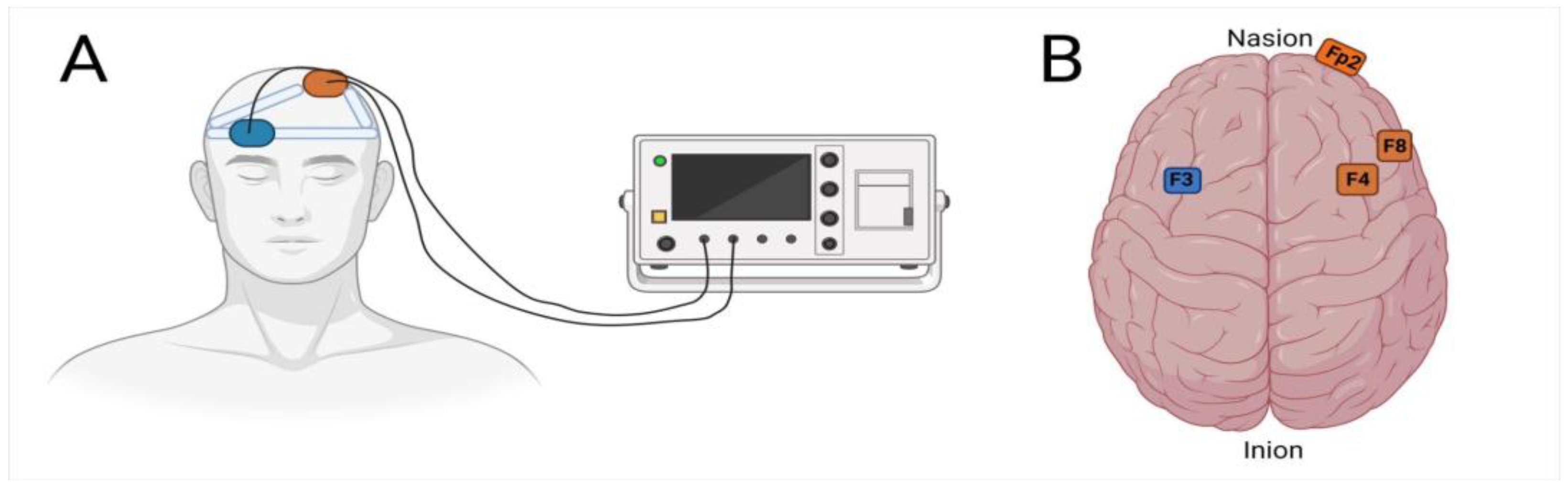
3.2. tDCS in Depression

tDCS is considered a new therapeutic approach for MDD with no significant adverse effects [49]. In MDD, the anodic electrode is usually placed over the area of the left dorsolateral prefrontal cortex, the activity of which is affected by depression. Although rTMS has the advantage of high temporal and spatial resolution, it is much more expensive and complicated to use than tDCS, which is portable and battery-powered. In addition, these two techniques have different mechanisms of action, and it is still unclear which of them would be more advantageous in terms of efficacy in the treatment of MDD [49].
In recent years, several researchers have studied the effects of tDCS on patients suffering from depressive disorders (Table 1). Specifically, the use of tDCS stimulation showed a significant improvement in symptoms in depressive patients compared to the control groups, as measured by the Hamilton Depression Rating Scale (HDRS) [53,54] or the Montgomery–Åsberg Depression Rating Scale (MADRS) [55]. On the other hand, a systematic review and meta-analysis on the use of tDCS in bipolar depression showed a reduction in depressive symptoms but also noted the risk of manic conversion, and the authors recommended future randomized control trials to investigate the effectiveness of tDCS versus placebo or mood stabilizers in bipolar depression [56].
tDCS is a potential treatment for people with major depression who cannot or do not want to take current first-line treatments. With high levels of acceptability, portability, and cost-effectiveness, tDCS is a potential first-line treatment for major depression [60].
Studies such as Woodham et al. [59] have demonstrated that tDCS can be administered at home, with supervised treatment, which in this way supports high adherence to the protocol, is a safe method without long-term adverse effects, and significantly reduces symptoms of depression. Compared to a control group, they demonstrated a higher response rate of 58.3% and remission of 44.9%, thus demonstrating the importance of the method, especially for cases resistant to standard treatment [59].
Used from the beginning of treatment, tDCS can help with quick intervention on the symptoms of depression and remission rate and help reduce the risk of the long-term impact of depression. Antidepressant medication treatment can change the course of brain deficits associated with MDD, so effective early intervention is significant for the prevention of brain deficiencies that may occur in patients with depression [61]. If psychiatrists traditionally start by prescribing a low dose for up to 8 weeks and then assess whether the treatment was effective or not and then increase the dose or change the treatment, recent evidence suggests that this long-term effect may have a significant long-term impact [62].

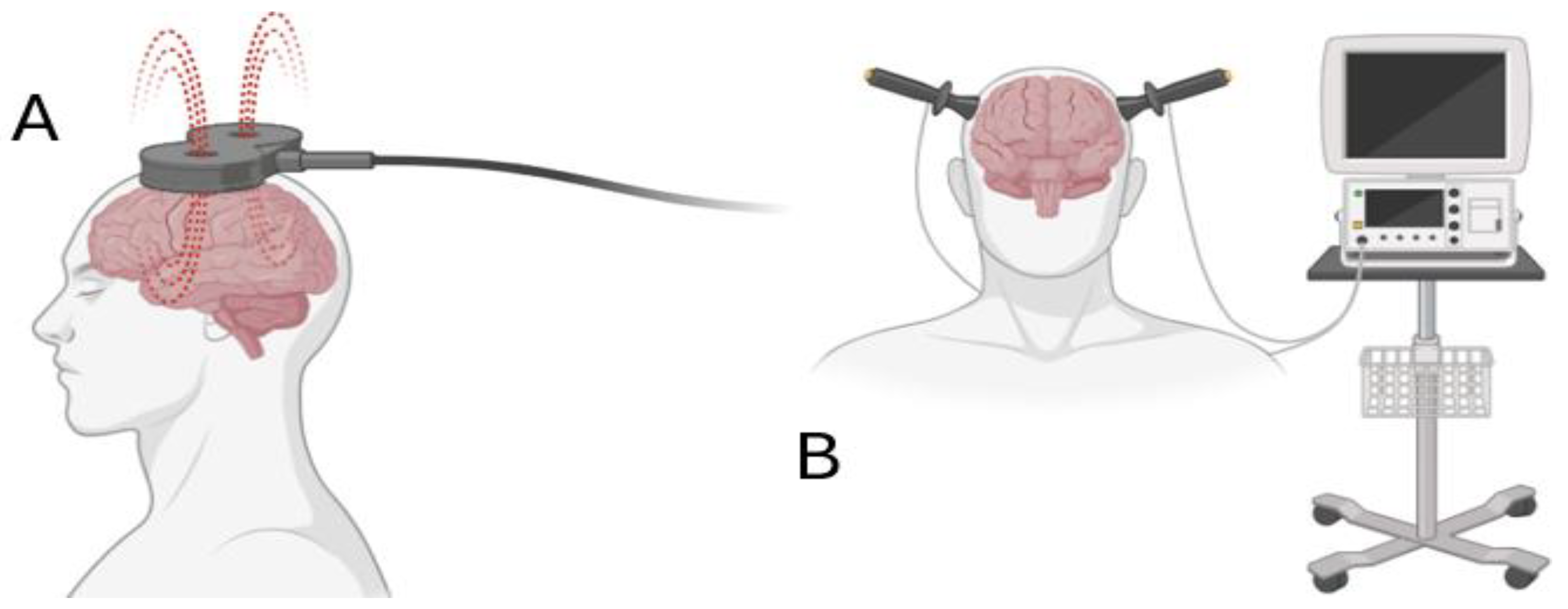
3.3. Exploring tMS as a Treatment Option for Depression

Repetitive transcranial magnetic stimulation (rTMS) has been approved for the treatment of major depression for both mild and treatment-resistant forms since 2008 in the US and since 2015 in the UK. Although its effectiveness has been demonstrated in clinical trials worldwide, the accessibility of rTMS varies significantly between countries. While this therapy is more widespread in North America, in other regions, such as Europe, its uptake is slower and uneven, influenced by factors such as resource availability and national clinical guidelines [63].
Studies have demonstrated excellent tolerability of tMS at a dosage of 6000 stimuli daily and 30,000 weekly, with a motor-evoked potential of approximately 120% without eliciting adverse events. The primary concern is the risk of epileptic seizures, which have been shown to occur more commonly within the motor cortex stimulation site [16].
In addition to pharmacotherapy, tMS treatment of depression is becoming a promising alternative for patients suffering from treatment-resistant depression. The neuroplasticity induced by tMS acts on both excitatory and inhibitory synapses. Several studies have shown an imbalance between excessive excitability and reduced cortical inhibition in patients with MDD, in addition to a reduced level of neuroplasticity in the PFC compared to healthy subjects [64].
The two most commonly used rTMS protocols in the literature are high-frequency left PFC stimulation and low-frequency right PFC stimulation. Both are effective in treating depression with comparable effects, but the low-frequency one is better tolerated and has a lower risk of seizures [65].
Although various studies and even meta-analyses reporting the clinical efficacy of rTMS in treating depression are often contradictory, rapid left frontal rTMS is generally supported as being effective, especially at higher doses and a greater number of sessions [66].

3.4. tDCS and tMS in Animal Models for Depression

The most common animal models for studying depression are based on the induction of anxiety-like and despair-like behaviors in response to stress. The Forced Swim Test, where rats are placed for a few minutes in a water-filled cylinder from which they cannot escape, tests withdrawal behavior. Induction of chronic stress through repeated exposure to a stressor for a long period induces long-lasting neurochemical, neuroimmune, and neuroendocrine changes, as well as behavioral changes manifested by increased stress and anhedonia [67].
In recent years, there has been increasing awareness and interest in tDCS and depression, thus appearing in various animal models (Table 2), specifically for rats [68] and mice [69]. These animals were experimentally subjected to classic depression paradigms such as the Open Field Test, Forced Swim Test, and Sucrose Preference Test, with tDCS (at the prefrontal, frontal, or neocortical level) and drugs as the intervention. Additionally, studies have included anxiety measures, such as the Elevated Plus Maze Test, as an additional investigation of comorbidities related to depression, reporting positive effects on both depression and anxiety symptoms [70]. These findings may be important for generalizing tDCS to human clinical settings and personal home use.
Jackson et al. [73] emphasized that animal-based tDCS research must carefully consider how electrode current density, electrode placement, and the specific animal model interact. Factors like the animal’s characteristics (e.g., weight, sex), electrode positioning, stimulation polarity, and exposure time all influence results. They also stressed that metrics like charge density alone are not sufficient—other variables, including duration of exposure and electrode size, are crucial.
Building on these findings, Chhatbar et al. [74] outlined the main parameters for defining a tDCS dose, including current (mA), duration (minutes), and electrode size (cm2), as well as derived factors like charge (C), current density (A/m2), and charge density (kC/m2). Although no strict consensus exists on what constitutes a dangerous dose capable of causing brain lesions, the researchers identified a current density threshold around 142.9 A/m2 (0.5 mA with a 3.5 mm2 electrode). Within a range of 142.9 A/m2 to 285.7 A/m2, lesion size increases in proportion to the charge density.
Studies performed on animals have shown the importance of astrocytes, indicating an astrocyte–neuron model involved in the complex neurobiological mechanisms of certain clinical diseases such as depression, Parkinson’s disease, chronic pain, stroke, and improved cognitive abilities [75]. Thus, the study realized by Monai and Hirase [76] proposes the notion that activation of astrocytic Ca2+ signaling could be a potent remedy for depression in a clinical setting.
Milighetti et al. [77] investigated the effects of tDCS on spontaneous spiking activity and concluded that at the current densities typically used in human or animal tDCS studies, the observed effects of applying electrical stimulation are most likely generated by mechanisms other than neuronal depolarization.
Long-term rTMS in rodent models has been shown to reverse anhedonic behavior and induce neurotrophic effects, including increased hippocampal cell proliferation and Brain-Derived Neurotrophic Factor (BDNF) expression [78].
Studies have shown long-lasting effects of rTMS protocols on both behavior and anhedonia, these being reversed, with effects maintained 2 weeks after the intervention. Comparing various frequencies of rTMS in an experimental model of chronic stress exposure showed that stimulation with a frequency of 25 Hz was more effective than with 5 Hz or 15 Hz. In addition to the antidepressant effect, rTMS has also shown improvements in neurogenesis, although this effect remains controversial. BDNF could play an important role as a key regulator in this case. It is positively influenced by rTMS and is known to play an important role in how antidepressants operate on neurogenesis and stress [67].
The following table (Table 3) summarizes the use of tDCSt and TMS in animal studies, including study protocols, tests conducted, and key outcomes observed.

3.5. Combining tDCS and tMS with Other Therapeutic Approaches in Depression

Research has shown that tDCS combined with medication therapy (Table 4) can improve the effectiveness of traditional treatment for depression without additional side effects. The combined approach to treating depression with selective serotonin reuptake inhibitors (SSRIs) and tDCS has shown better outcomes than when drug treatments are used alone. A study carried out on patients with moderate to severe, nonpsychotic, unipolar MDD showed that a combination of sertraline with tDCS increased treatment efficacy [25]. Brunoni et al. [25] interpreted that the lack of a statistically significant interaction between tDCS and medication treatment could be due to an additive clinical effect. Interestingly, the mechanism proposed for this combined therapeutic approach is that tDCS acts primarily through cortical activation, while SSRIs determine the downregulation of limbic hyperactivity [25]. The same group of researchers also tested the combined therapy of tDCS with another SSRI, such as escitalopram, in a single-center, double-blind trial, demonstrating the noninferiority of tDCS to escitalopram in the treatment of depression and side effects, such as skin redness, tinnitus, and nervousness [25]. In another study, researchers conducted a double-blind, randomized clinical trial and demonstrated that different drug therapies (e.g., mood stabilizers, lithium, sodium valproate, or carbamazepine) in combination with tDCS reduced depressive symptoms and improved response inhibition in patients with bipolar depression [83].
Another approach combines tDCS with neurobehavioral therapy, such as cognitive control therapy (CCT). Specifically, several studies showed a reduction in depressive symptoms when tDCS is combined with CCT [10]. Geriatric patients with depression, who are more prone to experience cognitive deficits, showed a significant improvement in cognitive tasks when exposed to tDCS and CCT [10].
Other methods, such as neurofeedback, may be used in conjunction with tDCS in the future to improve the outcome of antidepressant therapy. To date, a recent study indicated that neurofeedback interventions combined with tDCS could improve cognitive performance in healthy volunteers compared to using tDCS alone [84].
Although tMS is not yet widely used, when specialists choose it, it is because of treatment-resistant depression, a situation in which increasing the dose of antidepressants has not worked. In this situation, there is a lack of knowledge regarding optimal dosage and optimal strategy for changing the dosing of the antidepressants. Strategies used include reducing the dose of antidepressants used and introducing tMS, continuing with the same treatment and introducing tMS, or introducing an SNRI or second-generation antipsychotic in the antidepressant treatment along with tMS to accelerate treatment response and remission [85].
tMS is used concomitantly with antidepressants to potentiate changes in neuroplasticity, one of the mechanisms of action of psychiatric treatment. Together with the modulation of neurotransmitters and changes in network connectivity, both tMS and antidepressants address the main causes reported in major depression. Magnetic stimulation helps modify cortical excitability and neuronal potentiation in the long and short term. Thus, both tMS and antidepressants act on neuroplasticity and are considered to have an additive or synergistic effect [86].
Table 5 presents an overview of studies investigating the use of tMS in combination with other therapeutic approaches for the treatment of depression, including study protocols, tests conducted, and key outcomes.
Combining the two noninvasive approaches, tDCS and rTMS, which have shown great potential for treating depression, especially depression resistant to usual pharmacological treatment, results in a potential dual effect as a therapeutic advantage for rapid and effective action from their synergic action. Applied together, they have the strongest effect observed, both in the post-intervention period and in the follow-up period (scores measured with HDRS-240, without serious adverse effects being observed. The effect of tDCS is to precondition neuronal excitability by depolarizing the stimulated area, which subsequently, through the application of rTMS, facilitates deeper and lasting changes with a longer impact on brain plasticity [90].

4. Conclusions

This current review explores tDCS and tMS as effective alternatives or adjuncts to traditional therapies for MDD as a possible solution for those who are resistant to conventional treatments or cannot tolerate the side effects of sequestration. tDCS is a noninvasive, inexpensive method with no significant or short-term side effects.
tDCS treatment shows greater clinical efficacy, as measured by response rate, remission, and ongoing symptom assessment, compared with a placebo tDCS intervention. Recent studies have shown that tDCS improved depressive symptomatology and cognitive functions associated with depression, such as memory and attention.
However, despite significant advances in understanding its effects, tDCS is still not completely understood, particularly regarding its mechanisms and long-term efficacy. The lack of inferiority to antidepressant treatment has not yet been demonstrated. The mechanism proposed is neuroplasticity, with effects observed at the neuronal level. Evidence for neuroplastic effects mediating clinical outcomes in MDD is limited. Identifying predictive biomarkers is essential for understanding the pathophysiology of the disease and would help guide clinical decisions.
In the case of tMS, the efficacy is improved by lateralizing LF-TMS and HF-rTMS following the functional brain asymmetry that has been highlighted in the literature in the case of depression. However, not all patients have the same brain activity underlying depression. Individualizing tMS protocol parameters such as frequency and stimulated area may increase the antidepressant effect [91].
Future directions in studying tDCS should be focused on exploring the neurobiological mechanisms by which tDCS affects cognitive and emotional processes. This may help to identify the specific neural changes and pathways that are involved. Moreover, investigating the effects of tDCS combined with pharmacotherapy, psychotherapy, and cognitive rehabilitation could help to improve functional and cognitive outcomes in treatment-resistant patients. More studies in diverse demographic and clinical populations are essential, including studies in rural and low-resource settings where tDCS could be a valuable alternative because of its portability and affordability. These future directions could position tDCS as a more integrated component of treatment protocols for MDD, possibly offering patients a pathway to enhanced cognitive and emotional well-being.

Author Contributions

Conceptualization, F.Z. and B.M.R.; methodology, F.Z, D.-M.T. and C.D.; writing—original draft preparation, F.Z., D.-M.T., A.B. and C.D., writing—review and editing, B.M.R.; supervision, B.M.R. All authors have read and agreed to the published version of the manuscript.

Funding

No funding was received for conducting this study.

Institutional Review Board Statement

Not applicable.

Informed Consent Statement

Not applicable.

Data Availability Statement

No new data were created or analyzed in this study. Data sharing is not applicable to this article.

Conflicts of Interest

The authors declare no conflict of interest.

References

  1. Monroe, S.M.; Harkness, K.L. Major depression and its recurrences: Life course matters. Annu. Rev. Clin. Psychol. 2022, 18, 329–357. [Google Scholar] [CrossRef] [PubMed]
  2. American Psychiatric Association. Diagnostic and Statistical Manual of Mental Disorders, 5th ed.; text revision; American Psychiatric Publishing: Washington, DC, USA, 2022. [Google Scholar]
  3. World Health Organization. Depression and Other Common Mental Disorders: Global Health Estimates; WHO/MSD/MER/2017.2; World Health Organization: Geneva, Switzerland, 2017. [Google Scholar]
  4. Liu, Q.; He, H.; Yang, J.; Feng, X.; Zhao, F.; Lyu, J. Changes in the global burden of depression from 1990 to 2017: Findings from the Global Burden of Disease study. J. Psychiatr. Res. 2020, 126, 134–140. [Google Scholar] [CrossRef]
  5. Wang, J.; Luo, H.; Schülke, R.; Geng, X.; Sahakian, B.J.; Wang, S. Is transcranial direct current stimulation, alone or in combination with antidepressant medications or psychotherapies, effective in treating major depressive disorder? A systematic review and meta-analysis. BMC Med. 2021, 19, 319. [Google Scholar] [CrossRef] [PubMed]
  6. Borrione, L.; Bellini, H.; Razza, L.B.; Avila, A.G.; Baeken, C.; Brem, A.K.; Busatto, G.; Carvalho, A.F.; Chekroud, A.; Daskalakis, Z.J.; et al. Precision non-implantable neuromodulation therapies: A perspective for the depressed brain. Braz. J. Psychiatr. 2020, 42, 403–419. [Google Scholar] [CrossRef]
  7. Lam, R.W.; Kennedy, S.H.; Mclntyre, R.S.; Khullar, A. Cognitive dysfunction in major depressive disorder: Effects on psychosocial functioning and implications for treatment. Can. J. Psychiatry 2014, 59, 649–654. [Google Scholar] [CrossRef] [PubMed]
  8. Perini, G.; Cotta Ramusino, M.; Sinforiani, E.; Bernini, S.; Petrachi, R.; Costa, A. Cognitive impairment in depression: Recent advances and novel treatments. Neuropsychiatr. Dis. Treat 2019, 15, 1249–1258. [Google Scholar] [CrossRef]
  9. Zuckerman, H.; Pan, Z.; Park, C.; Brietzke, E.; Musial, N.; Shariq, A.S.; Iacobucci, M.; Yim, S.J.; Lui, L.; Rong, C.; et al. Recognition and Treatment of Cognitive Dysfunction in Major Depressive Disorder. Front. Psychiatry 2018, 9, 655. [Google Scholar] [CrossRef]
  10. Brunoni, A.R.; Boggio, P.S.; De Raedt, R.; Benseñor, I.M.; Lotufo, P.A.; Namur, V.; Valiengo, L.C.; Vanderhasselt, M.A. Cognitive control therapy and transcranial direct current stimulation for depression: A randomized, double-blinded, controlled trial. J. Affect. Disord. 2014, 162, 43–49. [Google Scholar] [CrossRef]
  11. Grimm, S.; Beck, J.; Schuepbach, D.; Hell, D.; Boesiger, P.; Bermpohl, F.; Niehaus, L.; Boeker, H.; Northoff, G. Imbalance between left and right dorsolateral prefrontal cortex in major depression is linked to negative emotional judgment: An fMRI study in severe major depressive disorder. Biolog. Psychiatry 2008, 63, 369–376. [Google Scholar] [CrossRef]
  12. Qiu, L.; Lui, S.; Kuang, W.; Huang, X.; Li, J.; Zhang, J.; Chen, H.; Sweeney, J.A.; Gong, Q. Regional increases of cortical thickness in untreated, first-episode major depressive disorder. Transl. Psychiatry. 2014, 4, e378. [Google Scholar] [CrossRef]
  13. Wise, T.; Radua, J.; Via, E.; Cardoner, N.; Abe, O.; Adams, T.M.; Amico, F.; Cheng, Y.; Cole, J.H.; de Azevedo Marques Périco, C.; et al. Common and distinct patterns of grey-matter volume alteration in major depression and bipolar disorder: Evidence from voxel-based meta-analysis. Mol. Psychiatry 2017, 22, 1455–1463. [Google Scholar] [CrossRef] [PubMed]
  14. Auer, D.P.; Pütz, B.; Kraft, E.; Lipinski, B.; Schill, J.; Holsboer, F. Reduced glutamate in the anterior cingulate cortex in depression: An in vivo proton magnetic resonance spectroscopy study. Biol. Psychiatry 2000, 47, 305–313. [Google Scholar] [CrossRef]
  15. Bench, C.J.; Frith, C.D.; Grasby, P.M.; Friston, K.J.; Paulesu, E.; Frackowiak, R.S.; Dolan, R.J. Investigations of the functional anatomy of attention using the Stroop test. Neuropsychologia 1993, 31, 907–922. [Google Scholar] [CrossRef]
  16. George, M.S.; Wassermann, E.M.; Kimbrell, T.A.; Little, J.T.; Williams, W.E.; Danielson, A.L.; Greenberg, B.D.; Hallett, M.; Post, R.M. Mood improvement following daily left prefrontal repetitive transcranial magnetic stimulation in patients with depression: A placebo-controlled crossover trial. Am. J. Psychiatry 1997, 154, 1752–1756. [Google Scholar] [CrossRef] [PubMed]
  17. Pfleiderer, B.; Michael, N.; Erfurth, A.; Ohrmann, P.; Homann, U.; Wolgast, M.; Fiebich, M.; Arolt, V.; Heindel, W. Effective electroconvulsive therapy reverses glutamate/glutamine deficit in the left anterior cingulum of unipolar depressed patients. Psychiatr. Res. Neuroimaging 2003, 122, 185–192. [Google Scholar] [CrossRef] [PubMed]
  18. Strafella, A.P.; Paus, T.; Barrett, J.; Dagher, A. Repetitive transcranial magnetic stimulation of the human prefrontal cortex induces dopamine release in the caudate nucleus. J. Neurosci. 2001, 21, 157. [Google Scholar] [CrossRef]
  19. Dubin, M. Imaging TMS: Antidepressant mechanisms and treatment optimization. Int. Rev. Psychiatry 2017, 29, 89–97. [Google Scholar] [CrossRef]
  20. Li, Q.; Fu, Y.; Liu, C.; Meng, Z. Transcranial direct current stimulation of the dorsolateral prefrontal cortex for treatment of neuropsychiatric disorders. Front. Behav. Neurosci. 2022, 16, 893955. [Google Scholar] [CrossRef]
  21. Gupta, A.; Burgess, R.; Drozd, M.; Gierula, J.; Witte, K.K.; Straw, S. The Surprise Question and clinician-predicted prognosis: Systematic review and meta-analysis. BMJ Support. Palliat. Care. 2024, 15, 12–35. [Google Scholar] [CrossRef]
  22. Iosifescu, D.V. Electroencephalography-derived biomarkers of antidepressant response. Harv. Rev. Psychiatry 2011, 19, 144–154. [Google Scholar] [CrossRef]
  23. Widge, A.S.; Bilge, M.T.; Montana, R.; Chang, W.; Rodriguez, C.I.; Deckersbach, T.; Carpenter, L.L.; Kalin, N.H.; Nemeroff, C.B. Electroencephalographic Biomarkers for Treatment Response Prediction in Major Depressive Illness: A Meta-Analysis. Am. J. Psychiatry 2019, 176, 44–56. [Google Scholar] [CrossRef] [PubMed]
  24. Breda, V.; Freire, R. Repetitive Transcranial Magnetic Stimulation (rTMS) in Major Depression. Adv. Exp. Med. Biol. 2024, 1456, 145–159. [Google Scholar] [CrossRef] [PubMed]
  25. Brunoni, A.R.; Valiengo, L.; Baccaro, A.; Zanão, T.A.; de Oliveira, J.F.; Goulart, A.; Boggio, P.S.; Lotufo, P.A.; Benseñor, I.M.; Fregni, F. The sertraline vs. electrical current therapy for treating depression clinical study: Results from a factorial, randomized, controlled trial. JAMA Psychiatry 2013, 70, 383–391. [Google Scholar] [CrossRef] [PubMed]
  26. Bai, S.; Dokos, S.; Ho, K.A.; Loo, C. A computational modeling study of transcranial direct current stimulation montages used in depression. Neuroimage 2014, 87, 332–344. [Google Scholar] [CrossRef]
  27. Bennabi, D.; Haffen, E. Transcranial Direct Current Stimulation (tDCS): A Promising Treatment for Major Depressive Disorder? Brain Sci. 2018, 8, 81. [Google Scholar] [CrossRef]
  28. Herrera-Melendez, A.L.; Bajbouj, M.; Aust, S. Application of Transcranial Direct Current Stimulation in Psychiatry. Neuropsychobiology 2020, 79, 372–383. [Google Scholar] [CrossRef]
  29. Antal, A.; Alekseichuk, I.; Bikson, M.; Brockmöller, J.; Brunoni, A.R.; Chen, R.; Cohen, L.G.; Dowthwaite, G.; Ellrich, J.; Flöel, A.; et al. Low intensity transcranial electric stimulation: Safety, ethical, legal regulatory and application guidelines. Clin. Neurophysiol. 2017, 128, 1774–1809. [Google Scholar] [CrossRef]
  30. Priori, A. Brain polarization in humans: A reappraisal of an old tool for prolonged non-invasive modulation of brain excitability. Clin. Neurophysiol. 2003, 114, 589–595. [Google Scholar] [CrossRef]
  31. Nitsche, M.A.; Paulus, W. Excitability changes induced in the human motor cortex by weak transcranial direct current stimulation. J. Physiol. 2000, 15, 527. [Google Scholar] [CrossRef]
  32. Matsumoto, H.; Ugawa, Y. Adverse events of tDCS and tACS: A review. Clin. Neurophysiol. Pract. 2016, 2, 19–25. [Google Scholar] [CrossRef]
  33. Terney, D.; Chaieb, L.; Moliadze, V.; Antal, A.; Paulus, W. Increasing human brain excitability by transcranial high-frequency random noise stimulation. J. Neurosci 2008, 28, 14147–14155. [Google Scholar] [CrossRef] [PubMed]
  34. Hallett, M. Transcranial magnetic stimulation and the human brain. Nature 2000, 406, 147–150. [Google Scholar] [CrossRef] [PubMed]
  35. Siebner, H.R.; Funke, K.; Aberra, A.S.; Antal, A.; Bestmann, S.; Chen, R.; Classen, J.; Davare, M.; Di Lazzaro, V.; Fox, P.T.; et al. Transcranial magnetic stimulation of the brain: What is stimulated?—A consensus and critical position paper. Clin. Neurophysiol. 2022, 140, 59–97. [Google Scholar] [CrossRef] [PubMed]
  36. Mutz, J.; Vipulananthan, V.; Carter, B.; Hurlemann, R.; Fu, C.H.Y.; Young, A.H. Comparative efficacy and acceptability of non-surgical brain stimulation for the acute treatment of major depressive episodes in adults: Systematic review and network meta-analysis. BMJ 2019, 364, l1079. [Google Scholar] [CrossRef]
  37. Goldberg, J.F. Electroconvulsive therapy: Still the gold standard for highly treatment-resistant mood disorders. CNS Spectrums. 2022, 27, 525–526. [Google Scholar] [CrossRef]
  38. Qi, S.; Cao, L.; Wang, Q.; Sheng, Y.; Yu, J.; Liang, Z. The Physiological Mechanisms of Transcranial Direct Current Stimulation to Enhance Motor Performance: A Narrative Review. Biology 2024, 13, 790. [Google Scholar] [CrossRef]
  39. Ornello, R.; Caponnetto, V.; Ratti, S.; D’Aurizio, G.; Rosignoli, C.; Pistoia, F.; Ferrara, M.; Sacco, S.; D’Atri, A. Which is the best transcranial direct current stimulation protocol for migraine prevention? A systematic review and critical appraisal of randomized controlled trials. J. Headache Pain. 2021, 22, 144. [Google Scholar] [CrossRef]
  40. Zatorre, R.J.; Fields, R.D.; Johansen-Berg, H. Plasticity in gray and white: Neuroimaging changes in brain structure during learning. Nat. Neurosci. 2012, 15, 528–536. [Google Scholar] [CrossRef]
  41. Luque-Casado, A.; Perakakis, P.; Hillman, C.H.; Kao, S.C.; Llorens, F.; Guerra, P.; Sanabria, D. Differences in Sustained Attention Capacity as a Function of Aerobic Fitness. Med. Sci. Sports Exerc. 2016, 48, 887–895. [Google Scholar] [CrossRef]
  42. Ponsoni, A.; Damiani Branco, L.; Cotrena, C.; Milman Shansis, F.; Fonseca, R.P. The effects of cognitive reserve and depressive symptoms on cognitive performance in major depression and bipolar disorder. J. Affect. Disord. 2020, 274, 813–818. [Google Scholar] [CrossRef]
  43. Brunoni, A.R.; Moffa, A.H.; Sampaio-Junior, B.; Borrione, L.; Moreno, M.L.; Fernandes, R.A.; Veronezi, B.P.; Nogueira, B.S.; Aparicio, L.V.M.; Razza, L.B.; et al. Trial of Electrical Direct-Current Therapy versus Escitalopram for Depression. N. Engl. J. Med. 2017, 376, 2523–2533. [Google Scholar] [CrossRef]
  44. Bliss, T.V.; Lomo, T. Long-lasting potentiation of synaptic transmission in the dentate area of the anaesthetized rabbit following stimulation of the perforant path. J. Physiol. 1973, 232, 331–356. [Google Scholar] [CrossRef] [PubMed]
  45. Froc, D.J.; Chapman, C.A.; Trepel, C.; Racine, R.J. Long-term depression and depotentiation in the sensorimotor cortex of the freely moving rat. J. Neurosci. 2000, 20, 438–445. [Google Scholar] [CrossRef] [PubMed]
  46. Monte-Silva, K.; Kuo, M.F.; Hessenthaler, S.; Fresnoza, S.; Liebetanz, D.; Paulus, W.; Nitsche, M.A. Induction of late LTP-like plasticity in the human motor cortex by repeated non-invasive brain stimulation. Brain Stimul. 2013, 6, 424–432. [Google Scholar] [CrossRef] [PubMed]
  47. Davidson, R.J.; McEwen, B.S. Social influences on neuroplasticity: Stress and interventions to promote well-being. Nat. Neurosci. 2012, 15, 689–695. [Google Scholar] [CrossRef]
  48. Keeser, D.; Meindl, T.; Bor, J.; Palm, U.; Pogarell, O.; Mulert, C.; Brunelin, J.; Möller, H.J.; Reiser, M.; Padberg, F. Prefrontal transcranial direct current stimulation changes connectivity of resting-state networks during fMRI. J. Neurosci. 2011, 31, 15284–15293. [Google Scholar] [CrossRef]
  49. Brunoni, A.R.; Nitsche, M.A.; Bolognini, N.; Bikson, M.; Wagner, T.; Merabet, L.; Edwards, D.J.; Valero-Cabre, A.; Rotenberg, A.; Pascual-Leone, A.; et al. Clinical research with transcranial direct current stimulation (tDCS): Challenges and future directions. Brain Stimul. 2012, 5, 175–195. [Google Scholar] [CrossRef]
  50. Liu, A.; Vöröslakos, M.; Kronberg, G.; Henin, S.; Krause, M.R.; Huang, Y.; Opitz, A.; Mehta, A.; Pack, C.C.; Krekelberg, B.; et al. Immediate neurophysiological effects of transcranial electrical stimulation. Nat. Commun. 2018, 9, 5092. [Google Scholar] [CrossRef]
  51. McDonnell, M.D.; Abbott, D. What is stochastic resonance? Definitions, misconceptions, debates, and its relevance to biology. PLoS Comput. Biol. 2009, 5, e1000348. [Google Scholar] [CrossRef]
  52. Kronberg, G.; Bridi, M.; Abel, T.; Bikson, M.; Parra, L.C. Direct Current Stimulation Modulates LTP and LTD: Activity Dependence and Dendritic Effects. Brain Stimul. 2017, 10, 51–58. [Google Scholar] [CrossRef]
  53. Boggio, P.S.; Rigonatti, S.P.; Ribeiro, R.B.; Myczkowski, M.L.; Nitsche, M.A.; Pascual-Leone, A.; Fregni, F. A randomized, double-blind clinical trial on the efficacy of cortical direct current stimulation for the treatment of major depression. Int. J. Neuropsychopharmacol. 2008, 11, 249–254. [Google Scholar] [CrossRef] [PubMed]
  54. Fregni, F.; Boggio, P.S.; Nitsche, M.A.; Marcolin, M.A.; Rigonatti, S.P.; Pascual-Leone, A. Treatment of major depression with transcranial direct current stimulation. Bipolar Disord. 2006, 8, 203–204. [Google Scholar] [CrossRef] [PubMed]
  55. Li, M.S.; Du, X.D.; Chu, H.C.; Liao, Y.Y.; Pan, W.; Li, Z.; Hung, G.C. Delayed effect of bifrontal transcranial direct current stimulation in patients with treatment-resistant depression: A pilot study. BMC Psychiatry 2019, 19, 180. [Google Scholar] [CrossRef]
  56. Dondé, C.; Amad, A.; Nieto, I.; Brunoni, A.R.; Neufeld, N.H.; Bellivier, F.; Poulet, E.; Geoffroy, P.A. Transcranial direct-current stimulation (tDCS) for bipolar depression: A systematic review and meta-analysis. Prog. Neuropsychopharmacol. Biol. Psychiatry 2017, 78, 123–131. [Google Scholar] [CrossRef]
  57. Loo, C.K.; Sachdev, P.; Martin, D.; Pigot, M.; Alonzo, A.; Malhi, G.S.; Lagopoulos, J.; Mitchell, P. A double-blind, sham-controlled trial of transcranial direct current stimulation for the treatment of depression. Int. J. Neuropsychopharmacol. 2010, 13, 61–69. [Google Scholar] [CrossRef]
  58. Rigonatti, S.P.; Boggio, P.S.; Myczkowski, M.L.; Otta, E.; Fiquer, J.T.; Ribeiro, R.B.; Fregni, F. Transcranial direct stimulation and fluoxetine for the treatment of depression. Eur. Psychiatry 2008, 23, 74–76. [Google Scholar] [CrossRef]
  59. Woodham, R.D.; Selvaraj, S.; Lajmi, N.; Hobday, H.; Sheehan, G.; Ghazi-Noori, A.R.; Lagerberg, P.J.; Rizvi, M.; Kwon, S.S.; Orhii, P.; et al. Home-based transcranial direct current stimulation treatment for major depressive disorder: A fully remote phase 2 randomized sham-controlled trial. Nat. Med. 2024, 31, 87–95. [Google Scholar] [CrossRef]
  60. Woodham, R.; Rimmer, R.M.; Mutz, J.; Fu, C.H.Y. Is tDCS a potential first-line treatment for major depression? Int. Rev. Psychiatry 2021, 33, 250–265. [Google Scholar] [CrossRef]
  61. Rotheneichner, P.; Lange, S.; O’Sullivan, A.; Marschallinger, J.; Zaunmair, P.; Geretsegger, C.; Aigner, L.; Couillard-Despres, S. Hippocampal neurogenesis and antidepressive therapy: Shocking relations. Neural Plast. 2014, 2014, 723915. [Google Scholar] [CrossRef]
  62. Habert, J.; Katzman, M.A.; Oluboka, O.J.; McIntyre, R.S.; McIntosh, D.; MacQueen, G.M.; Khullar, A.; Milev, R.V.; Kjernisted, K.D.; Chokka, P.R.; et al. Functional Recovery in Major Depressive Disorder: Focus on Early Optimized Treatment. Prim. Care Companion CNS Disord. 2016, 18, 24746. [Google Scholar] [CrossRef]
  63. Morriss, R.; Briley, P.M.; Webster, L.; Abdelghani, M.; Barber, S.; Bates, P.; Brookes, C.; Hall, B.; Ingram, L.; Kurkar, M.; et al. Connectivity-guided intermittent theta burst versus repetitive transcranial magnetic stimulation for treatment-resistant depression: A randomized controlled trial. Nat. Med. 2024, 30, 403–413. [Google Scholar] [CrossRef]
  64. Kinjo, M.; Wada, M.; Nakajima, S.; Tsugawa, S.; Nakahara, T.; Blumberger, D.M.; Mimura, M.; Noda, Y. Transcranial magnetic stimulation neurophysiology of patients with major depressive disorder: A systematic review and meta-analysis. Psychol. Med. 2021, 51, 1–10. [Google Scholar] [CrossRef]
  65. Mishra, B.R.; Sarkar, S.; Praharaj, S.K.; Mehta, V.S.; Diwedi, S.; Nizamie, S.H. Repetitive transcranial magnetic stimulation in psychiatry. Ann. Indian Acad. Neurol. 2021, 14, 245–251. [Google Scholar] [CrossRef]
  66. Anderson, I.M.; Delvai, N.A.; Ashim, B.; Lewin, C.; Singh, V.; Sturman, D.; Strickland, P.L. Adjunctive fast repetitive transcranial magnetic stimulation in depression. Br. J. Psychiatry 2007, 190, 533–534. [Google Scholar] [CrossRef] [PubMed]
  67. Tang, A.; Thickbroom, G.; Rodger, J. Repetitive Transcranial Magnetic Stimulation of the Brain. Neuroscientist 2016, 23, 82–94. [Google Scholar] [CrossRef]
  68. Gouveia, A.; de Oliveira Beleza, R.; Steculorum, S.M. AgRP neuronal activity across feeding-related behaviours. Eur. J. Neurosci. 2021, 54, 7458–7475. [Google Scholar] [CrossRef] [PubMed]
  69. Peanlikhit, T.; Van Waes, V.; Pedron, S.; Risold, P.Y.; Haffen, E.; Etiévant, A.; Monnin, J. The antidepressant-like effect of tDCS in mice: A behavioral and neurobiological characterization. Brain Stimul. 2017, 10, 748–756. [Google Scholar] [CrossRef]
  70. Walf, A.A.; Frye, C.A. The use of the elevated plus maze as an assay of anxiety-related behavior in rodents. Nat. Protoc. 2007, 2, 322–328. [Google Scholar] [CrossRef]
  71. Asgharian, A.F.; Vaghef, L. The effectiveness of high-frequency left DLPFC-rTMS on depression, response inhibition, and cognitive flexibility in female subjects with major depressive disorder. J. Psychiatr. Res. 2022, 149, 287–292. [Google Scholar] [CrossRef]
  72. Speer, A.M.; Wassermann, E.M.; Benson, B.E.; Herscovitch, P.; Post, R.M. Antidepressant efficacy of high and low frequency rTMS at 110% of motor threshold versus sham stimulation over left prefrontal cortex. Brain Stimul. 2014, 7, 36–41. [Google Scholar] [CrossRef]
  73. Jackson, M.P.; Bikson, M.; Liebetanz, D.; Nitsche, M. How to consider animal data in tDCS safety standards. Brain Stimul. 2017, 10, 1141–1142. [Google Scholar] [CrossRef]
  74. Chhatbar, P.Y.; Chen, R.; Deardorff, R.; Dellenbach, B.; Kautz, S.A.; George, M.S.; Feng, W. Safety and tolerability of transcranial direct current stimulation to stroke patients—A phase I current escalation study. Brain Stimul. 2017, 10, 553–559. [Google Scholar] [CrossRef] [PubMed]
  75. Saidi, M.; Firoozabadi, S.M. Glial cells have more important role in tDCS-induced brain activities rather than neurons. Med. Hypotheses 2021, 153, 110615. [Google Scholar] [CrossRef]
  76. Monai, H.; Hirase, H. Astrocytes as a target of transcranial direct current stimulation (tDCS) to treat depression. Neurosci. Res. 2018, 126, 15–21. [Google Scholar] [CrossRef] [PubMed]
  77. Milighetti, S.; Sterzi, S.; Fregni, F.; Hanlon, A.C.; Hayley, P.; Murphy, M.D.; Bundy, D.T.; Nudo, R.J.; Guggenmos, D.J. Effects of tDCS on spontaneous spike activity in a healthy ambulatory rat model. Brain Stimul. 2020, 13, 1566–1576. [Google Scholar] [CrossRef]
  78. Feng, S.F.; Shi, T.Y.; Fan-Yang; Wang, W.N.; Chen, Y.C.; Tan, Q.R. Long-lasting effects of chronic rTMS to treat chronic rodent model of depression. Behav. Brain Res. 2012, 232, 245–251. [Google Scholar] [CrossRef] [PubMed]
  79. Waye, S.C.; Dinesh, O.C.; Hasan, S.N.; Conway, J.D.; Raymond, R.; Nobrega, J.N.; Blundell, J.; Bambico, F.R. Antidepressant action of transcranial direct current stimulation in olfactory bulbectomised adolescent rats. J. Psychopharmacol. 2021, 35, 1003–1016. [Google Scholar] [CrossRef]
  80. Fang, G.; Wang, Y. Transcranial direct current stimulation (tDCS) produce anti-anxiety response in acute stress exposure rats via activation of amygdala CB1R. Behav. Brain Res. 2021, 400, 113050. [Google Scholar] [CrossRef]
  81. Peng, D.; Shi, F.; Li, G.; Fralick, D.; Shen, T.; Qiu, M.; Liu, J.; Jiang, K.; Shen, D.; Fang, Y. Surface vulnerability of cerebral cortex to major depressive disorder. PLoS ONE 2015, 10, e0120704. [Google Scholar] [CrossRef]
  82. Sachdev, P.S.; McBride, R.; Loo, C.; Mitchell, P.M.; Malhi, G.S.; Croker, V. Effects of different frequencies of transcranial magnetic stimulation (TMS) on the forced swim test model of depression in rats. Biol. Psychiatry 2022, 51, 474–479. [Google Scholar] [CrossRef]
  83. Mardani, P.; Zolghadriha, A.; Dadashi, M.; Javdani, H.; Mousavi, S.E. Effect of medication therapy combined with transcranial direct current stimulation on depression and response inhibition of patients with bipolar disorder type I: A clinical trial. BMC Psychiatry 2021, 21, 579. [Google Scholar] [CrossRef]
  84. Guleken, Z.; Eskikurt, G.; Karamürsel, S. Investigation of the effects of transcranial direct current stimulation and neurofeedback by the continuous performance test. Neurosci. Lett. 2020, 716, 134648. [Google Scholar] [CrossRef] [PubMed]
  85. Rakesh, G.; Cordero, P.; Khanal, R.; Himelhoch, S.S.; Rush, C.R. Optimally combining transcranial magnetic stimulation with antidepressants in major depressive disorder: A systematic review and Meta-analysis. J. Affect. Disord. 2024, 358, 432–439. [Google Scholar] [CrossRef]
  86. Minzenberg, M.J.; Leuchter, A.F. The effect of psychotropic drugs on cortical excitability and plasticity measured with transcranial magnetic stimulation: Implications for psychiatric treatment. J. Affect. Disord. 2019, 253, 126–140. [Google Scholar] [CrossRef]
  87. Bretlau, L.G.; Lunde, M.; Lindberg, L.; Undén, M.; Dissing, S.; Bech, P. Repetitive transcranial magnetic stimulation (rTMS) in combination with escitalopram in patients with treatment-resistant major depression: A double-blind, randomised, sham-controlled trial. Pharmacopsychiatry 2008, 41, 41–47. [Google Scholar] [CrossRef] [PubMed]
  88. Rossini, D.; Serretti, A.; Franchini, L.; Mandelli, L.; Smeraldi, E.; De Ronchi, D.; Zanardi, R. Sertraline versus fluvoxamine in the treatment of elderly patients with major depression: A double-blind, randomized trial. J. Clin. Psychopharmacol. 2005, 25, 471–475. [Google Scholar] [CrossRef]
  89. Fitzgerald, P.B.; Benitez, J.; de Castella, A.; Daskalakis, Z.J.; Brown, T.L.; Kulkarni, J. A randomized, controlled trial of sequential bilateral repetitive transcranial magnetic stimulation for treatment-resistant depression. Am. J. Psychiatry 2006, 163, 88–94. [Google Scholar] [CrossRef]
  90. Zhou, D.; Li, X.; Wei, S.; Yu, C.; Wang, D.; Li, Y.; Li, J.; Liu, J.; Li, S.; Zhuang, W.; et al. Transcranial Direct Current Stimulation Combined With Repetitive Transcranial Magnetic Stimulation for Depression: A Randomized Clinical Trial. JAMA Netw. Open. 2024, 7, e2444306. [Google Scholar] [CrossRef]
  91. Garcia-Toro, M.; Salva, J.; Daumal, J.; Andres, J.; Romera, M.; Lafau, O.; Echevarría, M.; Mestre, M.; Bosch, C.; Collado, C.; et al. High (20-Hz) and low (1-Hz) frequency transcranial magnetic stimulation as adjuvant treatment in medication-resistant depression. Psychiatry Res. 2006, 146, 53–57. [Google Scholar] [CrossRef]
Figure 1. (A) Example of electrode placement (anode, blue; reference electrode, orange) in transcranial direct current stimulation applied for a patient with depression/anxiety. The exact area for stimulation is determined based on the International 10/20 EEG system. (B) In MDD or BD, the stimulating electrodes can be positioned in different configurations: anode—F3 (left dorsolateral prefrontal cortex), reference electrode—F4 (right dorsolateral prefrontal cortex), Fp2 (right orbitofrontal cortex), or F8 (right inferior frontal gyrus) (generated with Biorender).
Applsci 15 04020 g001
Figure 2. (A) Example of tMS coil placement and electromagnetic flow. (B) Example of an Electroconvulsive therapy (ECT) device electrode placement (generated with Biorender).
Applsci 15 04020 g002
Table 1. tDCS in patients with depression.
Therapeutic ApproachType of DepressionDepression Rating ScalesOutcomeReferences
12 sessions;
2 mA for 30 min
(anode F3; cathode F4)
Unipolar depression
Bipolar depression
MADRSDepression scores ↘
(6th and 8th week)
Cognitive performance (e.g., paired association and social cognition) ↗
[55]
10 sessions;
2 mA, 20 min
(anode F3, cathode Fp2)
Unipolar major depression
H
HDRS, BDIDepression scores ↘
(t1, immediately after treatment; t2, 15 d after the end of treatment;
t3, 30 d after the end of treatment)
[53]
15 sessions;
2 mA for 20 min
(anode F3; cathode F4)
Unipolar depression
Bipolar depression
HDRS, MADRS, BDIDepression scores ↘
(10 sessions and 1 month)
[57]
10 sessions;
2 mA for 20 min
(anode F3; cathode F4)
Unipolar major DepressionBDIDepression scores ↘
(2nd, 4th and 8th week)
[58]
36 sessions (5 × 3 + 3 × 7)
2 mA for 30 min
(anode F3; cathode F4)
Major depressive DisorderHDRS, MADRSDepression scores ↘
(10-week mean −4.01)
[59]
Table 2. tMS in patients with depression.
Therapeutic ApproachType of DepressionDepression Rating ScalesOutcomeReferences
13 sessions;
10 Hz for 4–6 weeks
(left frontal rTMS)
Major depressive episode (on antidepressants)MADRS
HAD
MADRS effect size 0.86, ↘
HAD depression effect-size 0.92)↘
(4th and 6th week)
at 12th week, a significant effect remained
[66]
20 sessions
20 Hz for 2 weeks
(DLPFC rTMS)
Major depressive disorderBDI,
Go/NoGo,
WCST
Depression scores BDI↘
Enhanced accuracy and decrease reaction time at Go/NoGo task ↗
Errors and failures WCST↘
[71]
20 sessions
20 Hz for 2 weeks
(left DLPFC rTMS)
Major Depression unipolarHRSD-17Depression scores ↘[16]
20 sessions
20 Hz or 5 Hz
for 2 weeks
(left DLPFC rTMS)
Medication resistant
major depressive disorder or bipolar disorder
HRSD-17Depression scores ↘
Response rate for active rTMS was 60% ↗
[72]
Table 3. tDCS and tMS in animal studies.
Study DetailsProtocolTestOutcomeReferences
Adolescent male Sprague–Dawley ratstDCS
mPFC (AP: +2.2 mm to +4.7 mm)
Sucrose Preference Test (SPT), Open Field Test (OFT)Additive tDCS—paroxetine therapeutic action[79]
Adult male GEAS-W and Wistar tDCS,
Neocortex (AP: −1.5 mm, ML: ±3.0 mm)
The Open Field Test (OFT) Active-tDCS shows anxiolytic and antidepressant effects in epileptic rats.[68]
Adult female Swiss mice and adult male C57Bl/6 micetDCS,
Frontal cortex
Forced Swim Test (FST)tDCS induced long-lasting antidepressant-like effect[69]
Male Sprague Dawley ratstDCS,
Frontal cortex
Open Field Test (OFT), Elevated Plus Maze Test (EPMT)Anxiety reduction[80]
Rat models of depression and cortex-derived astrocytes from newborn ratstMS
1, 5, and 10 Hz
Open Field Test (OFT),
Forced Swim Test (FST)
Sucrose Preference Test (SPT)
Ameliorates depressive-like behavior and treats depression[81]
Rat models of depressionrTMS
1, 5, 15, 25, 100 Hz, 1000 stimuli each
Forced Swim Test (FST)Decreased immobility time in FST
antidepressant effects
[82]
CUMS rat model of depression
Male Sprague–Dawley rats
tMS
15 Hz for 3 wk, also venlafaxine
Sucrose Preference Test (SPT)
Open Field Test (OFT),
Forced Swim Test (FST)
Novelty-suppressed feeding test
Long-lasting effects and induce neuroplasticity[78]
Table 4. tDCS in combination with other therapeutic approaches in depression.
Therapeutic ApproachType of Depression 1OutcomeReferences
tDCS + SertralineMDDDepressive symptoms ↘[25]
tDCS + Mood stabilizers; Lithium; Sodium valproate; CarbamazepineBDDepressive symptoms ↘
Response inhibition ability ↗
[83]
tDCS + sertraline, hydrochloride, and escitalopramMDDDepressive symptoms ↘
response rate↗
[4]
tDCS + Cognitive control therapyMDDDepressive symptoms ↘
Cognitive tasks ↗
[10]
1 MDD—major depressive disorder; BD—bipolar depression.
Table 5. tMS, in combination with other therapeutic approaches to depression.
Therapeutic ApproachType of Depression 1OutcomeReferences
tMS + Escitalopram
15 Hz: 15 sessions—20 mg
MRMDDepressive symptoms ↘
HAM-D above 0.70
[87]
rTMS + venlafaxine/sertraline/escitalopram
15 Hz 2 weeks
MDDDepressive symptoms ↘
no difference between drugs
[88]
rTMS + SSRI/SNRI/Tricyclics
Right 1 HZ + Left 10 Hz 10 sessions +
further sessions up to 6 weeks if 10% reduction in MADRS weekly
MRMDDepressive symptoms ↘
MADRS ↗ 7.7
[89]
rTMS + clomipramine/sertraline/venlafaxine/reboxetine
1 Hz/2 Hz 10 sessions/2 weeks
LF-TMS and HF-rTMS
MRMDDepressive symptoms ↘
HamD
no difference between drugs
[88]
tMS
15 Hz for 3 wk, also venlafaxine
MDDDepressive symptoms ↘
no difference between drugs
[78]
1 MDD—major depressive disorder; MRMD—medication-resistant major depressive disorder.
Disclaimer/Publisher’s Note: The statements, opinions and data contained in all publications are solely those of the individual author(s) and contributor(s) and not of MDPI and/or the editor(s). MDPI and/or the editor(s) disclaim responsibility for any injury to people or property resulting from any ideas, methods, instructions or products referred to in the content.

