Article

Polyphenol Composition of Traditional Decoctions from Polygoni Cuspidati Rhizoma et Radix of Different Origin and Their Impact on Human Gingival Fibroblasts

1
Department of Pharmaceutical Biology and Biotechnology, Faculty of Pharmacy, Wroclaw Medical University, 50-556 Wroclaw, Poland
2
Department of Drug Form Technology, Wroclaw Medical University, Borowska 211 A, 50-556 Wroclaw, Poland
3
Department of Biostructure and Animal Physiology, Faculty of Veterinary Medicine, Wroclaw University of Environmental and Life Sciences, 50-375 Wroclaw, Poland
4
Centre Research and Innovation, Edmund Mach Foundation, 38098 San Michele All’Adige, TN, Italy
5
Department of Experimental Dentistry, Faculty of Dentistry, Wroclaw Medical University, 50-425 Wroclaw, Poland
6
Department of Dental Surgery, Wroclaw Medical University, 50-425 Wroclaw, Poland
*
Authors to whom correspondence should be addressed.
Appl. Sci. 2025, 15(4), 1914; https://doi.org/10.3390/app15041914
Submission received: 16 January 2025 / Revised: 2 February 2025 / Accepted: 5 February 2025 / Published: 12 February 2025

Abstract

:
Polygoni cuspidati rhizoma et radix (rhizomes of Reynoutria japonica Houtt.) have a long tradition of use in traditional Chinese medicine confirmed by numerous contemporary studies. Our earlier results implied the potential use of decoction of this raw material in oral wound improvement. In this study, we investigated Polygoni cuspidati rhizoma et radix traditionally prepared decoctions from European wildly growing plant (SC decoction; self-collected decoction) and from a pharmacopeial raw material (PH decoction) purchase from a certified pharmacy in Europe. We performed qualitative and quantitative phytochemical analysis and examined the effect of the decoctions and their major constituents on the viability of the human gingival fibroblast (HGF-1) cell line. The SC decoction caused a higher increase in cell viability in a wide range of concentrations 2.5–2000 µg/mL (from 100 µg/mL an increase of 35% and more, compared to control, at p ≤ 0.0001), while the decoction PH showed a statistically significant increase only at a concentration of 100 µg/mL (an increase of 24% compared to control, at p ≤ 0.001). Moreover, the PH decoction showed cytotoxic activity towards HGF-1 at higher concentrations (≥500 μg/mL), which was not observed in the SC decoction. Substantial differences in the chemical composition between the two decoctions were also observed. The SC decoction contained significantly more flavan-3-ols and procyanidin dimers and less stilbenes and anthraquinones than the PH decoction. For example, SC contained about 9 times more epicatechin and 3 times more catechin, as well as 4.5 times more procyanidin B1 and 9 times more procyanidin B2 and B4 than the PH decoction but about 7.5 times less resveratrol and 4 times less emodin. We concluded that the high content of flavan-3-ols and procyanidins with low cytotoxic potential towards HGF-1, as well as the correspondingly low content of some anthraquinones, had a beneficial effect on the activity of the SC decoction.

1. Introduction

Polygonum cuspidatum Siebold and Zucc. (syn. Reynoutria japonica Houtt, Fallopia japonica (Houtt.) Ronse Decr.) has two faces—it is listed by the World Conservation Union as one of the world’s worst invasive species [1] and at the same time is a highly valued traditional medicinal plant [2]. Since 1977, its dried rhizome (called hu zhang in Chinese), has been listed in the Pharmacopeia of the People’s Republic of China [3]. The use of this raw material for various ailments has a long tradition in East Asia. The first written record of the use of this species as a medicinal plant can be found in Mingyi Bielu, a famous monograph on traditional Chinese medicine written during the Han Dynasty, approximately 1800 years ago [4]. Over the years, this herb has been used for various indications: for normalizing gallbladders and to cure jaundice, to treat abdominal masses, stranguria, urethritis and postpartum blood stasis as well as for treatment of suppuration, sore throat, toothache, ulcer, hemorrhoids, chronic bronchitis, and other ailments [4]. Modern scientific research confirms the wide spectrum of biological activity of Polygoni cuspidati rhizoma et radix, having antimicrobial, antiviral, anti-inflammatory, estrogenic, neuroprotective, cardioprotective, and chemopreventive roles [5]. The inclusion of Polygoni cuspidati rhizoma et radix in the European Pharmacopoeia in 2017 [2] among herbal drugs created new possibilities for using the raw material, which is so abundant in Europe. One of the potential uses of this raw material is for oral hygiene and disease. From sources describing the traditional use of Polygoni cuspidati rhizoma et radix, we learn that it has long been used as a traditional medicine for burns and wounds [4,6]. In Korean folk medicine it is used to support oral hygiene [7]. Moreover, its wound-healing properties have been confirmed in animal studies [8].
The results of our previous studies also indicate its potential for healing wounds in the oral cavity. Extracts and decoctions obtained from European wildly growing plant stimulated human gingival fibroblast to proliferate, migrate, and increase collagen III synthesis [9,10]. The results of these studies encourage the use of this raw material in medicine, e.g., in regenerative dentistry. However, before we start using this raw material in formulations, it is worth finding out whether it is chemically and biologically equivalent to that considered a pharmacopeial raw material.
The aim of this study is to investigate the polyphenol composition in traditional decoctions of Polygoni cuspidati rhizoma et radix of different origins and their effect on human gingival fibroblasts. Decoctions were obtained from Polygoni cuspidati rhizoma et radix collected from a European wildly growing plant (self-collected decoction; SC decoction) and one from the Polygoni cuspidati rhizoma et radix purchased from a pharmacy in Europe as a pharmacopeial-quality raw material (PH decoction). We performed a qualitative and quantitative UHPLC-DAD-qTOF-MS analysis to compare the phytochemistry of the decoctions and UPLC/QqQ-MS/MS analysis for quantification of selected flavan-3-ols and procyanidins. So far, the biological activity of Polygoni cuspidati rhizoma et radix has been associated primarily with the presence of stilbenes and anthraquinones [11]. Our previous studies, however, indicate a significant content of flavan-3-ols and procyanidins in the extracts, which affect their antioxidant potential [12,13]. There is more and more evidence for the wide spectrum of activity of this class of compounds, especially useful in periodontal diseases or wounds [14,15,16]. Taking the above into account, in the research on gingival fibroblasts, we also included compounds found in significant amounts in the decoction, belonging to flavan-3-ols, procyanidins, and stilbenes.

2. Materials and Methods

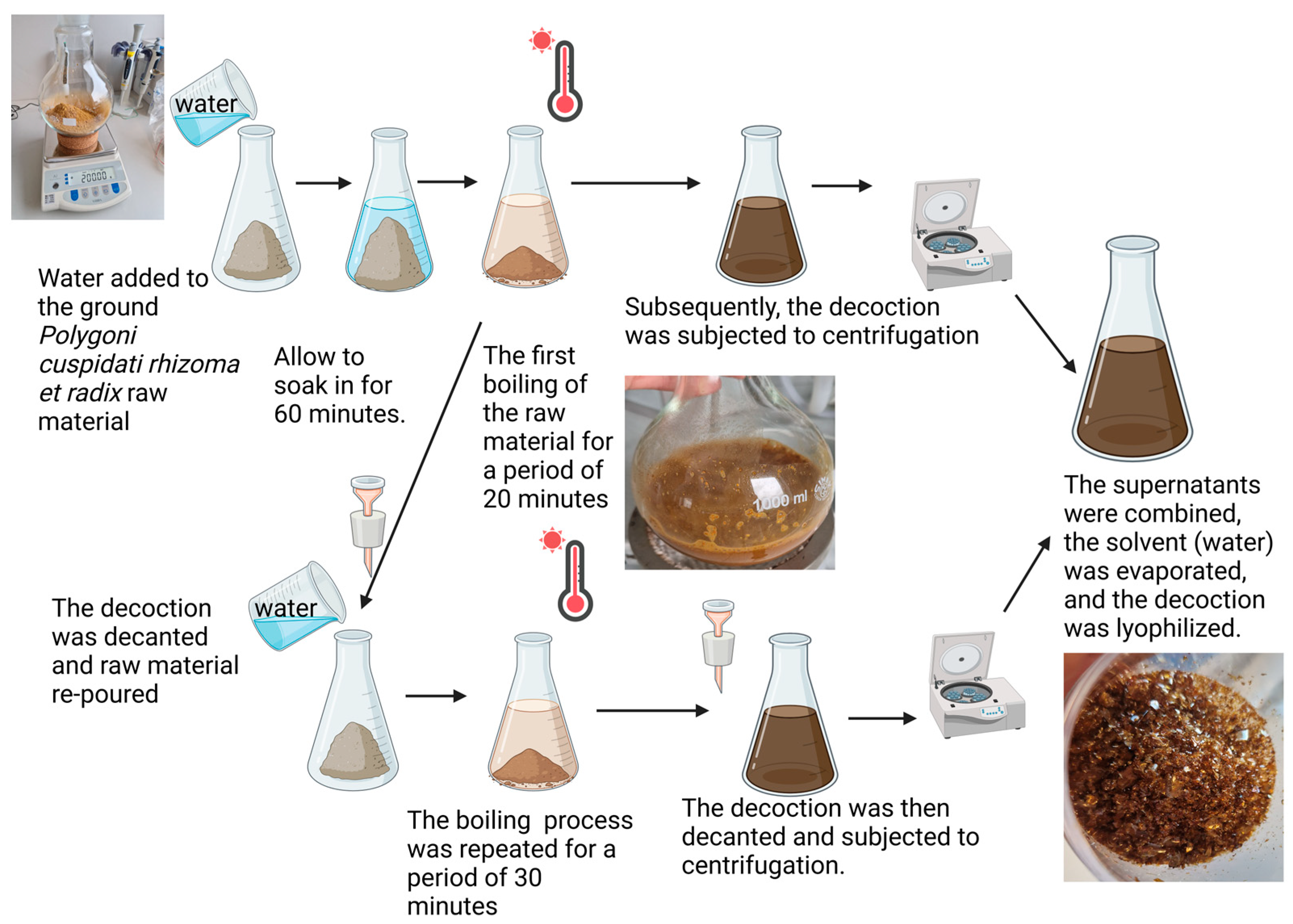
2.1. Plant Material and Decoction Preparation

The plant raw material was obtained from two following sources: collected from wild habitat (self-collected; SC) and purchased at a pharmacy (pharmacopeial raw material; PH). The rhizomes of P. cuspidatum (SC) were collected in the first week of October 2020 from urban environments near Wroclaw, Poland (coordinates: 51°07.404′ N, 17°04.146′ E). Only healthy, fully developed rhizomes with diameters ranging from 15 to 30 mm were selected for harvesting. The species identification is described elsewhere [10]. The second type was Polygoni cuspidati rhizoma et radix (hu zhang in pinyin Chinese) (PH), a pharmacopeial raw material, purchased from a Sonnen-Apotheke pharmacy in Germany (Bad Kotzting, Germany). The specification for this product is attached in the Supplementary Materials, along with information on the content of emodin and polydatin (piceid). The collection and curation of plant material is supervised by the Botanical garden of Medicinal Plants at the Wroclaw Medical University.
Decoctions were prepared according to the recommendations of traditional Chinese medicine [6]. Two weighted portions of 200 g of milled rhizomes of pharmacopeial-grade raw material (PH) and self-collected rhizomes of P. cuspidatum (SC) were soaked in 2000 mL of distilled water for 60 min. The mixture was then heated to a boil for 20 min. Each of the decoctions was centrifuged, and the remaining raw materials were soaked again in 1200 mL of water and brought to a boil for 30 min. The second batch of decoctions was also centrifuged. The supernatants from the first and second batch of decoctions were combined. The solvent was then evaporated under reduced pressure, and the dry residue was lyophilized. The process is illustrated in Figure 1.

2.2. UHPLC-DAD-qTOF-MS Qualitative and Quantitative Analysis

A measured amount of each decoction was dissolved in 80% methanol (MeOH, Merck/MilliporeSigma, Darmstadt, Germany) in a volumetric flask to achieve a concentration of 5 mg/mL. The prepared solutions were then filtered through a 0.22 μm syringe membrane (Chromafil, Macherey-Nagel, Düren, Germany) and transferred to vials, after which a 4 μL aliquot was injected into an ultra-high performance liquid chromatography coupled to diode array detection and a high-resolution quadrupole time-of-flight mass spectrometry (UHPLC-DAD-qTOF-MS) system with an autosampler. The same UHPLC-DAD-qTOF-MS system was used as well as the same qualitative analysis conditions as in our previous studies [9,10]. Briefly, an Ultimate 3000RS series system (Thermo Dionex, Sunnyvale, CA, USA) equipped with a low-pressure quaternary gradient pump was used, with vacuum degasser, an autosampler, a column compartment, a DAD, and a high-resolution quadrupole time-of-flight MS (Bruker qTOF Compact, Bruker Daltonik, Billerica, MA, USA) equipped with ESI. The system was controlled by Bruker Compass Hystar software, version 3.2 (Billerica, MA, USA). An analytical Kinetex C18 2.6 µm column (150 mm × 2.1 mm), (Phenomenex, Torrance, CA, USA) was maintained at 30 °C. Mobile phases A (H2O/HCOOH, 100:0.1, v/v) and B (acetonitrile/HCOOH, 100:0.1, v/v) were used in a following gradient program: 0–22 min 15–22% B, 22–33 min 22–95% B, followed by column equilibration with 15% B for 2 min between injections. The flow rate was 0.3 mL/min. Analysis of all samples was repeated four times as consecutive injections. UV-Vis spectra were recorded in the range of 200–450 nm. ESI-MS conditions were as follows: splitless, nebulizer pressure 30 psi; dry gas flow 8 L/min; dry temperature 250 °C; and capillary voltage 2.2 kV for negative ion mode. Mass spectra were recorded using the scan range (m/z) 50–2200. The collision energy was set automatically from 20 to 40 eV, depending on the m/z of the fragmented ion.
A previously developed, validated analytical method was used to quantify the piceid, resveratrol, vanicoside A, vanicoside B, emodin, and physcion [17]. Linearity, the LOD (limit of detection), and LOQ (limit of quantification) for all quantified compounds were presented in our previous study [17].

2.3. UPLC/QqQ-MS/MS Quantitative Analysis

A targeted metabolomics method, an ultrahigh performance liquid chromatography method coupled with triple quadrupole mass spectrometry (UPLC/QqQ-MS/MS) for quantification of flavan-3-ols and procyanidins, was performed according to the procedure described [18].
The separation was performed with an Exion LC system manufactured by AB Sciex LLC (Framingham, MA, USA) using an Acquity UPLC HSS T3 C18 (1.7 µm, 2.1 mm × 100 mm) column (Waters corporation, Milford, MA, USA) at 40 °C. The injection volume was 2 µL. The mobile phase consisted of water + 0.1% formic acid (A) and acetonitrile + 0.1% formic acid (B). An AB Sciex QTRAP 6500+ (Framingham, MA, USA) was operated in the negative ion multiple reaction monitoring (MRM) mode using a Turbo V ion source with the following settings: Curtain Gas (CR) 35 °C, IonSpray Voltage (IV) −4500 V, Temperature 400 °C, Collision Gas (CAD) Medium, Ion Source Gas 1 (GS1) 55 psi, and Ion Source Gas 2 (GS2) 45 psi. MultiQuant and Analyst from AB Sciex LLC (Framingham, MA, USA) were used for data acquisition and processing, respectively.
Lyophilized SC and PH decoctions as well as extracts obtained earlier and described in previous article [10]: 25% EtOH, 40% EtOH and 60% acetone were prepared for quantitative analysis as described below. In total, 10 mg of each sample was dissolved in 2 mL of 80% MeOH in a volumetric flask and then filtered through a 0.22 μm PVDF syringe membrane (Milipore Millex-GV, Milipore, Burlington, MA, USA) to vials. Samples were also tested after 20-fold dilution. For each sample the procedure was repeated at least once.

2.4. Cell Viability

2.4.1. Cell Line and Conditions

A human gingival fibroblast cell line (HGF-1), obtained from American Type Culture Collection (ATCC, Manassas, VA, USA), was selected for testing the biological activity of the fractions. These cells were cultured in Dulbecco’s Modified Eagle Media (DMEM medium). Furthermore, it was supplemented with 25 µg/mL gentamicin, 2 mM L-glutamine, and 10% FBS (Biological Industries, Beit-Haemek, Israel). Each cell line was cultured for 2 weeks before testing and detached with trypsin/EDTA solution and maintained at 37 °C, 5% CO2 with 95% humidity until the completion of experiments. The cell culture plastics used in the study were purchased from SPL Life Sciences (Pochon, Republic of Korea). The study used an ILC 180 SMART PRO incubator from POL-EKO (Wodzisław Slaski, Poland).

2.4.2. Cytotoxic Activity, MTT Assay

The cytotoxicity assay enabled the evaluation of human gingival fibroblast (HGF-1) cell viability based on mitochondrial enzyme activity. This method effectively quantifies cell proliferation and metabolic activity by reducing thiazolyl blue tetrazolium bromide (MTT), where purple formazan indicates viable cells. The linear relationship between cell number and absorbance allows for the accurate quantification of the cellular response to tested extracts. The HGF-1 cells were seeded in a 96-well flat-bottom microplate and maintained at 37 °C in 95% humidity and 5% CO2 overnight. The decoctions were used in the concentration range from 2.5 µg/mL to 2000 µg/mL. Four chemical compounds were also tested at concentrations ranging from 5 µg/mL to 100 µg/mL: epicatechin, procyanidin B2, resveratrol, and piceid. The concentration of DMSO in the samples was equal to or less than 2%. The cells were then incubated for 24 h under the same conditions. The MTT solution (1 mg/mL thiazolyl blue solution; Pol-Aura, Dywity, Poland) was prepared in RPMI 1640 medium without L-glutamine and phenol red (Capricorn Scientific, Ebsdorfergrund, Germany) under a laminar flow hood. After incubation, 50 µL of MTT solution was added to each well. The plates were returned to the incubator for two hours at 37 °C. After incubation, the MTT solution was removed, and 100 µL of isopropanol was added to each well to dissolve the formazan crystals. The MTT assay was performed in accordance with the ISO 10993-5:2009 (E) standard [19].

2.5. Statistical Analysis

All assays were performed in at least triplicate, and results are presented as the mean of the replicates ± SD. To assess the distribution of results, one of the following tests was used: Shapiro–Wilk test, D’Agostino–Pearson test or Kolmogorov–Smirnov test. Two-way ANOVA and Tukey’s multiple comparisons tests (GraphPad Prism v10, San Diego, CA, USA) were used to evaluate significant differences between the obtained values.

3. Results

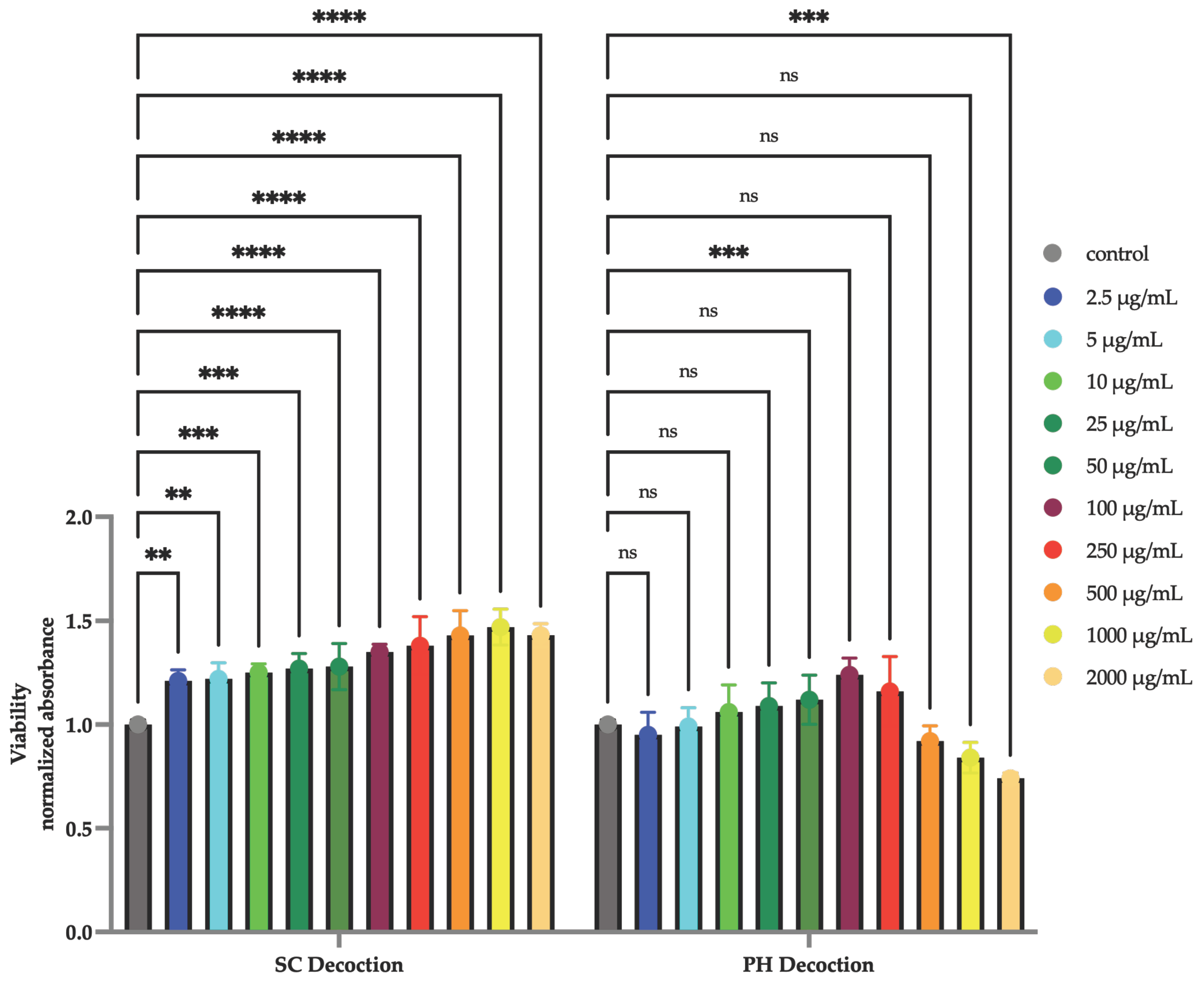
3.1. Cell Viability—MTT Assay

The MTT assay results showed differences in the effects of the decoctions on HGF-1 (Figure 2). The SC decoction was not cytotoxic to HGF-1 cells at any of the tested concentrations. In the PH decoction, the highest tested concentrations caused a decrease in cell viability. Moreover, the SC decoction caused a higher increase in cell viability at all tested concentrations, while the decoction PH showed a statistically significant increase only at a concentration of 100 µg/mL.
All tested compounds showed an increase in HGF-1 cell viability, but in a different range of concentrations (Figure 3). Resveratrol had the narrowest range of concentrations in which it stimulated cells to divide. It was the only one to also showed statistically significant cytotoxicity at a dose of 100 µg/mL.

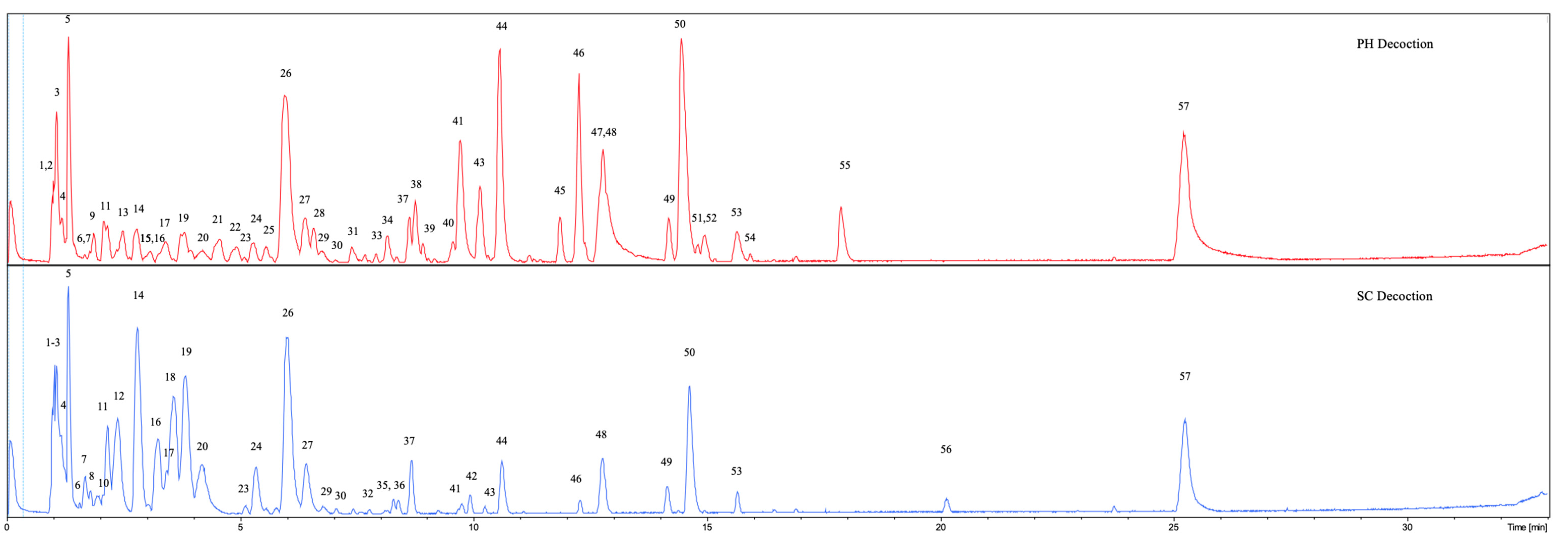
3.2. UHPLC-DAD-qTOF-MS Qualitative Analysis

UHPLC-DAD-qTOF-MS analysis revealed differences between the decoction obtained from the raw material collected from the natural environment and the raw material purchased in the pharmacy (Table 1). Chromatograms of decoctions analyzed at the same concentrations are presented below (Figure 4). At first glance, it can be seen that the SC decoction chromatogram shows higher peaks between 1 and 6 min of retention time than the chromatogram of the PH decoction. Most of these compounds belong to flavan-3-ols and procyanidins (peak numbers 7 (Procyanidin dimer), 8 (Procyanidin trimer), 10 (Procyanidin dimer), 11 (Catechin), 12 (Procyanidin dimer), 14 (Epicatechin), and 20 (Procyanidin dimer monogallate). In turn, in the PH chromatogram, we can see several very high peaks that are much lower in the SC chromatogram (peaks numbers 38 (Resveratrol-(galloylglucoside), 41 (Emodin-glucoside), 43 (Emodin-hexose-sulfate), 44 (Resveratrol), 45 (Emodin-hexose-sulfate), 46 (Torachrysone-hexoside), 47 (Emodin-hexose-sulfate), 50 (Sulfonyl torachrysone/isomer), 51 (Emodin-glucoside), 52 (Physcionin), and 55 (Sulfemodin). Most of these peaks belong to anthraquinones. A clear difference between the decoctions is also the significantly greater amount of sulfate compounds in the PH decoction than in the SC decoction: 13 compounds (peak numbers 9, 13, 15, 16, 21, 22, 24, 28, 33, 43, 47, 50, and 55) and 5 compounds (16, 18, 24, 43 (very low peak), and 50 (much lower peak)), respectively.

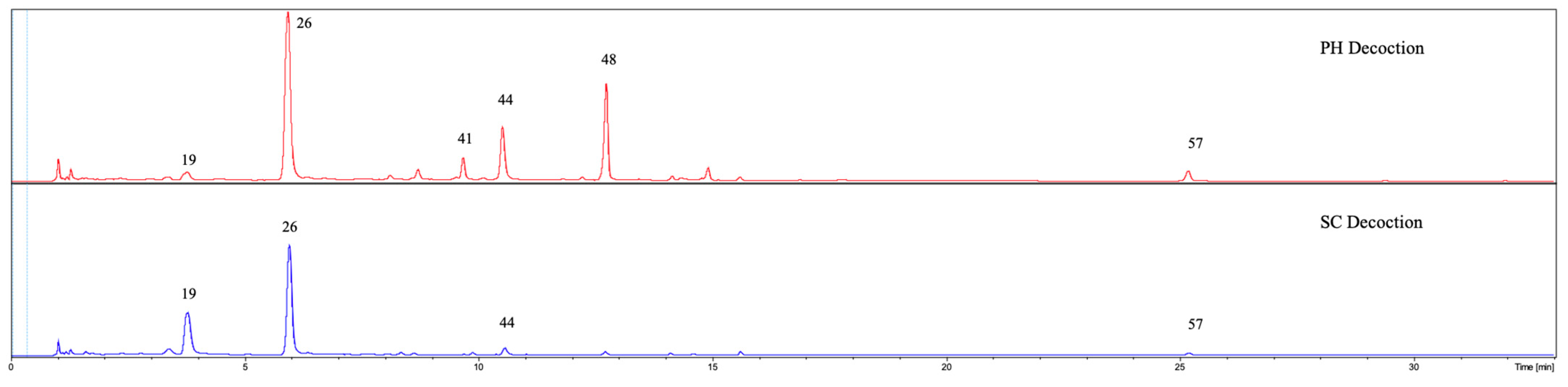
3.3. UHPLC-DAD-qTOF-MS Quantitative Analysis

A previously developed, validated analytical method was used to quantify six compounds: piceid, resveratrol, vanicoside A, vanicoside B, emodin, and physcion. Apart from vanicosides, which were absent in the decoctions, all other compounds were present in significantly higher amounts in the PH decoction than in the SC decoction (Figure 5, Table 2). Physcion was present in very small amounts allowing detection but not quantification.

3.4. UPLC/QqQ-MS/MS Quantitative Analysis of Flavan-3-Ols and Procyanidins

In order to confirm the differences in the content of flavan-3-ols and procyanidins, we used a previously developed and validated UPLC/QqQ-MS/MS method [18] that allows for very precise determination of the content of several selected flavan-3-ols and procyanidins. In addition to the decoctions, we also analyzed the extracts obtained earlier from the same raw material collected from the wild habitat and described in the previous article [10]: 25% EtOH, 40% EtOH, and 60% acetone. The results are presented in Figure 6 and Table 3.
Figure 6 shows a difference in the content of the compounds depending on the solvent used for the extraction (25% EtOH, 40%EtOH, 60% acetone). There are also significant differences between decoctions prepared in the same way but obtained from raw materials from different sources (PH decoction, SC decoction). In the case of most of the analyzed compounds, all procyanidins, and most flavan-3-ols, their content was much higher in the SC decoction than in the PH decoction. The results of the quantitative analysis are consistent with the qualitative analysis described above. We observe a very significant difference in the amount of epicatechin, catechin as well as all analyzed procyanidins. Interestingly, although the use of ethanol and acetone for extraction resulted in higher contents of most of the tested compounds, some of them, mainly galloylated ones, were observed only in decoctions (catechin gallate, epigallocatechin gallate, epigallocatechin, gallocatechin, Table 3). It is also interesting that catechin and epicatechin act differently. While the epicatechin content increases with the ethanol and acetone content, the catechin content is highest in the SC decoction. The same tendency is seen for catechin gallate and epicatechin gallate.

4. Discussion

The results from MTT assay showed differences in the effects of the decoctions on human gingival fibroblasts (Figure 2). The SC decoction was not cytotoxic to HGF-1 cells and caused a high increase in cell viability at all tested concentrations. On the other hand, the PH decoction showed a statistically significant increase in cell viability only at a concentration of 100 µg/mL. Moreover, the highest tested concentrations caused a decrease in cell viability. The differences in activity of decoctions are due to the different chemical composition of them. The SC decoction is much richer in flavan-3-ols and procyanidins. Qualitative and quantitative analysis revealed a higher content of procyanidin monomers, such as catechin and epicatechin, as well as procyanidin dimers. Taking the above into account, we conducted studies examining the influence of epicatechin (monomer) and procyanidin B2 (dimer) present in the decoctions on the viability of gingival fibroblasts. We have observed that these compounds have a wide range of concentrations that stimulate cell viability (5–100 µg/mL). They did not show cytotoxic effects at any of the tested concentrations (Figure 2). The content of these compounds is much higher in the SC decoction than the PH decoction. Similarly, there are significantly more of the other procyanidins in SC decoction: procyanidin B1, B3, B4, and catechin. The presence of these compounds in appropriate concentrations may have a significant influence on the observed effect of the SC decoction on gingival fibroblasts. Data from other studies confirm the stimulating effect of flavan-3-ols such as catechin [25] and procyanidins [26] on the viability and proliferation of fibroblasts (human dermal fibroblasts) or keratinocytes as well as their migration and collagen production, which may significantly accelerate wound healing. Flavan-3-ols and procyanidins may stimulate fibroblast proliferation through the activation of the PI3K/Akt/mTOR signaling pathway, which plays a crucial role in regulating cell growth and survival. Research indicates that certain flavonoids can modulate this pathway, leading to increased cellular proliferation [27]. Currently, research is also being conducted on animals using preparations such as ointments containing procyanidins to accelerate wound healing. The results of these studies are promising [28]. Studies confirm also the lack of cytotoxicity in a wide range of catechin and epicatechin concentrations towards gingival fibroblasts. Babich et al. [29] indicated different cytotoxicity of flavan-3-ols towards normal human gingival fibroblast (HGF-2) cells. The simple flavan-3-ols, such as (+)-epicatechin, (−)-catechin, and (−)-epigallocatechin showed the lowest cytotoxicity with midpoint cytotoxicity—NR50 values above 500 µM. Conversely, the flavan-3-ols with a galloyl moiety: (−)-catechin gallate, (−)-epicatechin gallate, and (−)-epigallocatechin gallate showed cytotoxicity at lower concentrations ((NR50) = 131 µM, 100 µM, and 256 µM, respectively). In our decoctions, among the galloyl monomers, only epicatechin gallate was present in relatively high concentrations, with no significant differences between the decoctions.
The observed differences in the activity of the decoctions could also be due to different concentrations of stilbenes, including resveratrol and piceid. Both piceid and resveratrol are present in the PH decoction in significantly higher concentrations (Table 2). While piceid revealed a stimulating effect on gingival fibroblast viability at all tested concentrations (5–100 µg/mL), resveratrol showed this effect only at a dose of 10 µg/mL (~44 µM) and was cytotoxic to the cells at the highest tested concentration-100 µg/mL (~439 µM), (Figure 3). Similar results were obtained in the study by Chin et al. [30], where resveratrol increased the viability of human gingival fibroblasts after 24 h incubation at a concentration of 10 µM but showed cytotoxicity at higher concentrations of 100 and 200 µM. Our results are also consistent with other studies that have shown that the viability and proliferation rate of gingival fibroblasts is dependent on the resveratrol concentration [31,32] and it is cytotoxic at higher doses. The compounds found in greater amounts in the PH decoction are also anthraquinones (Table 1 and Table 2), mainly emodin and its derivatives. Considering the reports of cytotoxicity of emodin, even at low concentrations (10 µM), towards fibroblasts [33,34], it seems that this compound could also have contributed to the weaker activity of the PH decoction. The PH decoction also contains a significantly higher amount of emodin hexosides (compound 41, 43, 45, 47, and 51) and torachrysone hexoside (compound 46). Several studies have shown the toxicity of emodin hexosides and torachrysone hexoside toward hepatocytes [23]. Moreover, these compounds showed severe toxicity against zebrafish embryos and appear to be responsible for Polygonum multiflorum-induced idiosyncratic liver injury (IDILI) [35]. According to traditional Chinese medicine, plant materials should be properly processed before used, which affects the chemical composition, activity, as well as the toxicity of the final preparation. Specific recommendations are included in the Chinese Pharmacopoeia [36]. Regarding Polygonum multiflorum (PM), the Chinese Pharmacopoeia (2015 edition) recommends steaming the root of PM with black soybean decoction [23]. According to Abd Hamid et al. [23], such processing significantly reduced the cytotoxicity of the PM final preparation and was associated with a reduction in the amounts of these two potentially toxic compounds (emodin-8-O-glucoside and torachrysone-O-hexose) in it. Another important aspect is that, the main form of application in traditional Chinese pharmacotherapy is the decoction [36,37]. Our studies conducted on gingival fibroblasts are in line with these recommendations, showing that the decoction is a preparation with activity higher or equal to ethanol and acetone extracts, but it has significantly lower cytotoxicity [9,10]. In our earlier study [10], ethanol extracts (25% EtOH and 40% EtOH) and an acetone extract (60% acetone) prepared from the same raw material that was used to prepare the SC decoction showed a narrower range of concentrations stimulating the viability of gingival fibroblasts than the SC decoction. After 24 h of incubation, these extracts stimulated fibroblast viability at a concentration of 50 μg/mL, but starting from 500 μg/mL, cytotoxic activity was observed. We included these extracts in the current study to examine the content of procyanidins and flavan-3-ols (Figure 6, Table 3). Ethanol and acetone extracts contained significantly higher epicatechin content than the decoction, but not catechin. The highest content of the procyanidins tested was observed in 60% acetone, which is consistent with the literature that it is a good solvent for procyanidin extraction [38]. A decrease in the content of these compounds was observed with a decrease in the amount of ethanol in the solvent: 40%EtOH > 25%EtOH > decoction. However, the amount of procyanidin B3 in the decoction was higher than in the ethanol extracts. It should be remembered that here we have only quantitatively examined flavan-3-ol monomers and procyanidins dimers. We know from previously performed tests that the total amount of tannins and, therefore, also the more polymerized procyanidins, is much higher in acetone and ethanol extracts than in a decoction [9,10]. Despite the higher content of procyanidins and flavan-3-ols with proven fibroblast proliferation-stimulating activity in these ethanol and acetone extracts, these preparations did not give a more beneficial effect than decoctions. Similarly, in the case of the PH decoction, other compounds present in higher concentrations in these products, e.g., the previously mentioned resveratrol or emodin and its derivatives, could have a negative effect on the tested activity. According to a previous study [10], the content of emodin and resveratrol in ethanol and acetone extracts is significant higher than in the SC decoction. Emodin per gram of extract is 1.47 mg (25%EtOH), 3.68 mg (40% EtOH), and 9.63 mg (60% acetone), and for resveratrol it is 3.95 mg (25%EtOH), 3.05 mg (40%EtOH), and 1.47 mg (60% acetone) [10]. It appears that these compounds may be responsible for the cytotoxic effects of the extracts at high concentrations. Moreover, it is also possible that high concentration of procyanidins or flavan-3-ols in extracts may have a negative effect on fibroblasts.
In summary, it should be stated that although the PH decoction showed stimulation of the viability of gingival fibroblasts, this activity was weaker than that of the SC decoction. The PH decoction showed cytotoxic activity at higher concentrations, which was not observed in the SC decoction. The differences in activity resulted from different chemical composition. The SC decoction contained significantly more flavan-3-ols and procyanidin dimers and less stilbenes and anthraquinones than the PH decoction, which probably had a positive effect on the observed activity. It is worth mentioning that decoctions usually have a high content of saccharides, which we have already observed in the rhizomes of Polygonum cuspidatum [9], and they can also interact synergistically with other compounds, influencing the final activity. We are currently testing the activity of this group of compounds.
The major differences in the composition of decoctions may be due to environmental factors but may also result from genetic variation resulting from a possible crossing and repeated uncontrolled backcrossing with related species. In Europe, Reynoutria × bohemica J. Chrtek & A. Chrtkova, a hybrid of Reynoutria japonica and Reynoutria sachalinensis, is common, and the genetic structure of hybrid populations is unresolved. These hybrids have often a varied spectrum of morphological traits that may lead to unintended admixture to the material collected from wild habitats. The identification problems and the proposed solution are discussed extensively in our other study [39]. The possibilities of pharmacological use of a hybrid derived from two plants used in traditional medicine [12] should be tackled in further studies.
Considering the results of this study it seems that the plants growing in Europe provide a good material for the development of pharmaceutical formulations used, for example, in the healing of wounds in the oral cavity. However, there are still a few issues to be clarified. One of them is the probable lack of meeting the European Pharmacopoeia criteria of the raw material collected from the natural environment, which requires an emodin content of 1.0% and polydatin (piceid) content of 1.5% of dried plant material [2]. These criteria (and the quantitative methods described therein) differ somewhat from the requirements of the Chinese Pharmacopoeia, where the minimum contents are 0.6% and 0.15%, respectively [11]. From the data provided on the pharmacopeial raw material, (supplement) we learn that it meets the requirements of the Chinese Pharmacopoeia, containing 0.81% emodin and 1.45% polydatin. Considering the much lower emodin content in the SC decoction, this raw material might not meet these requirements. However, cytotoxicity of anthraquinones is currently being discussed more and more often, and raw materials containing them are becoming the subject of toxicological interest, and restrictions are being introduced [40,41]. It seems that this raw material or the preparations that are made from it should also be reviewed from this perspective.
It should be noted that this study has limitations, one of them is the small number of samples (raw materials) taken for testing both from the natural environment and from the pharmacy. The results of the study should be confirmed on a larger number of samples. Another limitation is the results from the in vitro study, which do not always translate into the results of studies on animals and humans.

5. Conclusions

Research has shown that the decoction obtained from raw material collected from the wild environment in Europe has a superior profile of tested biological activity in comparison to the raw material purchased in a pharmacy. Significant differences were observed in the composition of compounds in the decoctions. The SC decoction contained significantly more flavan-3-ols (e.g., about 9 times more epicatechin and 3 times more catechin) and procyanidin dimers (e.g., about 4.5 times more procyanidin B1 and 9 times more procyanidin B2 and B4), as well as about 7.5 times less resveratrol and anthraquinones (about 4 times less emodin). We concluded that such a ratio of compounds had a positive effect on the observed viability of fibroblasts. The study shows that it is worth taking an interest in the medical use of a plant that occurs so abundantly in Europe. However, extended research is necessary on the significant differences in the chemical composition of raw materials.

Supplementary Materials

The following supporting information can be downloaded at: https://www.mdpi.com/article/10.3390/app15041914/s1, Certificate for material purchased from a pharmacy.

Author Contributions

Conceptualization, J.H. and I.N.-H.; methodology, I.N.-H., T.G. and G.Z.; validation, I.N.-H. and G.Z.; formal analysis, I.N.-H. and G.Z.; investigation, I.N.-H., M.F., T.G. and G.Z.; resources, T.G. and I.N.-H.; data curation, I.N.-H.; writing—original draft preparation, I.N.-H. and J.H.; writing—review and editing, A.M. and S.M.; visualization, M.F. and P.S.; supervision, I.N.-H.; project administration, J.H. and I.N.-H.; funding acquisition, I.N.-H. and A.M. All authors have read and agreed to the published version of the manuscript.

Funding

This research was funded by Wroclaw Medical University project No: SUBZ.D030.24.081. The curation of plant material is funded by the Ministry of Research and Higher Education of Poland via Botanical Garden of Medicinal Plant at the Wroclaw Medical University grant for special research facility decision No. 28/598769/SPUB/SP/2024.

Institutional Review Board Statement

Not applicable.

Informed Consent Statement

Not applicable.

Data Availability Statement

The original contributions presented in this study are included in the article. Further inquiries can be directed to the corresponding author.

Acknowledgments

We would like to thank Hanna Czapor-Irzabek for the collection of LC/MS data. The measurements were carried out in the Laboratory of Elemental Analysis and Structural Research at Wroclaw Medical University.

Conflicts of Interest

The authors declare no conflicts of interest.

References

  1. Global Invasive Species Database. Species Profile: Polygonum Cuspidatum. Available online: https://www.iucngisd.org/gisd/speciesname/Polygonum+cuspidatum (accessed on 23 November 2024).
  2. Nawrot-Hadzik, I.; Hadzik, J.; Fleischer, M.; Choromańska, A.; Sterczała, B.; Kubasiewicz-Ross, P.; Saczko, J.; Gałczyńska-Rusin, M.; Gedrange, T.; Matkowski, A. Chemical composition of east Asian invasive knotweeds, their cytotoxicity and antimicrobial efficacy against cariogenic pathogens: An in-vitro study. Med. Sci. Monit. 2019, 25, 3279–3287. [Google Scholar] [CrossRef] [PubMed]
  3. Editorial Committee of Chinese Pharmacopoeia. Chinese Pharmacopoeia, 2010th ed.; Medical Science and Technology Press: Beijing, China, 2010; pp. 194–195. [Google Scholar]
  4. Peng, W.; Qin, R.; Li, X.; Zhou, H. Botany, phytochemistry, pharmacology, and potential application of Polygonum cuspidatum Sieb.et Zucc.: A review. J. Ethnopharmacol. 2013, 148, 729–745. [Google Scholar] [CrossRef] [PubMed]
  5. Zhang, H.; Li, C.; Kwok, S.T.; Zhang, Q.W.; Chan, S.W. A review of the pharmacological effects of the dried root of Polygonum cuspidatum (Hu Zhang) and its constituents. Evid.-Based Complement. Altern. Med. 2013, 2013, 208349. [Google Scholar] [CrossRef]
  6. Hempen, C.-H.; Fischer, T. A Materia Medica for Chinese Medicine: Plants, Minerals, and Animal Products; Churchill Livingstone: London, UK, 2009; ISBN 9780443100949. [Google Scholar]
  7. Song, J.H.; Kim, S.K.; Chang, K.W.; Han, S.K.; Yi, H.K.; Jeon, J.G. In vitro inhibitory effects of Polygonum cuspidatum on bacterial viability and virulence factors of Streptococcus mutans and Streptococcus sobrinus. Arch. Oral Biol. 2006, 51, 1131–1140. [Google Scholar] [CrossRef] [PubMed]
  8. Wu, X.B.; Luo, X.Q.; Gu, S.Y.; Xu, J.H. The effects of Polygonum cuspidatum extract on wound healing in rats. J. Ethnopharmacol. 2012, 141, 934–937. [Google Scholar] [CrossRef]
  9. Hadzik, J.; Choromańska, A.; Karolewicz, B.; Matkowski, A.; Dominiak, M.; Złocińska, A.; Nawrot-Hadzik, I. Oral Wound Healing Potential of Polygoni Cuspidati Rhizoma et Radix Decoction-In Vitro Study. Pharmaceuticals 2023, 16, 267. [Google Scholar] [CrossRef]
  10. Nawrot-Hadzik, I.; Matkowski, A.; Pitułaj, A.; Sterczała, B.; Olchowy, C.; Szewczyk, A.; Choromańska, A. In Vitro Gingival Wound Healing Activity of Extracts from Reynoutria japonica Houtt Rhizomes. Pharmaceutics 2021, 13, 1764. [Google Scholar] [CrossRef]
  11. Alperth, F.; Melinz, L.; Fladerer, J.P.; Bucar, F. Uhplc analysis of reynoutria japonica houtt. Rhizome preparations regarding stilbene and anthranoid composition and their antimycobacterial activity evaluation. Plants 2021, 10, 1809. [Google Scholar] [CrossRef]
  12. Nawrot-Hadzik, I.; Slusarczyk, S.; Granica, S.; Hadzik, J.; Matkowski, A. Phytochemical Diversity in Rhizomes of Three Reynoutria Species and their Antioxidant Activity Correlations Elucidated by LC-ESI-MS/MS Analysis. Molecules 2019, 24, 1136. [Google Scholar] [CrossRef]
  13. Magacz, M.; Oszajca, M.; Nawrot-Hadzik, I.; Drożdż, R.; Jurczak, A.; Hadzik, J.; Smakosz, A.; Krzyściak, W. Phenolic compounds of Reynoutria sp. As modulators of oral cavity lactoperoxidase system. Antioxidants 2021, 10, 676. [Google Scholar] [CrossRef]
  14. Chen, H.; Wang, W.; Yu, S.; Wang, H.; Tian, Z.; Zhu, S. Procyanidins and Their Therapeutic Potential against Oral Diseases. Molecules 2022, 27, 2932. [Google Scholar] [CrossRef] [PubMed]
  15. Nawrot-Hadzik, I.; Matkowski, A.; Hadzik, J.; Dobrowolska-Czopor, B.; Olchowy, C.; Dominiak, M.; Kubasiewicz-Ross, P. Proanthocyanidins and Flavan-3-Ols in the Prevention and Treatment of Periodontitis—Antibacterial Effects. Nutrients 2021, 13, 165. [Google Scholar] [CrossRef] [PubMed]
  16. Nawrot-Hadzik, I.; Matkowski, A.; Kubasiewicz-Ross, P.; Hadzik, J. Proanthocyanidins and Flavan-3-ols in the Prevention and Treatment of Periodontitis-Immunomodulatory Effects, Animal and Clinical Studies. Nutrients 2021, 13, 239. [Google Scholar] [CrossRef] [PubMed]
  17. Nawrot-Hadzik, I.; Granica, S.; Domaradzki, K.; Pecio, Ł.; Matkowski, A. Isolation and Determination of Phenolic Glycosides and Anthraquinones from Rhizomes of Various Reynoutria Species. Planta Med. 2018, 84, 1118–1126. [Google Scholar] [CrossRef] [PubMed]
  18. Vrhovsek, U.; Masuero, D.; Gasperotti, M.; Franceschi, P.; Caputi, L.; Viola, R.; Mattivi, F. A Versatile Targeted Metabolomics Method for the Rapid. J. Agric. Food Chem. 2012, 60, 8831–8840. [Google Scholar] [CrossRef] [PubMed]
  19. ISO 10993-5:2009; Biological Evaluation of Medical Devices—Part 5: Tests for In Vitro Cytotoxicity. Organization for Standardization: Geneva, Switzerland, 2009.
  20. Xiao, K.; Xuan, L.; Xu, Y.; Bai, D.; Zhong, D. Constituents from Polygonum cuspidatum. Chem. Pharm. Bull. 2002, 50, 605–608. [Google Scholar] [CrossRef] [PubMed]
  21. Xiao, K.; Xuan, L.; Xu, Y.; Bai, D. Stilbene Glycoside Sulfates from Polygonum cuspidatum. J. Nat. Prod. 2000, 63, 1373–1376. [Google Scholar] [CrossRef]
  22. Krenn, L.; Presser, A.; Pradhan, R.; Bahr, B.; Paper, D.H.; Mayer, K.K.; Kopp, B. Sulfemodin 8-O-β-d-Glucoside, a New Sulfated Anthraquinone Glycoside, and Antioxidant Phenolic Compounds from Rheum emodi. J. Nat. Prod. 2003, 66, 1107–1109. [Google Scholar] [CrossRef] [PubMed]
  23. Abd Hamid, R.; Xiao, K.; Liu, C.; Wang, Y.; Han, L.; Wang, P.; Zhao, Q.; Zheng, F.; Dou, Z.; Yang, W.; et al. Rapid Discovery of the Potential Toxic Compounds in Polygonum multiflorum by UHPLC/Q-Orbitrap-MS-Based Metabolomics and Correlation Analysis. Front. Pharmacol. 2019, 10, 329. [Google Scholar] [CrossRef]
  24. Ye, M.; Han, J.; Chen, H.; Zheng, J.; Guo, D. Analysis of phenolic compounds in rhubarbs using liquid chromatography coupled with electrospray ionization mass spectrometry. J. Am. Soc. Mass Spectrom. 2007, 18, 82–91. [Google Scholar] [CrossRef] [PubMed]
  25. Chaniad Id, P.; Tewtrakul, S.; Sudsai, T.; Langyanai, S.; Kaewdana, K. Anti-inflammatory, wound healing and antioxidant potential of compounds from Dioscorea bulbifera L. bulbils. PLoS ONE 2020, 15, e0243632. [Google Scholar] [CrossRef] [PubMed]
  26. Wang, R.; Lechtenberg, M.; Sendker, J.; Petereit, F.; Deters, A.; Hensel, A. Wound-healing plants from TCM: In vitro investigations on selected TCM plants and their influence on human dermal fibroblasts and keratinocytes. Fitoterapia 2013, 84, 308–317. [Google Scholar] [CrossRef] [PubMed]
  27. Zughaibi, T.A.; Suhail, M.; Tarique, M.; Tabrez, S. Targeting PI3K/Akt/mTOR Pathway by Different Flavonoids: A Cancer Chemopreventive Approach. Int. J. Mol. Sci. 2021, 22, 12455. [Google Scholar] [CrossRef] [PubMed]
  28. Rajakumari, R.; Volova, T.; Oluwafemi, O.S.; Rajeshkumar, S.; Thomas, S.; Kalarikkal, N. Nano formulated proanthocyanidins as an effective wound healing component. Mater. Sci. Eng. C 2020, 106, 110056. [Google Scholar] [CrossRef]
  29. Babich, H.; Krupka, M.E.; Nissim, H.A.; Zuckerbraun, H.L. Differential in vitro cytotoxicity of (-)-epicatechin gallate (ECG) to cancer and normal cells from the human oral cavity. Toxicol. In Vitro 2005, 19, 231–242. [Google Scholar] [CrossRef] [PubMed]
  30. Chin, Y.T.; Hsieh, M.T.; Lin, C.Y.; Kuo, P.J.; Yang, Y.C.S.H.; Shih, Y.J.; Lai, H.Y.; Cheng, G.Y.; Tang, H.Y.; Lee, C.C.; et al. 2,3,5,4′-tetrahydroxystilbene-2-O-β-glucoside isolated from Polygoni Multiflori ameliorates the development of periodontitis. Mediat. Inflamm. 2016, 2016, 6953459. [Google Scholar] [CrossRef]
  31. D’Amico, E.; Pierfelice, T.V.; Amoroso, R.; Cacciatore, I.; D’Arcangelo, C.; Lepore, S.; D’Ercole, S.; Di Pietro, N.; Di Rienzo, A.; Petrini, M.; et al. Emerging Effects of Resveratrol Derivatives in Cells Involved in Oral Wound Healing: A Preliminary Study. Int. J. Mol. Sci. 2023, 24, 3276. [Google Scholar] [CrossRef] [PubMed]
  32. Birar, V.C.; Sheerin, A.N.; Ostler, E.L.; Faragher, R.G.A. Novel resveratrol derivatives have diverse effects on the survival, proliferation and senescence of primary human fibroblasts. Biogerontology 2020, 21, 817–826. [Google Scholar] [CrossRef] [PubMed]
  33. Xiong, G.; Chen, H.; Wan, Q.; Dai, J.; Sun, Y.; Wang, J.; Li, X. Emodin promotes fibroblast apoptosis and prevents epidural fibrosis through PERK pathway in rats. J. Orthop. Surg. Res. 2019, 14, 319. [Google Scholar] [CrossRef]
  34. Baczewska, I.; Hawrylak-Nowak, B.; Zagórska-Dziok, M.; Ziemlewska, A.; Nizioł-Łukaszewska, Z.; Borowski, G.; Dresler, S. Towards the Use of Lichens as a Source of Bioactive Substances for Topical Applications. Molecules 2024, 29, 4352. [Google Scholar] [CrossRef]
  35. Yang, J.B.; Li, W.F.; Liu, Y.; Wang, Q.; Cheng, X.L.; Wei, F.; Wang, A.G.; Jin, H.T.; Ma, S.C. Acute toxicity screening of different extractions, components and constituents of Polygonum multiflorum Thunb. on zebrafish (Danio rerio) embryos in vivo. Biomed. Pharmacother. 2018, 99, 205–213. [Google Scholar] [CrossRef] [PubMed]
  36. Wang, M.; Franz, G. The role of the European Pharmacopoeia (Ph Eur) in quality control of traditional Chinese herbal medicine in European member states. World J. Tradit. Chin. Med. 2015, 1, 5–15. [Google Scholar] [CrossRef]
  37. Sheridan, H.; Krenn, L.; Jiang, R.; Sutherland, I.; Ignatova, S.; Marmann, A.; Liang, X.; Sendker, J. The potential of metabolic fingerprinting as a tool for the modernisation of TCM preparations. J. Ethnopharmacol. 2012, 140, 482–491. [Google Scholar] [CrossRef] [PubMed]
  38. Downey, M.O.; Hanlin, R.L. Comparison of Ethanol and Acetone Mixtures for Extraction of Condensed Tannin from Grape Skin. S. Afr. J. Enol. Vitic. 2016, 31, 154–159. [Google Scholar] [CrossRef]
  39. Stafiniak, M.; Bielecka, M.; Kujawa, K.; Jezierska-Domaradzaka, A.; Pencakowski, B.; Basiak, A.; Matkowski, A.; Nawrot-Hadzik, I. Integrative morphological, phytochemical, and molecular identification of three invasive and medicinal Reynoutria species. Res. Sq. 2024. [Google Scholar] [CrossRef]
  40. Loschi, F.; Faggian, M.; Sut, S.; Ferrarese, I.; Maccari, E.; Peron, G.; Dall’acqua, S. Development of an LC–DAD–MS-Based Method for the Analysis of Hydroxyanthracene Derivatives in Food Supplements and Plant Materials. Molecules 2022, 27, 1932. [Google Scholar] [CrossRef] [PubMed]
  41. Turck, D.; Bohn, T.; Castenmiller, J.; De Henauw, S.; Hirsch-Ernst, K.I.; Knutsen, H.K.; Mangelsdorf, I.; McArdle, H.J.; Naska, A.; Pentieva, K.; et al. Scientific Opinion on additional scientific data related to the safety of preparations of Rheum palmatum L., Rheum officinale Baill. and their hybrids, Rhamnus purshiana DC., Rhamnus frangula L. and Cassia senna L., submitted pursuant to Article 8(4) of R. EFSA J. 2024, 22, e8766. [Google Scholar] [CrossRef] [PubMed]
Figure 1. A diagrammatic representation of the traditional method of preparing the decoction. Created in BioRender.com.
Applsci 15 01914 g001
Figure 2. HGF-1 viability after 24 h incubation with the different concentrations of the decoctions. Presented error bars are means ± SD for n ≥ 5, ** Statistically significant compared to control (untreated cells) at p ≤ 0.01, *** for p ≤ 0.001, and **** for p ≤ 0.0001; ns—not statistically significant.
Applsci 15 01914 g002
Figure 3. HGF-1 viability after 24 h incubation with the different compounds. Presented error bars are means ± SD for n ≥ 5. ** Statistically significant compared to control (untreated cells) at p ≤ 0.01, *** for p ≤ 0.001, and **** for p ≤ 0.0001; ns—not statistically significant.
Applsci 15 01914 g003
Figure 4. ESI-MS (negative mode) chromatograms of decoctions from Polygoni cuspidati rhizoma et radix (PH decoction (red) and SC decoction (blue). Key to peak identity as in Table 1.
Applsci 15 01914 g004
Figure 5. UV-HPLC chromatograms of PH decoction (red) and SC decoction (blue) with detection at 298 nm.
Applsci 15 01914 g005
Figure 6. Content [mg/g dry weight] of flavan-3-ols and procyanidins in the studied PH and SC decoctions (water was used as a solvent to prepare the PH decoction and the SC decoction) and extracts obtained by using different solvents, described more precisely in the previous article [10]: 25% EtOH, 40% EtOH, and 60% acetone. Values that share the same letter are not significantly different as determined by Tukey’s post hoc test. Presented error bars are means ± SD. p ≤ 0.05 for statistically significant differences.
Applsci 15 01914 g006
Table 1. The table shows the compounds tentatively identified in Polygoni cuspidati rhizoma et radix decoctions and their retention times, UV λ max, MS data, and ionic formulas.
Nr.CompoundRt. [min]UV Max
[nm]
m/z
[M-H]
Error (ppm)Ion
Formula
MS2 Main-Ion
(Relative Intensity %)
MS2 Fragments
(Relative Intensity %)
References
1Unknown carbohydrate1.0ND341.1091−0.6C12H21O11113.0258 (100)101 (66), 119 (63),
179 (18), 173 (16)
HMDB0000258
2Unknown carbohydrate1.05ND719.20173.2C30H39O20377.0868 (100)379 (29), 341 (13), 215 (2.0), 179 (0.4)-
3Organic acid, e.g., citric acid1.1ND191.01951.2C6H7O7111.0083 (100) HMDB0000094
4Organic acid, e.g., malic acid1.15ND133.0145−1.6C4H5O5115.0014 (100) HMDB0000156
5Organic acid, e.g., citric acid1.3ND191.0199−0.7C6H7O7111.0083 (100) HMDB0000094
6Unknown1.57225, 280443.1928−1.2C21H31O10443.1941 (100)189 (17), 101 (9), 113 (8), 119 (8)
7Procyanidin dimer, Type B1.6–1.7225, 280577.13470.7C30H25O12289.0722 (100)407 (60), 125 (33), 425 (18)[12]
8Procyanidin trimer, Type B1.8225, 280865.2002−2.0C45H37O18575.1211 (100)577 (81), 287 (69), 695 (48), 713 (46)[12]
9Unknown/dihydroxyphenylvaleric acid sulfate1.9280289.0389−0.5C11H13O7S96.9585 (100)149 (5), 209 (2)HMDB0240447
10Procyanidin dimer, Type B1.9–2.0225, 280577.1363−2.0C30H25O12289.0719 (100)407 (68), 125 (42), 425 (20)[12]
11Catechin2.15225, 280289.0722−1.7C15H13O6109.0280 (100)123 (89), 203 (71), 221 (64),151 (63)[12]
12Procyanidin dimer, Type B2.4225, 280577.13500.3C30H25O12289.0744 (100)407 (60), 125 (34), 425 (19)[12]
13Unknown sulfate derivative 2.5280313.0027−0.9C12H9O8S189.0561 (100)233 (4)
14Epicatechin2.8225, 280289.0723−0.8C15H13O6109.0292 (100)123 (93), 221 (80), 125 (76)[12]
15Piceid sulfate3.08280, 320469.0825−3.3C20H21O11S227.0717 (100)307 (4), 243 (1), 269 (1), 389 (0.5)HMDB0240553
16Lynioresinol 2a-sulfate3.25280499.1286−1.3C22H27O11S499.1292 (100)96.9585 (12), 110.9755 (6), 453.0840 (2)HMDB0039926
[20]
17Piceatannol glucoside3.4220, 290, 318405.1202−2.8C20H21O9243.0672 (100)201 (1), 225 (0.5)[17]
18Isolariciresinol-2a-sulfate3.6280439.1081−2.8C20H23O9S439.1085 (100)96.9585 (24),
359.1514 (2),
110.9749 (1)
HMDB0240703
[20]
19Resveratrolside3.8220, 304, 315389.1237, 435.1291 [M+COO]1.2C20H21O8227.0713 (100)228 (16), 225 (9), 185 (2)[17]
20Procyanidin dimer monogallate4.2225, 280729.14580.4C37H29O16407.0789 (100)289 (62), 577 (45), 441 (28), 451 (28)[12]
21Resveratrol sulfoglucoside4.5290, 320469.0827−3.2C20H21O11S469.0827 (100)241 (45), 96.96 (12), 227 (2)HMDB0037075
[21]
22Resveratrol sulfoglucoside isomer4.9290, 320469.0824−3.1C20H21O11S241.0027 (100)227 (13), 96.95 (7), 138.96 (2)HMDB0037075
[21]
23Unknown5.1270233.04530.9C12H9O5109.0280 (100)123 (89), 203 (71)-
24Unknown sulfate derivative5.35280269.01230.7C11H9O6S189.0556 (100)147 (0.5)
25Luteolin-7-O-glucoside5.6220, 282447.0940−1.6C21H19O11285.0411 (100)447 (75), 327 (3)HMDB0035588
26Piceid6.0220, 304, 315389.1245−0.8C20H21O8227.0716 (100)228 (13), 185 (2), 225 (0.5)[17]
27Epicatechin-3-O-gallate6.4220, 279441.0828−0.2C22H17O10169.0141 (100)289 (51), 125 (22), 245 (14)[17]
28Unknown sulfate derivative6.6280285.0076−0.4C11H9O7S205.0514 (100)190 (46)
29Epicatechin-O-gallate isomer6.78220, 279441.0842−3.3C22H17O10169.0153 (100)289 (53), 125 (25), 245 (13)[17]
30Procyanidin trimer monogallate7.0225, 2801017.2127−3.1C52H41O22729.1431 (100)577 (31), 865 (30), 287 (28), 441 (19)[12]
31Unknown/Citrusin A7.4280537.1987−1.8C26H33O12329.1397 (100)341 (56), 385 (27)HMDB0039230
32Resveratrol hexoside7.8220, 304, 315389.1244−0.5C20H21O8227.0712 (100)228 (15), 185 (2)[17]
33Emodin-hexose-sulfate7.9220, 280511.0559−1.3C21H19O13S241.0027 (100)269 (25), 96 (10),
431 (5)
[22,23]
34Resveratrol-(galloylglucoside)8.2220, 280, 320541.1362−1.9C27H25O12541.1377 (100)313 (25), 169 (7), 227 (7)HMDB0039341
35Unknown, Azelaic acid?8.3-187.0978−1.0C9H15O4125.0960 (100)97 (14), 169 (9)
36Resveratrol derivative8.4220, 282, 325431.1355−1.8C22H23O9227.0722 (100)228 (14), 185 (1)[17]
37Resveratrol hexoside8.6220, 304, 315389.1246−0.9C20H21O8227.0721 (100)228 (16), 185 (1)[17]
38Resveratrol-(galloylglucoside) isomer8.7220, 280, 320541.1367−2.9C27H25O12313.0577 (100)169 (10), 227 (9)HMDB0039341
39Aloesone hexoside8.9220, 270, 420393.1196−1.4C19H21O9231.0666 (100)232 (13), 187 (0,3)HMDB0035734
40Resveratrol-(galloylglucoside) isomer9.6220, 284, 320541.1364−2.4C27H25O12541.1376 (100)313 (33), 169 (28), 227 (11)HMDB0039341
41Emodin-glucoside9.7217, 252, 284, 423431.0995−2.7C21H19O10431.0994 (100)269 (81), 240 (8)[17]
42Lapathoside D9.9213, 285, 315633.1830−0.7C30H33O15145.0298 (100)487 (44), 633 (34), 469 (7)[10]
43Emodin-hexose-sulfate10.1220, 280511.0562−2.0C21H19O13S269.0463 (100)431 (18), 241 (3)[22,23]
44Resveratrol10.6220, 279, 307227.0718−2.1C14H11O3143.0506 (100)185 (85), 227 (47), 144 (11)[17]
45Emodin-hexose-sulfate11.8220, 280511.0563−2.3C21H19O13S241.0034 (100)431 (47), 269 (30)[22,23]
46Torachrysone-hexoside12.3226, 266, 325407.1359−2.7C20H23O9245.0825 (100)246 (14), 230 (11)[17]
47Emodin-hexose-sulfate11.8220, 280511.0559−1.4C21H19O13S241.0034 (100)269 (32), 431 (10)[22,23]
48Emodin-glucoside12.8221, 269, 281, 423431.0989−1.2C21H19O10269.0454 (100)431 (50), 311 (5)[17]
49Emodin-8-O-(6′-O-malonyl)-glucoside14.2220, 281, 423517.09980.0C24H21O13473.1102 (100)269 (66), 311 (4)[17]
50Sulfonyl torachrysone/isomer14.6220, 312325.0392−1.5C14H13O7S245.0823 (100)230 (32), 215 (1)[11]
51Emodin-glucoside14.8221, 269, 281, 423431.0984−0.1C21H19O10269.0461 (100)282 (12), 431 (3), 311 (2)[17]
52Physcionin/Rheochrysin15.0221, 272, 423445.1147−1.6C22H21O10283.0616 (100)307 (3), 240 (3)HMDB0040511/HMDB35931
53Hydropiperoside15.7222, 290, 313779.21791.7C39H39O17779.2200 (100)145 (92), 633 (62),
453 (7), 615 (5)
[17]
54Aloe-emodin 8-O-(6-O-acetyl)-glucoside15.9220, 281, 423473.1094−1.0C23H21O11269.0466 (100)473 (78), 311 (5), 293 (1)[24]
55Sulfemodin 17.9220, 265, 286, 316349.0037−3.9C15H9O8S269.0467 (100)225 (0.5)PubChem SID
274505204
56Questin20.1222, 286, 313,
430
283.0614−0.6C16H11O5240.0431 (100)269 (0.6)[17]
57Emodin25.2221, 248, 267, 288, 430269.0458−0.9C15H9O5269.0461 (100)225 (28), 241 (10), 197 (2), 181 (1)[17]
HMDB ID: Human Metabolome Database.
Table 2. Content of analyzed compounds in studied decoctions.
Analyte(μg/mL of Liquid Decotion)
SD
(mg/g of Dry Decoction)
SD
SC DecoctionPH DecoctionSC DecoctionPH Decoction
Piceid104.26
±0.42
173.86
±11.39
20.85
±0.08
34.77 *
±2.27
Resveratrol3.11
±0.017
23.08
±1.54
0.62
±0.003
4.62 *
±0.31
Emodin3.95
±0.03
14.85
±0.96
0.78
±0.006
2.97 *
±0.19
Physcion1.24 a
±0.01
2.09 a
±0.14
0.25
±0.003
0.42 ns
±0.02
Vanicosides A and B were not detected in the decoctions. a level below LOQ (limit of quantification) but above LOD (limit of detection). * Statistically significant SC decoction compared to the PH decoction at p ≤ 0.05; ns—not statistically significant.
Table 3. Content of analyzed compounds in the studied decoctions and extracts.
Sample Namemg/gLOQPH DecoctionSC Decoction25% EtOH40% EtOH60% Acetone
catechin gallatemg/g0.0010.3510.212<LOQ<LOQ<LOQ
SD 0.0500.044---
catechinmg/g0.021.4674.4492.3392.5953.030
SD 0.2550.3290.2950.6400.149
epicatechin gallatemg/g0.0012.0962.3342.9876.9989.213
SD 0.0830.0500.0610.1090.019
epicatechinmg/g0.0011.26811.35517.10320.09824.611
SD 0.0400.3090.2500.3830.145
epigallocatechin gallatemg/g0.0010.0110.004<LOQ<LOQ<LOQ
SD 0.0020.000---
epigallocatechinmg/g0.0010.0040.008<LOQ<LOQ<LOQ
SD 0.0010.001---
gallocatechin gallatemg/g0.001<LOQ<LOQ<LOQ<LOQ<LOQ
SD -----
gallocatechinmg/g0.0010.0040.010<LOQ<LOQ<LOQ
SD 0.0000.000---
procyanidin A2mg/g0.0010.0040.0240.1000.0970.131
SD 0.0020.0090.0060.0080.035
procyanidin B1mg/g0.0011.4366.4217.4667.8038.129
SD 0.0240.0080.1760.0800.361
procyanidin B2+B4mg/g0.0011.49913.78018.69919.42222.817
SD 0.1000.3740.4171.1751.167
procyanidin B3mg/g0.0010.6152.3151.4971.7652.438
SD 0.0330.2010.1150.1210.107
Disclaimer/Publisher’s Note: The statements, opinions and data contained in all publications are solely those of the individual author(s) and contributor(s) and not of MDPI and/or the editor(s). MDPI and/or the editor(s) disclaim responsibility for any injury to people or property resulting from any ideas, methods, instructions or products referred to in the content.

