



Comparing 2D and 3D Feature Extraction Methods for Lung Adenocarcinoma Prediction Using CT Scans: A Cross-Cohort Study

 , , ,
, , ,  and
and 
Simple Summary
Abstract
1. Introduction
- The classification between adenocarcinoma and non-adenocarcinoma, with the negative class including two distinct histological subtypes;
- A detailed description of the cases excluded from the datasets, along with the pre-processing steps applied to the CT scans, ensuring transparency and reproducibility;
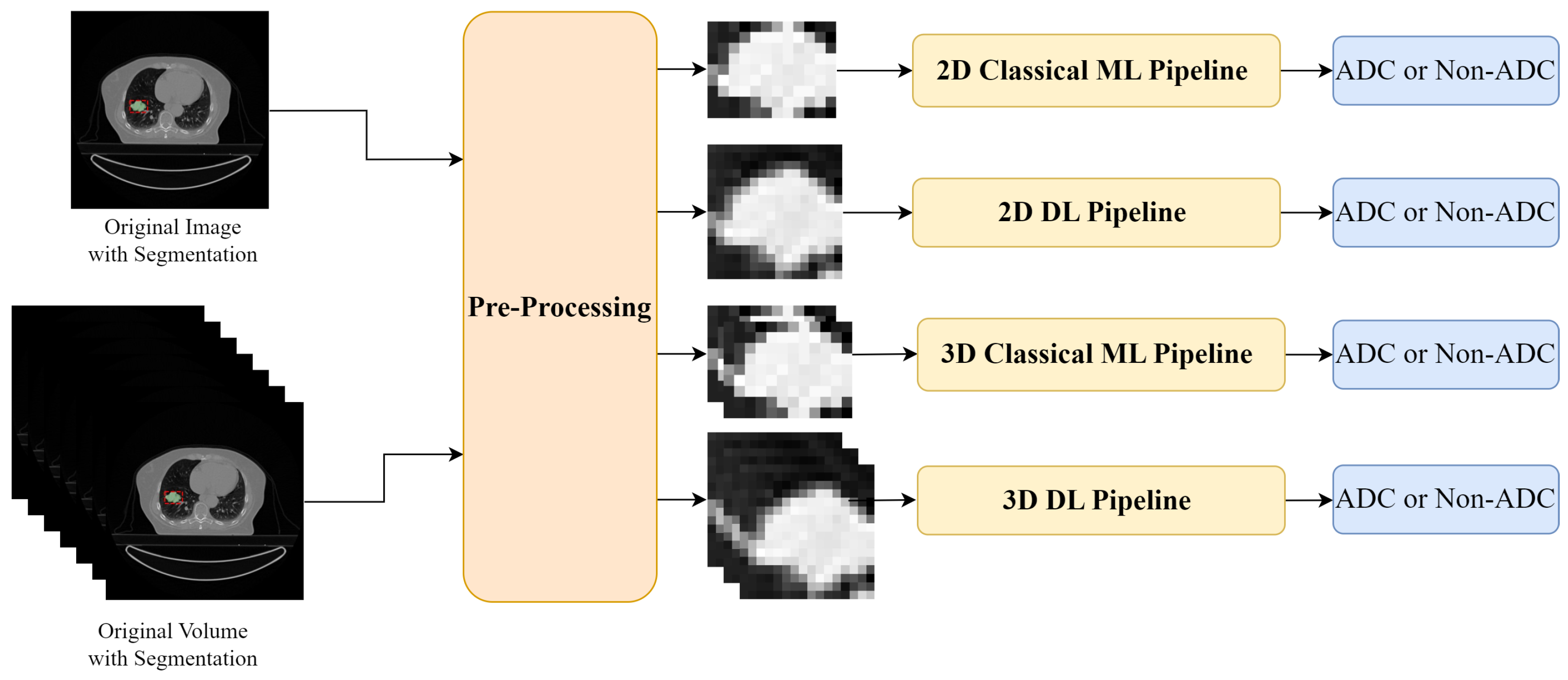
- A comparative analysis of two pipelines, one based on radiomic features and the other on deep features, for each pipeline two different classifiers were compared;
- An exploration of different input forms, specifically 2D slices and 3D volumes from CT scans;
- A cross-cohort study where one public dataset was used for training the models, which were then evaluated in two external public datasets.
2. Related Work
2.1. Radiomic Features
2.2. Deep Learning Approaches
2.3. Overview
3. Materials and Methods
3.1. Datasets
3.1.1. Lung-PET-CT-Dx
3.1.2. NSCLC-Radiomics
3.1.3. NSCLC-Radiogenomics
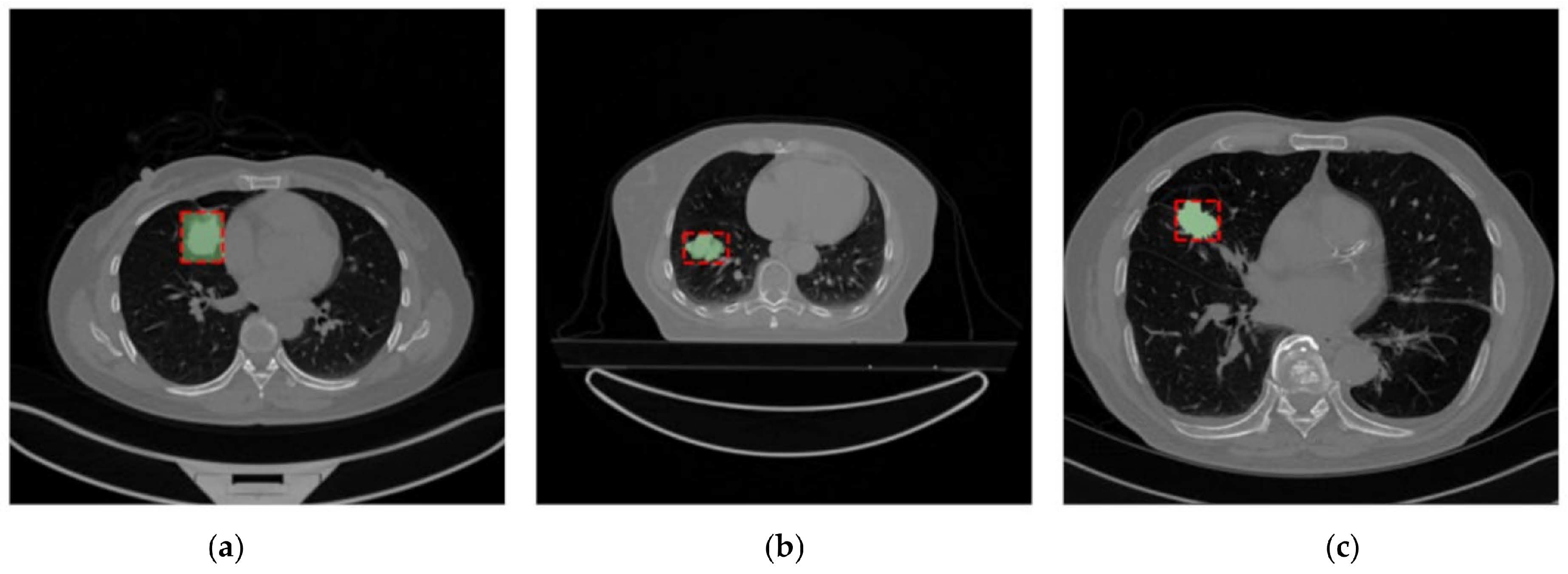
3.1.4. Datasets Uniformization
3.2. Classic Machine Learning Pipeline
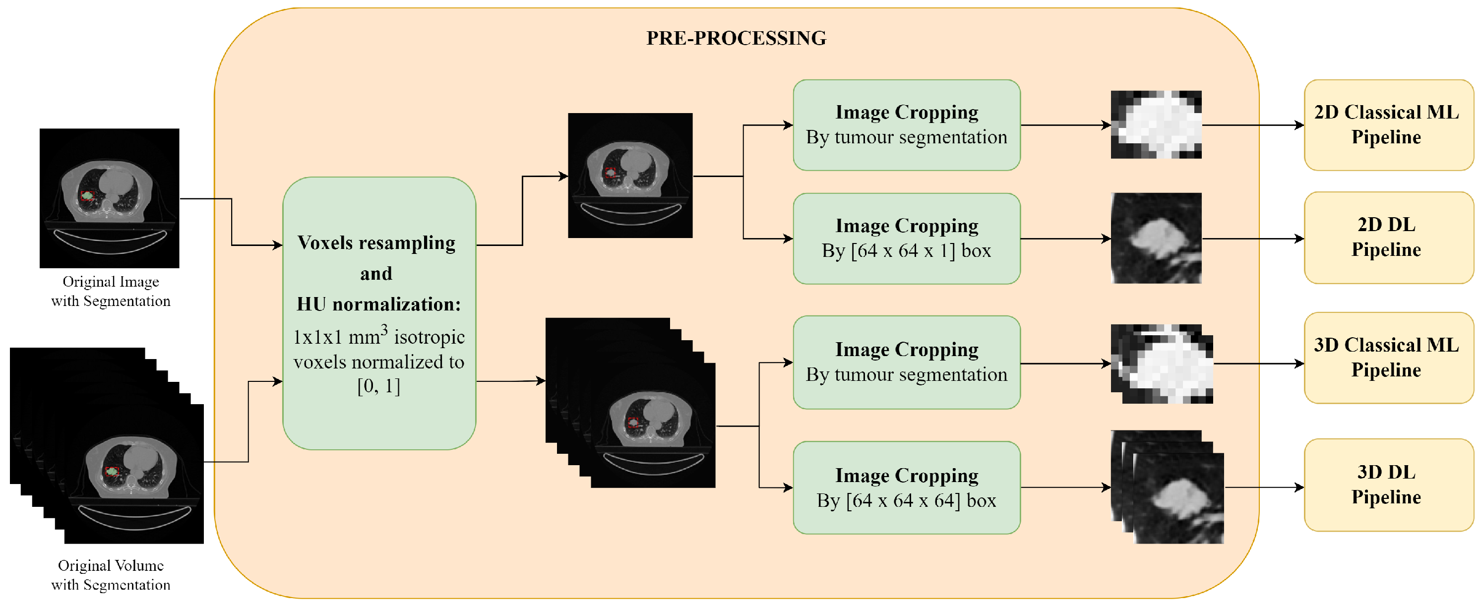
3.2.1. Pre-Processing
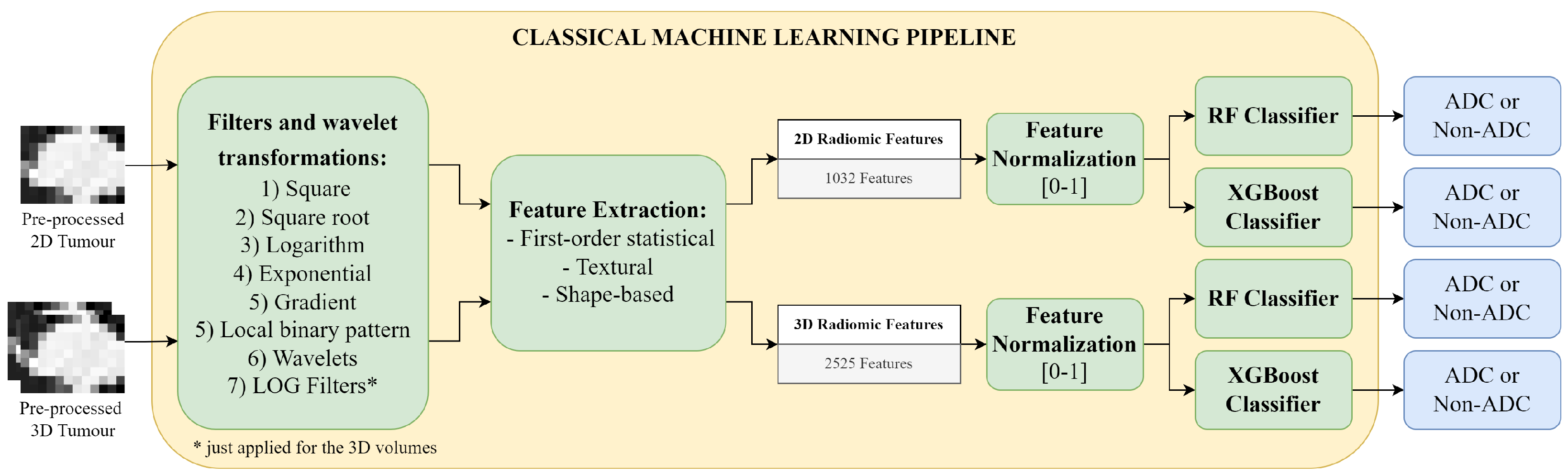
3.2.2. Radiomic Feature Extraction
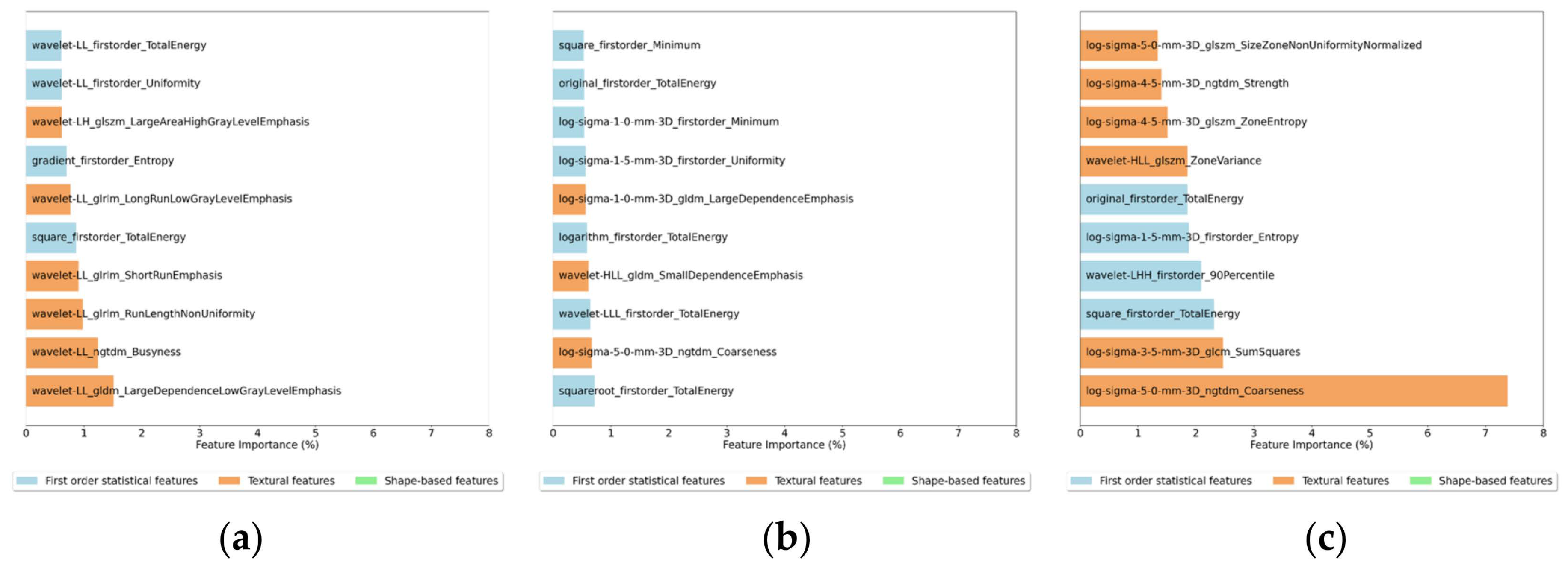
- First order statistical features—describe the variations of pixel’s intensity inside the defined ROI, and include metrics such as the energy, entropy or median of the pixel intensities;
- Textural features—can describe the tumour heterogeneity and the inter-voxel relations in the image ROI. These relations can be quantified using: the Grey Level Co-occurrence Matrix (GLCM), the Grey Level Run Length Matrix (GLRLM), the Grey Level Size Zone (GLSZM), the Grey Level Dependence Matrix (GLDM) and the Neighbouring Grey Tone Difference Matrix (NGTDM).
- Shape-based—describe the tumour size and shape in 2D or 3D.
3.2.3. Synthetic Minority Oversampling Technique
3.2.4. Random Forest Classifier
3.2.5. XGBoost Classifier
3.3. Deep Learning Pipeline
3.3.1. Pre-Processing
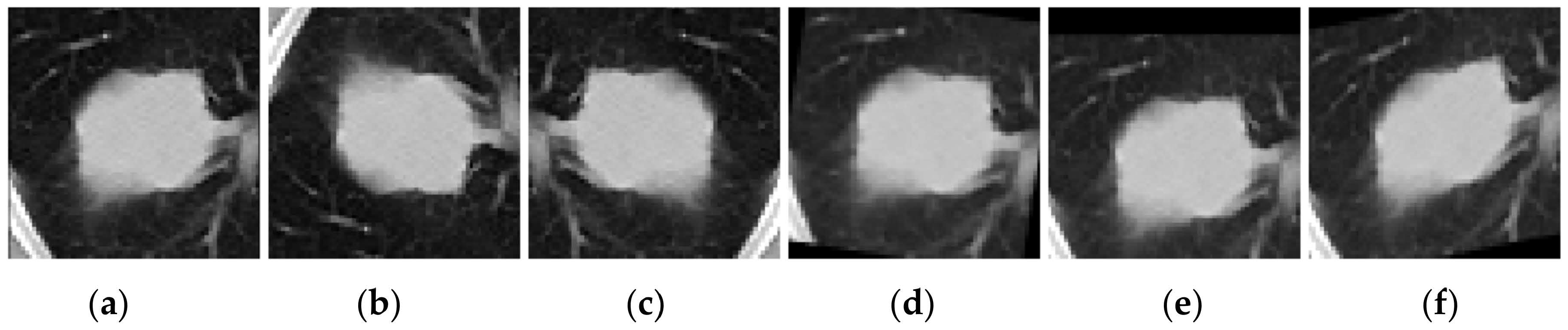
3.3.2. Data Augmentation
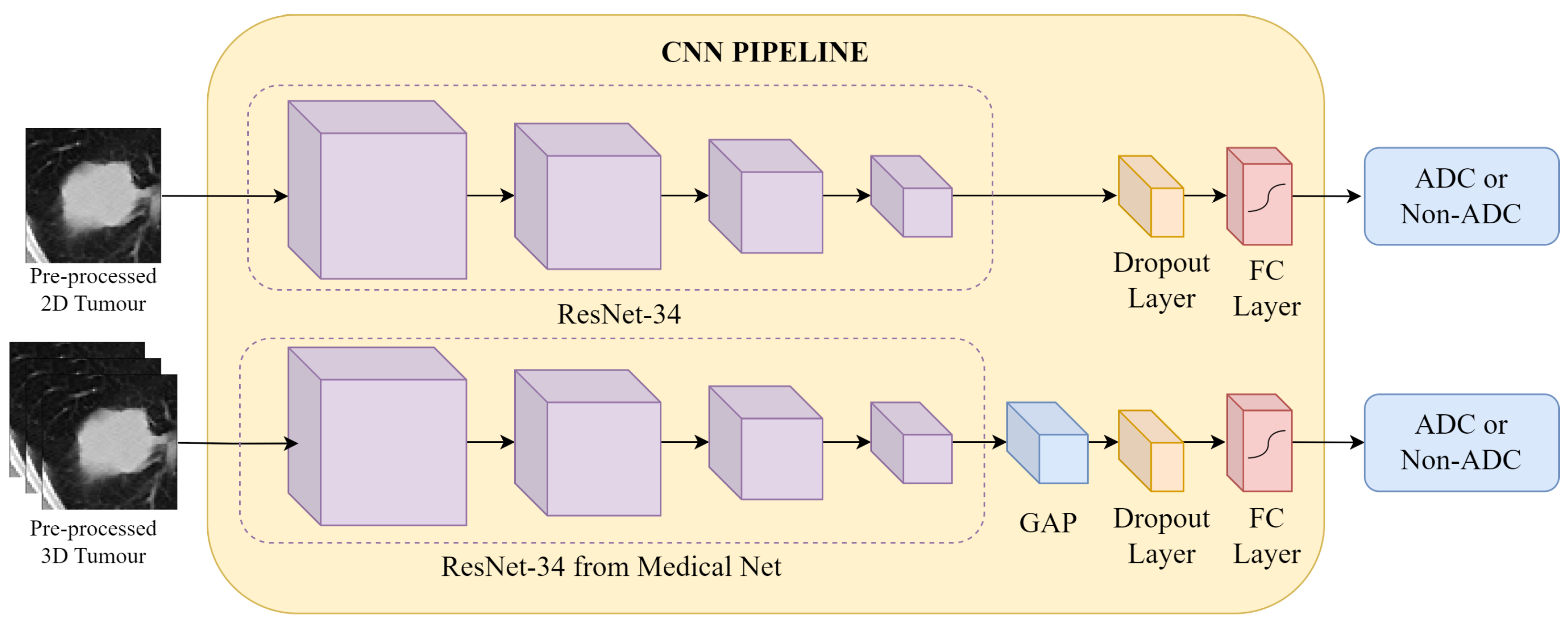
3.3.3. CNN Approach
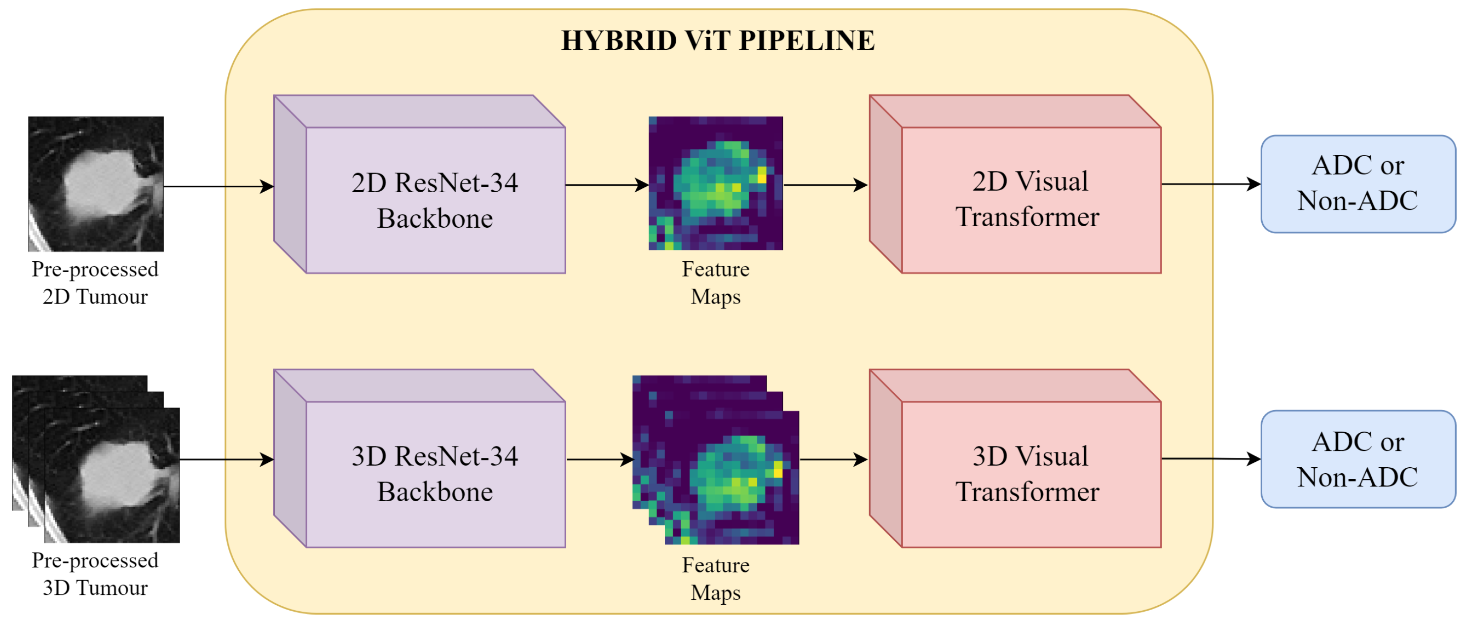
3.3.4. Hybrid ViT Approach
4. Results and Discussion
4.1. 2D vs 3D
4.2. Classical Machine Learning Pipeline
4.3. Deep Learning Pipeline
4.4. Classic Machine Learning vs Deep Learning
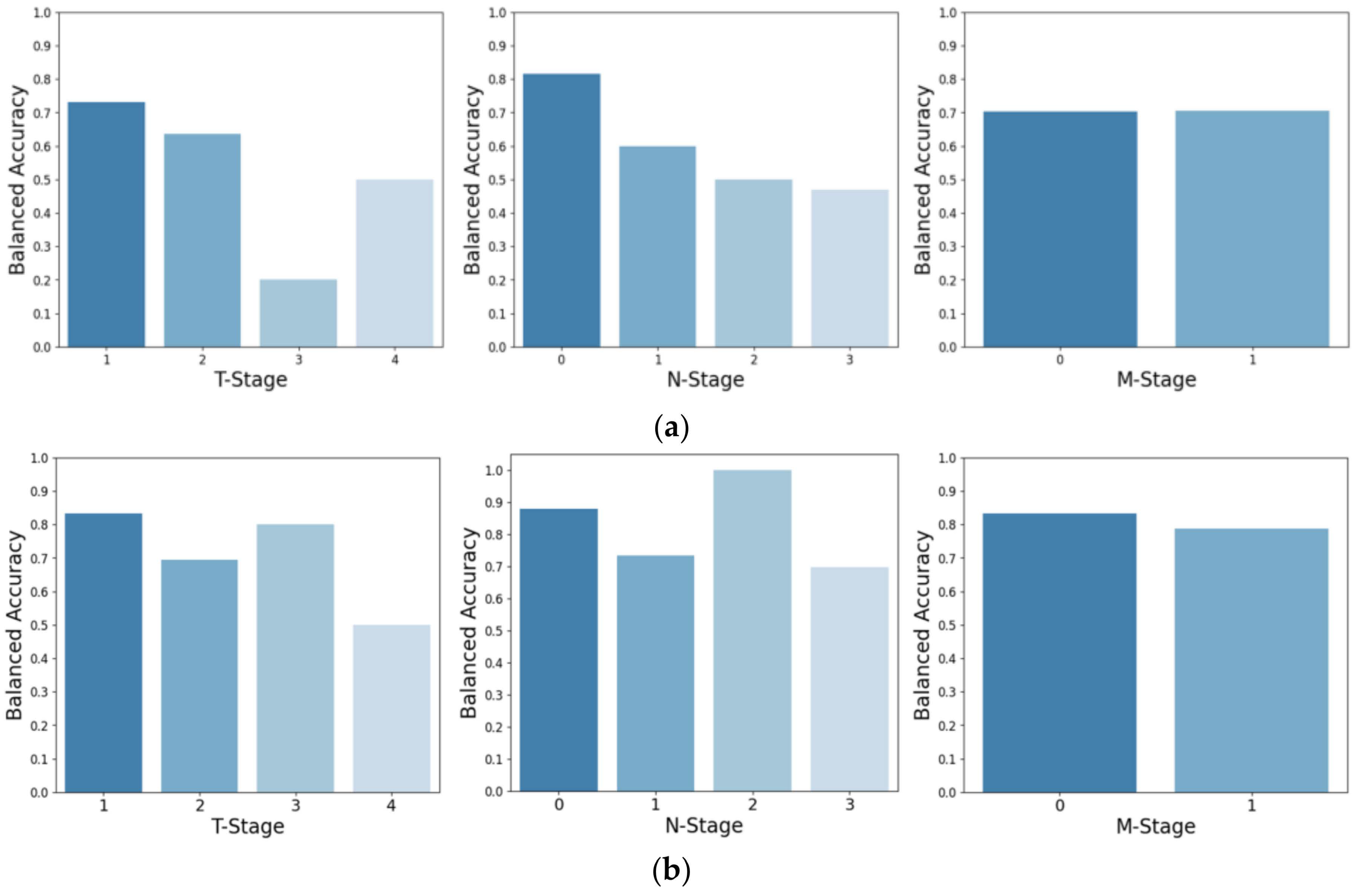
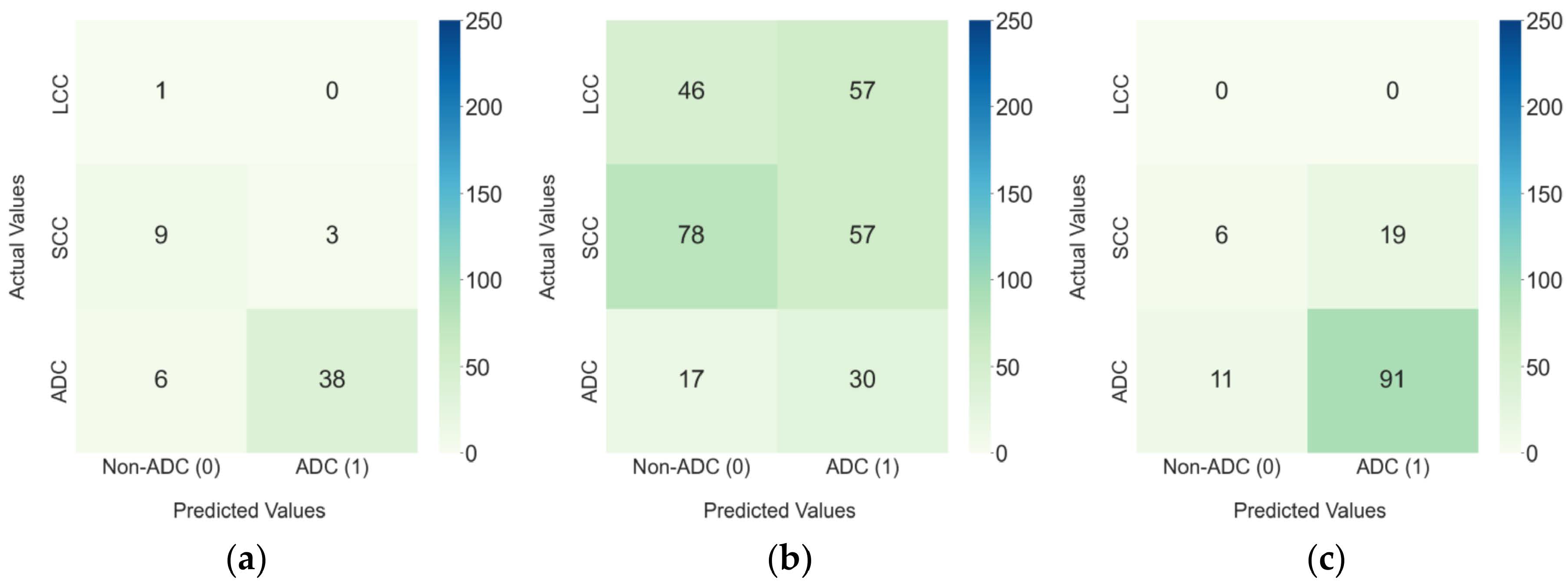
4.5. External Test Datasets
4.6. Limitations
5. Conclusions
Author Contributions
Funding
Institutional Review Board Statement
Informed Consent Statement
Data Availability Statement
Acknowledgments
Conflicts of Interest
Abbreviations
| ADC | Adenocarcinoma |
| AUC | Area Under the Curve |
| BCE | Binary Cross Entropy |
| CNN | Convolutional Neural Networks |
| CT | Computed Tomography |
| DL | Deep Learning |
| FC | Fully Connected |
| GAP | Global Average Pooling |
| GLCM | Grey Level Co-occurrence Matrix |
| GLDM | Grey Level Dependence Matrix |
| GLRLM | Grey Level Run Length Matrix |
| GLSZM | Grey Level Size Zone |
| GTV | Gross Tumour Volume |
| HU | Hounsfield Units |
| IBSI | Image Biomarker Standardization Initiative |
| KNN | K-Nearest Neighbors |
| LCC | Large Cell Carcinoma |
| LOG | Laplacian Gaussian Filters |
| ML | Machine Learning |
| MLP | Multi-Layer Perceptron |
| NGTDM | Neighbouring Grey Tone Difference Matrix |
| NOS | Not Otherwise Specified |
| NSCLC | Non-Small-Cell Lung Cancer |
| ResNet | Residual Neural Network |
| RF | Random Forest |
| ROI | Region Of Interest |
| SCC | Squamous Cell Carcinoma |
| SCLC | Small-Cell Lung Cancer |
| SMOTE | Synthetic Minority Oversampling Technique |
| SVM | Support-Vector Machine |
| ViT | Vision Transformer |
| WHO | World Health Organization |
| XGBoost | eXtreme Gradient Boosting |
Appendix A. Criteria Used for Dataset Uniformization
- non-original DICOM series;
- series without tumour annotation;
- series with non-continual tumour annotation;
- series with a slice thickness larger than 5 mm.
- cases without complete information (e.g., lack annotation of the GTV or lack of histopathological diagnosis);
- cases without an “Original” DICOM series;
- cases in which the GTV segmentation is not continuous (e.g., presence of multiple or divided nodules);
- cases where the GTV is located in other anatomical structures (e.g., located in the ribs);
- cases in which the nodule is located outside the lung parenchyma (e.g., in mediastinal lymph nodes).
References
- World Health Organization. Global Cancer Burden Growing, Amidst Mounting Need for Services. 2024. Available online: https://www.who.int/news/item/01-02-2024-global-cancer-burden-growing--amidst-mounting-need-for-services (accessed on 6 November 2024).
- Sung, H.; Ferlay, J.; Siegel, R.L.; Laversanne, M.; Soerjomataram, I.; Jemal, A.; Bray, F. Global Cancer Statistics 2020: GLOBOCAN Estimates of Incidence and Mortality Worldwide for 36 Cancers in 185 Countries. CA Cancer J. Clin. 2021, 71, 209–249. [Google Scholar] [CrossRef] [Scilit] [PubMed]
- Schabath, M.B.; Cote, M.L. Cancer Progress and Priorities: Lung Cancer. Cancer Epidemiol. Biomark. Prev. 2019, 28, 1563–1579. [Google Scholar] [CrossRef] [Scilit]
- Luo, W. Nasopharyngeal carcinoma ecology theory: Cancer as multidimensional spatiotemporal “unity of ecology and evolution” pathological ecosystem. Theranostics 2023, 1607–1631. [Google Scholar] [CrossRef] [Scilit]
- Khodabakhshi, Z.; Mostafaei, S.; Arabi, H.; Oveisi, M.; Shiri, I.; Zaidi, H. Non-small cell lung carcinoma histopathological subtype phenotyping using high-dimensional multinomial multiclass CT radiomics signature. Comput. Biol. Med. 2021, 136, 104752. [Google Scholar] [CrossRef] [Scilit]
- Patil, R.; Mahadevaiah, G.; Dekker, A. An Approach Toward Automatic Classification of Tumor Histopathology of Non–Small Cell Lung Cancer Based on Radiomic Features. Tomography 2016, 2, 374–377. [Google Scholar] [CrossRef] [Scilit]
- D’Arnese, E.; di Donato, G.W.; del Sozzo, E.; Santambrogio, M.D. Towards an Automatic Imaging Biopsy of Non-Small Cell Lung Cancer. In Proceedings of the 2019 IEEE EMBS International Conference on Biomedical & Health Informatics (BHI), Chicago, IL, USA, 19–22 May 2019; pp. 1–4, ISSN 2641-3604. [Google Scholar] [CrossRef] [Scilit]
- Marentakis, P.; Karaiskos, P.; Kouloulias, V.; Kelekis, N.; Argentos, S.; Oikonomopoulos, N.; Loukas, C. Lung cancer histology classification from CT images based on radiomics and deep learning models. Med. Biol. Eng. Comput. 2021, 59, 215–226. [Google Scholar] [CrossRef] [Scilit]
- Chen, X.; Feng, B.; Chen, Y.; Liu, K.; Li, K.; Duan, X.; Hao, Y.; Cui, E.; Liu, Z.; Zhang, C.; et al. A CT-based radiomics nomogram for prediction of lung adenocarcinomas and granulomatous lesions in patient with solitary sub-centimeter solid nodules. Cancer Imaging 2020, 20, 45. [Google Scholar] [CrossRef] [Scilit]
- Avanzo, M.; Stancanello, J.; Pirrone, G.; Sartor, G. Radiomics and deep learning in lung cancer. Strahlenther. Und Onkol. 2020, 196, 879–887. [Google Scholar] [CrossRef] [Scilit]
- Thawani, R.; McLane, M.; Beig, N.; Ghose, S.; Prasanna, P.; Velcheti, V.; Madabhushi, A. Radiomics and radiogenomics in lung cancer: A review for the clinician. Lung Cancer 2018, 115, 34–41. [Google Scholar] [CrossRef] [Scilit]
- Saikia, T.; Hansdah, M.; Singh, K.K.; Bajpai, M.K. Classification of Lung Nodules based on Transfer Learning with K-Nearest Neighbor (KNN). In Proceedings of the 2022 IEEE International Conference on Imaging Systems and Techniques (IST), Virtual Conference, 21–23 June 2022; pp. 1–2809. [Google Scholar] [CrossRef] [Scilit]
- Pang, S.; Zhang, Y.; Ding, M.; Wang, X.; Xie, X. A Deep Model for Lung Cancer Type Identification by Densely Connected Convolutional Networks and Adaptive Boosting. IEEE Access 2020, 8, 4799–4805. [Google Scholar] [CrossRef] [Scilit]
- Chaunzwa, T.L.; Hosny, A.; Xu, Y.; Shafer, A.; Diao, N.; Lanuti, M.; Christiani, D.C.; Mak, R.H.; Aerts, H.J.W.L. Deep learning classification of lung cancer histology using CT images. Sci. Rep. 2021, 11, 5471. [Google Scholar] [CrossRef] [Scilit] [PubMed]
- Liu, H.; Jiao, Z.; Han, W.; Jing, B. Identifying the histologic subtypes of non-small cell lung cancer with computed tomography imaging: A comparative study of capsule net, convolutional neural network, and radiomics. Quant. Imaging Med. Surg. 2021, 11, 2756765. [Google Scholar] [CrossRef] [Scilit]
- Guo, Y.; Song, Q.; Jiang, M.; Guo, Y.; Xu, P.; Zhang, Y.; Fu, C.C.; Fang, Q.; Zeng, M.; Yao, X. Histological Subtypes Classification of Lung Cancers on CT Images Using 3D Deep Learning and Radiomics. Acad. Radiol. 2021, 28, e258–e266. [Google Scholar] [CrossRef] [Scilit]
- Malaviya, N.; Rahevar, M.; Virani, A.; Ganatra, A.; Bhuva, K. LViT: Vision Transformer for Lung cancer Detection. In Proceedings of the 2023 International Conference on Artificial Intelligence and Smart Communication (AISC), Greater Noida, India, 27–29 January 2023; pp. 93–98. [Google Scholar] [CrossRef] [Scilit]
- Nejad, R.R.; Hooshmand, S. HViT4Lung: Hybrid Vision Transformers Augmented by Transfer Learning to Enhance Lung Cancer Diagnosis. In Proceedings of the 2023 5th International Conference on Bio-Engineering for Smart Technologies (BioSMART), Paris, France, 7–9 June 2023; pp. 1–7. [Google Scholar] [CrossRef] [Scilit]
- Pandey, A.; Kumar, A. Deep Features Based Automated Multimodel System for Classification of Non-Small Cell Lung Cancer. In Proceedings of the 2022 IEEE Delhi Section Conference (DELCON), New Delhi, India, 11–13 February 2022; pp. 1–7. [Google Scholar] [CrossRef] [Scilit]
- Moitra, D.; Mandal, R.K. Prediction of Non-small Cell Lung Cancer Histology by a Deep Ensemble of Convolutional and Bidirectional Recurrent Neural Network. J. Digit. Imaging 2020, 33, 895–902. [Google Scholar] [CrossRef] [Scilit]
- Wang, R.; Zhang, Y.; Yang, J. TransPND: A Transformer Based Pulmonary Nodule Diagnosis Method on CT Image. In Proceedings of the Pattern Recognition and Computer Vision, Shenzhen, China, 4–7 November 2022; Yu, S., Zhang, Z., Yuen, P.C., Han, J., Tan, T., Guo, Y., Lai, J., Zhang, J., Eds.; Lecture Notes in Computer Science; Springer Nature: Cham, Switzerland, 2022; pp. 348–360. [Google Scholar] [CrossRef] [Scilit]
- Ubaldi, L.; Valenti, V.; Borgese, R.F.; Collura, G.; Fantacci, M.E.; Ferrera, G.; Iacoviello, G.; Abbate, B.F.; Laruina, F.; Tripoli, A.; et al. Strategies to develop radiomics and machine learning models for lung cancer stage and histology prediction using small data samples. Phys. Med. 2021, 90, 13–22. [Google Scholar] [CrossRef] [Scilit]
- Zhu, X.; Dong, D.; Chen, Z.; Fang, M.; Zhang, L.; Song, J.; Yu, D.; Zang, Y.; Liu, Z.; Shi, J.; et al. Radiomic signature as a diagnostic factor for histologic subtype classification of non-small cell lung cancer. Eur. Radiol. 2018, 28, 2772–2778. [Google Scholar] [CrossRef] [Scilit]
- Bianconi, F.; Palumbo, I.; Spanu, A.; Nuvoli, S.; Fravolini, M.L.; Palumbo, B. PET/CT Radiomics in Lung Cancer: An Overview. Appl. Sci. 2020, 10, 1718. [Google Scholar] [CrossRef] [Scilit]
- Egger, J.; Gsaxner, C.; Pepe, A.; Pomykala, K.L.; Jonske, F.; Kurz, M.; Li, J.; Kleesiek, J. Medical deep learning—A systematic meta-review. Comput. Methods Programs Biomed. 2022, 221, 106874. [Google Scholar] [CrossRef] [Scilit]
- Tomassini, S.; Falcionelli, N.; Sernani, P.; Burattini, L.; Dragoni, A.F. Lung nodule diagnosis and cancer histology classification from computed tomography data by convolutional neural networks: A survey. Comput. Biol. Med. 2022, 146, 105691. [Google Scholar] [CrossRef] [Scilit]
- Tashtoush, Y.; Obeidat, R.; Al-Shorman, A.; Darwish, O.; Al-Ramahi, M.; Darweesh, D. Enhanced convolutional neural network for non-small cell lung cancer classification. Int. J. Electr. Comput. Eng. 2023, 13, 1024–1038. [Google Scholar] [CrossRef] [Scilit]
- Ma, C.; Yue, S. Minimum Sample Size Estimate for Classifying Invasive Lung Adenocarcinoma. Appl. Sci. 2022, 12, 8469. [Google Scholar] [CrossRef] [Scilit]
- Wang, C.; Shao, J.; Lv, J.; Cao, Y.; Zhu, C.; Li, J.; Shen, W.; Shi, L.; Liu, D.; Li, W. Deep learning for predicting subtype classification and survival of lung adenocarcinoma on computed tomography. Transl. Oncol. 2021, 14, 101141. [Google Scholar] [CrossRef] [Scilit]
- Parvaiz, A.; Khalid, M.A.; Zafar, R.; Ameer, H.; Ali, M.; Fraz, M.M. Vision Transformers in medical computer vision—A contemplative retrospection. Eng. Appl. Artif. Intell. 2023, 122, 106126. [Google Scholar] [CrossRef] [Scilit]
- Dosovitskiy, A.; Beyer, L.; Kolesnikov, A.; Weissenborn, D.; Zhai, X.; Unterthiner, T.; Dehghani, M.; Minderer, M.; Heigold, G.; Gelly, S.; et al. An Image is Worth 16x16 Words: Transformers for Image Recognition at Scale. arXiv 2020, arXiv:2010.11929. [Google Scholar]
- Guo, J.; Han, K.; Wu, H.; Tang, Y.; Chen, X.; Wang, Y.; Xu, C. CMT: Convolutional Neural Networks Meet Vision Transformers. In Proceedings of the 2022 IEEE/CVF Conference on Computer Vision and Pattern Recognition (CVPR), New Orleans, LA, USA, 18–24 June 2022; pp. 12165–12175, ISSN 2575-7075. [Google Scholar] [CrossRef] [Scilit]
- Varoquaux, G.; Cheplygina, V. Machine learning for medical imaging: Methodological failures and recommendations for the future. npj Digit. Med. 2022, 5, 48. [Google Scholar] [CrossRef] [Scilit]
- Swensen, S.; Silverstein, M.; Ilstrup, D.; Schleck, C.; Edell, E. The probability of malignancy in solitary pulmonary nodules. Application to small radiologically indeterminate nodules. Arch. Intern. Med. 1997, 157, 849–855. [Google Scholar]
- Li, P.; Wang, S.; Li, T.; Lu, J.; HuangFu, Y.; Wang, D. A Large-Scale CT and PET/CT Dataset for Lung Cancer Diagnosis; The Cancer Imaging Archive: Bethesda, MD, USA, 2020. [Google Scholar] [CrossRef]
- Aerts, H.J.W.L.; Wee, L.; Rios Velazquez, E.; Leijenaar, R.T.H.; Parmar, C.; Grossmann, P.; Carvalho, S.; Bussink, J.; Monshouwer, R.; Haibe-Kains, B.; et al. Data from NSCLC-Radiomics; The Cancer Imaging Archive: Bethesda, MD, USA, 2019. [Google Scholar] [CrossRef] [Scilit]
- Bakr, S.; Gevaert, O.; Echegaray, S.; Ayers, K.; Zhou, M.; Shafiq, M.; Zheng, H.; Zhang, W.; Leung, A.; Kadoch, M.; et al. Data for NSCLC Radiogenomics Collection; The Cancer Imaging Archive: Bethesda, MD, USA, 2017. [Google Scholar] [CrossRef]
- Clark, K.; Vendt, B.; Smith, K.; Freymann, J.; Kirby, J.; Koppel, P.; Moore, S.; Phillips, S.; Maffitt, D.; Pringle, M.; et al. The Cancer Imaging Archive (TCIA): Maintaining and Operating a Public Information Repository. J. Digit. Imaging 2013, 26, 1045–1057. [Google Scholar] [CrossRef] [Scilit]
- Chen, L.; Qi, H.; Lu, D.; Zhai, J.; Cai, K.; Wang, L.; Liang, G.; Zhang, Z. Machine vision-assisted identification of the lung adenocarcinoma category and high-risk tumor area based on CT images. Patterns 2022, 3, 100464. [Google Scholar] [CrossRef] [Scilit]
- van Griethuysen, J.J.M.; Fedorov, A.; Parmar, C.; Hosny, A.; Aucoin, N.; Narayan, V.; Beets-Tan, R.G.H.; Fillion-Robin, J.C.; Pieper, S.; Aerts, H.J.W.L. Computational Radiomics System to Decode the Radiographic Phenotype. Cancer Res. 2017, 77, e104–e107. [Google Scholar] [CrossRef] [Scilit]
- Zwanenburg, A.; Leger, S.; Vallières, M.; Löck, S. Image biomarker standardisation initiative. Radiology 2020, 295, 328–338. [Google Scholar] [CrossRef] [Scilit]
- Chawla, N.V.; Bowyer, K.W.; Hall, L.O.; Kegelmeyer, W.P. SMOTE: Synthetic Minority Over-sampling Technique. J. Artif. Intell. Res. 2002, 16, 321–357. [Google Scholar] [CrossRef] [Scilit]
- Lemaître, G.; Nogueira, F.; Aridas, C.K. Imbalanced-learn: A python toolbox to tackle the curse of imbalanced datasets in machine learning. J. Mach. Learn. Res. 2017, 18, 559–563. [Google Scholar]
- Pedregosa, F.; Varoquaux, G.; Gramfort, A.; Michel, V.; Thirion, B.; Grisel, O.; Blondel, M.; Prettenhofer, P.; Weiss, R.; Dubourg, V.; et al. Scikit-learn: Machine Learning in Python. J. Mach. Learn. Res. 2011, 12, 2825–2830. [Google Scholar]
- Cardoso, M.J.; Li, W.; Brown, R.; Ma, N.; Kerfoot, E.; Wang, Y.; Murrey, B.; Myronenko, A.; Zhao, C.; Yang, D.; et al. MONAI: An open-source framework for deep learning in healthcare. arXiv 2022, arXiv:2211.02701. [Google Scholar] [CrossRef] [Scilit]
- He, K.; Zhang, X.; Ren, S.; Sun, J. Deep Residual Learning for Image Recognition. arXiv 2015, arXiv:1512.03385. [Google Scholar] [CrossRef] [Scilit]
- Paszke, A.; Gross, S.; Massa, F.; Lerer, A.; Bradbury, J.; Chanan, G.; Killeen, T.; Lin, Z.; Gimelshein, N.; Antiga, L.; et al. PyTorch: An Imperative Style, High-Performance Deep Learning Library. In Proceedings of the Advances in Neural Information Processing Systems 32, Vancouver, BC, Canada, 8–14 December 2019; Wallach, H., Larochelle, H., Beygelzimer, A., d’Alché Buc, F., Fox, E., Garnett, R., Eds.; Curran Associates, Inc.: Red Hook, NY, USA, 2019; pp. 8024–8035. [Google Scholar]
- Chen, S.; Ma, K.; Zheng, Y. Med3D: Transfer Learning for 3D Medical Image Analysis. arXiv 2019, arXiv:1904.00625. [Google Scholar]
- Lucidrains GitHub. Lucidrains/Vit-Pytorch: Implementation of Vision Transformer, a Simple Way to Achieve SOTA in Vision Classification With Only a Single Transformer Encoder, in Pytorch. Available online: https://github.com/lucidrains/vit-pytorch/ (accessed on 6 September 2024).
- Rami-Porta, R. Future Perspectives on the TNM Staging for Lung Cancer. Cancers 2021, 13, 1940. [Google Scholar] [CrossRef] [Scilit]
- Dunn, B.; Pierobon, M.; Wei, Q. Automated Classification of Lung Cancer Subtypes Using Deep Learning and CT-Scan Based Radiomic Analysis. Bioengineering 2023, 10, 690. [Google Scholar] [CrossRef] [Scilit]

| Dataset | Class | Total | Train | Validation | Test |
|---|---|---|---|---|---|
| Lung-PET-CT-Dx | ADC | 221 | 146 | 31 | 44 |
| Non-ADC | 64 | 38 | 13 | 13 | |
| Total | 285 | 184 | 44 | 57 | |
| NSCLC-Radiomics | ADC | 47 | - | - | 47 |
| Non-ADC | 238 | - | - | 238 | |
| Total | 285 | - | - | 285 | |
| NSCLC-Radiogenomics | ADC | 102 | - | - | 102 |
| Non-ADC | 25 | - | - | 25 | |
| Total | 127 | - | - | 127 |
| Hyper-Parameter | Values |
|---|---|
| NEstimators | {50, 100, 150, 200, 250, 300, 400, 500, 750, 1000} |
| MaxDepth | {None, 1, 10, 50, 100, 150, 200, 300} |
| MinSamplesSplit | {2, 10, 15, 20, 25, 30, 50, 75, 100} |
| MinSamplesLeaf | {1, 2, 4, 6, 8, 10} |
| Criterion | {gini, entropy, log_loss} |
| Hyper-Parameter | Values |
|---|---|
| NEstimators | {50, 100, 150, 200, 250, 300, 400, 500, 750, 1000} |
| MaxDepth | {None, 1, 10, 50, 100, 150, 200, 300} |
| MinChildWeight | {1, 2, 4, 6} |
| {0.001, 0.01, 0.1} | |
| {0, 0.5, 1, 2} | |
| {0, 0.1, 0.2, 0.3} |
| Transformations | Range |
|---|---|
| Flips | - |
| Rotation | [−20, 20]° |
| Shift | [−15, 15]% |
| Shear | [−20, 20]% |
| Hyper-Parameter | Values |
|---|---|
| Dropout | {0, 0.5} |
| Learning Rate | {0.000005, 0.000001, 0.00001} |
| Weight Decay | {0, 0.001, 0.01} |
| Hyper-Parameter | Values |
|---|---|
| ViT Layers | {6, 8, 10} |
| Learning Rate | {0.00001, 0.00005, 0.0001} |
| Weight Decay | {0, 0.001, 0.01} |
| Classifier | Input | Test Set | AUC | Balanced Accuracy | Precision | Recall | Specificity |
|---|---|---|---|---|---|---|---|
| Random Forest | 2D | Lung-PET-CT-Dx | 0.806 | 0.712 | 0.867 | 0.886 | 0.538 |
| NSCLC-Radiomics | 0.543 | 0.497 | 0.164 | 0.936 | 0.059 | ||
| NSCLC-Radiogenomics | 0.659 | 0.531 | 0.814 | 0.941 | 0.120 | ||
| 3D | Lung-PET-CT-Dx | 0.836 | 0.762 | 0.889 | 0.909 | 0.615 | |
| NSCLC-Radiomics | 0.560 | 0.509 | 0.168 | 0.830 | 0.189 | ||
| NSCLC-Radiogenomics | 0.676 | 0.555 | 0.821 | 0.990 | 0.120 | ||
| ResNet-34 | 2D | Lung-PET-CT-Dx | 0.783 | 0.660 | 0.861 | 0.705 | 0.615 |
| NSCLC-Radiomics | 0.577 | 0.598 | 0.222 | 0.638 | 0.559 | ||
| NSCLC-Radiogenomics | 0.495 | 0.527 | 0.814 | 0.814 | 0.240 | ||
| 3D | Lung-PET-CT-Dx | 0.841 | 0.700 | 0.955 | 0.477 | 0.923 | |
| NSCLC-Radiomics | 0.576 | 0.550 | 0.219 | 0.340 | 0.761 | ||
| NSCLC-Radiogenomics | 0.589 | 0.555 | 0.839 | 0.510 | 0.600 |
| Input | Classifier | Test Set | AUC | Balanced Accuracy | Precision | Recall | Specificity |
|---|---|---|---|---|---|---|---|
| 3D | XGBoost | Lung-PET-CT-Dx | 0.853 | 0.801 | 0.909 | 0.909 | 0.692 |
| NSCLC-Radiomics | 0.570 | 0.533 | 0.176 | 0.872 | 0.193 | ||
| NSCLC-Radiogenomics | 0.745 | 0.550 | 0.820 | 0.980 | 0.120 | ||
| Hybrid ViT | Lung-PET-CT-Dx | 0.869 | 0.816 | 0.927 | 0.864 | 0.769 | |
| NSCLC-Radiomics | 0.617 | 0.580 | 0.208 | 0.638 | 0.521 | ||
| NSCLC-Radiogenomics | 0.613 | 0.567 | 0.827 | 0.892 | 0.240 |
| Model | Type of Features | ||
|---|---|---|---|
| First Order Statistical | Textural | Shape-Based | |
| 2D Random Forest | 34.6 % | 64.7 % | 0.6 % |
| 3D Random Forest | 28.3 % | 70.7 % | 0.9 % |
| 3D XGBoost | 24.8 % | 73.8 % | 1.4 % |
Disclaimer/Publisher’s Note: The statements, opinions and data contained in all publications are solely those of the individual author(s) and contributor(s) and not of MDPI and/or the editor(s). MDPI and/or the editor(s) disclaim responsibility for any injury to people or property resulting from any ideas, methods, instructions or products referred to in the content. |
© 2025 by the authors. Licensee MDPI, Basel, Switzerland. This article is an open access article distributed under the terms and conditions of the Creative Commons Attribution (CC BY) license (https://creativecommons.org/licenses/by/4.0/).
Share and Cite
Gouveia, M.; Mendes, T.; Rodrigues, E.M.; P. Oliveira, H.; Pereira, T. Comparing 2D and 3D Feature Extraction Methods for Lung Adenocarcinoma Prediction Using CT Scans: A Cross-Cohort Study. Appl. Sci. 2025, 15, 1148. https://doi.org/10.3390/app15031148
Gouveia M, Mendes T, Rodrigues EM, P. Oliveira H, Pereira T. Comparing 2D and 3D Feature Extraction Methods for Lung Adenocarcinoma Prediction Using CT Scans: A Cross-Cohort Study. Applied Sciences. 2025; 15(3):1148. https://doi.org/10.3390/app15031148
Chicago/Turabian StyleGouveia, Margarida, Tânia Mendes, Eduardo M. Rodrigues, Hélder P. Oliveira, and Tania Pereira. 2025. "Comparing 2D and 3D Feature Extraction Methods for Lung Adenocarcinoma Prediction Using CT Scans: A Cross-Cohort Study" Applied Sciences 15, no. 3: 1148. https://doi.org/10.3390/app15031148
APA StyleGouveia, M., Mendes, T., Rodrigues, E. M., P. Oliveira, H., & Pereira, T. (2025). Comparing 2D and 3D Feature Extraction Methods for Lung Adenocarcinoma Prediction Using CT Scans: A Cross-Cohort Study. Applied Sciences, 15(3), 1148. https://doi.org/10.3390/app15031148

