Article

In Vitro-Derived Vitis labrusca var. Isabella Juices Restore Intestinal Epithelial Integrity via Antioxidant and Anti-Inflammatory Actions

by
Vanessa Dalla Costa
1,
Carolina Frison
2,
Raffaella Filippini
1 and
Paola Brun
2,*
1
Department of Pharmaceutical and Pharmacological Sciences, University of Padova, Via Marzolo, 5, 35131 Padua, Italy
2
Department of Molecular Medicine, University of Padova, Via Gabelli 63, 35121 Padova, Italy
*
Author to whom correspondence should be addressed.
Appl. Sci. 2025, 15(24), 13192; https://doi.org/10.3390/app152413192
Submission received: 7 November 2025 / Revised: 12 December 2025 / Accepted: 13 December 2025 / Published: 16 December 2025
(This article belongs to the Section Chemical and Molecular Sciences)

Featured Application

These findings support the use of Vitis labrusca var. Isabella cell-culture juices as natural products with beneficial potential in gut inflammatory disorders, offering a renewable and scalable support to conventional anti-inflammatory agents.

Abstract

Inflammatory bowel disease is characterised by chronic mucosal inflammation, oxidative stress, and impaired epithelial barrier function. Current therapies primarily suppress inflammation but do not effectively restore epithelial integrity. In this study, we established in vitro cell cultures of Vitis labrusca var. Isabella to obtain juices that were chemically characterised and assessed for antioxidant and anti-inflammatory activities in human intestinal epithelial cell lines (i.e., Caco-2). Chemical analysis revealed variable levels of stilbenoids, including trans-resveratrol and resveratrol diglucosides depending on culture conditions. The suspension-derived juice grown in darkness (SVMD) significantly reduced lipopolysaccharide-induced IL-1β and TNF-α release and mitigated oxidative stress in Caco-2 cells by lowering levels of intracellular reactive oxygen species. In Caco-2 monolayers infected with Salmonella enterica, SVMD preserved transepithelial electrical resistance, indicating protection of epithelial barrier integrity, without exerting direct antibacterial effects. These findings demonstrate that V. labrusca cell-culture juices exert potent antioxidant and anti-inflammatory actions and promote epithelial protection through modulation of redox balance. Overall, this study highlights the potential of sustainable cell-culture-derived materials as promising natural products for supporting intestinal homeostasis and managing gut inflammatory disorders.

1. Introduction

Inflammatory bowel disease (IBD) comprises chronic, relapsing inflammatory disorders of the gastrointestinal tract, mainly Crohn’s disease (CD) and ulcerative colitis (UC), which arise from a dysregulated mucosal immune response to unidentified antigens or luminal microbes. The incidence and prevalence of IBD continue to rise globally, with over seven million people currently affected [1]. Patients suffering from IBD commonly experience abdominal pain, diarrhoea, weight loss, fatigue, and anorexia [2,3]. Histologically, UC presents with continuous, superficial mucosal inflammation limited to the colon, whereas CD can involve any gastrointestinal segment and is characterised by transmural inflammation, deep ulcerations, and fistulae [4]. These features of chronic immune-mediated damage highlight the central role of anti-inflammatory therapies in IBD management.
Conventional treatments for IBD rely on anti-inflammatory and immunomodulatory agents targeting immune-driven pathways [5]. Biologic therapies such as anti-TNF agents effectively induce and maintain remission; however, up to 30% of patients report primary non-response or develop anti-drug antibodies [6]. Other biologics, including anti-integrin and anti-IL-12/23 antibodies, and small molecules such as JAK inhibitors (tofacitinib, upadacitinib, filgotinib) or S1P receptor modulators (ozanimod, etrasimod) have expanded oral therapeutic options, though their use may be limited by safety concerns, including cardiovascular risks [7]. Despite these advances, the long-term effectiveness of the current therapies remains uncertain, as they primarily suppress immune activity and do not directly restore epithelial integrity or redox balance. Indeed, oxidative stress plays a central role in the pathogenesis of IBD. During active inflammation, infiltrating neutrophils and macrophages generate excessive reactive oxygen species (ROS), which overwhelm local antioxidant defences and contribute to epithelial injury. Elevated ROS levels disrupt tight junctions, impair mitochondrial function, and amplify cytokine production [5]. The imbalance between ROS generation and antioxidant capacity highlights the need for alternative treatment strategies to control both inflammation and mucosal repair [8]. In this context, bioactive plant-derived compounds endowed with antioxidant activities could emerge as promising candidates [9].
Among natural sources of anti-inflammatory and antioxidant molecules, grape fruits and leaves are among the most extensively investigated systems. Moreover, grapes are widely consumed worldwide as fresh fruit and processed products such as wine and juices [10,11,12,13]. While Vitis vinifera is the main species used in winemaking, Vitis labrusca has gained attention as an important dietary source of polyphenols, particularly in the Americas [14,15,16,17]. Their phytochemical profile includes catechin, epicatechin, quercetin, caffeic acid, and resveratrol, compounds known for antioxidant and anti-inflammatory activities in epithelial and hepatic models [13,18,19]. V. labrusca juices are especially rich in anthocyanins, flavanols, and stilbenes, which correlate with strong radical-scavenging capacity and inhibition of lipid peroxidation [14]. Grape-derived extracts have also been shown to reduce inflammation and support intestinal barrier function in epithelial cells [20,21], suggesting that V. labrusca may modulate oxidative and inflammatory pathways relevant to colonic health. In our previous work, we established two in vitro callus lines of V. labrusca var. Isabella as a promising and sustainable source of phytochemicals [22]. Here, we further characterised calli and suspension cultures grown in darkness, an advantageous condition for industrial use, and analysed their chemical profiles. We then evaluated the antioxidant and anti-inflammatory activities of the juices derived from these cultures, demonstrating their ability to restore epithelial barrier integrity.
Since almost 80% of the world’s population relies on plant-derived components for their crucial health and wellness [23,24], the application of plant cell culture techniques offers a range of opportunities for sustainable access to healthy products, for which the supply from natural sources is constrained by seasonal, geographical, and biodiversity loss issues.

2. Materials and Methods

2.1. Chemicals

HPLC-grade solvents were purchased from VWR-BDH Chemicals (Milan, Italy), and ultrapure water was withdrawn from a Sartorius Arium system (Sartorius Italy, Varedo, Italy). Trans-resveratrol was purchased from Merck (Milan, Italy). All the medium components were purchased from Duchefa (Micropoli, Milan, Italy).

2.2. Callus and Suspension Obtainment

VM and VB callus cell lines were previously obtained from leaf explants of Vitis labrusca var. Isabella on MS [25] and B5 [26] agarized media supplemented with plant growth regulators: 2,4-dichlorophenoxyacetic acid (1.3 mg/L), kinetin (0.25 mg/L), and naphthaleneacetic acid (0.25 mg/L) as reported in Dalla Costa et al. [22]. VM is the cell line grown in MSA medium, and VB is the cell line grown in B5A medium, both under photoperiod, as previously described [22].
VMD and VBD calli cell lines, VM and VB lines grown in dark conditions, respectively, were obtained by separating the biomass from the parental calli during subcultures and maintaining them in total darkness in the same media as the photoperiod material, at 25 ± 1 °C. The calli were subcultured every 5 weeks.
SVMD suspensions were obtained by transferring VM calli into flasks containing MSA medium [22] without the gellifying agent. Suspensions were kept at 100 rpm and 25 ± 1 °C under a 16/8 h photoperiod. After five cultures, to allow the material stabilisation in the liquid culture, half of the suspension cells were transferred to dark conditions at 100 rpm and 25 ± 1 °C. After a period of stabilisation, SVMD (suspension-cells under dark conditions) were used for the following experiments.

2.3. Chemical Analysis

2.3.1. Juice Preparation

The chemical analysis was done on the 28-day-old calli and 13-day-old suspensions. The juices from calli were obtained as previously described [22]. Briefly, the harvested material was frozen, then thawed, and squeezed and sonicated in an ultrasound bath. After centrifugation at 13,200 rpm, the upper phase (juice) was collected for analysis. The suspension juices were obtained using the same extraction method as for calli, except for the harvesting protocol. The suspensions were filtered, washed with distilled water, and left dry at room temperature until the frosting [27,28]. Once thawed, the juice was extracted. The juices were named for the material they derived from: VM and VB, and VMD and VBD are the juices from calli grown under photoperiod and dark conditions, respectively, and SVMD is the juice from cell suspensions maintained under darkness.

2.3.2. Quantitative Analysis of Stilbenoids Expressed as Trans-Resveratrol Equivalents

Following the previous compound identification and quantification for the VM and VB cell lines [22], in this work, we used the same analytical protocol. Briefly, an Agilent 1100 HPLC Series System (Agilent, Santa Clara, CA, USA) equipped with a degasser, a quaternary gradient pump, a column thermostat, and a diode array detector was used for the quantitative analysis. The separation was performed on a C6-Phenyl Gemini column (5 μm, 250 × 4.6 mm) from Phenomenex (Torrance, CA, USA), maintained at 40 °C. The mobile phase consisted of 0.15% acetic acid in water (A) and acetonitrile (B), with the already reported [22,29] elution program: 97% A at 0–6 min, 75% A at 15 min, 75% A at 20 min, 20% A at 30 min, and 97% A at 40 min. The flow rate was set to 1 mL/min, and the injection volume was 10 μL; chromatograms were acquired at 325 nm. Trans-resveratrol standard solutions were used to build the calibration curve (R2 = 0.999) with six concentration levels. Peak areas were plotted against the trans-resveratrol, and the total stilbenoid content was expressed as trans-resveratrol equivalents. The analysis was performed in duplicate, and the results are expressed as the mean ± standard deviation (SD).

2.4. Biological Activity Assays

2.4.1. Cell Culture

Caco-2 (HTB-37™) and HT29 (HTB-38™) human enterocyte cell lines, both derived from colorectal adenocarcinoma, and T84 (CCL-248™) colonocytes, derived from colorectal carcinoma, were obtained from ATCC (via LGC Standards S.r.L., Sesto San Giovanni, Milan, Italy). Caco-2 cells were maintained in Eagle’s Minimum Essential Medium (EMEM) supplemented with 20% v/v foetal bovine serum (FBS); HT29 cells in Dulbecco’s Modified Eagle’s Medium (DMEM) supplemented with 10% v/v FBS; and T84 cells in a 1:1 mixture of DMEM and Ham’s F-12 Medium supplemented with 10% v/v FBS, antibiotic-antimycotic, and L-glutamine. Media and supplements for the cell cultures were purchased from Thermo Fisher Scientific (Milan, Italy). Cells were grown at 37 °C in a humidified atmosphere containing 5% CO2 and seeded at a density of 1 × 10⁵ cells/mL in sterile plastic culture plates (Sarstedt S.r.l., Trezzano sul Naviglio, Milan, Italy).

2.4.2. Bacterial Culture

Salmonella enterica serovar Typhimurium (ATCC 14028) was used for infection experiments. Bacteria were routinely cultured in Luria–Bertani (LB) broth (Thermo Fisher Scientific) at 37 °C under constant agitation (180 rpm) until reaching mid-logarithmic phase, corresponding to an optical density at 600 nm (OD600) of approximately 0.8. Bacterial cells were then harvested by centrifugation at 4000× g for 10 min, washed twice in sterile phosphate-buffered saline (PBS), and resuspended in antibiotic-free Eagle’s Minimum Essential Medium (EMEM). Bacterial suspensions were prepared in LB medium and subsequently spread onto Hektoen Enteric agar plates (Thermo Fisher Scientific). The plates were incubated at 37 °C for 24 h for bacterial colony enumeration.

2.4.3. Cell Viability Assay

To evaluate potential cytotoxic effects and identify the highest non-toxic concentrations of the tested juices, Caco-2 cells were incubated for 24 h with increasing concentrations of the juices (0–25% v/v). Following exposure, cell viability was determined through the MTT [3-(4,5-dimethylthiazol-2-yl)-2,5-diphenyltetrazolium bromide] assay (Merck, Milan, Italy). An MTT solution (5 mg/mL) was added to each well, and the plates were incubated at 37 °C for 4 h to enable the formation of formazan crystals. The crystals were subsequently dissolved for 16 h in 10% (w/v) sodium dodecyl sulfate containing 0.01 N HCl. Absorbance was recorded at 590 nm using a microplate reader (Victor X2 Multiplate Reader; Perkin Elmer, Milan, Italy). Cell viability was expressed as a percentage relative to untreated control cells. To exclude potential cytotoxic effects, the highest non-toxic juice concentrations identified in Caco-2 cells were subsequently tested in HT29 and T84 cells.

2.4.4. Enzyme-Linked Immunosorbent Assay

Cells were exposed to the juices for 16 h, either with or without 100 ng/mL lipopolysaccharide (LPS) from Salmonella enterica serotype Typhimurium (Merck, Milan, Italy). After incubation, conditioned media were collected and clarified by centrifugation (1600 rpm, 6 min, 4 °C). Concentrations of interleukin (IL)-1β and tumour necrosis factor (TNF)-α were determined using commercially available enzyme-linked immunosorbent assay (ELISA) kits (Thermo Fisher Scientific). The detection limits of the IL-1β and TNF-α ELISA kits were 2 pg/mL and 0.13 pg/mL, respectively. The reactions were visualized with the 3,3′,5,5′-tetramethylbenzidine (TMB) substrate, and optical density was measured at 450 nm using a Victor X2 Multiplate Reader (Perkin Elmer, Milan, Italy).

2.4.5. Detection of Intracellular Reactive Oxygen Species Levels

Intracellular reactive oxygen species (ROS) production was evaluated using the cell-permeable probe 2′,7′-dichlorodihydrofluorescein diacetate (H2DCFDA; Molecular Probes, Invitrogen, Milan, Italy). Once internalized, H2DCFDA is hydrolyzed by cellular esterases and subsequently oxidized to form the fluorescent molecule 2′,7′-dichlorofluorescein (DCF), whose fluorescence intensity reflects the intracellular ROS concentration. To detect ROS generation, we incubated cells with the test juices for 16 h. At the 15-h time point, the stimuli were refreshed, and following a 30-min interval, cells were exposed to 50 μM H2DCFDA in pre-heated phosphate-buffered saline (PBS). Cells not loaded with H2DCFDA served as a negative control to set the autofluorescent signal. Where indicated, along with the juices, cells were incubated with 5 μM MitoTEMPO [(2-(2,2,6,6-tetramethylpiperidin-1-oxyl-4-ylamino)-2-oxoethyl)triphenylphosphonium chloride; Merck], a mitochondria-targeted superoxide scavenger, or with 10 μM MnTBAP (Merck), a superoxide dismutase mimetic and peroxynitrite scavenger. After 30 min of incubation in the dark at 37 °C with H2DCFDA, cells were challenged with 25 μM hydrogen peroxide (H2O2; Merck) for 10 min, then washed, detached with Trypsin-EDTA, and immediately analysed using a BD FACSCanto™ flow cytometer (BD Biosciences, San Jose, CA, USA). A total of 10,000 events per sample were collected. Data were processed and analysed using the Floreada.io software FCS3.0 platform (https://floreada.io/analysis, accessed on 2 September 2025).

2.4.6. Transepithelial Electrical Resistance Assay

To evaluate the protective effects of the juices on epithelial barrier integrity, Caco-2 cells were cultured on permeable Transwell® inserts (polyester membrane, 0.4 μm pore size, growth area 1.12 cm2; Corning Costar, Sial, Rome, Italy) at a seeding density of 2 × 10⁵ cells/cm2. Cells were maintained in complete Eagle’s Minimum Essential Medium (EMEM) supplemented as described above, with medium changes every 3 days in both apical and basolateral chambers. Monolayers were allowed to differentiate for 21 days, and the formation of a tight epithelial barrier was monitored by measuring transepithelial electrical resistance (TEER). TEER values were recorded in Hank’s Balanced Salt solution (HBSS, ThermoFisher) after 20 min equilibration period at room temperature, using a Millicell ERS-2 Voltohmmeter connected to a pair of chopstick electrodes (Millipore; Milan, Italy). Cell monolayers exhibiting TEER values above 500 Ω·cm2 were considered fully differentiated and suitable for experiments. The apical chambers were added with the juices and, 16 h later, infected with Salmonella enterica serovar Typhimurium (multiplicity of infection, MOI 1:10) in antibiotic-free medium. Control wells received the juices alone. After 4 h of infection, the contents of the apical chamber were collected and bacteria were appropriately diluted and plated. The Caco-2 cell inserts were washed and incubated in fresh complete medium containing gentamicin (100 μg/mL) to kill any remaining extracellular bacteria. TEER measurements were determined at 0, 2, 4, and 24 h post-infection. Barrier protection was expressed as the percentage of TEER relative to baseline values and compared among untreated, infected, and juice-treated infected groups.

2.5. Statistical Analysis

Results of the chemical analyses are expressed as the mean ± standard deviation (SD) from two independent experiments, each performed in duplicate. The choice to conduct two independent experimental runs was driven by the need to monitor the stability of the material over the stabilisation period. The low variability observed (coefficient of variation <5%) indicates good analytical precision and supports the reliability of the measurements obtained in both independent experiments. In addition, the limited number of experiments aligns with one of the main objectives of our study, the sustainability, as we aimed to minimise the consumption of time, materials, and energy while ensuring that the data remained robust and reliable. Statistical analyses were performed using one-way ANOVA in GraphPad Prism v7.05 (San Diego, CA, USA). Differences were considered statistically significant at p < 0.05. Distinct Latin letters were assigned in alphabetical order to indicate significant differences among samples, from the richest to the poorest.
Data from biological assays are presented as the mean ± standard error of the mean (SEM) from three independent experiments, each conducted in triplicate. Graphs were prepared using GraphPad Prism version 7.05 (San Diego, CA, USA). Statistical analyses were performed using one-way analysis of variance (ANOVA) followed by Bonferroni’s multiple comparison post hoc test. Differences were considered statistically significant at p < 0.05.

3. Results and Discussion

3.1. Obtainment and Chemical Analysis of Calli in Dark Conditions

3.1.1. Obtainment of Callus Cell Lines

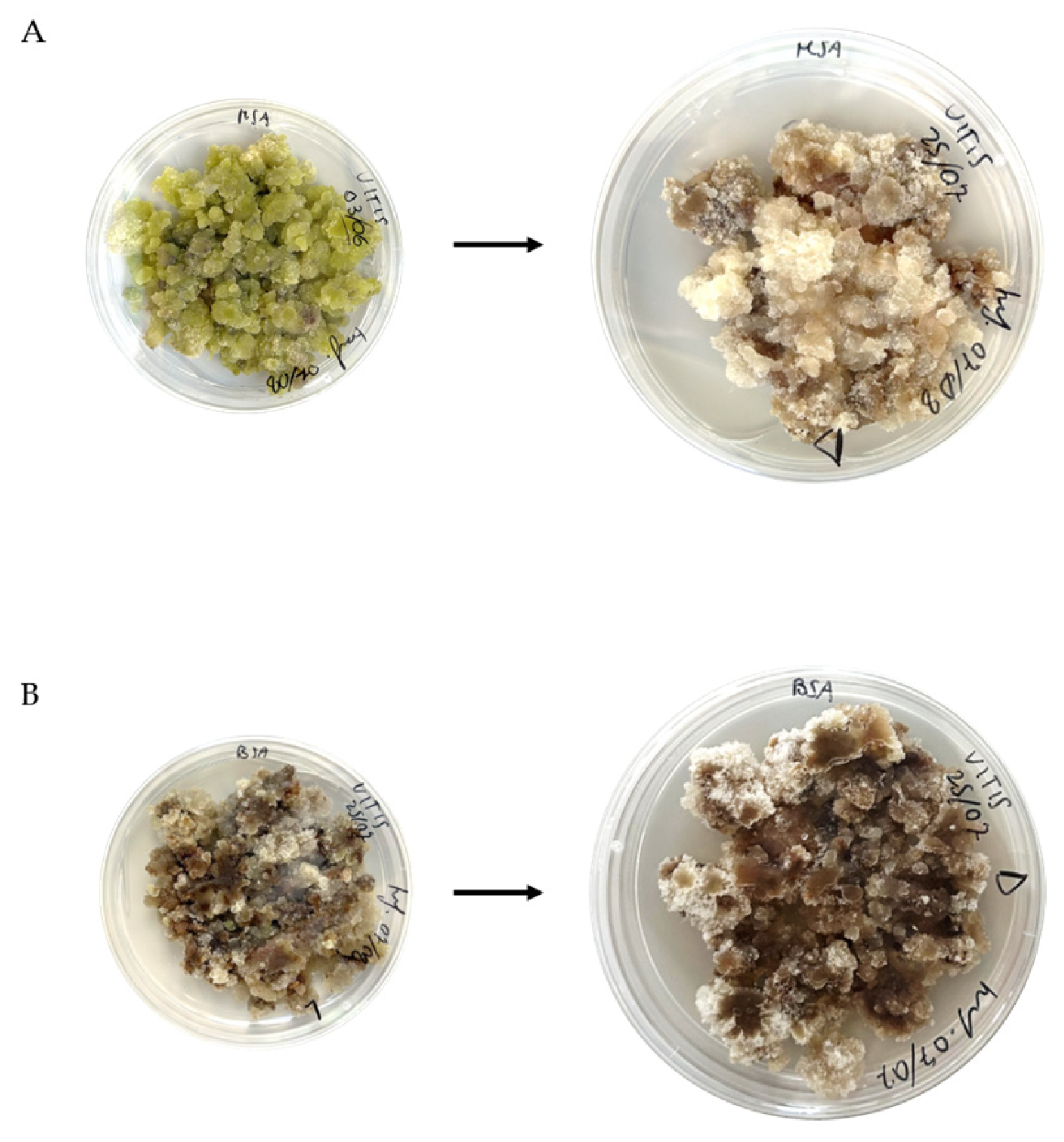
Calli grown in darkness were generated from parental calli previously maintained under photoperiod by transferring selected clumps to the same media but in the complete absence of light. Their morphology was monitored during the dark-growth period and subsequent subcultures. Calli on MSA medium (VMD) exhibited marked morphological changes compared to the parental material [22], becoming white-yellowish, with cells transitioning from dark green to brownish–red (Figure 1A). In contrast, calli on B5A medium in darkness (VBD) largely retained the characteristics and growth rate of the parental cells (Figure 1B).
Different light conditions, such as duration, quality, and intensity, can influence plant physiology, developmental, and chemical pathways, affecting growth and development both in vivo and in vitro [30,31]. Our results confirm the morphological changes triggered by the absence of light, especially in the VM cell lines, characterised by green cells due to the presence of chloroplasts, organelles that are inactive in the absence of light, as in VMD.

3.1.2. Qualitative-Quantitative Analysis of Stilbenoids in Calli

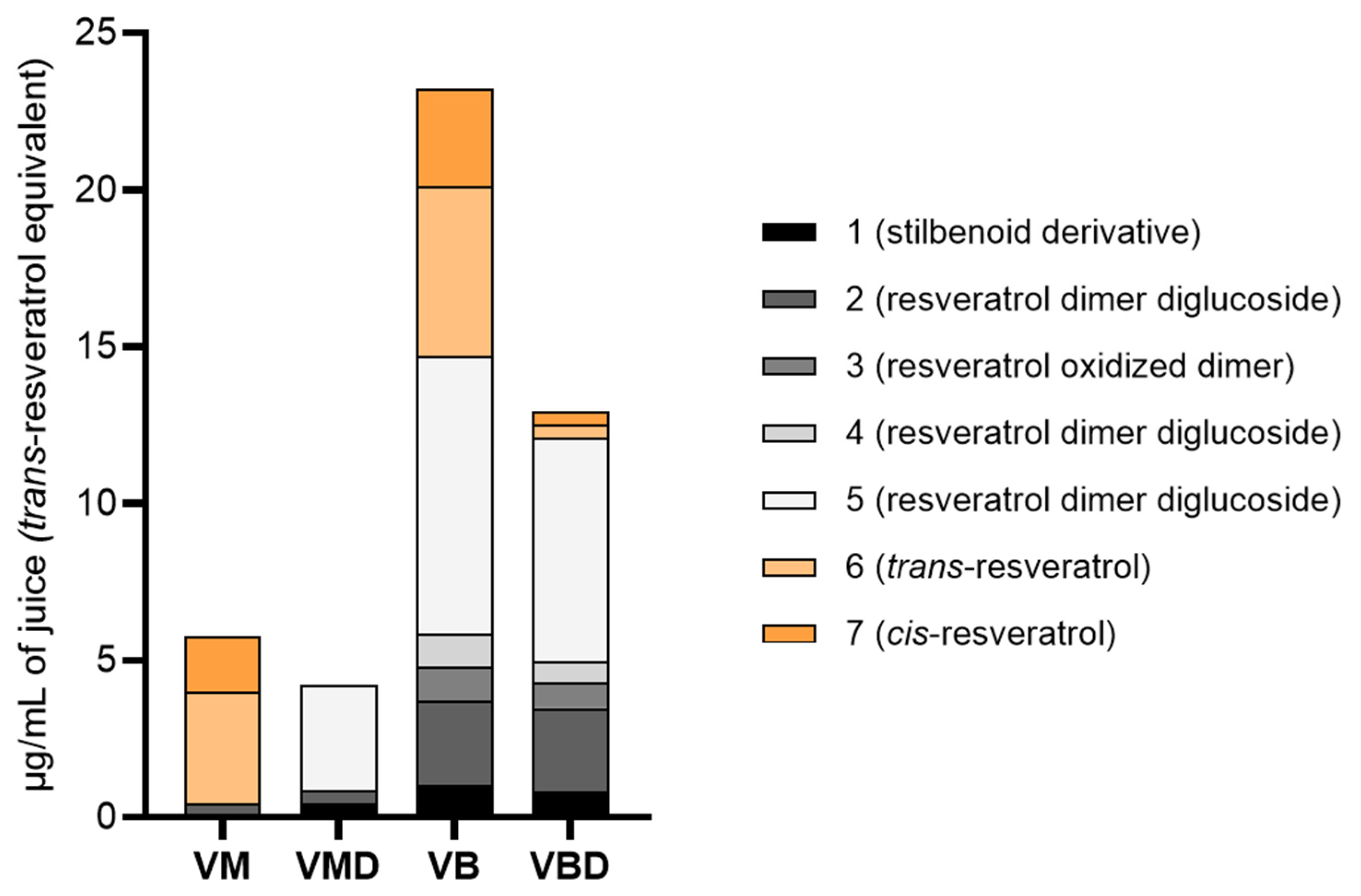
The quali-quantitative analysis was performed on calli harvested on the 28th day of the growth cycle, based on the main stilbenoid content of the material at that time point for the one grown under photoperiod [22], and also for the one in darkness. The analysis was done on the main compound class in the juices, the stilbenoids. The chromatograms were acquired at 325 nm, based on the maximum peaks of the stilbenoids, which occur around 300–330 nm [32]. The areas of the selected compounds were plotted against the trans-resveratrol standard curve. Table 1 reports the total stilbenoid content for each cell line’s juice. To compare the relative composition across cell lines and growth conditions (with or without photoperiod), the mean relative abundance of each compound is shown in Figure 2. Compounds are numbered according to Dalla Costa et al. [22].
The VB cell line showed the highest total stilbenoid content (p ≤ 0.0001), followed by VBD, whereas VMD contained significantly less stilbenoids than VM (p ≤ 0.02) and was the poorest overall. These results confirm that light strongly influences secondary-metabolite accumulation in plant cell cultures. Consistent with our observations, fluorescent light is commonly used to enhance metabolite biosynthesis, and photoperiod is known to regulate cell growth and production of secondary metabolism [30,33], even if only a limited number of studies relate the light to stilbene production [34]. In the relative profiles (Figure 2), cis- and trans-resveratrol disappeared in VMD and were nearly absent in VBD. At the same time, glucosylated derivatives were largely retained in VBD, with only slight decreases (e.g., compound 2:2.68 µg/mL in VB vs. 2.66 µg/mL in VBD; compound 5:8.86 to 7.12 µg/mL). Notably, compound 5 was still detectable in VMD, though absent in VM. To our knowledge, no studies have examined calli of V. labrusca var. Isabella or their stilbenoid profiles under different light conditions. Comparisons with V. vinifera and V. rupestris studies show inconsistent light-dependent effects on stilbenoid biosynthesis across species and cultivars [32,35,36], reinforcing that metabolite responses to growth conditions are highly genotype-specific and must be empirically determined [37,38,39].

3.2. Biological Activities of Juices Obtained from Calli

3.2.1. Cell Viability in Intestinal Epithelial Cell Lines

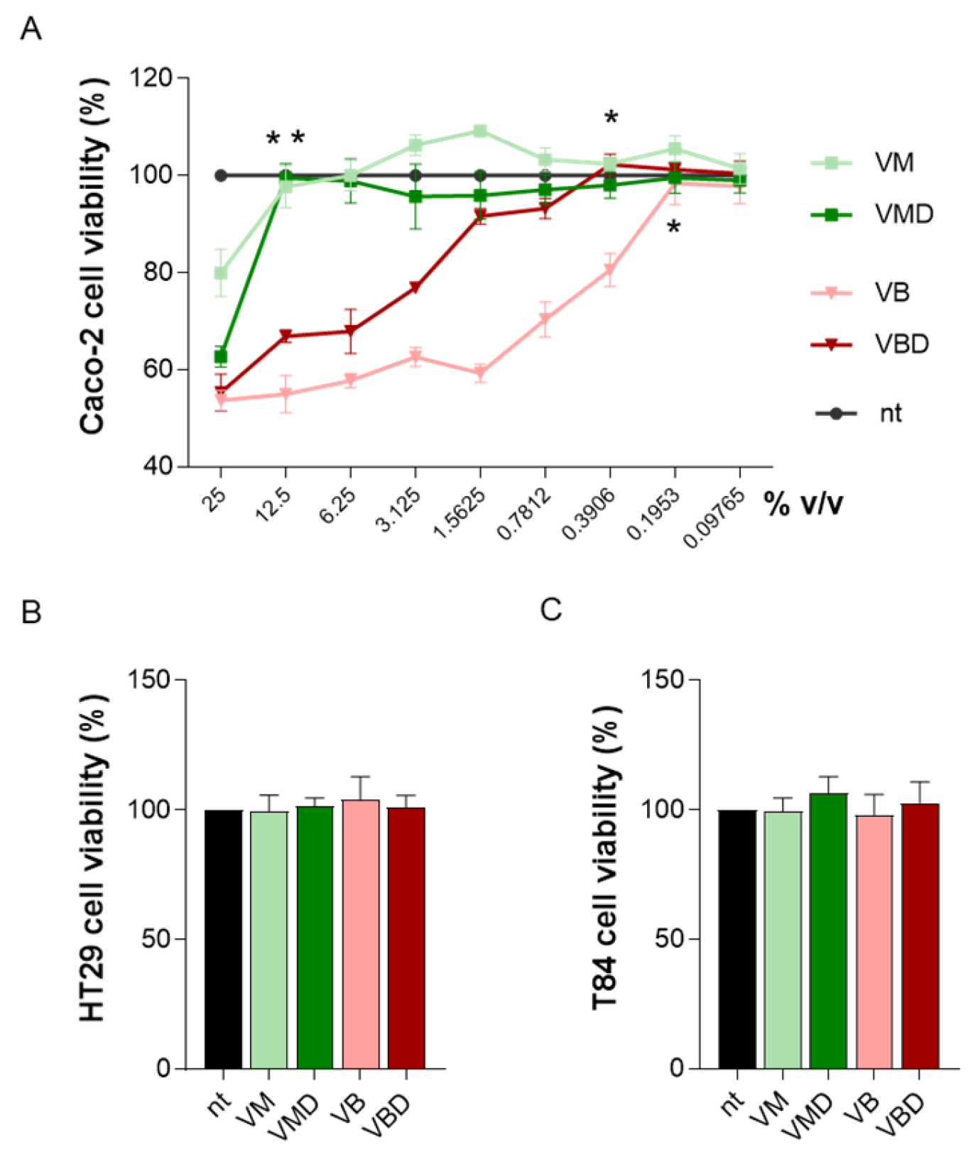
The cytotoxic potential of the juices was first evaluated using the MTT assay in Caco-2 cells after a 24-h incubation with increasing concentrations of the juices. As shown in Figure 3A, treatment with 12.5% v/v VM and VMD maintained cell viability above 90%, indicating the absence of detectable cytotoxicity (p < 0.05 vs. 25% v/v of the respective juices). In contrast, VB and VBD markedly reduced cell viability. Viability values became comparable to those of non-treated cells at 0.19% v/v for VB (p < 0.05 vs. 0.39% v/v) and at 0.39% v/v for VBD (p < 0.05 vs. 0.78% v/v). At these concentrations, no toxic effects were observed in HT29 or T84 cells, too (Figure 3B,C). Based on these results, 12.5% v/v was selected as the highest non-toxic concentration for VM and VMD, while 0.19% v/v was used for VB and VBD in subsequent experiments. However, to ensure consistency across conditions, VM and VMD were also tested at 0.19% v/v, the non-toxic concentration detected for VB and VBD.

3.2.2. Anti-Inflammatory Effects of the Juices

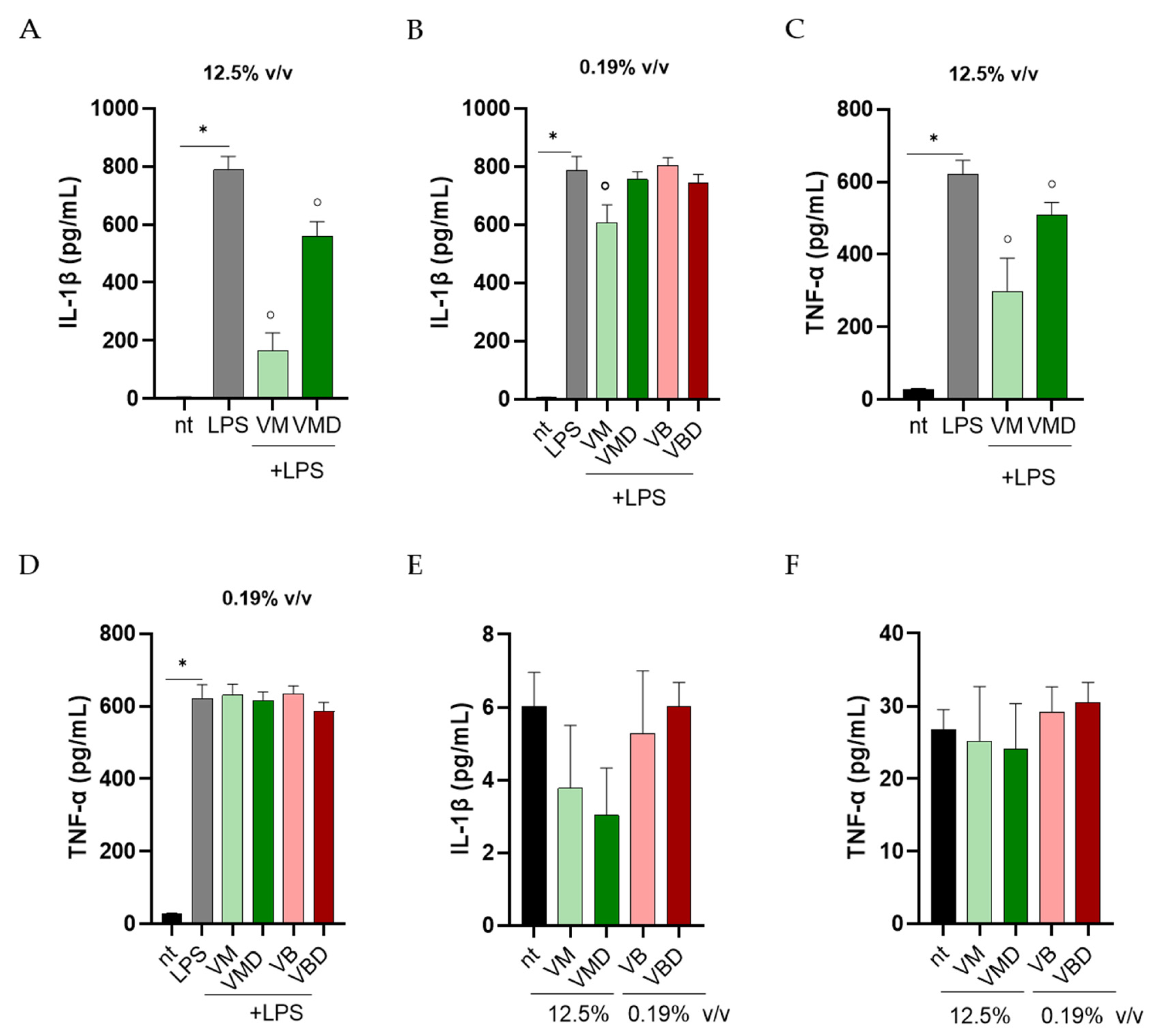
During intestinal inflammation, microbial components such as LPS stimulate epithelial cells to release pro-inflammatory cytokines, including IL-1β and TNF-α, leading to oxidative stress and barrier dysfunction [40]. To assess the anti-inflammatory activity of the juices, Caco-2 cells were exposed to LPS with or without the treatments, and cytokine levels were quantified by ELISA. As shown in Figure 4A–C, LPS (100 ng/mL) significantly increased IL-1β and TNF-α secretion (p < 0.02). At 12.5% v/v, both VM and VMD markedly reduced this LPS-induced cytokine release (p < 0.05). To match the non-toxic concentrations used for VB and VBD, VM and VMD were also tested at 0.19% v/v. At this lower dose (Figure 4B,D), only VM significantly decreased IL-1β and TNF-α production (p < 0.05).
Similar results were obtained in HT29 and T84 cells stimulated with 0.19% v/v juices (Table 2). LPS strongly increased IL-1β and TNF-α secretion in HT29 and T84 cell lines. Only VM (0.19% v/v) significantly attenuated the cytokine induction (p < 0.05 vs. LPS), whereas VB and VBD did not alter cytokine production. Collectively, these findings demonstrate that VM possesses the most pronounced anti-inflammatory activity among the tested samples, effectively counteracting LPS-induced cytokine release in human intestinal epithelial cells. Importantly, when Caco-2 cells were treated with the juices alone, no significant changes in IL-1β or TNF-α levels were observed (Figure 4E,F), confirming that the juices per se do not elicit an inflammatory response.

3.2.3. Effects of the Juices on Intracellular ROS Production

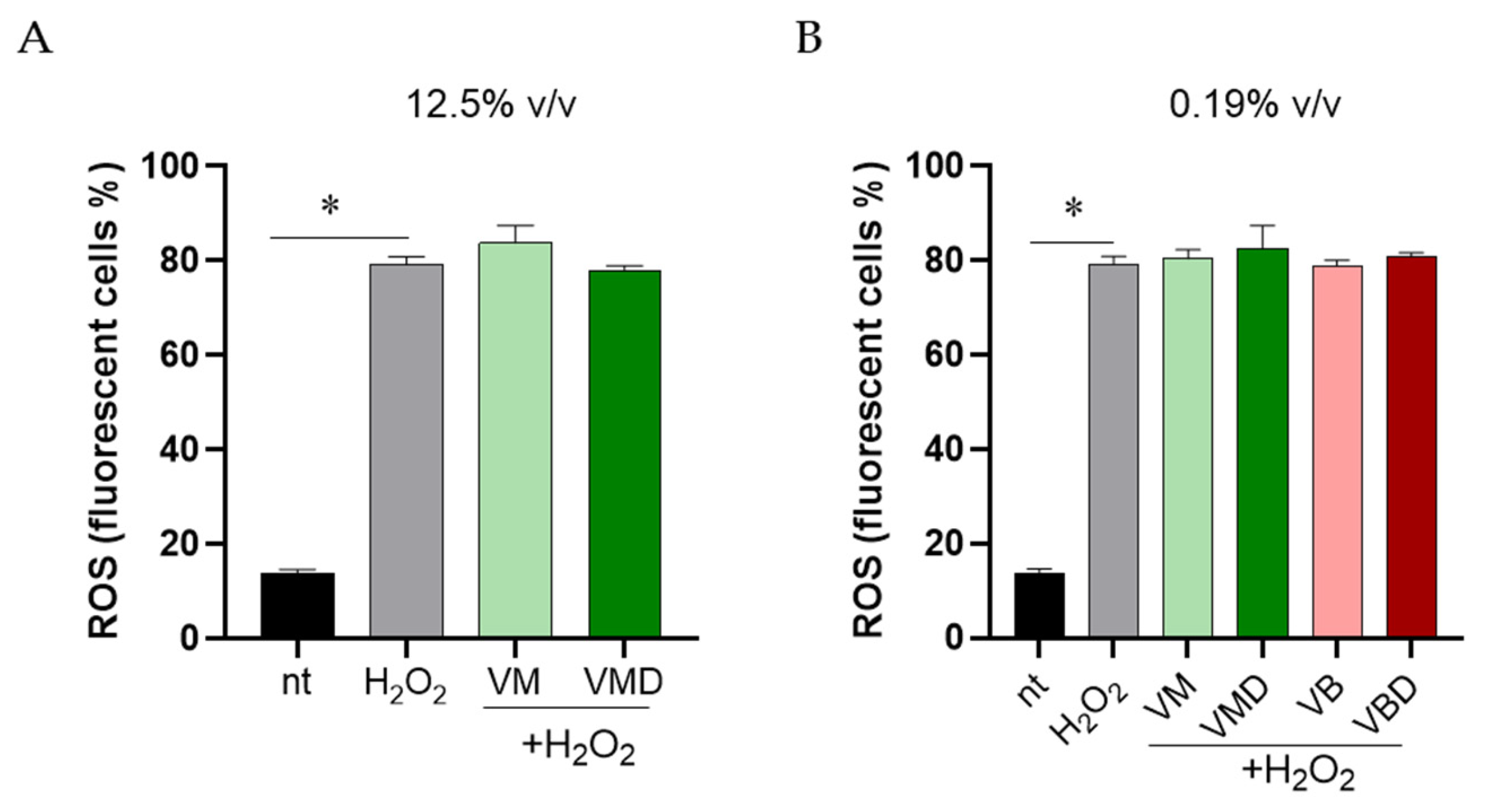
Intracellular ROS production was evaluated using the H2DCFDA probe after oxidative challenge with 25 µM H2O2. Briefly, cells were incubated with the juices for 16 h and then loaded with the H2DCFDA probe for 30 min. Ten minutes before the analysis at the flow cytometer, cells were challenged with H2O2. As expected, we did not detect a fluorescent signal in cells not loaded with H2DCFDA. Exposure to H2O2 markedly increased intracellular ROS levels in Caco-2 cells (p < 0.02; Figure 5A,B). Among all the tested samples, no one significantly reduced H2O2-induced ROS accumulation in Caco-2 cells when applied at both 12.5% and 0.19% v/v.

3.3. Establishment and Chemical Analysis of Cell Suspensions in Dark Conditions

3.3.1. Establishment of Cell Suspensions

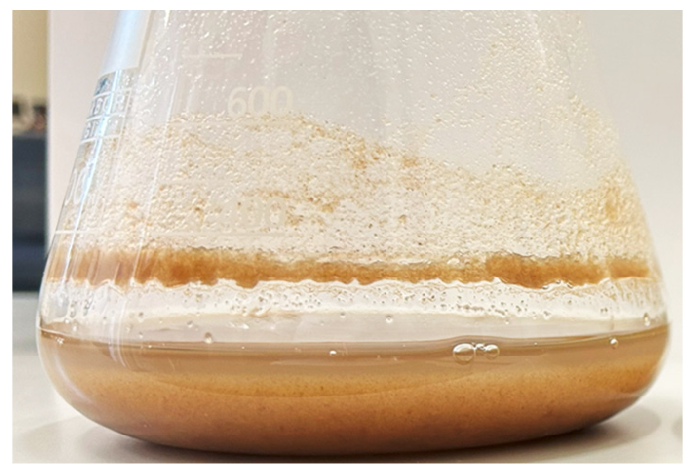
Based on the results of the VM cell line and the respective cell line grown in darkness (VMD) on the anti-inflammatory activity assays, we decided to establish cell suspension cultures starting from the VM cell line. The suspensions in dark conditions (SVMD) were obtained by dividing the suspensions derived from VM calli, which were kept under photoperiod. After a period of stabilisation, we obtained SVMD suspensions (an example flask is shown in Figure 6), which were characterised by appropriate cell growth and density.
Cell suspensions in a liquid medium exhibit several advantages over calli growing on solid media, which can be quite heterogeneous with respect to cell biochemical properties. Suspensions can be scaled up in various bioreactor systems, resulting in faster growth, lower costs, and less labor-intensive processes, with a smaller production area requirement [41,42]. We also evaluated whether our suspension cultures could grow in darkness, as continuous light or photoperiod conditions require additional equipment, higher energy consumption, and greater costs [43]. Scaling up plant cell cultures to industrial bioreactors is already challenging because these systems demand precise environmental control and rarely permit light exposure [44]. Since plant cells can obtain energy from sugar-rich media and do not strictly require light, most bioreactors operate in dark conditions, unlike photobioreactors used for photosynthetic microorganisms [45]. Although light can stimulate secondary-metabolite production and complete darkness may require precautions [46], we tested dark cultivation to determine whether our cell suspensions could remain biosynthetically active under conditions more compatible with large-scale production.

3.3.2. Qualitative-Quantitative Analysis of Stilbenoids of Cell Suspensions

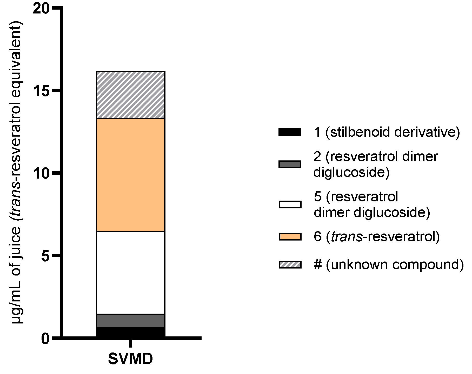
Juice extracted from a 13-day-old cell suspension was used for quantitative analysis. The growth cycle of suspension cultures tends to be shorter than that of calli on solid media; they are fast-growing cells that can synthesise secondary metabolites in either the stationary or logarithmic phase [47,48]. After determining the growth curve of SVMD suspensions, according to the cell volume after the sedimentation method, the suspension cells on the 13th day of the growth cycle were found to be at the logarithmic phase [27,46], corresponding to the growth phase (24th day) of the calli on solid medium. Thus, the juice extracted from 13-day-old suspension cells was selected for chemical comparison with the callus juices. Figure 7 shows the total stilbenoid content and the relative composition of SVMD.
SVMD showed a total stilbenoid content of 16.18 ± 0.18 µg/mL in juice statistically higher than those in VM, VMD, and VBD juices (p ≤ 0.0001), and lower only to VB (p ≤ 0.01). The total stilbenoid content of SVMD juice was approximately 3 times that measured in VM, and almost 4 times that in VMD. The passage to liquid cultures showed higher potential of single cells, or a few clustered cells, compared to calli cells on solid medium. In addition, another compound eluted after trans-resveratol (named #) was found, which is probably a resveratrol derivative, based on its UV spectrum (λmax 224–322). The compound 5, a resveratrol dimer diglucoside, increased its concentration up to 5.02 µg/mL of juice, much higher than in VMD juice. In SVMD, we also observed the reappearance of trans-resveratrol (compound 6), which was totally absent in the VMD juice, and with the highest concentration of 6.83 µg/mL of juice. The finding of different secondary metabolite productions and concentrations in suspensions than in the calli confirms that every single change in the cultivation condition may have effects on the material, both in terms of morphology, chemistry, as well as biosynthetic pathway activation, fostering the possibility of in vitro biotechnology to be used for several purposes depending on the intended aim and scope.

3.4. Biological Activities of Juice Obtained from Cell Suspension in Dark Conditions

3.4.1. SVMD Treatment Reduces LPS-Mediated Inflammation in Intestinal Cells

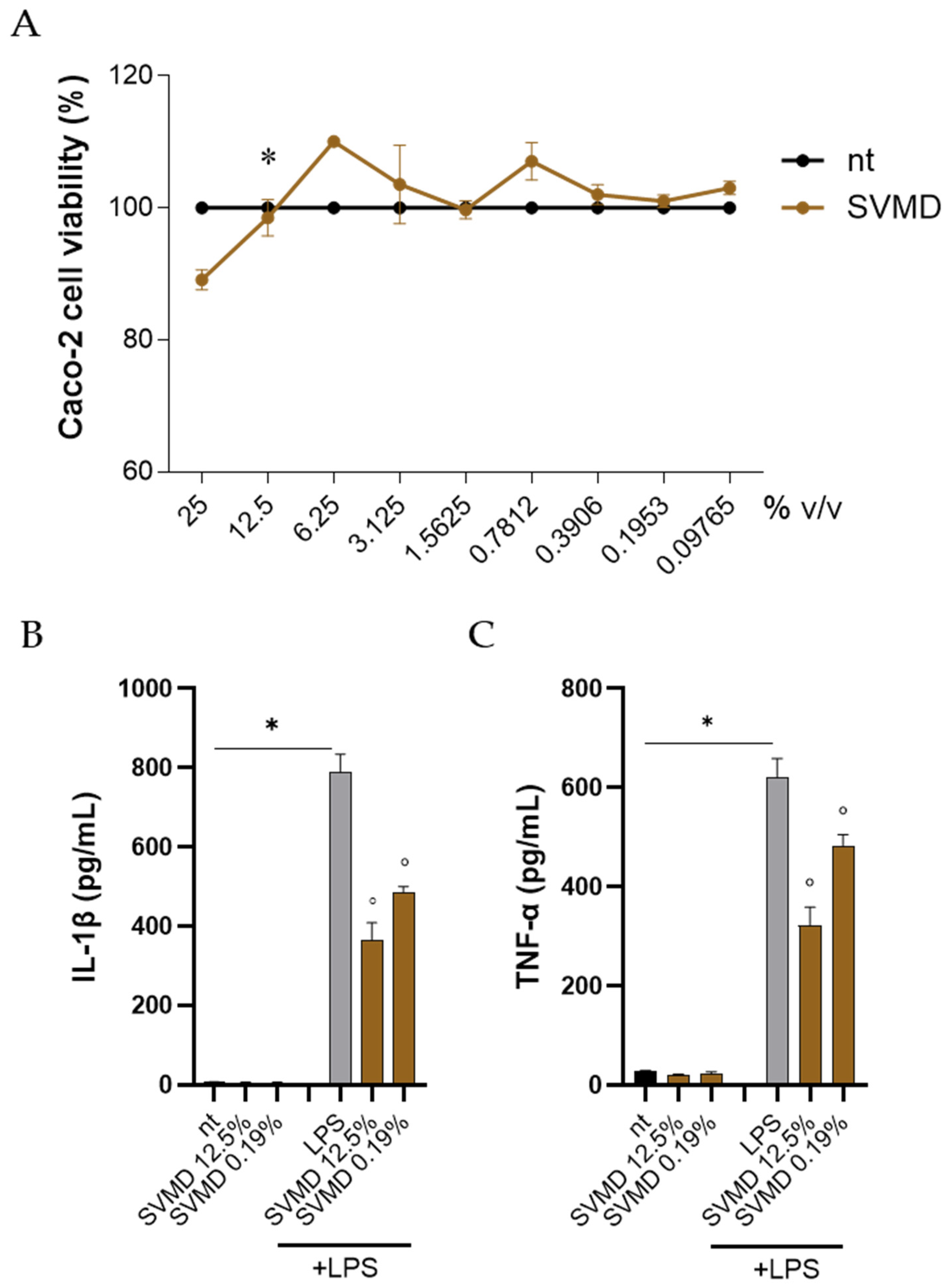
The SVMD juice was first investigated to exclude any possible toxic effects on intestinal cell lines. Cells were cultivated for 24 h with increasing concentrations of SVMD, and cell viability was assessed using the MTT assay. As reported in Figure 8A, treatment with SVMD 12.5% v/v did not exert toxic effects on Caco-2 cells (p < 0.05 vs. 25% v/v SVMD). Indeed, treatment with SVMD resulted in a higher proportion of viable cells, although we did not observe a trend consistent with a dose–response pattern (Figure 8A). Similar results were observed in HT29 and T84 cell lines.
SVMD significantly reduced LPS-induced IL-1β and TNF-α production in Caco-2 cells at both 12.5% and 0.19% v/v. At 12.5% v/v, SVMD decreased IL-1β by 2.15-fold and TNF-α by 1.93-fold, while at 0.19% v/v, it lowered TNF-α by 1.62-fold (p < 0.05; Figure 8B). Comparable effects were observed in HT29 and T84 cells: in HT29, SVMD (0.19% v/v) reduced IL-1β and TNF-α by 1.74- and 1.57-fold, respectively; in T84, it decreased IL-1β by 1.42-fold and TNF-α by 2.40-fold.

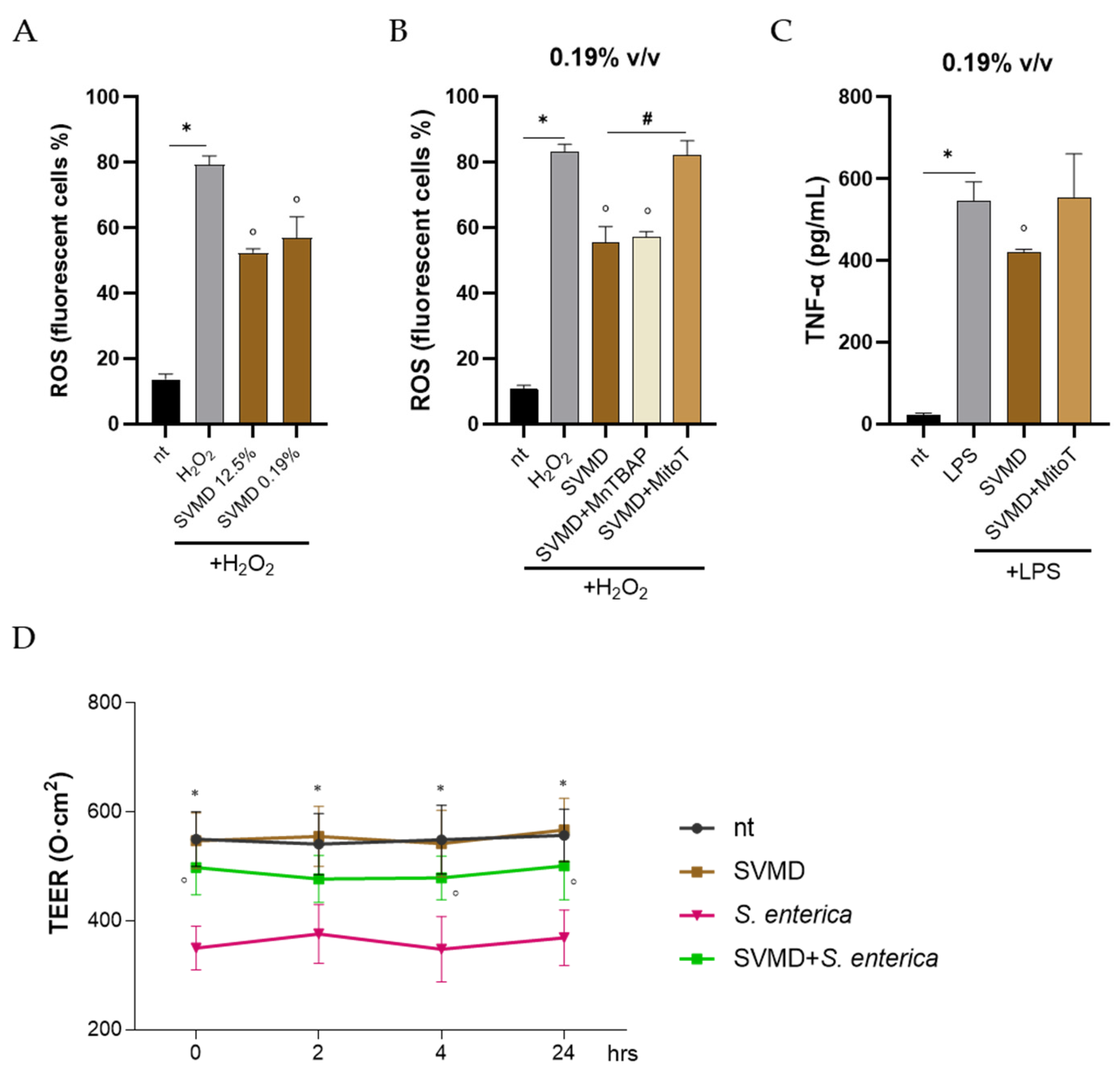
3.4.2. SVMD Treatment Dampens ROS Production in Cells and Restores Barrier Integrity

Caco-2 cells were treated with SVMD, challenged with H2O2 and loaded with H2DCFDA probe to evaluate intracellular ROS generation. As reported in Figure 9A, SVMD significantly reduced ROS generation in H2O2-treated cells (p < 0.05) when applied at both 12.5% and 0.19% v/v. No fluorescent signal was detected in cells not loaded with H2DCFDA. Stimulation of cells with H2O2 generates ROS via multiple pathways. Indeed, H2O2 could increase mitochondrial membrane potential and electron leakage, thus boosting mitochondrial ROS output [49]. However, the H2O2-mediated oxidative stress in cells also involves the impairment of peroxisomal β-oxidation, protein misfolding, activation of NOX enzymes, and impairment of calcium flux [50,51,52]. To investigate the mechanisms underlying SVMD’s antioxidant activity, Caco-2 cells were pretreated with MitoTEMPO, a mitochondrial ROS scavenger, or MnTBAP, a superoxide dismutase mimetic. MnTBAP did not affect SVMD’s antioxidant effect, whereas MitoTEMPO completely abolished it (Figure 9B), indicating a prominent role for SVMD in mitochondrial-derived ROS. MitoTEMPO also reduced SVMD’s anti-inflammatory activity (Figure 9C), further linking mitochondrial redox balance to inflammatory responses [53,54]. To assess whether SVMB’s antioxidant and anti-inflammatory effects translate into functional epithelial protection, TEER was measured in differentiated Caco-2 monolayers [55,56]. Baseline TEER values were stable (550 ± 50 Ω·cm2), and SVMB (0.19% v/v) alone did not alter barrier integrity over 24 h (Figure 9D). Infection with Salmonella enterica (MOI 1:10) caused a rapid TEER drop to 350 ± 40 Ω·cm2 within 4 h, whereas SVMB pretreatment markedly preserved TEER, maintaining values around 640 ± 45 Ω·cm2 (p < 0.05 vs. infected cells). SVMB did not reduce bacterial counts recovered from infected monolayers, indicating no direct antibacterial activity.
Several points limit the findings of our study. All experiments were performed using intestinal epithelial cell lines, which are usual models for the colonic cancerogenic process. While these systems are widely employed to study epithelial responses, they do not fully recapitulate the complexity of the intestinal mucosa in vivo, where immune cells, stromal components, microbiota, and vascular signals collectively shape inflammatory and redox pathways. Consequently, the translational relevance of the observed anti-inflammatory and antioxidant effects requires validation in intestinal organoids or appropriate animal models of intestinal inflammation. Second, the mechanistic insights provided here are necessarily limited. We focused primarily on two readouts, namely pro-inflammatory cytokine and intracellular ROS levels. We did not investigate downstream signalling cascades such as NF-κB, MAPKs, or Nrf2, which are known to regulate redox homeostasis and inflammation. In addition, although the juices contain a mixture of stilbenoids and other molecules, the specific compounds responsible for the observed biological activities were not individually isolated or assessed. Future studies integrating comprehensive molecular analyses and in vivo approaches will be required to fully define the mechanisms of action and therapeutic potential of V. labrusca var. Isabella cell-culture juices.
Overall, our data identify SVMB as a promising bioactive juice with antioxidant and anti-inflammatory activity in intestinal epithelial cells. Several complementary mechanisms may underlie its ability to modulate the intracellular redox state. Stilbenoids can directly scavenge ROS via their hydroxylated aromatic structure, and they are known to activate the Nrf2–ARE pathway, thereby inducing endogenous antioxidant enzymes such as SOD2, catalase, and glutathione-related systems, many of which operate within the mitochondria. In addition, polyphenols can enhance mitochondrial respiratory efficiency, reducing electron leakage from complexes I and III and consequently lowering mitochondrial ROS production. Based on these mechanisms, we propose that the SVMD juice, by attenuating ROS generation, dampens pro-inflammatory cytokine release and preserves epithelial barrier integrity. Interestingly, SVMD juice induces a decrease in ROS even though the stilbenoid content is lower than that of the VB, the richest juice, which showed no activity. On the other hand, the SVMD showed a statistically higher stilbenoid content compared to the parental calli (both under photoperiod and darkness), and a completely different relative composition as well. The juices tested, unlike conventional extracts obtained with a more or less selective solvent, exhibit an extremely complex composition, referring not only to secondary metabolites but also to primary metabolites, which, although considered of marginal importance in terms of activity, could contribute to modulating the activity of secondary metabolites. The suspensions and calli examined, although derived from the same cell line or from the same explant type, are in different culture conditions (different basal media, illumination conditions, liquid medium in agitated flasks and solid in static conditions), which could likely have a dramatic impact on the regulation of biochemical processes and, therefore, on the final composition of the juice. Further studies will be required to identify the specific bioactive components responsible and to clarify the molecular pathways engaged, particularly redox-sensitive signalling cascades such as NF-κB and Nrf2.

Author Contributions

Conceptualization, V.D.C., R.F. and P.B.; methodology, V.D.C. and C.F.; software, V.D.C. and C.F.; validation, V.D.C., R.F. and P.B.; formal analysis, V.D.C. and P.B.; investigation, V.D.C., R.F. and P.B.; resources, R.F. and P.B.; data curation, V.D.C., R.F. and P.B.; writing—original draft preparation, V.D.C. and P.B.; writing—review and editing, R.F. and P.B.; visualization, V.D.C., R.F. and P.B.; supervision, R.F. and P.B. All authors have read and agreed to the published version of the manuscript.

Funding

This research received no external funding.

Institutional Review Board Statement

Not applicable.

Informed Consent Statement

Not applicable.

Data Availability Statement

The original contributions presented in this study are included in the article. Further inquiries can be directed to the corresponding author.

Conflicts of Interest

The authors declare no conflicts of interest.

References

  1. Heydari, K.; Rahnavard, M.; Ghahramani, S.; Hoseini, A.; Alizadeh-Navaei, R.; Rafati, S.; Raei, M.; Vahidipour, M.; Salehi, F.; Motafeghi, F.; et al. Global Prevalence and Incidence of Inflammatory Bowel Disease (IBD): A Systematic Review and Meta-Analysis of Population-Based Studies. Gastroenterol. Hepatol. Bed Bench 2025, 18, 132–146. [Google Scholar] [CrossRef] [PubMed]
  2. Ungaro, R.; Mehandru, S.; Allen, P.B.; Peyrin-Biroulet, L.; Colombel, J.-F. Ulcerative Colitis. Lancet 2017, 389, 1756–1770. [Google Scholar] [CrossRef]
  3. Torres, J.; Mehandru, S.; Colombel, J.-F.; Peyrin-Biroulet, L. Crohn’s Disease. Lancet 2017, 389, 1741–1755. [Google Scholar] [CrossRef]
  4. Di Vincenzo, F.; Quintero, M.A.; Serigado, J.M.; Koru-Sengul, T.; Killian, R.M.; Poveda, J.; England, J.; Damas, O.; Kerman, D.; Deshpande, A.; et al. Histologic and Endoscopic Findings Are Highly Correlated in a Prospective Cohort of Patients with Inflammatory Bowel Diseases. J. Crohn’s Colitis 2025, 19, jjae141. [Google Scholar] [CrossRef]
  5. Lamb, C.A.; Kennedy, N.A.; Raine, T.; Hendy, P.A.; Smith, P.J.; Limdi, J.K.; Hayee, B.; Lomer, M.C.E.; Parkes, G.C.; Selinger, C.; et al. British Society of Gastroenterology Consensus Guidelines on the Management of Inflammatory Bowel Disease in Adults. Gut 2019, 68, s1–s106. [Google Scholar] [CrossRef]
  6. Selinger, C.P.; Rosiou, K.; Lenti, M.V. Biological Therapy for Inflammatory Bowel Disease: Cyclical Rather than Lifelong Treatment? BMJ Open Gastroenterol 2024, 11, e001225. [Google Scholar] [CrossRef] [PubMed]
  7. Santiago, P.; Braga-Neto, M.B., Jr.; Loftus, E.V. Novel Therapies for Patients With Inflammatory Bowel Disease. Gastroenterol. Hepatol. 2022, 18, 453–465. [Google Scholar]
  8. Singh, U.P.; Singh, N.P.; Busbee, B.; Guan, H.; Singh, B.; Price, R.L.; Taub, D.D.; Mishra, M.K.; Nagarkatti, M.; Nagarkatti, P.S. Alternative Medicines as Emerging Therapies for Inflammatory Bowel Diseases. Int. Rev. Immunol. 2012, 31, 66–84. [Google Scholar] [CrossRef] [PubMed]
  9. Davila, M.M.; Papada, E. The Role of Plant-Derived Natural Products in the Management of Inflammatory Bowel Disease—What Is the Clinical Evidence So Far? Life 2023, 13, 1703. [Google Scholar] [CrossRef]
  10. Topalović, A.; Knežević, M.; Bajagić, B.; Ivanović, L.; Milašević, I.; Đurović, D.; Mugoša, B.; Podolski-Renić, A.; Pešić, M. Grape (Vitis vinifera L.): Health Benefits and Effects of Growing Conditions on Quality Parameters. In Biodiversity and Biomedicine; Elsevier: Amsterdam, The Netherlands, 2020; pp. 385–401. ISBN 978-0-12-819541-3. [Google Scholar]
  11. Toaldo, I.M.; Fogolari, O.; Pimentel, G.C.; De Gois, J.S.; Borges, D.L.G.; Caliari, V.; Bordignon-Luiz, M. Effect of Grape Seeds on the Polyphenol Bioactive Content and Elemental Composition by ICP-MS of Grape Juices from Vitis labrusca L. LWT Food Sci. Technol. 2013, 53, 1–8. [Google Scholar] [CrossRef]
  12. Yamamoto, L.Y.; De Assis, A.M.; Roberto, S.R.; Bovolenta, Y.R.; Nixdorf, S.L.; García-Romero, E.; Gómez-Alonso, S.; Hermosín-Gutiérrez, I. Application of Abscisic Acid (S-ABA) to Cv. Isabel Grapes (Vitis vinifera × Vitis labrusca) for Color Improvement: Effects on Color, Phenolic Composition and Antioxidant Capacity of Their Grape Juice. Food Res. Int. 2015, 77, 572–583. [Google Scholar] [CrossRef]
  13. Pacifico, S.; D’Abrosca, B.; Scognamiglio, M.; Gallicchio, M.; Potenza, N.; Piccolella, S.; Russo, A.; Monaco, P.; Fiorentino, A. Metabolic Profiling of Strawberry Grape (Vitis × Labruscana Cv. ‘Isabella’) Components by Nuclear Magnetic Resonance (NMR) and Evaluation of Their Antioxidant and Antiproliferative Properties. J. Agric. Food Chem. 2011, 59, 7679–7687. [Google Scholar] [CrossRef] [PubMed]
  14. Toaldo, I.M.; Cruz, F.A.; Alves, T.D.L.; De Gois, J.S.; Borges, D.L.G.; Cunha, H.P.; Da Silva, E.L.; Bordignon-Luiz, M.T. Bioactive Potential of Vitis labrusca L. Grape Juices from the Southern Region of Brazil: Phenolic and Elemental Composition and Effect on Lipid Peroxidation in Healthy Subjects. Food Chem. 2015, 173, 527–535. [Google Scholar] [CrossRef] [PubMed]
  15. Kurt-Celebi, A.; Colak, N.; Hayirlioglu-Ayaz, S.; Kostadinović Veličkovska, S.; Ilieva, F.; Esatbeyoglu, T.; Ayaz, F.A. Accumulation of Phenolic Compounds and Antioxidant Capacity during Berry Development in Black ‘Isabel’ Grape (Vitis vinifera L. × Vitis labrusca L.). Molecules 2020, 25, 3845. [Google Scholar] [CrossRef]
  16. Ozkan, K.; Karadag, A.; Sagdic, O.; Ozcan, F.S.; Ozer, H. The Effects of Different Drying Methods on the Sugar, Organic Acid, Volatile Composition, and Textural Properties of Black ‘Isabel’ Grape. Food Meas. 2023, 17, 1852–1861. [Google Scholar] [CrossRef]
  17. Keskin, N.; Bilir Ekbic, H.; Kaya, O.; Keskin, S. Antioxidant Activity and Biochemical Compounds of Vitis vinifera L. (Cv. ‘Katıkara’) and Vitis labrusca L. (Cv. ‘Isabella’) Grown in Black Sea Coast of Turkey. Erwerbs-Obstbau 2021, 63, 115–122. [Google Scholar] [CrossRef]
  18. Pacifico, S.; D’Abrosca, B.; Scognamiglio, M.; Gallicchio, M.; Galasso, S.; Monaco, P.; Fiorentino, A. Antioxidant Polyphenolic Constituents of Vitis × Labruscana Cv. ‘Isabella’ Leaves. Open Nat. Prod. J. 2013, 5, 5–11. [Google Scholar] [CrossRef][Green Version]
  19. Nunes, S.; Danesi, F.; Del Rio, D.; Silva, P. Resveratrol and Inflammatory Bowel Disease: The Evidence so Far. Nutr. Res. Rev. 2018, 31, 85–97. [Google Scholar] [CrossRef]
  20. Recinella, L.; Chiavaroli, A.; Veschi, S.; Cama, A.; Acquaviva, A.; Libero, M.L.; Leone, S.; Di Simone, S.C.; Pagano, E.; Zengin, G.; et al. A Grape (Vitis vinifera L.) Pomace Water Extract Modulates Inflammatory and Immune Response in SW-480 Cells and Isolated Mouse Colon. Phytother. Res. 2022, 36, 4620–4630. [Google Scholar] [CrossRef]
  21. Taladrid, D.; González De Llano, D.; Zorraquín-Peña, I.; Tamargo, A.; Silva, M.; Molinero, N.; Moreno-Arribas, M.V.; Bartolomé, B. Gastrointestinal Digestion of a Grape Pomace Extract: Impact on Intestinal Barrier Permeability and Interaction with Gut Microbiome. Nutrients 2021, 13, 2467. [Google Scholar] [CrossRef] [PubMed]
  22. Dalla Costa, V.; Piovan, A.; Brun, P.; Filippini, R. Unconventional Material from In Vitro Plant Cell Cultures: Vitis labrusca Var. Isabella Case Study. Appl. Sci. 2025, 15, 9139. [Google Scholar] [CrossRef]
  23. Wawrosch, C.; Zotchev, S.B. Production of Bioactive Plant Secondary Metabolites through in Vitro Technologies—Status and Outlook. Appl. Microbiol. Biotechnol. 2021, 105, 6649–6668. [Google Scholar] [CrossRef]
  24. Latif, R.; Nawaz, T. Medicinal Plants and Human Health: A Comprehensive Review of Bioactive Compounds, Therapeutic Effects, and Applications. Phytochem. Rev. 2025. [Google Scholar] [CrossRef]
  25. Murashige, T.; Skoog, F. A Revised Medium for Rapid Growth and Bioassays with Tobacco Tissue Cultures. Physiol. Plant. 1962, 15, 473–497. [Google Scholar] [CrossRef]
  26. Gamborg, O.L.; Miller, R.A.; Ojima, K. Nutrient requirements of suspension cultures of soybean root cells. Exp. Cell Res. 1968, 50, 151–158. [Google Scholar] [CrossRef]
  27. Dalla Costa, V.; Piovan, A.; Brun, P.; Filippini, R. Morus alba L. Cell Cultures as Sources of Antioxidant and Anti-Inflammatory Stilbenoids for Food Supplement Development. Molecules 2025, 30, 2073. [Google Scholar] [CrossRef]
  28. Dalla Costa, V.; Piovan, A.; Varfaj, I.; Marcotullio, M.C.; Brun, P.; Filippini, R. From “Maraschino” to Cell Cultures: A Deep Study on Prunus cerasus L. Cell Culture Juices. Molecules 2025, 30, 1089. [Google Scholar] [CrossRef] [PubMed]
  29. Dalla Costa, V.; Piovan, A.; Filippini, R.; Brun, P. From Ethnobotany to Biotechnology: Wound Healing and Anti-Inflammatory Properties of Sedum telephium L. In Vitro Cultures. Molecules 2024, 29, 2472. [Google Scholar] [CrossRef] [PubMed]
  30. Murthy, H.N.; Joseph, K.S.; Paek, K.Y.; Park, S.Y. Light as an Elicitor for Enhanced Production of Secondary Metabolites in Plant Cell, Tissue, and Organ Cultures. Plant Growth Regul. 2024, 104, 31–49. [Google Scholar] [CrossRef]
  31. Batista, D.S.; Felipe, S.H.S.; Silva, T.D.; De Castro, K.M.; Mamedes-Rodrigues, T.C.; Miranda, N.A.; Ríos-Ríos, A.M.; Faria, D.V.; Fortini, E.A.; Chagas, K.; et al. Light Quality in Plant Tissue Culture: Does It Matter? In Vitro Cell. Dev. Biol. Plant 2018, 54, 195–215. [Google Scholar] [CrossRef]
  32. Taurino, M.; Ingrosso, I.; D’amico, L.; De Domenico, S.; Nicoletti, I.; Corradini, D.; Santino, A.; Giovinazzo, G. Jasmonates Elicit Different Sets of Stilbenes in Vitis vinifera Cv. Negramaro Cell Cultures. SpringerPlus 2015, 4, 49. [Google Scholar] [CrossRef]
  33. Cavallaro, V.; Muleo, R. The Effects of LED Light Spectra and Intensities on Plant Growth. Plants 2022, 11, 1911. [Google Scholar] [CrossRef] [PubMed]
  34. Valletta, A.; Iozia, L.M.; Leonelli, F. Impact of Environmental Factors on Stilbene Biosynthesis. Plants 2021, 10, 90. [Google Scholar] [CrossRef]
  35. Donati, L.; Ferretti, L.; Frallicciardi, J.; Rosciani, R.; Valletta, A.; Pasqua, G. Stilbene Biosynthesis and Gene Expression in Response to Methyl Jasmonate and Continuous Light Treatment in Vitis vinifera Cv. Malvasia Del Lazio and Vitis rupestris Du Lot Cell Cultures. Physiol. Plant. 2019, 166, 646–662. [Google Scholar] [CrossRef]
  36. Andi, S.A.; Gholami, M.; Ford, C.M. The Effect of Methyl Jasmonate and Light Irradiation Treatments on the Stilbenoid Biosynthetic Pathway in Vitis vinifera Cell Suspension Cultures. Nat. Prod. Res. 2018, 32, 909–917. [Google Scholar] [CrossRef]
  37. Dixon, R.A. (Ed.) Isolation and Maintenance of Callus and Cell Suspension Cultures. In Plant Cell Culture—A Pratical Approach; Oxford Academic: Oxford, UK, 1985. [Google Scholar]
  38. Gray, D.J.; Trigiano, R.N. Introducing to Plant Tissue Culture. In Plant Tissue Culture Concepts and Laboratory Exercises, 2nd ed.; Routledge: New York, NY, USA, 2000; Chapter 1; p. 5. [Google Scholar]
  39. Pasternak, T.P.; Steinmacher, D. Plant Growth Regulation in Cell and Tissue Culture In Vitro. Plants 2024, 13, 327. [Google Scholar] [CrossRef] [PubMed]
  40. Stephens, M.; Von Der Weid, P.-Y. Lipopolysaccharides Modulate Intestinal Epithelial Permeability and Inflammation in a Species-Specific Manner. Gut Microbes 2020, 11, 421–432. [Google Scholar] [CrossRef] [PubMed]
  41. Chandran, H.; Meena, M.; Barupal, T.; Sharma, K. Plant Tissue Culture as a Perpetual Source for Production of Industrially Important Bioactive Compounds. Biotechnol. Rep. 2020, 26, e00450. [Google Scholar] [CrossRef]
  42. Krasteva, G.; Georgiev, V.; Pavlov, A. Recent Applications of Plant Cell Culture Technology in Cosmetics and Foods. Eng. Life Sci. 2021, 21, 68–76. [Google Scholar] [CrossRef]
  43. Häkkinen, S.T.; Nygren, H.; Nohynek, L.; Puupponen-Pimiä, R.; Heiniö, R.-L.; Maiorova, N.; Rischer, H.; Ritala, A. Plant Cell Cultures as Food—Aspects of Sustainability and Safety. Plant Cell Rep. 2020, 39, 1655–1668. [Google Scholar] [CrossRef]
  44. Titova, M.; Popova, E.; Nosov, A. Bioreactor Systems for Plant Cell Cultivation at the Institute of Plant Physiology of the Russian Academy of Sciences: 50 Years of Technology Evolution from Laboratory to Industrial Implications. Plants 2024, 13, 430. [Google Scholar] [CrossRef] [PubMed]
  45. Verdú-Navarro, F.; Moreno-Cid, J.A.; Weiss, J.; Egea-Cortines, M. The Advent of Plant Cells in Bioreactors. Front. Plant Sci. 2023, 14, 1310405. [Google Scholar] [CrossRef]
  46. Mustafa, N.R.; De Winter, W.; Van Iren, F.; Verpoorte, R. Initiation, Growth and Cryopreservation of Plant Cell Suspension Cultures. Nat. Protoc. 2011, 6, 715–742. [Google Scholar] [CrossRef] [PubMed]
  47. Bourgaud, F.; Gravot, A.; Milesi, S.; Gontier, E. Production of Plant Secondary Metabolites: A Historical Perspective. Plant Sci. 2001, 161, 839–851. [Google Scholar] [CrossRef]
  48. Chattopadhyay, S.; Farkya, S.; Srivastava, A.K.; Bisaria, V.S. Bioprocess Considerations for Production of Secondary Metabolites by Plant Cell Suspension Cultures. Biotechnol. Bioprocess Eng. 2002, 7, 138–149. [Google Scholar] [CrossRef]
  49. Zhang, X.; Lee, M.D.; Wilson, C.; McCarron, J.G. Hydrogen Peroxide Depolarizes Mitochondria and Inhibits IP3-Evoked Ca2+ Release in the Endothelium of Intact Arteries. Cell Calcium 2019, 84, 102108. [Google Scholar] [CrossRef]
  50. Zhang, M.; Zhang, G.; Meng, X.; Wang, X.; Xie, J.; Wang, S.; Wang, B.; Wang, J.; Liu, S.; Huang, Q.; et al. Reduction of the Oxidative Damage to H2O2 -Induced HepG2 Cells via the Nrf2 Signalling Pathway by Plant Flavonoids Quercetin and Hyperoside. Food Sci. Hum. Wellness 2024, 13, 1864–1876. [Google Scholar] [CrossRef]
  51. Zenin, V.; Ivanova, J.; Pugovkina, N.; Shatrova, A.; Aksenov, N.; Tyuryaeva, I.; Kirpichnikova, K.; Kuneev, I.; Zhuravlev, A.; Osyaeva, E.; et al. Resistance to H2O2-Induced Oxidative Stress in Human Cells of Different Phenotypes. Redox Biol. 2022, 50, 102245. [Google Scholar] [CrossRef]
  52. Ransy, C.; Vaz, C.; Lombès, A.; Bouillaud, F. Use of H2O2 to Cause Oxidative Stress, the Catalase Issue. Int. J. Mol. Sci. 2020, 21, 9149. [Google Scholar] [CrossRef]
  53. Li, L.; Peng, P.; Ding, N.; Jia, W.; Huang, C.; Tang, Y. Oxidative Stress, Inflammation, Gut Dysbiosis: What Can Polyphenols Do in Inflammatory Bowel Disease? Antioxidants 2023, 12, 967. [Google Scholar] [CrossRef]
  54. Muro, P.; Zhang, L.; Li, S.; Zhao, Z.; Jin, T.; Mao, F.; Mao, Z. The Emerging Role of Oxidative Stress in Inflammatory Bowel Disease. Front. Endocrinol. 2024, 15, 1390351. [Google Scholar] [CrossRef] [PubMed]
  55. Dunleavy, K.A.; Raffals, L.E.; Camilleri, M. Intestinal Barrier Dysfunction in Inflammatory Bowel Disease: Underpinning Pathogenesis and Therapeutics. Dig. Dis. Sci. 2023, 68, 4306–4320. [Google Scholar] [CrossRef] [PubMed]
  56. Ghosh, S.S.; Wang, J.; Yannie, P.J.; Ghosh, S. Intestinal Barrier Dysfunction, LPS Translocation, and Disease Development. J. Endocr. Soc. 2020, 4, bvz039. [Google Scholar] [CrossRef] [PubMed]
Figure 1. (A) VM green cell line, which became whitish-coloured in dark conditions (VMD); (B) VB cell line, which kept almost the same brownish-coloured in dark conditions (VBD).
Applsci 15 13192 g001
Figure 2. Relative content of each compound in VM, VMD, VB, and VBD juices. The results are reported as means, and the compounds are numbered following the order reported in Dalla Costa et al. [22]. The two identified aglycones are highlighted in pale (trans-resveratrol) and dark orange (cis-resveratrol).
Applsci 15 13192 g002
Figure 3. Cell viability of intestinal epithelial cell lines exposed to juices. (A) Caco-2 cells were incubated with juices at different concentrations for 24 h. (B) HT29 and (C) T84 cells were incubated with juices at 0.19% v/v for 24 h. Cell viability was assessed using the MTT assay and expressed as a percentage relative to untreated control cells (nt). Data are reported as the mean ± SEM of three independent experiments performed in triplicate. * denotes p < 0.05 vs. the immediately higher concentration of the respective juices.
Applsci 15 13192 g003
Figure 4. Effects of the juices on pro-inflammatory cytokine release in Caco-2 cells. Caco-2 cells were incubated for 16 h with the indicated juices at a concentration of 12.5% v/v (A,C) or 0.19% v/v (B,D) in the presence of 100 ng/mL lipopolysaccharide (LPS) or not (E,F). The levels of interleukin (IL)-1β and tumour necrosis factor (TNF)-α were quantified in the conditioned media using ELISA. Data are expressed as the mean ± SEM of three independent experiments performed in triplicate. * indicates p < 0.05 vs. non treated cells (nt); ° indicates p < 0.05 vs. LPS stimulated cells.
Applsci 15 13192 g004
Figure 5. Juices did not affect intracellular ROS generation. Caco-2 cells were incubated for 16 h with juices at the concentrations 12.5% v/v (A) or 0.19% v/v (B). Cells were exposed to 25 µM H2O2, and the generation of ROS was quantified using the H2DCFDA probe. Cells were analysed using a flow cytometer and data are expressed as the percentage of fluorescent cells relative to the total cell population. Data represent mean ± SEM from three independent experiments performed in triplicate. * indicates p < 0.05 vs. non-treated cells (nt).
Applsci 15 13192 g005
Figure 6. Cell suspension in dark conditions (SVMD).
Applsci 15 13192 g006
Figure 7. Relative content of each compound in SVMD juice. The results are reported as means, and the compounds are numbered following the order reported in Dalla Costa et al. [22]. Trans-resveratrol aglycone is highlighted in pale orange.
Applsci 15 13192 g007
Figure 8. (A) Caco-2 cells were incubated with SVMD at different concentrations for 24 h. Cell viability was assessed using the MTT assay and expressed as a percentage relative to untreated control cells (nt). Data are reported as the mean ± SEM of three independent experiments performed in triplicate. * denotes p < 0.05 vs. the immediately higher concentration of SVMD. (B,C) Caco-2 cells were incubated for 16 h with SVMD at 12.5% or 0.19% v/v in the presence of 100 ng/mL lipopolysaccharide (LPS) or not. The levels of interleukin (IL)-1β and tumour necrosis factor (TNF)-α were quantified in the conditioned media using ELISA. Data are expressed as the mean ± SEM of three independent experiments performed in triplicate. * indicates p < 0.05 vs. non treated cells (nt); ° indicates p < 0.05 vs. LPS stimulated cells.
Applsci 15 13192 g008
Figure 9. (A) Caco-2 cells were incubated for 16 h with SVMB at the concentrations 12.5% v/v or 0.19% v/v. Cells were exposed to 25 µM H2O2 and ROS generation was quantified using the H2DCFDA probe. Cells were analysed using a flow cytometer and data are expressed as the percentage of fluorescent cells relative to the total cell population. (B) To assess the source of intracellular ROS, cells were treated with the scavengers MnTBAP (10 µM) or MitoTEMPO (5 µM) for 16 h, along with the juice. ROS were generated by incubation with 25 µM H2O2 and ROS generation was quantified as described above. (C) TNF-α levels were measured by ELISA in LPS-stimulated Caco-2 cells treated with SVMB (0.19% v/v) with or without MitoTEMPO (5 µM). Data represent mean ± SEM from three independent experiments performed in triplicate. * p < 0.02 vs. untreated cells (nt); ° denotes p < 0.05 vs. H2O2 or LPS treated cells; # denotes p < 0.05 vs. cells incubated with SVMB and challenged with H2O2. (D) Caco-2 cells grown on transwell inserts were incubated with SVMB (0.19% v/v) for 16 h and subsequently infected apically with S. enterica serovar Typhimurium (MOI 1:10) for 4 h. Transepithelial electrical resistance (TEER) was monitored at 0, 2, 4, and 24 h post-infection using a voltohmmeter equipped with electrodes. Data are reported as the mean ± SEM from three independent experiments, each performed in duplicate. * denotes p < 0.02 in non-treated (nt) cells vs. cells infected with S. enterica; ° denotes p < 0.05 in cells treated with SVMB and S. enterica vs. S. enterica-infected cells.
Applsci 15 13192 g009
Table 1. Total stilbenoid contents, expressed as the sum of the single compounds, of VM, VMD, VB, and VBD juices. The results are reported as means ± SD. The significant differences at p < 0.05 are denoted by different Latin letters in alphabetic order, from the highest to the lowest.
Cell LineTotal Stilbenoid Content
VM5.76 ± 0.17 c
VMD4.22 ± 0.12 d
VB23.24 ± 0.46 a
VBD12.97 ± 0.18 b
Table 2. Effects of the juices on IL-1β and TNF-α release in HT29 and T84 cells. HT29 and T84 cells were stimulated for 16 h with 100 ng/mL LPS in the presence or absence of the indicated juices (0.19% v/v). Cytokines released were determined in the conditioned media by ELISA. Data are reported as the mean ± SEM (pg/mL) from three independent experiments performed in triplicate. * indicates p < 0.05 vs. non treated cells (nt); ° indicates p < 0.05 vs. LPS stimulated cells.
HT29T84
IL-1βTNF-αIL-1βTNF-α
nt4.38 ± 1.1216.12 ± 2.372.33 ± 1.543.42 ± 0.43
LPS574.32 ± 16.43 *677.35 ± 11.53 *328.94 ± 11.53 *236.94 ± 5.94 *
LPS + VM451.68 ± 21.72 °652.84 ± 12.38331.05 ± 12.54225.47 ± 7.43
LPS + VMD569.04 ± 10.83669.04 ± 8.74319.05 ± 9.56231.91 ± 10.23
LPS + VB552.50 ± 14.02671.83 ± 12.36330.47 ± 9.31227.48 ± 6.84
LPS + VBD565.88 ± 17.63649.43 ± 16.43312.67 ± 15.42231.43 ± 8.75
Disclaimer/Publisher’s Note: The statements, opinions and data contained in all publications are solely those of the individual author(s) and contributor(s) and not of MDPI and/or the editor(s). MDPI and/or the editor(s) disclaim responsibility for any injury to people or property resulting from any ideas, methods, instructions or products referred to in the content.