Article

Complex Network Responses to Regulation of a Brain-Computer Interface During Semi-Naturalistic Behavior

by
Tengfei Feng
1,2,3,*,
Halim Ibrahim Baqapuri
1,4,
Jana Zweerings
1,3 and
Klaus Mathiak
1,3,*
1
Department of Psychiatry, Psychotherapy and Psychosomatics, Faculty of Medicine, RWTH Aachen University, 52074 Aachen, Germany
2
School of Biomedical Engineering, Faculty of Medicine, Dalian University of Technology, Dalian 116620, China
3
JARA-Translational Brain Medicine, RWTH Aachen University, 52074 Aachen, Germany
4
Department of Epileptology and Neurology, Medical Faculty, RWTH Aachen University, 52074 Aachen, Germany
*
Authors to whom correspondence should be addressed.
Appl. Sci. 2025, 15(23), 12583; https://doi.org/10.3390/app152312583
Submission received: 31 October 2025 / Revised: 25 November 2025 / Accepted: 26 November 2025 / Published: 27 November 2025
(This article belongs to the Special Issue Brain-Computer Interfaces: Development, Applications, and Challenges)

Abstract

Brain–computer interfaces (BCIs) can be used to monitor and provide real-time feedback on brain signals, directly influencing external systems, such as virtual environments (VE), to support self-regulation. We piloted a novel immersive, first-person shooting BCI-VE during which the avatars’ movement speed was directly influenced by neural activity in the supplementary motor area (SMA). Previous analyses revealed behavioral and localized neural effects for active versus reduced contingency neurofeedback in a randomized controlled trial design. However, the modeling of neural dynamics during such complex tasks challenges traditional event-related approaches. To overcome this limitation, we employed a data-driven framework utilizing group-level independent networks derived from BOLD-specific components of the multi-echo fMRI data obtained during the BCI regulation. Individual responses were estimated through dual regression. The spatial independent components corresponded to established cognitive networks and task-specific networks related to gaming actions. Compared to reduced contingency neurofeedback, active regulation induced significantly elevated fractional amplitude of low-frequency fluctuations (fALFF) in a frontoparietal control network, and spatial reweighting of a salience/ventral attention network, with stronger expression in SMA, prefrontal cortex, inferior parietal lobule, and occipital regions. These findings underscore the distributed network engagement of BCI regulation during a behavioral task in an immersive virtual environment.

1. Introduction

Brain–computer interfaces (BCIs) translate neural signals into control commands for self-regulating or operating external devices or virtual avatars [1,2]. Real-time functional magnetic resonance imaging (rt-fMRI) has been used to implement BCIs that provide participants with continuous, blood-oxygen-level-dependent (BOLD) -based feedback to support self-regulation of targeted brain regions [3]. Such fMRI-based regulation has been applied to modulate emotion systems, motor circuits, and cognitive-control networks in both healthy and clinical populations [4,5,6]. Recent reviews and meta-analyses emphasize the potential clinical utility of rt-fMRI regulation, while also noting variability across protocols and outcomes that motivates methodological refinement [7,8]. Training success often depends not only on neural plasticity but also on participant engagement, motivation, and the ecological validity of the feedback context [9,10]. These insights suggest that fMRI-based regulation is promising but that more immersive, behaviorally rich designs may help reduce variability and improve outcomes.
In parallel with the development of self-regulation paradigms, a large body of work has established neural decoding as a core foundation for BCI research across recording modalities. Recent surveys synthesize how multivariate models and machine-learning approaches applied to fMRI can decode perceptual and cognitive state information and how these capabilities have been used as the basis for fMRI-based BCIs [11,12,13]. Work in fMRI decoding has shown that distributed activation patterns can be used to reconstruct perceptual content from the visual cortex, even when participants engage with a realistic virtual environment (VE), demonstrating the feasibility of extracting meaningful internal states from high-dimensional fMRI signals [14,15]. Related advances in EEG-based BCIs have shown how temporal information from large-scale oscillatory patterns can be integrated into decoding pipelines for motor imagery estimation [16]. While non-invasive EEG systems and invasive technologies (like ECoG) have both leveraged multivariate and deep models, the superior spatial and temporal fidelity of invasive approaches allows for more accurate decoding of user intentions [17,18]. Together, these developments demonstrate a broad literature on neural decoding across fMRI, EEG, and invasive modalities, providing essential context for interpreting network-level mechanisms engaged during immersive fMRI-BCI tasks.
Immersive virtual environments could increase ecological validity and participant engagement by situating feedback inside interactive contexts [19]. Virtual and game-like environments can amplify motivation, reduce boredom, and create intuitive sensorimotor mappings between neural signals and in-task effects [20]. EEG-based neurofeedback under VE displayed efficacy in increasing motivation, interest, and adherence through enhanced immersion and a greater sense of presence [21]. Recent studies showed that closed-loop fMRI can be embedded into rich, dynamic virtual tasks and still deliver reliable, participant-specific feedback [22,23]. Our applied pilot study demonstrated VE-based fMRI regulation in paradigms of motor control regions, suggesting that immersive contexts can successfully support self-regulation performance [9]. These findings highlight the potential of immersive BCI paradigms to study regulation under semi-naturalistic behavior.
However, analyzing neural responses during paradigms using complex, immersive environments presents new challenges. Conventional event-related fMRI approaches rely on well-defined stimulus timings and model-based regressors, which are difficult to construct in continuous, interactive environments [24,25]. Region-of-interest (ROI) analyses and fixed-design GLMs can miss distributed, nonstationary network responses that are central to understanding how regulation shifts large-scale brain function. Brain activity during regulation is dynamic and distributed, requiring analytic strategies that capture network-level reconfigurations rather than only localized activations [26]. For immersive BCI regulation, thus, data-driven network approaches are promising to reveal the spatiotemporal patterns that underlie successful self-regulation.
Independent component analysis (ICA) has become an essential tool for data-driven mapping of functional brain networks. Group ICA allows the decomposition of fMRI data into spatially independent components that align with canonical systems such as the default mode, salience, and executive control networks [27]. Dual regression then projects group-level components back to subjects, providing participant-specific time courses and spatial maps for statistical comparison [28]. Scalable pipelines for group ICA are now available and have been applied to detect reproducible networks facing both healthy and different psychiatric disorders [29,30,31]. Multi-echo EPI with TE-dependent denoising improves BOLD sensitivity and model fits relative to single-echo acquisitions in designs relevant to real-time applications [32,33]. As the innovative resource for BCI focusing on physiological BOLD-like signals, recent work from our group has also demonstrated that a group ICA pipeline on BOLD-specific response from multi-echo data enables discovery and inference on a naturalistic dataset with a depression cohort [25]. Together, these methods form a rigorous analytic framework for investigating how BCI regulation shapes whole-brain networks at the neural level.
The present study builds directly on our pilot work introducing an immersive first-person virtual environment for rt-fMRI regulation [9]. In that paradigm, movement speed was modulated by activity in the supplementary motor area (SMA). Despite the progress, modeling neural dynamics during freely paced, complex behavior in closed-loop settings remains challenging for traditional event-related analyses. Prior fMRI-BCI studies have largely focused on ROI feedback and univariate outcomes, whereas distributed, system-level changes during self-regulation in immersive environments are less well characterized. Here, we extended that work by applying group ICA on BOLD-specific networks from individual multi-echo data and dual regression to capture individual whole-brain network responses. Specifically, we aim to identify group-level networks engaged during BCI regulation, assess differences in network dynamics between active and reduced contingency regulation groups, and explore regulation-related network shifts based on individual spatial maps. These analyses aim to reveal how BCI regulation during semi-naturalistic behavior affects distributed brain systems.

2. Materials and Methods

2.1. Participants and Experimental Design

We present a secondary neuroimaging analysis of a randomized controlled trial previously published BCI regulation paradigm that integrated real-time fMRI with an immersive virtual environment (see [9] for full details). Briefly, the paradigm employed a first-person shooting (FPS) task implemented in Unreal Engine 4 (version 4.12.5; Epic Games, Cary, NC, USA). The environment was adapted from the “Shooter Game” template to create an arena-style task optimized for use in the scanner. Game difficulty was adjusted to reduce cognitive load while retaining scoring rules for eliminating enemies and avoiding being eliminated. Neural activity modulated the player’s movement speed, which served as the neurofeedback (NF) modality.
During scanning, real-time fMRI signals were extracted from the SMA. Participants were blindly allocated to two groups. In the active NF group, feedback was provided by mapping a 1% change in BOLD activity to a 100% change in movement speed. In the reduced contingency neurofeedback (rc-NF) group, feedback was scaled such that a 10% change in BOLD activity corresponded to a 100% change in speed. Online processing was implemented with a custom MATLAB (R2016a) toolbox, which handled signal extraction, feedback processing, and real-time communication with the paradigm.
Twenty-four right-handed participants (five females; mean age = 24.0 ± 2.8 years) completed all planned scanning sessions and were included in the analysis. All participants had at least five years of video gaming experience, including ≥1 h/week of shooter gameplay. Exclusion criteria included acute psychiatric, neurological, or medical conditions, as well as MRI contraindications. The study protocol was approved by the Independent Ethics Committee of the University Hospital RWTH Aachen (EK 188/17), and written informed consent was obtained from all participants. Participants were instructed to maximize their task score while regulating their brain activity through feedback-driven modulation of movement speed.

2.2. MRI Data Acquisition and Preprocessing

MRI data were acquired on a Siemens 3 Tesla Prisma-fit scanner (Siemens Medical Systems, Erlangen, Germany) equipped with a 20-channel head coil. Functional images were collected using a multiband multi-echo echo-planar imaging (ME-EPI) sequence with parallel imaging. Four echoes (TEs = 12.7, 27.6, 42.5, and 57.4 ms) were acquired to optimize signal quality across brain regions. Acquisition parameters were as follows: repetition time (TR) = 1 s, flip angle = 67°, matrix size = 64 × 64, 36 transverse slices, threefold multiband acceleration, and iPAT factor = 2. For each session, 610 volumes were obtained. For co-registration, high-resolution anatomical images were collected using a T1-weighted MPRAGE sequence (TR = 2000 ms, TE = 3.03 ms, TI = 900 ms, voxel size = 1 mm isotropic, matrix = 256 × 256, 176 sagittal slices).
Data preprocessing and denoising followed a tensor-ICA denoising approach [32]. The first 10 volumes of each run were discarded to minimize T1 saturation effects. Motion correction was applied to the first-echo images (TE = 12.7 ms), and the estimated motion parameters were used to realign all four echo series. Functional data were spatially smoothed with a 4 mm full-width at half-maximum (FWHM) Gaussian kernel before tensor-ICA. Tensor-ICA decomposition was performed with the Multivariate Exploratory Linear Optimized Decomposition into Independent Components (MELODIC, v3.15) toolbox in FSL (fsl.fmrib.ox.ac.uk/fsl). The number of components was estimated automatically using the minimum description length (MDL) criterion.
Components were classified as BOLD or non-BOLD based on their echo-time dependence, and non-BOLD components were regressed out of all four echoes. The denoised data were combined using weighted averaging across echoes (weights = [1, 2, 2, 2]). Finally, the cleaned data were normalized into standard space and smoothed with an 8 mm FWHM Gaussian kernel for group analysis. Preprocessing prior to and after ICA was conducted in SPM12 (Wellcome Trust Centre for Neuroimaging, London, UK), implemented in MATLAB.

2.3. Group Networks Under BCI Regulation and Dual Regression for Comparison

There are several ways to perform group ICA that have been introduced into the neuroimaging field for extracting group networks [27,31]. In fMRI studies, the temporal concatenation method is widely applied. Data reduction alongside the temporal domain is always performed prior to the ICA to reduce the computational complexity [29,30,34]. To investigate whole-brain network-level responses during neurofeedback, we applied a group ICA framework based on ME data (Figure 1B,C). First, tensor-ICA was used at the individual level to reduce the temporal dimensionality of multi-echo data while preserving the BOLD-related network [32]. The individual-level components were then concatenated across participants to form the input for the group ICA, which was performed in MELODIC with automatic estimation of the number of components using the MDL criterion.
Group-level spatial maps were back-projected to individuals using dual regression, yielding subject-specific time series and spatial maps for each network. To characterize the network dynamics, we computed the fractional amplitude of low-frequency fluctuations (fALFF) for each component’s time course. fALFF was calculated as the ratio of power in 0.01–0.08 Hz to the total power across 0–0.25 Hz after demeaning and linear detrending [35]. Group differences (NF vs. rc-NF) were tested with two-sample t-tests across 72 runs (corrected for the number of components. The p-values were corrected for multiple comparisons by accounting for the number of components. Voxel-wise group differences in spatial maps were tested using nonparametric permutation testing (5000 permutations) implemented in the statistical nonparametric mapping (SnPM) toolbox. Statistical significance was defined as p < 0.001 at the voxel level and p < 0.05 family-wise error (FWE) corrected at the cluster level. Multiple comparisons by accounting for 2 times the number of components considered to determine the threshold for the significant clusters.

3. Results

3.1. Group Networks Under BCI Regulation

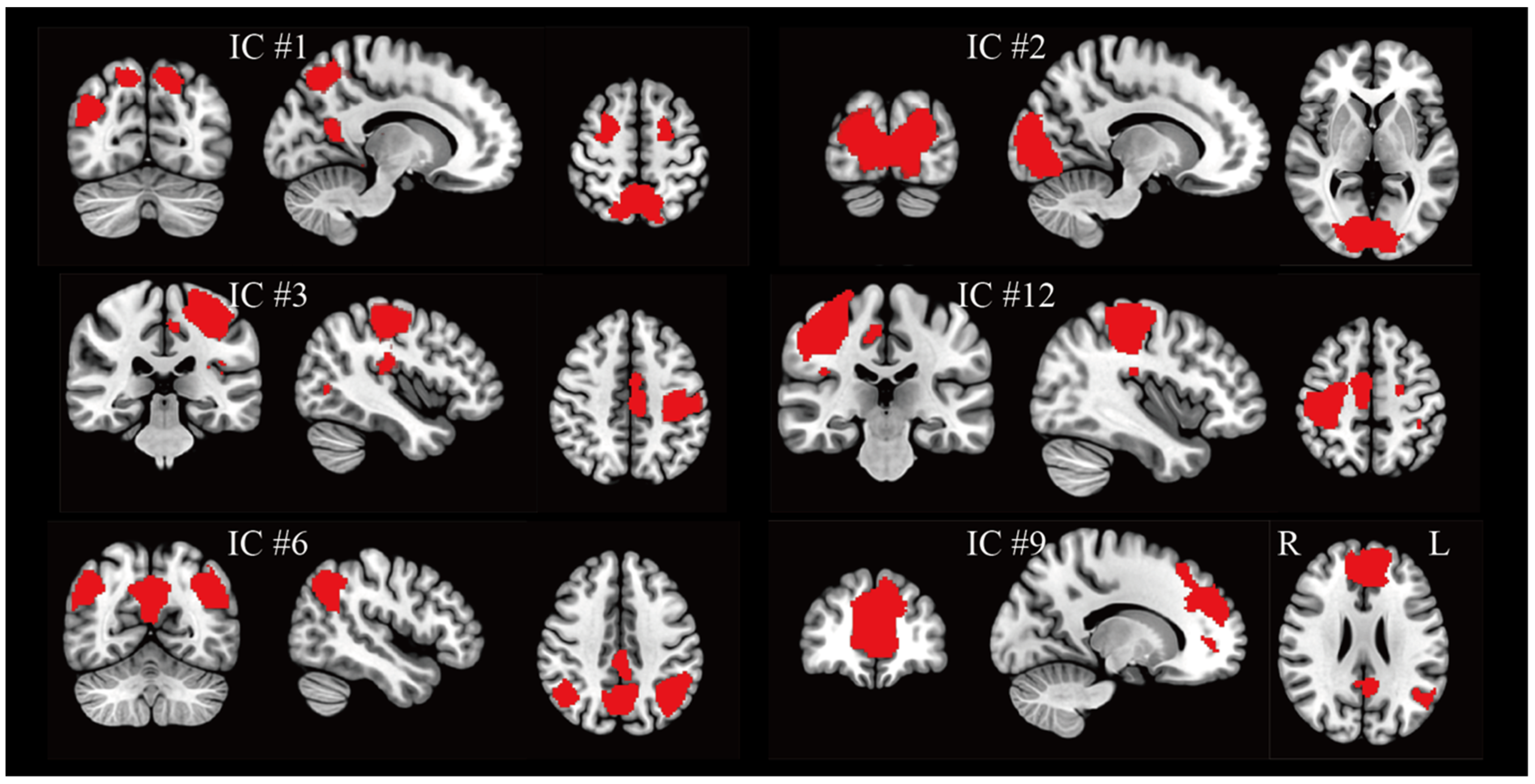
Sixteen BCI regulation-related group components were extracted across runs and participants. Visual inspection and anatomical labeling indicated that the vast majority corresponded to well-known large-scale systems engaged during naturalistic, visually guided motor behavior (Figure 2). A dorsal attention network component (IC#1) encompassed bilateral superior and middle frontal gyri with parietal extensions, consistent with intraparietal/superior parietal nodes implicated in goal-directed orienting. A primary visual component (IC#2) covered the occipital cortex with peaks in the lingual gyrus and intracalcarine cortex, reflecting sustained visual processing in the immersive environment. Two lateralized sensorimotor components (IC#3 left, IC#12 right) captured the precentral and postcentral gyri and portions of SMA, showing clear hemispheric segregation aligned with typical somatomotor representations. The default mode network was partitioned into two complementary components (IC#6 and IC#9) that together recapitulated the medial prefrontal, posterior cingulate/precuneus, and lateral parietal hubs. In addition, the behavioral relevance of the data-driven components was established by testing their dual regression time series against in-game events. The left-lateralized sensorimotor network (IC#3) showed a reliable coupling with discrete player actions. Its time series correlated with responses to shooting events in 61 out of 72 runs after multiple-comparison correction (mean r = 0.38 ± 0.16).

3.2. Group Difference in Network Dynamics

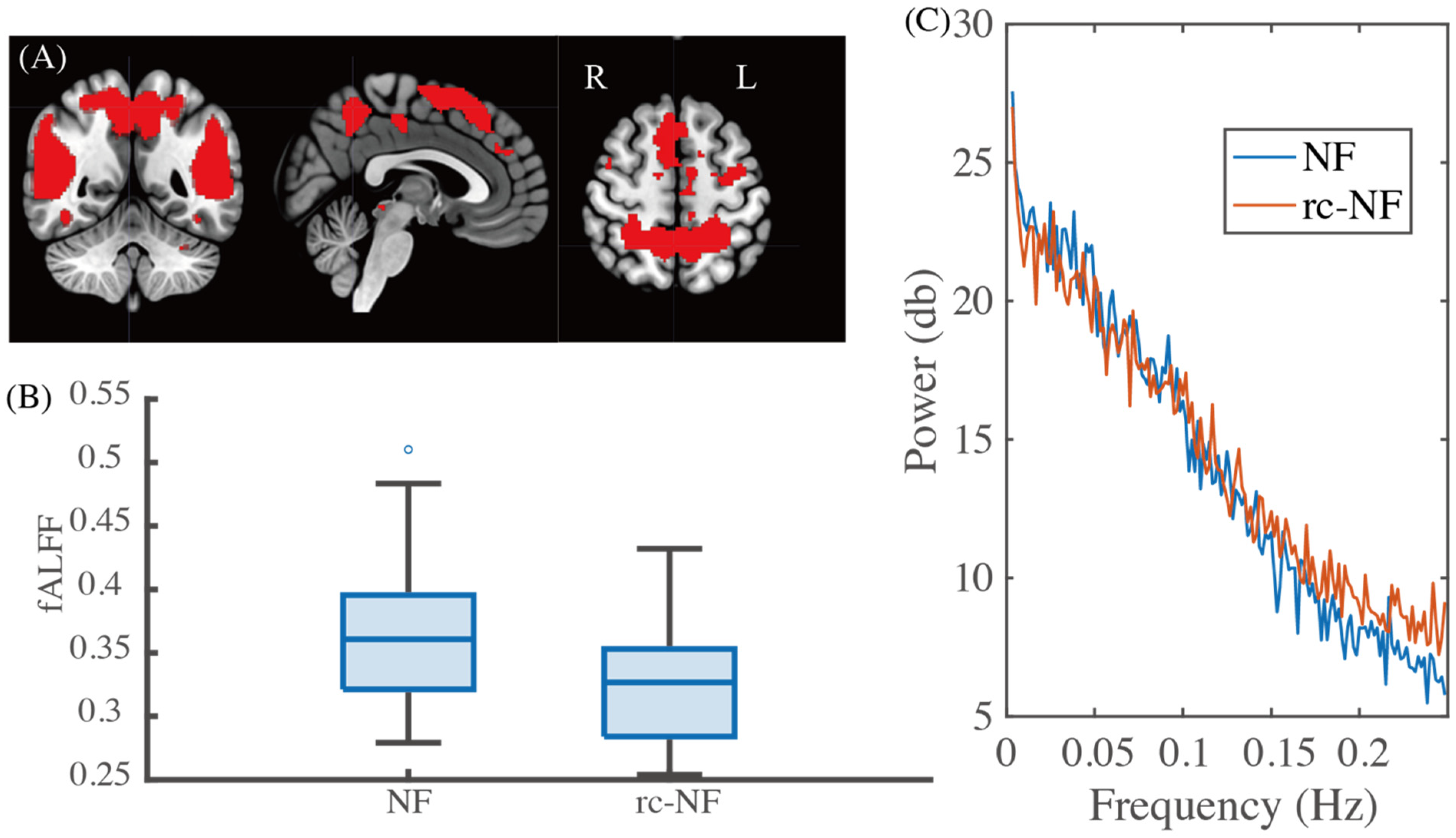
To assess how neurofeedback altered ongoing network dynamics, we quantified the fALFF for each component’s time series. After controlling for multiple comparisons across the sixteen components, a single effect survived correction (Figure 3). The frontoparietal control network (IC#15) exhibiting the expected dorsolateral prefrontal–inferior parietal topographies expressed significantly higher fALFF in the NF group than in the rc-NF group (corrected p = 0.036). Higher involvement of the low-frequency band 0.01~0.08 Hz is observed (Figure 3C) in the NF group. This indicated stronger low-frequency engagement of executive control processes under veridical feedback. No other components showed corrected between-group differences in fALFF, suggesting that neurofeedback selectively enhanced slow-control dynamics rather than globally elevating low-frequency power across networks.

3.3. Group Differences in Spatial Expressions

Voxel-wise comparisons of dual regression spatial maps revealed a complementary effect on network topology. The salience/ventral attention component (IC#13) displayed clusters with significant differences between NF and rc-NF groups (FWE corrected p < 0.05; Figure 4). Three clusters survived after correction (Table 1), with peaks located in the left inferior parietal, postcentral, middle, and superior frontal gyrus, and left occipital lobe. Around 20% of the target region of SMA is contained in the large cluster #2. Hence, the distribution of these clusters overlaps both the neurofeedback target (SMA) and associative control regions, while extending into the early visual cortex. This pattern indicates a reweighting of salience-network expression that spans regulatory, sensorimotor, and sensory nodes during visually guided action. No other components exhibited significant spatial differences under the threshold, considering the multiple comparisons.

4. Discussion

We used a data-driven, multi-echo-based on group ICA framework and dual regression to examine whole-brain network responses during BCI regulation embedded in an immersive first-person shooting virtual environment. Group decomposition recovered sixteen components that corresponded to common large-scale systems (e.g., visual, dorsal attention, default mode, sensorimotor, salience, and control networks) as well as task-specific sensorimotor maps related to VE actions. At the network dynamics level, the control-related component (IC#15) showed significantly higher fALFF values during active BCI regulation compared with the reduced contingency control. At the spatial level, IC#13, which most closely resembled the salience-network topology, exhibited voxel-wise group differences with clusters near the left inferior parietal, postcentral, middle, and superior frontal gyrus, and left occipital lobe. Around 20% of the target region of SMA is contained in the large cluster #2. Together, these results point to distributed, network-level modulation associated with successful regulation under an immersive BCI task rather than purely local effects limited to the SMA.
Control network dynamics and self-regulation frontoparietal control networks (FPCN) flexibly integrate task rules, goals, and sensory evidence to support adaptive behavior [36,37]. Increased fALFF within FPCN during active neurofeedback suggests stronger engagement of slow-control dynamics that track self-monitoring, strategy maintenance, and error-driven updating, consistent with sustained attention and executive monitoring demands in neurofeedback [36,38,39]. fALFF has been interpreted as a proxy for spontaneous low-frequency activity in resting-state studies [35]. Elevated fALFF under continuous tasks may indicate persistent, slow control processes required to maintain a regulation set. The salience network (SAL), anchored in the anterior insula and the dorsal anterior cingulate with extensions into prefrontal and parietal cortices, is thought to detect behaviorally relevant events and orchestrate switching between internally oriented (default mode) and externally oriented (control and attentional) systems [36,40,41]. The observed spatial reweighting in an SAL/ventral attention component across SMA, lateral prefrontal, inferior parietal, and occipital regions suggests that veridical feedback enhanced SAL’s integration with sensorimotor and visual circuits that are critical for visually guided action and speed control. This aligns with proposals that SAL facilitates rapid reallocation of control resources and maintains readiness to exploit informative feedback during continuous behavior. Notably, significant clusters included SMA and precentral gyrus that engaged during motor imagery and action selection [42], supporting the view that the BCI target is embedded within a broader salience-control-sensorimotor ensemble.
Our prior ROI-based analyses in this paradigm established that participants could modulate SMA activity and behavior (movement speed) under active feedback [9]. The network perspective here extends those findings in two ways. First, it shows that veridical feedback amplifies slow FPCN dynamics, consistent with more effective executive control and sustained attention. Second, it reveals an SAL-centered redistribution of spatial expression across regulatory, motor, and early visual territories. This organization is well suited to a task where participants must integrate internal regulation goals with rapid sensory–motor decisions. These results resonate with the “triple-network” framework, in which interactions among default mode, salience, and control networks shape cognitive control and its dysfunction [36]. In our immersive BCI, veridical feedback appears to bias the system toward an SAL-FPCN configuration supportive of regulation.
These observations verified the feasibility of the design of future BCI-based regulation with immersive, semi-naturalistic contexts. Immersive VEs embed neurofeedback in meaningful sensorimotor contingencies, which can heighten motivation and clarify the link between brain activity and behavior [9,19,23]. Naturalistic stimuli typically recruit broad, reliable networks and stabilize inter-individual patterns of activity [24,43]. In our FPS task, movement speed proved to be an intuitive, high-leverage mapping for participants, where it immediately affects navigation, risk, and reward, potentially increasing adherence to regulation strategies [9,44]. Besides the VE, we tested the analysis framework based on multi-echo data together with group ICA and dual regression. For continuous, interactive designs where conventional GLMs struggle, ICA identifies meaningful networks and dual regression yields per-subject time courses and maps suitable for both dynamic and spatial inference. Together, these methods form clear implications for the design and analysis of future BCI regulation.
At the same time, several limitations temper the conclusions. While effects survived conservative correction, the sample is modest. Replication in larger cohorts is needed to refine effect sizes and to probe inter-individual variability (e.g., gaming expertise). Because the virtual environment entails continuous motor actions, visual input, and decision-making, disentangling the specific contribution of neurofeedback from task demands remains challenging. Individual differences in strategy, motivation, or baseline neural states may have further influenced the magnitude of network changes. Expanding targets beyond SMA, like prefrontal or limbic regions, which are less accessible to non-invasive stimulation, may open new therapeutic avenues. In addition, this study is primarily descriptive at the network level and was not designed to benchmark supervised decoding accuracy. Future work may integrate machine-/deep-learning models that can exploit temporal dependencies and multiscale structure to decode latent states from VR-BCI data and to test generalization across tasks and individuals [12,13,14]. Such approaches are complementary to the present network-centric analysis and may yield predictors that can be used for adaptive or personalized neurofeedback. Finally, moving from offline network metrics toward real-time, network-level feedback represents an exciting, albeit technically demanding, direction that could close the loop on network-adaptive BCIs.

5. Conclusions

In an immersive fMRI-BCI, SMA-based neurofeedback was associated with selective modulation of large-scale networks, reflected by higher low-frequency engagement within a frontoparietal control component and a reweighting of salience-network expression encompassing SMA, prefrontal, parietal, and occipital regions. These results move beyond localized SMA modulation to a systems-level account of how BCIs may shape brain function during real-time behavior. The present findings indicate involvement of distributed networks; however, they cannot prove specific causal mechanisms. Future studies incorporating decoding-based models or perturbation methods may further clarify the functional roles of these networks.

Author Contributions

Conceptualization, T.F., H.I.B., J.Z. and K.M.; methodology, T.F. and K.M.; software, T.F.; validation, T.F., J.Z. and K.M.; formal analysis, T.F.; investigation, T.F. and J.Z.; resources, T.F., H.I.B. and K.M.; data curation, T.F.; writing—original draft preparation, T.F.; writing—review and editing, T.F., H.I.B., J.Z. and K.M.; visualization, T.F., H.I.B., J.Z. and K.M.; supervision, K.M.; project administration, T.F. and K.M.; funding acquisition, K.M. All authors have read and agreed to the published version of the manuscript.

Funding

This research was funded by the German Research Foundation (DFG—IRTG 2150; TRR 379 B03 and Q02, 512007073) and the German Ministry for Education and Research (BMBF; APIC: 01EE1405C). T.F. was funded by the scholarships from China Scholarship Council (No. 202106060064). The study sponsors were not involved in the study design and in the collection, analysis, and interpretation of data; in the writing of the manuscript; or in the decision to submit the manuscript for publication.

Institutional Review Board Statement

The study was conducted in accordance with the Declaration of Helsinki and approved by the Independent Ethics Committee of the University Hospital RWTH Aachen (protocol code EK 188/17, 25 September 2017).

Informed Consent Statement

Informed consent was obtained from all subjects involved in the study.

Data Availability Statement

The data and the scripts for reproducing the results are available upon request if not limited by concerns of the ethical commission.

Acknowledgments

This work was supported by the Brain Imaging Facility of the Interdisciplinary Center for Clinical Research (IZKF) Aachen within the Faculty of Medicine at RWTH Aachen University.

Conflicts of Interest

The authors declare no conflicts of interest.

Abbreviations

The following abbreviations are used in this manuscript:
BCIBrain–computer interface
SMAsupplementary motor area
fALFFfractional amplitude of low-frequency fluctuations
rt-fMRIReal-time functional magnetic resonance imaging
BOLDblood-oxygen-level-dependent
VEvirtual environment
ROIRegion-of-interest
ICAIndependent component analysis
NFNeurofeedback

References

  1. Zhang, H.; Jiao, L.; Yang, S.; Li, H.; Jiang, X.; Feng, J.; Zou, S.; Xu, Q.; Gu, J.; Wang, X.; et al. Brain–Computer Interfaces: The Innovative Key to Unlocking Neurological Conditions. Int. J. Surg. 2024, 110, 5745–5762. [Google Scholar] [CrossRef] [PubMed]
  2. Gao, X.; Wang, Y.; Chen, X.; Gao, S. Interface, Interaction, and Intelligence in Generalized Brain–Computer Interfaces. Trends Cogn. Sci. 2021, 25, 671–684. [Google Scholar] [CrossRef] [PubMed]
  3. Sitaram, R.; Ros, T.; Stoeckel, L.; Haller, S.; Scharnowski, F.; Lewis-Peacock, J.; Weiskopf, N.; Blefari, M.L.; Rana, M.; Oblak, E.; et al. Closed-Loop Brain Training: The Science of Neurofeedback. Nat. Rev. Neurosci. 2017, 18, 86–100. [Google Scholar] [CrossRef]
  4. Sarkheil, P.; Zilverstand, A.; Kilian-Hütten, N.; Schneider, F.; Goebel, R.; Mathiak, K. fMRI Feedback Enhances Emotion Regulation as Evidenced by a Reduced Amygdala Response. Behav. Brain Res. 2015, 281, 326–332. [Google Scholar] [CrossRef]
  5. Zweerings, J.; Sarkheil, P.; Keller, M.; Dyck, M.; Klasen, M.; Becker, B.; Gaebler, A.J.; Ibrahim, C.N.; Turetsky, B.I.; Zvyagintsev, M.; et al. Rt-fMRI Neurofeedback-Guided Cognitive Reappraisal Training Modulates Amygdala Responsivity in Posttraumatic Stress Disorder. NeuroImage Clin. 2020, 28, 102483. [Google Scholar] [CrossRef]
  6. Mel’nikov, M.Y.; Bezmaternykh, D.D.; Savelov, A.A.; Petrovskiy, E.D.; Kozlova, L.I.; Natarova, K.A.; Larina, T.D.; Andamova, T.M.; Zvyagintsev, M.; Shtark, M.B.; et al. Real-Time fMRI Neurofeedback Compared to Cognitive Behavioral Therapy in a Pilot Study for the Treatment of Mild and Moderate Depression. Eur. Arch. Psychiatry Clin. Neurosci. 2023, 273, 1139–1149. [Google Scholar] [CrossRef]
  7. Zhang, Y.; Becker, B.; Kendrick, K.M.; Zhang, Q.; Yao, S. Self-Navigating the “Island of Reil”: A Systematic Review of Real-Time fMRI Neurofeedback Training of Insula Activity. Transl. Psychiatry 2025, 15, 170. [Google Scholar] [CrossRef]
  8. Pindi, P.; Houenou, J.; Piguet, C.; Favre, P. Real-Time fMRI Neurofeedback as a New Treatment for Psychiatric Disorders: A Meta-Analysis. Prog. Neuro-Psychopharmacol. Biol. Psychiatry 2022, 119, 110605. [Google Scholar] [CrossRef]
  9. Baqapuri, H.I.; Roes, L.D.; Zvyagintsev, M.; Ramadan, S.; Kell, M.; Roecher, E.; Zweerings, J.; Klasen, M.; Gur, R.C.; Mathiak, K. A Novel Brain–Computer Interface Virtual Environment for Neurofeedback During Functional MRI. Front. Neurosci. 2021, 14, 593854. [Google Scholar] [CrossRef] [PubMed]
  10. Watve, A.; Haugg, A.; Frei, N.; Koush, Y.; Willinger, D.; Bruehl, A.B.; Stämpfli, P.; Scharnowski, F.; Sladky, R. Facing Emotions: Real-Time fMRI-Based Neurofeedback Using Dynamic Emotional Faces to Modulate Amygdala Activity. Front. Neurosci. 2024, 17, 1286665. [Google Scholar] [CrossRef]
  11. Du, B.; Cheng, X.; Duan, Y.; Ning, H. fMRI Brain Decoding and Its Applications in Brain–Computer Interface: A Survey. Brain Sci. 2022, 12, 228. [Google Scholar] [CrossRef]
  12. Wen, D.; Wei, Z.; Zhou, Y.; Li, G.; Zhang, X.; Han, W. Deep Learning Methods to Process fMRI Data and Their Application in the Diagnosis of Cognitive Impairment: A Brief Overview and Our Opinion. Front. Neuroinform. 2018, 12, 23. [Google Scholar] [CrossRef]
  13. Cui, W.; Wang, Z.; Ma, L. SED-GPT: A Non-Invasive Method for Long-Sequence Fine-Grained Semantics and Emotions Decoding. Appl. Sci. 2025, 15, 11100. [Google Scholar] [CrossRef]
  14. Floren, A.; Naylor, B.; Miikkulainen, R.; Ress, D. Accurately Decoding Visual Information from fMRI Data Obtained in a Realistic Virtual Environment. Front. Hum. Neurosci. 2015, 9, 327. [Google Scholar] [CrossRef]
  15. Guo, J.; Yi, C.; Li, F.; Xu, P.; Tian, Y. MindLDM: Reconstruct Visual Stimuli from fMRI Using Latent Diffusion Model. In Proceedings of the 2024 IEEE International Conference on Computational Intelligence and Virtual Environments for Measurement Systems and Applications (CIVEMSA), Xi’an, China, 14–16 June 2024; pp. 1–6. [Google Scholar]
  16. Rithwik, P.; Benzy, V.K.; Vinod, A.P. High Accuracy Decoding of Motor Imagery Directions from EEG-Based Brain Computer Interface Using Filter Bank Spatially Regularised Common Spatial Pattern Method. Biomed. Signal Process. Control 2022, 72, 103241. [Google Scholar] [CrossRef]
  17. Liu, F.; Meamardoost, S.; Gunawan, R.; Komiyama, T.; Mewes, C.; Zhang, Y.; Hwang, E.; Wang, L. Deep Learning for Neural Decoding in Motor Cortex. J. Neural Eng. 2022, 19, 056021. [Google Scholar] [CrossRef] [PubMed]
  18. Candelori, B.; Bardella, G.; Spinelli, I.; Ramawat, S.; Pani, P.; Ferraina, S.; Scardapane, S. Spatio-Temporal Transformers for Decoding Neural Movement Control. J. Neural Eng. 2025, 22, 016023. [Google Scholar] [CrossRef] [PubMed]
  19. Kober, S.E.; Wood, G.; Berger, L.M. Controlling Virtual Reality With Brain Signals: State of the Art of Using VR-Based Feedback in Neurofeedback Applications. Appl. Psychophysiol. Biofeedback 2024. [Google Scholar] [CrossRef]
  20. Piszcz, A.; Rojek, I.; Mikołajewski, D. Impact of Virtual Reality on Brain–Computer Interface Performance in IoT Control—Review of Current State of Knowledge. Appl. Sci. 2024, 14, 10541. [Google Scholar] [CrossRef]
  21. Castanho, L.; Martinho, D.V.; Saial, A.C.; Gouveia, B.R.; Gouveia, É.R.; Ribeiro, F. The Efficacy of Virtual Reality-Based EEG Neurofeedback in Health-Related Symptoms Relief: A Systematic Review. Appl. Psychophysiol. Biofeedback 2025. [Google Scholar] [CrossRef]
  22. Pinheiro, J.; de Almeida, R.S.; Marques, A. Emotional Self-Regulation, Virtual Reality and Neurofeedback. Comput. Hum. Behav. Rep. 2021, 4, 100101. [Google Scholar] [CrossRef]
  23. Li, K.; Huang, W.; Gao, W.; Guan, Z.; Huang, Q.; Yu, J.-G.; Yu, Z.L.; Li, Y. An Electroencephalography-Based Brain-Computer Interface for Emotion Regulation With Virtual Reality Neurofeedback. IEEE Trans. Cogn. Dev. Syst. 2024, 16, 1405–1417. [Google Scholar] [CrossRef]
  24. Cong, F.; Puoliväli, T.; Alluri, V.; Sipola, T.; Burunat, I.; Toiviainen, P.; Nandi, A.K.; Brattico, E.; Ristaniemi, T. Key Issues in Decomposing fMRI during Naturalistic and Continuous Music Experience with Independent Component Analysis. J. Neurosci. Methods 2014, 223, 74–84. [Google Scholar] [CrossRef]
  25. Feng, T.; Gaebler, A.J.; Keller, M.; Zweerings, J.; Li, H.; Cong, F.; Mathiak, K. Basic Stimulus Processing Alterations from Top-down Cognitive Control in Depression Drive Independent Temporal Components of Multi-Echo Naturalistic fMRI Data. Transl. Psychiatry 2025, 15, 171. [Google Scholar] [CrossRef]
  26. Koush, Y.; Masala, N.; Scharnowski, F.; Van De Ville, D. Data-Driven Tensor Independent Component Analysis for Model-Based Connectivity Neurofeedback. NeuroImage 2019, 184, 214–226. [Google Scholar] [CrossRef]
  27. Filippini, N.; MacIntosh, B.J.; Hough, M.G.; Goodwin, G.M.; Frisoni, G.B.; Smith, S.M.; Matthews, P.M.; Beckmann, C.F.; Mackay, C.E. Distinct Patterns of Brain Activity in Young Carriers of the APOE-Ε4 Allele. Proc. Natl. Acad. Sci. USA 2009, 106, 7209–7214. [Google Scholar] [CrossRef] [PubMed]
  28. Nickerson, L.D.; Smith, S.M.; Öngür, D.; Beckmann, C.F. Using Dual Regression to Investigate Network Shape and Amplitude in Functional Connectivity Analyses. Front. Neurosci. 2017, 11, 115. [Google Scholar] [CrossRef]
  29. Salman, M.S.; Du, Y.; Lin, D.; Fu, Z.; Fedorov, A.; Damaraju, E.; Sui, J.; Chen, J.; Mayer, A.R.; Posse, S.; et al. Group ICA for Identifying Biomarkers in Schizophrenia: ‘Adaptive’ Networks via Spatially Constrained ICA Show More Sensitivity to Group Differences than Spatio-Temporal Regression. NeuroImage Clin. 2019, 22, 101747. [Google Scholar] [CrossRef] [PubMed]
  30. Calhoun, V.D.; Silva, R.F.; Adalı, T.; Rachakonda, S. Comparison of PCA Approaches for Very Large Group ICA. NeuroImage 2015, 118, 662–666. [Google Scholar] [CrossRef]
  31. Hu, G.; Waters, A.B.; Aslan, S.; Frederick, B.; Cong, F.; Nickerson, L.D. Snowball ICA: A Model Order Free Independent Component Analysis Strategy for Functional Magnetic Resonance Imaging Data. Front. Neurosci. 2020, 14, 569657. [Google Scholar] [CrossRef]
  32. Feng, T.; Baqapuri, H.I.; Zweerings, J.; Li, H.; Cong, F.; Mathiak, K. Characterizing the Distribution of Neural and Non-Neural Components in Multi-Echo EPI Data across Echo Times Based on Tensor-ICA. NeuroImage 2025, 311, 121199. [Google Scholar] [CrossRef]
  33. Bhavsar, S.; Zvyagintsev, M.; Mathiak, K. BOLD Sensitivity and SNR Characteristics of Parallel Imaging-Accelerated Single-Shot Multi-Echo EPI for fMRI. NeuroImage 2014, 84, 65–75. [Google Scholar] [CrossRef]
  34. Erhardt, E.B.; Rachakonda, S.; Bedrick, E.J.; Allen, E.A.; Adali, T.; Calhoun, V.D. Comparison of Multi-Subject ICA Methods for Analysis of fMRI Data. Hum. Brain Mapp. 2011, 32, 2075–2095. [Google Scholar] [CrossRef]
  35. Zou, Q.-H.; Zhu, C.-Z.; Yang, Y.; Zuo, X.-N.; Long, X.-Y.; Cao, Q.-J.; Wang, Y.-F.; Zang, Y.-F. An Improved Approach to Detection of Amplitude of Low-Frequency Fluctuation (ALFF) for Resting-State fMRI: Fractional ALFF. J. Neurosci. Methods 2008, 172, 137–141. [Google Scholar] [CrossRef]
  36. Menon, V. Large-Scale Brain Networks and Psychopathology: A Unifying Triple Network Model. Trends Cogn. Sci. 2011, 15, 483–506. [Google Scholar] [CrossRef]
  37. Cole, M.W.; Reynolds, J.R.; Power, J.D.; Repovs, G.; Anticevic, A.; Braver, T.S. Multi-Task Connectivity Reveals Flexible Hubs for Adaptive Task Control. Nat. Neurosci. 2013, 16, 1348–1355. [Google Scholar] [CrossRef]
  38. Sulzer, J.; Haller, S.; Scharnowski, F.; Weiskopf, N.; Birbaumer, N.; Blefari, M.L.; Bruehl, A.B.; Cohen, L.G.; deCharms, R.C.; Gassert, R.; et al. Real-Time fMRI Neurofeedback: Progress and Challenges. NeuroImage 2013, 76, 386–399. [Google Scholar] [CrossRef] [PubMed]
  39. Sridharan, D.; Levitin, D.J.; Menon, V. A Critical Role for the Right Fronto-Insular Cortex in Switching between Central-Executive and Default-Mode Networks. Proc. Natl. Acad. Sci. USA 2008, 105, 12569–12574. [Google Scholar] [CrossRef] [PubMed]
  40. Stein, J.; Korb, F.M.; Goschke, T.; Zwosta, K. Salience Network Resting-State Functional Connectivity Predicts Self-Controlled Decision-Making. Sci. Rep. 2025, 15, 16332. [Google Scholar] [CrossRef]
  41. Schimmelpfennig, J.; Topczewski, J.; Zajkowski, W.; Jankowiak-Siuda, K. The Role of the Salience Network in Cognitive and Affective Deficits. Front. Hum. Neurosci. 2023, 17, 1133367. [Google Scholar] [CrossRef] [PubMed]
  42. Nachev, P.; Kennard, C.; Husain, M. Functional Role of the Supplementary and Pre-Supplementary Motor Areas. Nat. Rev. Neurosci. 2008, 9, 856–869. [Google Scholar] [CrossRef] [PubMed]
  43. Sonkusare, S.; Breakspear, M.; Guo, C. Naturalistic Stimuli in Neuroscience: Critically Acclaimed. Trends Cogn. Sci. 2019, 23, 699–714. [Google Scholar] [CrossRef] [PubMed]
  44. Mathiak, K.; Weber, R. Toward Brain Correlates of Natural Behavior: fMRI during Violent Video Games. Hum. Brain Mapp. 2006, 27, 948–956. [Google Scholar] [CrossRef] [PubMed]
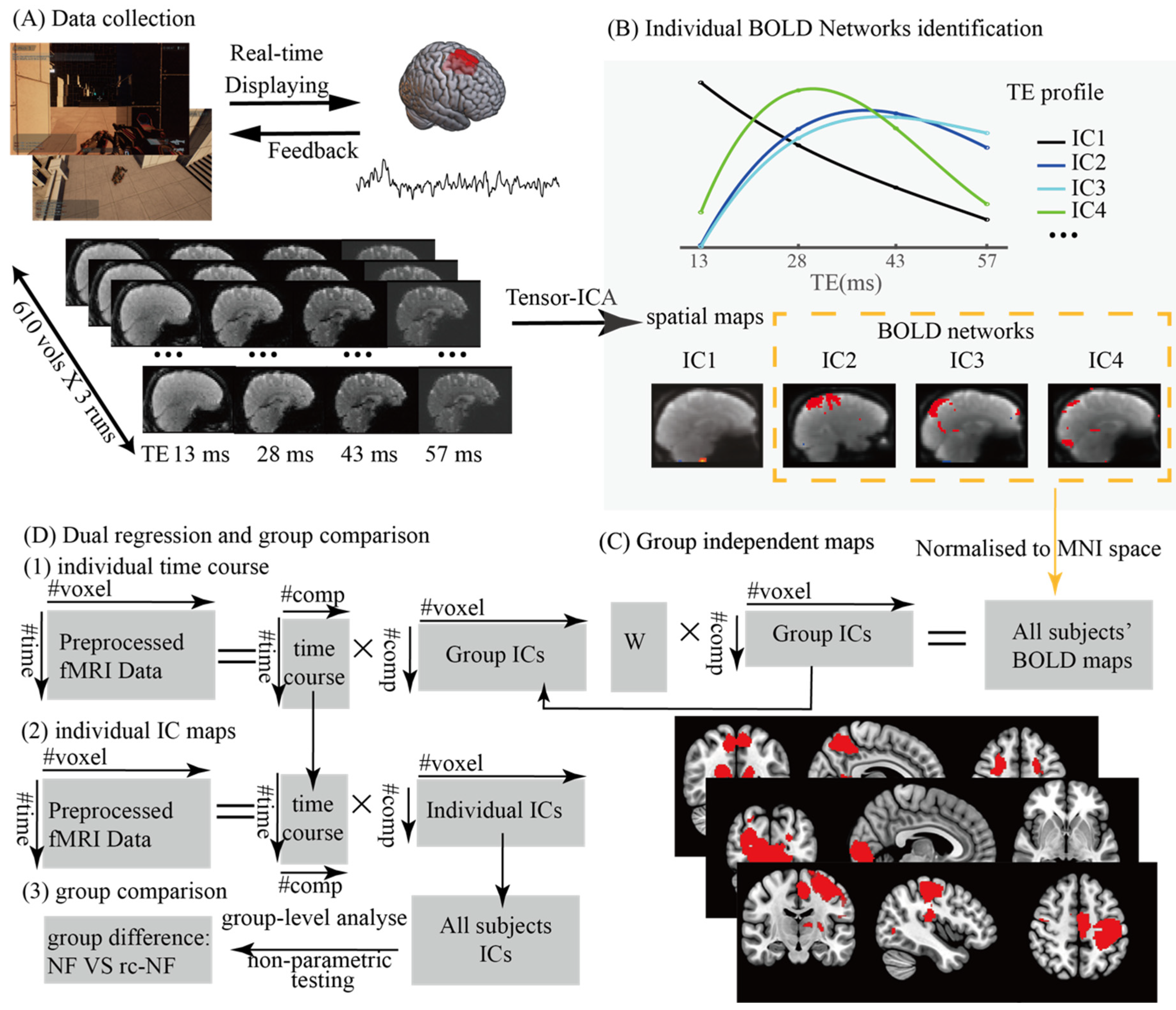
Figure 1. Overview of experimental design and analysis pipeline. (A) Immersive first-person shooting virtual environment used in the scanner. Participant navigates arena and shoots, while their movement speed is modulated by neural activity in the supplementary motor area (SMA). Multi-echo EPI data with four echo times were acquired at the same time. (B) Individual BOLD networks identification based on three-way decomposition of the ME-EPI data. Components exhibiting BOLD patterns (bell-shaped) across TEs were identified as BOLD networks. (C) Estimation of group independent networks from concatenated spatial maps from individuals. (D) Dual regression for individual spatial maps and time series, and further comparisons.
Applsci 15 12583 g001
Figure 2. Representative spatial maps of BCI regulation-related group components (red, Z > 2.3). Sixteen independent components were derived from the group ICA, and they corresponded to common functional systems. IC#1 reflected part of the dorsal attention network. IC#2 demonstrated the engagement of the visual network (occipital cortex). IC#3 and IC#12 represented left and right sensorimotor networks, and the time courses of IC#3 are correlated with discrete in-game shooting events. IC#6 and IC#9 showed patterns of the default mode network.
Applsci 15 12583 g002
Figure 3. Differences in network dynamics of one control network-related component between active and reduced contingency neurofeedback groups. (A) IC#15 displayed the dorsolateral prefrontal–inferior parietal topographies (red, Z > 2.3). (B) Boxplot of component fALFF (0.01−0.08 Hz) shows higher low-frequency engagement in NF versus rc-NF. Between-group difference survived correction across components (corrected p = 0.036). (C) Average power spectra of individual time courses by group illustrate selectively elevated power in the low-frequency band under active feedback.
Applsci 15 12583 g003
Figure 4. Differences between active and reduced contingency neurofeedback groups in spatial expression of a salience/ventral attention component. Spatial map of the salience/ventral attention-like component (IC#13) derived from group ICA is shown in the top line (red). Three clusters with group difference were observed near the left inferior parietal, postcentral, middle, and superior frontal gyrus, and the left occipital lobe (green). Around 20% of the target region of SMA is contained in the large cluster.
Applsci 15 12583 g004
Table 1. Clusters with a significant difference between active and reduced contingency neurofeedback groups.
Cluster #Brain LabelPeaks’ MNI CoordinatestkE
xyz
1Left inferior parietal−48−46366.172532
Left postcentral−40−36505.74
Left inferior parietal−46−44525.32
2Left middle frontal−2636265.786704
Left superior frontal20−30385.71
Left superior frontal−26−2−545.53
3Left calcarine16−8085.251354
Left middle occipital−44−6804.54
Left inferior occipital−34−74−83.78
The regions were labeled according to the automatic anatomical labeling atlas (AAL3). Cluster-level threshold according to pFWE < 0.0016, considering the multiple comparisons after voxel-level threshold p < 0.001.
Disclaimer/Publisher’s Note: The statements, opinions and data contained in all publications are solely those of the individual author(s) and contributor(s) and not of MDPI and/or the editor(s). MDPI and/or the editor(s) disclaim responsibility for any injury to people or property resulting from any ideas, methods, instructions or products referred to in the content.