Review

Fracture Toughness of CAD/CAM Resin-Based Materials vs. Direct Composite Resins: A Scoping Review

by
Socratis Thomaidis
1,
Eftychia Pappa
1 and
Maria Antoniadou
1,2,*
1
Department of Operative Dentistry, School of Health Sciences, National and Kapodistrian University of Athens, 11527 Athens, Greece
2
Certified Systemic Management Program (CSAP), University of Piraeus, 18534 Piraeus, Greece
*
Author to whom correspondence should be addressed.
Appl. Sci. 2025, 15(22), 12308; https://doi.org/10.3390/app152212308
Submission received: 17 October 2025 / Revised: 10 November 2025 / Accepted: 11 November 2025 / Published: 20 November 2025

Featured Application

Large partial restorations can be performed directly with composite resins or by means of CAD/CAM restorations. Fracture toughness can give some input on the crack propagation resistance of these restorations and therefore facilitate the decision of using direct or indirect restorations.

Abstract

Objective: To compare the fracture toughness of CAD/CAM resin-based restorative materials and direct composite resins. Materials and Methods: A systematic search was conducted in July 2025 across PubMed/MEDLINE, EBSCO, Scopus, ISI Web of Science, and grey literature. Eligible studies were only in vitro investigations evaluating fracture toughness of direct composite resins and CAD/CAM hybrid materials. Study selection, data extraction, and risk-of-bias assessment were performed independently by two reviewers. A systematic review and meta-analysis were not performed due to methodological heterogeneity, and findings were synthesized qualitatively. Results: Fifty-two studies met the inclusion criteria, including 16 assessing CAD/CAM restorative materials. Considerable variability in fracture toughness values was observed, even within the same material type. Most studies were judged to present a medium risk of bias. Short fiber-reinforced composites exhibited higher fracture toughness, whereas other CAD/CAM resin-based materials showed values comparable to direct composites. Conclusions: Current evidence does not confirm superior fracture toughness of CAD/CAM resin-based materials compared with direct composites. Short fiber-reinforced composites appear to offer improved resistance to crack propagation. Standardized testing protocols are needed to enable more reliable comparisons.

1. Introduction

Composite resins have been extensively used for small- and medium-sized restorations for decades, showing proven long-term clinical success [1,2]. Nevertheless, bulk fracture remains the most common reason for failure [1], followed closely by marginal chipping [2]. Because all composite resin restorations inherently contain flaws such as pores, fracture toughness has been identified as the most critical mechanical property for their evaluation [3]. The incorporation of fillers, while intended to reinforce the resin matrix, may also introduce microstructural heterogeneities and defects such as pores and filler agglomerates, both of which can act as critical triggers for fracture [4].
When characterizing composite resins according to their mechanical properties, compressive strength, flexural strength, diametral strength, and many other properties can be tested [5]. These tests can give an insight into their resistance to fracture. Most materials, though, have intrinsic cracks or flaws that can lead to premature failure during their application/service [6,7]. Fracture toughness is a property that gives information on the resistance to the propagation of an intrinsic crack or flaw of a material, which will lead to failure [5]. Fracture toughness differs fundamentally from strength, as it is an intrinsic material property that should not vary according to specimen geometry or test configuration [4,5]. It is defined as the resistance of a material to crack propagation from a pre-existing flaw [6]. Because virtually all restorative materials contain such flaws, clinical failure often occurs at lower loads than predicted from their measured strength values [6]. To account for this, fracture toughness testing requires specimens to be fabricated with a predefined crack, from which failure propagates under loading [5]. This crack can either be incorporated during specimen fabrication or introduced after curing by cutting [7]. The most reliable method is to subject a notched specimen to cyclic fatigue loading, which produces an infinitely sharp notch or, even better, a sharp notch ending to a formed “pre-crack” and ensures accurate results [5,7].
The clinical importance of fracture toughness has been repeatedly demonstrated. A systematic review by Heintze et al. [8] reported a weak positive correlation (rho = 0.34) between fracture toughness and clinical fractures of posterior composite restorations across 31 different materials. In contrast, no correlations were observed between compressive strength, flexural modulus, or flexural strength and restoration survival [8]. Ferracane and Condon [9] also found an association between fracture toughness and both marginal breakdown and wear of composites, while Tyas [10], much earlier, identified fracture toughness as the property most strongly associated with failure in Class IV cavities.
Direct composite resins are widely applied in both small and relatively large cavities [11]. For larger restorations, indirect techniques can also be employed [12]. Traditional indirect composites, fabricated on stone casts and polymerized under heat and pressure after initial light curing, were designed to reduce polymerization shrinkage [13]; however, they still presented internal defects and pores similar to direct composites [14]. The advent of intraoral scanning and CAD/CAM technology has since transformed indirect restorative workflows, enabling the fabrication of dense, highly cross-linked resin-based blocks with improved homogeneity and fewer internal flaws [15,16,17]. CAD/CAM systems initially gained popularity through the use of glass ceramics, which consistently demonstrated superior mechanical properties compared with conventional composite resins [18,19,20]. More recently, resin-based CAD/CAM restorative blocks, including resin nanoceramics and polymer-infiltrated ceramic networks (PICNs),have been introduced into the restorative material portfolio [18].
Several in vitro investigations have assessed the fracture toughness of these novel materials, reporting both promising results and variability in performance [5,9,17,18,21]. Clinical evidence has also confirmed their applicability. A retrospective clinical study by Borgia et al. [20] demonstrated the long-term survival of indirect composite restorations. Systematic reviews and meta-analyses have further reinforced the feasibility of both direct and indirect composites: Angeletaki et al. [21] highlighted the effectiveness of direct versus indirect restorations for posterior teeth, Azeem et al. [22] confirmed their clinical comparability, McGrath et al. [23] synthesized long-term performance data, while Moussa et al. [24] and Tennert et al. [25] supported their use in large Class II cavities, including cusp coverage. From another systematic review and meta-analysis on clinical performance of direct composite resin versus indirect restorations on endodontically treated posterior teeth, it was found that for the short term (2.5 to 3 years), low-quality evidence suggested no difference in tooth survival or restoration quality [26]. Shafik Elmoselhy et al. [27] in a two-year controlled clinical trial reported that the CAD/CAM nano-hybrid composite blocks were as reliable as Lithium disilicate for restoring mutilated vital teeth. Moreover, Hassan et al. [28], in a randomized clinical split-mouth study, reported that IPS e.max CAD and hybrid RNC exhibit nearly identical clinical performance when evaluated over a 2-year period as per the modified USPHS criteria. On the other hand, El Din et al. [29], in a randomized controlled clinical trial that evaluated the restoration of non-vital posterior teeth with endocrowns, found that lithium disilicate IPS e.max CAD was the material of choice, while nano ceramic hybrid was less recommended. In the longitudinal clinical study of Hofsteenge et al. [30], with a 14-year follow-up period, studying the performance of premolars restored with direct or indirect cusp-replacing resin composite restorations, no statistically significant difference in survival rates between direct and indirect composite cusp-replacing restorations was mentioned.
Despite this growing body of evidence, methodological heterogeneity and limited standardization across studies restrict firm conclusions [20,23,25,31]. Moreover, although CAD/CAM resin-based restorative materials are increasingly popular, their potential advantages over direct composites in terms of fracture toughness remain uncertain [15,16]. To date, no structured evidence synthesis has comprehensively mapped the available studies on this topic. Accordingly, this scoping review was conducted to examine whether CAD/CAM resin-based restorative materials, particularly resin nanoceramics and polymer-infiltrated ceramic networks, exhibit higher fracture toughness than direct composite resins.

2. Materials and Methods

Our study followed the PRISMA Extension for Scoping Reviews (PRISMA-ScR) [32] and the Cochrane Handbook for Systematic Reviews [32]. The research question of this scoping review was structured according to the population-concept-context (PCC) framework. The population of interest comprised CAD/CAM resin-based materials used for indirect restorations, particularly resin nanoceramics and polymer-infiltrated ceramic networks. The primary outcome assessed (concept) was fracture toughness (K_IC), which, as we described briefly in the introduction part, reflects a material’s resistance to crack initiation and propagation and is regarded as a critical indicator of long-term clinical performance. The concept under evaluation included resin-infiltrated ceramics and other hybrid CAD/CAM restorative materials. The comparison group (context) consisted of direct composite resins routinely used in clinical practice.
Accordingly, the guiding question of this scoping review was as follows:
“Do CAD/CAM resin-based restorative materials, including resin nanoceramics and polymer-infiltrated ceramic networks, demonstrate higher fracture toughness than direct composite resins?”

2.1. Sources and Search Strategy

The MEDLINE via PubMed, ISI Web of Science, Scopus, and EBSCO Medline electronic databases were searched to identify eligible studies. The Cochrane Library was not included, as this database mainly indexes controlled clinical trials, whereas the present review focused on laboratory investigations. The initial search strategy combined terms related to composite resins, CAD/CAM restorative materials, and fracture toughness, and was subsequently refined to broaden coverage.
In PubMed, the following search strategy was applied:
((“Composite Resins” OR “Resin Cements” OR “composite resin”[All Fields] OR “resin-based material”[All Fields]) AND (“Computer-Aided Design” OR “Computer-Aided Manufacturing” OR “CAD/CAM”[All Fields] OR “digital dentistry”[All Fields]) AND (“Fracture, Mechanical” OR “fracture toughness”[All Fields] OR “crack propagation”[All Fields] OR “K_IC”[All Fields])).
In Web of Science, the query used was as follows:
TS = (composite resin OR resin-based material OR resin cement) AND TS = (CAD/CAM OR computer aided design OR computer aided manufacturing OR digital dentistry) AND TS = (fracture toughness OR crack propagation OR K_IC).
In Scopus, the search expression was as follows:
TITLE-ABS-KEY ((“composite resin” OR “resin-based material” OR “resin cement”) AND (“CAD/CAM” OR “computer aided design” OR “computer aided manufacturing” OR “digital dentistry”) AND (“fracture toughness” OR “crack propagation” OR “K_IC”)).
In EBSCOhost MEDLINE Complete, Medical Subject Headings (MH) and text words were combined as follows:
(MH “Composite Resins” OR MH “Resin Cements”) AND (MH “Computer-Aided Design” OR MH “Computer-Aided Manufacturing” OR “CAD/CAM” OR “digital dentistry”) AND (“fracture toughness” OR “crack propagation” OR “K_IC” OR “mechanical fracture”).
To capture grey literature, additional research was performed in Google Scholar, ProQuest Dissertations and Theses, and conference proceedings indexed in Web of Science. Reference lists of all included articles and relevant reviews were hand-searched to identify further eligible studies. All searches were conducted with restrictions on publication date (2000–2025) and language (only articles in English) and were last updated in June 2025. Duplicate records retrieved from the four databases were removed using Zotero (Zotero.org).

2.2. Selection, Inclusion, and Exclusion Criteria

Only in vitro studies evaluating the fracture toughness (K_IC) of commercially available direct composite resins or CAD/CAM resin-based materials were included [33,34]. Clinical trials, case reports, narrative or systematic reviews, and opinion articles were excluded, as well as studies examining other properties (e.g., bond strength, marginal adaptation, polymerization heat) or using non-commercial experimental materials. Finally, studies published in languages other than English were excluded.
To better clarify the study selection process, clinical trials were excluded, as fracture toughness is an intrinsic material property that can only be determined through laboratory testing under controlled conditions. Computational approaches such as Finite Element Analysis (FEA) were also excluded. Although FEA is a valuable numerical method for simulating stress distribution and predicting fracture behavior in restorative systems, its accuracy depends entirely on the validity of the input parameters, such as material properties, boundary conditions, and geometric assumptions. When these inputs are incomplete or unrealistic, the resulting analysis can produce misleading outcomes [35]. Moreover, FEA models represent idealized and simplified versions of clinical situations, which may fail to capture material defects, interface behavior, or environmental factors observed in experimental setups [36].
Therefore, only laboratory studies directly measuring fracture toughness (K_IC) through standardized mechanical testing were considered eligible for inclusion. The primary outcome of this scoping review was the range and distribution of reported K_IC values across different resin-based restorative materials. Titles and abstracts were independently screened by two reviewers, and eligible studies were selected by consensus for full-text assessment. Reference lists of the included papers were manually searched to identify additional studies [36]. Disagreements were resolved through discussion with a third reviewer [37], and inter-reviewer agreement was substantial (Cohen’s κ = 0.84) [38].
Finally, no quantitative synthesis or meta-analysis was performed, as the purpose of this scoping review was to map the methodological diversity and summarize trends rather than to estimate pooled effects. Any references to subgrouping or comparative evaluation (e.g., by material type or testing condition) refer to qualitative descriptive grouping, used solely to illustrate consistency and variability within the evidence base. Because the review was exploratory and non-comparative, no pre-registration or analytic protocol was required.

2.3. Data Extraction

Two reviewers extracted data into a standardized Excel form (Microsoft Office Excel 2013, Redmond, WA, USA). Extracted variables included (1) author and year of publication, (2) fracture toughness testing method, (3) curing protocol (light source, irradiance, curing time), (4) number of specimens per group, (5) material description (type, manufacturer), (6) reported fracture toughness values (mean ± SD in parentheses). When data was incomplete, the corresponding author was contacted. Studies with insufficient reporting were excluded from quantitative synthesis.

2.4. Risk of Bias Assessment

Two reviewers independently assessed the risk of bias of the included in vitro studies using the QUIN (https://plos.figshare.com/articles/dataset/QUIN_tool_risk_of_bias_tool_for_assessing_i_in_vitro_i_studies_/26542423?file=48360766, accessed on 10 November 2025) tool for laboratory dental research [39]. The original QUIN checklist includes items that are not applicable to fracture toughness experiments (e.g., detailed sampling technique, outcome assessor blinding in a clinical sense). These items were prospectively marked as “not applicable” and were excluded from the denominator when calculating the final score, so that studies were not penalized for study-design features that cannot be implemented in in vitro settings. The following items were retained and judged as “yes/no”: clearly stated aim, sample size justification (or predefinition), adequate description of materials and comparison groups, reproducible methodology (including curing protocol and fracture test), operator/reporting details, and appropriate statistical analysis and presentation of results. Each study’s proportion of “yes” answers over applicable items was then used to classify it into three categories: low risk of bias (≥75% of applicable items fulfilled), moderate risk (50–74%), and high risk (<50%). Disagreements were resolved by discussion.

2.5. Studies Included in the Study

A total of 1438 records were retrieved from the four electronic databases searched: PubMed (n = 364), Scopus (n = 566), Web of Science (n = 412), and EBSCOhost MEDLINE Complete (n = 96). After the removal of 465 duplicate records, 973 unique articles remained for title and abstract screening. During title and abstract screening, 801 records were excluded for not meeting the inclusion criteria. These mainly consisted of studies assessing other mechanical or physical properties (e.g., flexural strength, bond strength, marginal adaptation), using experimental or non-commercial composites, or presenting non-laboratory designs such as reviews, clinical trials, or finite element analyses. The remaining 172 articles were assessed at the full-text level. After a detailed eligibility evaluation, 115 studies were excluded for reasons such as incomplete reporting of fracture toughness data, non-standardized testing, or duplicate datasets. Consequently, 57 studies were initially retained for retrieval; 5 could not be retrieved in full text, leaving 52 eligible studies for final inclusion. Among the included studies, 16 investigated CAD/CAM resin-based materials and 36 examined direct composite resins. The screening and selection process is summarized in Figure 1, which presents the PRISMA flow diagram for this scoping review.
Table 1 summarizes the in vitro studies that met the inclusion criteria and were analyzed in this review.

3. Results

Marked variability was observed in the fracture toughness values reported across the included studies, with differences of up to two-fold or more for the same material. Because of this heterogeneity and the limited consistency of available data, a quantitative synthesis through meta-analysis was not feasible. Furthermore, all included studies were laboratory investigations evaluating the fracture toughness of composite resins intended for direct or indirect use, as well as hybrid CAD/CAM resin-based materials designed for inlays, onlays, and overlays. Consequently, methodological details typically applied to clinical trials, such as sampling strategies, operator blinding, or randomization, were not applicable to the present in vitro context. However, when extracted teeth were used as substrates, criteria such as specimen preparation, operator calibration, and assessment protocols should be considered for risk of bias.
Risk of bias was assessed using the QUIN tool [38]. Twelve criteria were evaluated and scored as adequately specified (2 points), inadequately specified (1 point), not specified (0 points), or not applicable (excluded from calculation) (Table S1) (Abbreviated form is seen in Appendix A). The overall scores were then used to classify studies as having low risk of bias (>70%), medium risk of bias (50–70%), or high risk of bias (<50%).

3.1. Descriptive Statistics

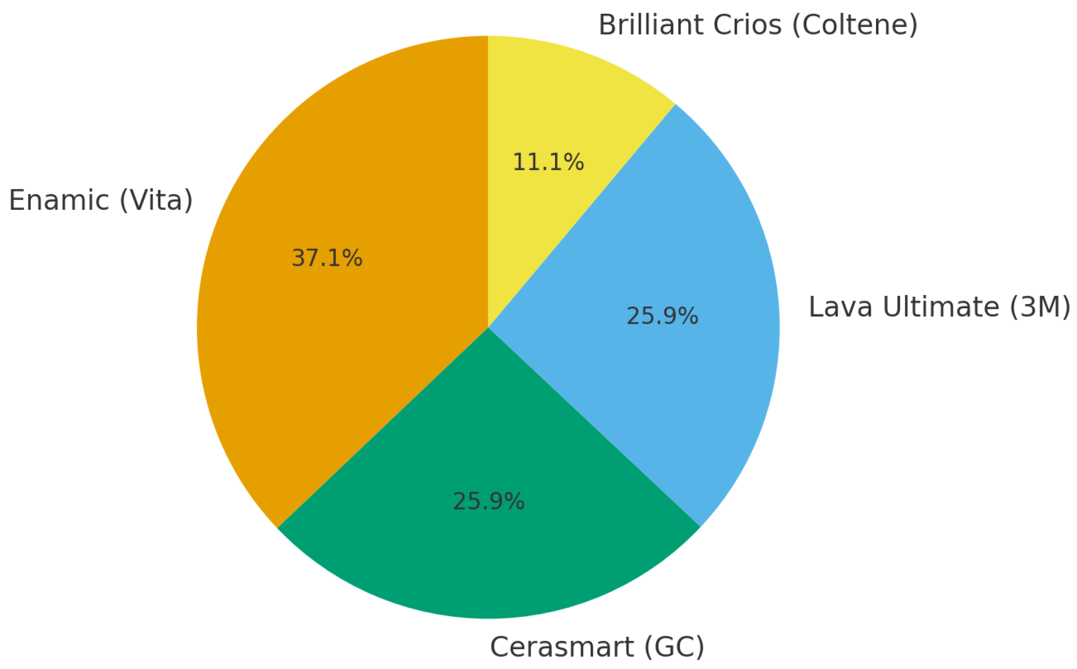
The most common materials used for CAD CAM restorations were Enamic (Vita) (n = 10, 37.0%) [15,16,79,82,83,85,86,93,94,96], and Lava Ultimate (3M/ESPE) (n = 7, 25.9%) [15,16,82,86,89,93,97], Cerasmart (GC) (n = 7, 25.9%) [15,79,82,86,88,97] and Brilliant Crios (Coltene) (n = 3, 11.1.%) [15,91,97]. Because several studies tested more than one material, these proportions are not mutually exclusive and therefore exceed 100% when summed.
The most common test used for fracture toughness evaluation was SENB (n = 37) [15,16,40,42,44,46,47,48,50,51,52,53,54,55,56,57,60,61,62,63,64,65,67,68,69,71,75,77,78,79,80,83,86,89,91,96] followed by Notch less triangular prism (n = 5) [58,88,90,92,94], Compact tension specimen (n = 4) [59,69,72,97], Brazilian test (n = 3) [74,78,81], crack length measurement in hardness tests (n = 2) [76,93], and fractographic analysis (n = 1) [85]. To visualize the frequency of materials evaluated, the distribution of CAD/CAM resin-based restorative blocks investigated in the included studies is shown in Figure 2. The majority of the evidence is concentrated on Enamic (Vita), followed by Lava Ultimate (3M) and Cerasmart (GC), whereas Brilliant Crios (Coltene) has been less frequently tested. This uneven distribution indicates that conclusions regarding CAD/CAM composites are largely driven by data from a limited number of material types, highlighting the need for broader comparative studies.
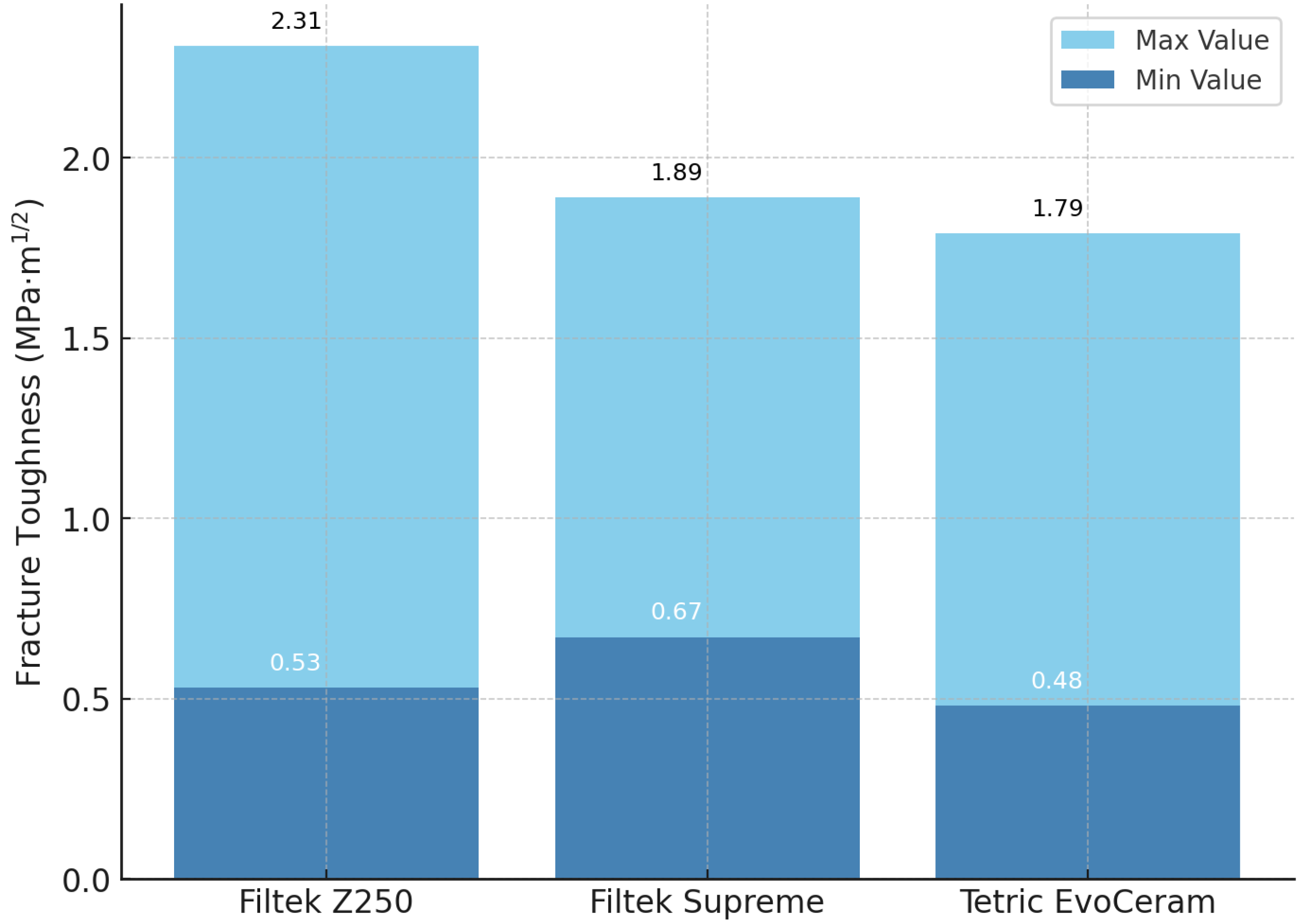
Fracture toughness values demonstrated a broad range across the included studies, often differing more than two-fold for the same commercial material. For example, Filtek Z250 showed values from as low as 0.53 MPa·m1/2 [81] up to 2.31 MPa·m1/2 [57]. Similarly, Filtek Supreme presented a spread between 0.67 MPa·m1/2 [64] and 1.89 MPa·m1/2 [57]. For Tetric EvoCeram, reported values ranged from 0.48 MPa·m1/2 [40] to 1.79 MPa·m1/2 [80]. This level of variability highlights the significant influence of testing protocols, specimen preparation, and environmental conditions on the measured outcomes.

3.2. Subgroup Analysis by Material Type

When grouped by restorative material category, the studies showed clear differences in fracture toughness behavior. Short fiber-reinforced composites consistently demonstrated the highest values, often exceeding 2.5 MPa·m1/2, as reported by Garoushi et al. [54] and confirmed in the experimental work of Suzaki et al. [95] Both groups observed that fiber reinforcement provided enhanced crack-bridging and more stable fracture patterns compared with conventional matrices.
In contrast, conventional direct composites such as Filtek Z250, Tetric EvoCeram, and Filtek Supreme showed moderate values, frequently ranging between 0.5 and 2.0 MPa·m1/2. Watanabe et al. [81] reported low values for Z250, whereas Ilie et al. found substantially higher results for the same material under different conditions. Similarly, Abdulhameed et al. [40] reported the lower end of values for Tetric EvoCeram, while Yap et al. demonstrated higher values under alternative testing conditions. For Filtek Supreme, Lien et al. [64] observed relatively low toughness, whereas Ilie et al. measured nearly threefold higher performance.
Bulk-fill composites generally produced results comparable to those of conventional composites. Ilie et al. [57] reported that bulk-fill resins such as Tetric EvoCeram Bulk-Fill and Filtek Bulk-Fill Flowable achieved fracture toughness values within the same range as their conventional analogues, without consistent evidence of superiority. Finally, for CAD/CAM materials, resin nanoceramics such as Lava Ultimate and Cerasmart were tested in multiple studies. Hampe et al. [15] and Ilie et al. [57] found that their fracture toughness values largely overlapped with those of direct composites, suggesting no significant mechanical advantage. Similarly, polymer-infiltrated ceramic networks (PICNs) such as Enamic were evaluated by Harada et al. [16] and Ilie et al. [57], both reporting values comparable to those of conventional resins. In order to better illustrate the variability reported across studies, the ranges of fracture toughness values for three commonly tested direct composites are presented in Figure 3.

3.3. Effect of Testing Method and Environment

The method of fracture toughness evaluation exerted a significant influence on the reported outcomes. The single-edge notched beam (SENB) test was by far the most frequently employed, being used in more than two-thirds of the included studies (e.g., Ilie et al., 2012 [57]; Garoushi et al., 2013 [54]; Harada et al., 2015 [16]; Hampe et al., 2019 [15]; Suzaki et al., 2020 [95]; Alsarani et al., 2024 [82]). SENB generally provided stable ranges of values for both direct and CAD/CAM composites, allowing more reliable comparisons. By contrast, the notchless triangular prism (NTP) test was reported less frequently (Ilie et al., 2021 [58]; Karaer et al., 2020 [88]; 2020; Lucsanszky et al., 2020 [90]; Nguyen et al., 2012 [92]; Sulaiman et al., 2022 [94]), and typically produced lower absolute values, reflecting its sensitivity to notch geometry and crack initiation.
The compact tension-C(T)-method, adopted by Knobloch et al. [59]; Messe et al. [69]; Ribeiro et al. [72]; and Wendler et al. [97], yielded values broadly comparable with SENB but required larger specimens and more complex preparation, limiting its use. The Brazilian disk test, applied in studies such as Scherrer et al. [74], Watanabe et al. [81], and Thomaidis et al. [78], often generated lower and less reproducible values, with wide intra-material variability. Indentation-based approaches, where fracture toughness was estimated from crack length around Vickers hardness indentations [76,93], generally underestimated fracture toughness compared with SENB. Three studies [40,58,78] uniquely incorporated fractographic analysis, directly examining fracture surfaces to validate crack initiation points, which provided valuable qualitative insights not captured by mechanical testing alone. To facilitate comparison across methodologies, the main fracture toughness testing methods identified in the included studies, together with their strengths, weaknesses, and typical outcomes, are summarized in Table 2.
As shown in the previous table, SENB was the most frequently applied and generally yielded reproducible fracture toughness values, while NTP and Brazilian disk tests tended to underestimate or produce more variable results. C(T) provided values comparable to SENB but required larger specimens, limiting its use. Indentation methods consistently underestimated fracture toughness, and fractography offered only qualitative insights. These differences prove the importance of standardized methodology when comparing outcomes across studies.
The testing environment also played a critical role. Several investigations conducted under dry conditions at room temperature [40,54,71,72] tended to report higher fracture toughness values. In contrast, studies performed under wet conditions at 37 °C [15,42,57,60,61,69,95] or artificial saliva [58,65,75,85] consistently reported lower values, likely due to water sorption, hydrolytic degradation, and plasticization of the resin matrix. Aging protocols further affected results: thermocycling [15,65,74,93,94] and bleaching solution [51,82] generally reduced fracture toughness, while heat/pressure post-curing [59,74] sometimes enhanced it. In this part, these findings highlight the necessity of standardized test setups and controlled environmental conditions for meaningful inter-study comparisons.

3.4. Risk of Bias Summary

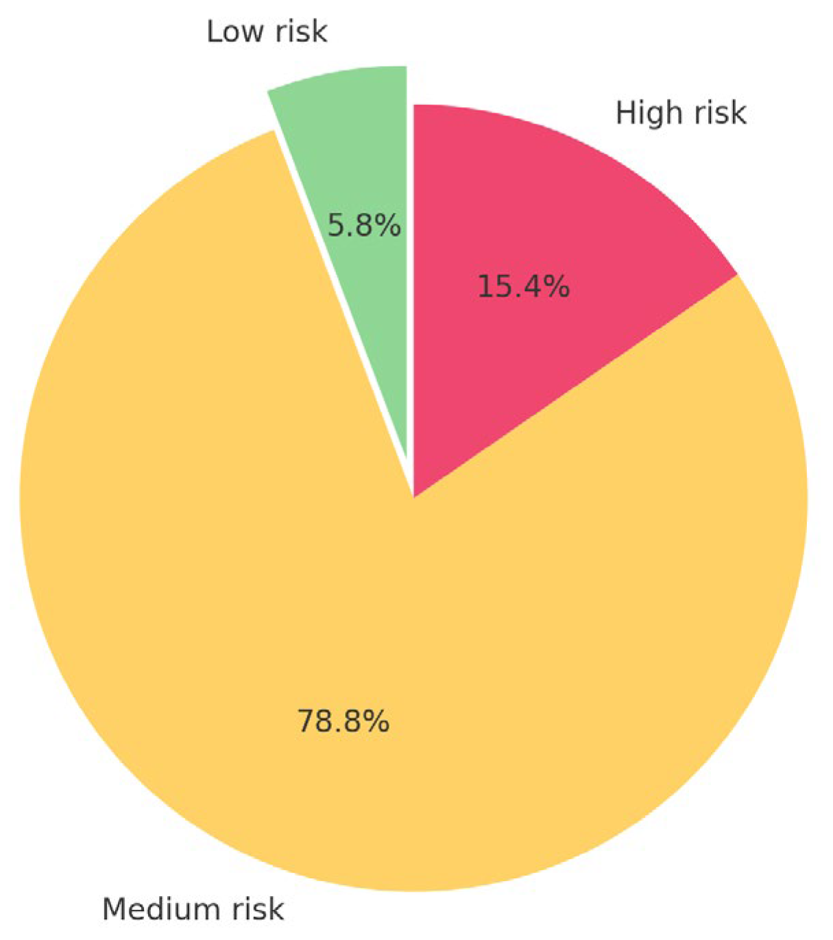
The detailed scoring according to the Quinn tool [38] is summarized in Table S1, and in an abbreviated way in Appendix A. The majority of studies were judged to have a medium risk of bias (n = 41) (79%), with only a small number classified as low risk (n = 3) (6%) and the remainder as high risk (n = 8) (15%). Also, methodological shortcomings were frequent across the dataset. Blinding of assessors was not reported in any of the studies (e.g., Ilie et al., 2012 [57]; Hampe et al., 2019 [15]; Harada et al., 2015 [16]), reflecting a general limitation in laboratory testing of dental materials. Randomization procedures were described in only two investigations [16,68], while most others (e.g., Watanabe et al., 2008 [81]; Yap et al., 2004 [80]; Lien et al., 2010 [64]) did not specify allocation methods for specimen grouping. Operator details were almost universally omitted, including in studies by Abdulhameed et al. (2020) [40], which complicates reproducibility. Only three studies performed a priori sample size analysis (Kamourieh et al., 2024 [19]; Alsarani et al., 2024 [82]; and Lucsanszky et al., 2020 [90]), while the rest relied on convenience samples without justification. In conclusion, while the reporting of basic specimen preparation and testing protocols was generally adequate, the absence of randomization, blinding, and sample size calculations limits the internal validity of the evidence base and underlines the need for stronger and more reliable methodological standards in laboratory evaluations of dental materials. The distribution of risk-of-bias ratings across the 52 studies is illustrated in Figure 4.

3.5. Summary of Results

Across the 52 included studies, reported fracture toughness values displayed substantial heterogeneity, often differing by more than twofold even for the same commercial product. Conventional direct composites (e.g., Filtek Z250, Filtek Supreme, Tetric EvoCeram) exhibited moderate toughness values with broad variability, and bulk-fill composites performed within similar ranges. In contrast, short fiber-reinforced composites consistently demonstrated higher fracture toughness across testing protocols, whereas resin nanoceramics and polymer-infiltrated ceramic networks (e.g., Enamic, Lava Ultimate, Cerasmart) did not show consistent advantages over direct composites. Furthermore, among testing approaches, the single edge notched beam (SENB) method was most frequently employed and produced the most reproducible values, while the Notched Triangular Prism (NTP), Brazilian disk, and indentation methods generally yielded lower or less reliable results. Testing under wet conditions at 37 °C or following artificial aging procedures (thermocycling, ethanol storage) typically reduced fracture toughness compared with dry, room-temperature testing, underscoring the sensitivity of results to environmental parameters. Also, risk-of-bias assessment revealed that most studies were of moderate quality, with only two classified as low risk and eight as high risk. The principal sources of bias were the absence of blinding, limited randomization, incomplete reporting of operators, and infrequent justification of sample size.
Finally, a descriptive sensitivity check, excluding non-standardized methods and limiting inclusion to studies conducted under physiological conditions, did not alter the overall trend. This suggests that the superior performance of short fiber-reinforced composites was not driven by a single methodological subset. However, this observation remains qualitative, as no formal statistical sensitivity analysis was undertaken.

4. Discussion

This scoping review highlights the inherent complexity of assessing fracture toughness in resin-based restorative materials. Despite decades of development in both direct and CAD/CAM composites, reported values remain inconsistent across studies [54,85,94], reflecting the strong influence of testing protocols, notch geometry, and environmental conditions rather than intrinsic material properties [3,7,8]. Although various international standards have been cited in the literature (e.g., ASTM E399, ASTM D5045-14, ISO 20795-1:2013, ISO 13586:2018) [41,45,49,66,70,73,87,98], none are specifically designed for dental composites. This lack of material-specific standardization renders cross-study comparisons problematic and should temper any attempt to categorically rank materials. Moreover, fracture toughness alone cannot reliably predict clinical performance; rather, it should be interpreted alongside other correlated properties that influence the failure of brittle materials, such as flexural strength [3,7,9]. The parameter is also affected by microstructural and compositional factors, including filler morphology [58,60,61,99,100,101,102,103], polymer matrix chemistry [104,105,106], and aging resistance [55,95]. Nevertheless, fracture toughness remains one of the few mechanical parameters repeatedly associated with restoration survival and bulk fracture in clinical investigations [8,11], confirming its continuing relevance as a benchmark property for evaluating restorative materials.
However, the heterogeneity in testing setups, environmental conditions, and specimen geometries substantially limits the comparability of absolute K_IC values across studies. Therefore, any references to higher or lower performance among material classes (for instance, the generally higher values reported for short fiber-reinforced composites) should be interpreted as descriptive trends rather than quantitative superiority. These tendencies likely reflect differences in experimental methodology as much as, if not more than, true intrinsic material behavior.
Mechanistic interpretations, such as the presumed role of fiber reinforcement, filler morphology, or crack deflection mechanisms in polymer-infiltrated ceramic networks, provide plausible explanations for observed trends but should not be read as demonstrated causal relationships, given the absence of standardized comparative testing. The present synthesis, therefore, aims to map the state of the evidence rather than to establish ranked hierarchies of materials.
In summary, while general patterns can be observed, the strength of the evidence remains inherently limited by the in vitro, highly heterogeneous, and method-dependent nature of the included studies.

4.1. Fracture Toughness Tests

The studies included in this review applied a range of international standards for fracture toughness evaluation [41,45,49,66,70,73,87,98], including ASTM E399, ASTM D5045-14, ISO 13586:2018, and ISO 20795-1:2013, even though none of these protocols were specifically designed for dental composites. As a result, substantial variability was observed between methods. The single-edge notched beam (SENB) technique was most frequently used [15,16,40,42,44,46,47,48,50,51,52,53,54,55,56,57,60,61,62,63,64,65,67,68,71,75,77,78,79,80,82,83,85,86,89,91,94,97] and generally produced reliable, intermediate values, although differences in notch width, sharpening, and pre-crack preparation can introduce significant error [99,100]. The compact tension-C(T)-method [59,69,72,97], although less common, offers easier notch preparation and can provide insights into crack propagation [7,99]. By contrast, the notchless triangular prism (NTP) [58,88,90,92,94] and Brazilian disk methods [74,78,81] often underestimated fracture toughness and suffered from low reproducibility. The indentation method, suitable for brittle ceramics, proved unreliable for composite resins, systematically underestimating toughness values [76,93]. Finally, fractographic analysis gave only qualitative information, with high dispersion due to difficulties in measuring flaw size [4]. The way the notch is formed or sectioned, as well as the sharpening of it or not, can give great variations in the results. The ideal technique for fracture toughness calculation is to load the notched specimen in tension, in order to create an infinitely sharp pre-crack. Creating a pre-crack in composite resin specimens is very difficult because they are brittle and have small dimensions, and specimens can break before testing. Therefore, a narrow notch can be made with the use of a razor blade in the mold for specimen fabrication, or by sharpening the notch with a razor blade and abrasive [7].
Finally, we should mention that none of the tests used are exclusively designed for composite resins or resin-infiltrated ceramic blocks intended for CAD CAM use [41,45,49,66,70,73,87,98]. So, overall, our findings emphasize the need for a standardized, composite-specific fracture toughness test that balances accuracy with clinical relevance. Finally, we should point out that the high variability in the fracture toughness values is mainly attributed to the lack of homogeneous test conditions across the included studies.

4.2. Materials for CAD/CAM Restorations

CAD/CAM blocks for indirect restorations include resin nanoceramics (e.g., Lava Ultimate, Cerasmart, Brilliant Crios, Katana Avencia, Paradigm MZ100) and polymer-infiltrated ceramic networks (PICNs) such as Enamic. These materials are industrially polymerized under controlled temperature and pressure, producing a dense, highly cross-linked matrix with fewer internal defects than chairside light-cured composites [15,16,89]. Enamic consists of a feldspathic ceramic skeleton infiltrated with a UDMA/TEGDMA resin, whereas Lava Ultimate contains approximately 85 wt.% silica/zirconia nanofillers dispersed in a Bis-GMA/UDMA/TEGDMA matrix [15,82,86,93]. Comparable hybrid formulations are found in Cerasmart and Brilliant Crios, although variations in filler morphology, particle size distribution, and matrix chemistry have been linked to the observed variability in reported mechanical performance [15,86,88,89,90].
Despite these optimized microstructural features, fracture toughness values reported for CAD/CAM composites remain within the range of conventional direct resins [85,90,94]. The enhanced polymerization and reduced porosity achieved through industrial processing appear to improve defect uniformity but do not systematically translate into higher K_IC values across studies. Short fiber-reinforced CAD/CAM blocks, such as Trinia or everX Posterior, have occasionally shown higher apparent toughness, particularly when fibers are oriented parallel to the loading direction [55,62,63,95]. However, these findings derive from a limited number of in vitro experiments with differing test geometries, making quantitative comparisons tentative.
In conclusion, current evidence suggests that CAD/CAM resin-based materials achieve comparable fracture toughness to direct composites, with mechanical performance largely governed by formulation and testing methodology rather than processing route. While CAD/CAM manufacturing may enhance homogeneity and reduce voids, it does not inherently ensure superior fracture resistance, highlighting the need for standardized evaluation methods before drawing definitive inter-material conclusions [12,17,18,33].

4.3. Crack Propagation and Inorganic Fillers

Filler characteristics have been widely reported to influence the fracture toughness of resin composites by altering crack-propagation pathways. Increased filler loading may enhance crack deflection, branching, and limited plastic deformation of the surrounding resin matrix, thereby facilitating energy dissipation and delaying catastrophic failure [58,60,61,101,102]. Microcracks forming ahead of the main crack tip can redistribute stress and reduce its intensity, which in turn appears to improve overall toughness [100,101].
Filler morphology also plays an important role. Spherical or well-rounded fillers tend to promote smoother crack deflection and higher packing density, whereas irregular particles generate rougher fracture surfaces and increase resistance to crack growth [57,60]. Conversely, pre-polymerized filler particles may reduce the effective inorganic content and consequently lower measured toughness [57,103]. We should further mention that the optimal filler volume fraction has been suggested to lie between 55% and 60%, beyond which the contribution to toughness plateaus or even declines because of matrix embrittlement or interfacial defects [58,59,60,102].
Taken together, these findings indicate that reported differences in fracture toughness among composites are not purely material-intrinsic but reflect a multifactorial interplay between filler content, morphology, and dispersion within the polymer network, factors that vary substantially across formulations and testing methodologies. Consequently, the observed variability in K_IC values across studies must be interpreted as an outcome of both material composition and experimental context rather than a direct measure of material superiority.

4.4. Crack Propagation in PICN

Polymer-infiltrated ceramic networks (PICNs) exhibit fracture mechanisms distinct from those of conventional resin composites. Instead of crack propagation occurring solely through the resin or filler phase, cracks in PICNs interact with the dual polymer–ceramic network, frequently deflecting or blunting at the interfacial boundaries [86,96,103]. This interaction promotes energy dissipation through mechanisms such as crack-tip bridging, interface sliding, and localized plastic deformation, leading to a mixed elastic–plastic fracture response under loading [94,103].
Experimental in vitro studies employing indentation and microfracture techniques have shown that PICNs can undergo creep-like deformation under forces comparable to masticatory loads, with cracks penetrating the ceramic backbone but subsequently deflecting along polymer-rich zones [85,90,103]. Such behavior suggests that the polymer phase contributes to crack arrest and redistribution of stress, thereby modifying the fracture path rather than substantially increasing the absolute fracture toughness values (K_IC). Reported K_IC ranges for PICNs remain similar to those of high-performance resin composites, indicating that their apparent toughness advantage arises from altered failure modes rather than higher numerical toughness.

4.5. Influence of Resin Matrix and Composite Formulation

The chemical composition of the resin matrix appears to exert a substantial influence on the measured fracture toughness of resin-based composites. Composites containing UDMA-based matrices often exhibit higher reported toughness than those formulated with Bis-GMA, possibly due to lower viscosity, greater chain flexibility, and enhanced cross-linking potential [58,104,105,106]. Silorane-based systems have also been reported to show greater resistance to crack propagation compared with conventional methacrylate resins, although their clinical adoption has been limited [64]. Furthermore, fracture toughness (K_IC) and energy absorption to failure values tended to increase when specimens were photo-polymerized at higher radiant exitance and/or for longer exposure times [40,53,58,63,64,76]. These effects, however, are highly dependent on experimental design, light source characteristics, and specimen geometry, emphasizing the sensitivity of K_IC to curing variables rather than reflecting inherent material superiority.
Beyond matrix chemistry, marked variability in toughness values was observed across different composite formulations. Reported values for direct resins ranged from 0.42 to 2.4 MPa·m½, with hybrid composites generally showing higher means than microfilled or flowable variants [57,80,81]. Bulk-fill composites demonstrated performance comparable to conventional hybrids, though outcomes varied across brands and curing protocols [37,39,47,53,54,62,79]. Also, short fiber-reinforced composites frequently achieved the highest K_IC values, with EverX Posterior and EverX Flow often exceeding 2.5 MPa·m1/2 [55,62,63,95]. This apparent improvement is attributed to fiber bridging and crack-deflection phenomena, which provide extrinsic toughening mechanisms beyond filler effects alone [19,58]. Nevertheless, such advantages should be interpreted cautiously, as inter-study differences in fiber orientation, test geometry, and environmental conditions limit direct comparability.
In summary, advances in resin chemistry and filler design have broadened the performance spectrum of composite materials, but due to the strong influence of testing methodology, only qualitative trends, rather than definitive rankings, can be established. Short fiber reinforcement remains the most consistently reported formulation feature associated with higher in vitro fracture toughness, although further standardized testing is required to confirm its clinical relevance.

4.6. Fracture Toughness and Clinical Performance of CAD/CAM Materials

CAD/CAM resin-based materials, including polymer-infiltrated ceramic networks (PICNs) such as Enamic, and resin nanoceramics such as Lava Ultimate, Cerasmart, Brilliant Crios, Katana Avencia, and Paradigm MZ100, typically reported fracture toughness values between 0.6 and 1.7 MPa·m1/2, within the same range as conventional direct composites [15,85,86,97]. Although industrial polymerization processes are expected to reduce porosity and enhance cross-linking density, the available in vitro data does not show a consistent or systematic improvement in toughness. Occasional higher values reported for Cerasmart and Brilliant Crios [88,91] appear to reflect study-specific testing conditions rather than intrinsic material superiority.
Short fiber-reinforced CAD/CAM blocks (e.g., Trinia, everX Posterior) have occasionally exhibited higher apparent fracture toughness (above 3 MPa·m1/2), particularly when fibers were aligned with the loading direction [55,62,63,95]. However, these findings derive from limited experimental subsets using diverse testing protocols, and direct comparison with other materials remains methodologically uncertain.
From a clinical standpoint, fracture toughness represents one of several laboratory indicators that may correlate with restoration survival and resistance to bulk fracture [3,8]. Nonetheless, the relationship between in vitro toughness and clinical longevity is indirect, and existing clinical evidence comparing CAD/CAM composites to conventional restorative materials remains limited and heterogeneous. Meta-analyses have suggested that Enamic may perform less favorably than lithium disilicate in terms of fracture-related failures, although 5-year survival rates still exceed 95% [107]. Similarly, randomized clinical trials report comparable mid-term outcomes for Lava Ultimate onlays and leucite-reinforced ceramics [105], while prospective trials of Enamic inlays show 97% survival after three years, with bulk fracture as the predominant failure mode [108,109,110]. A recent comparison of direct and CAD/CAM composites (Gradia SO vs. Gradia Block) likewise demonstrated favorable 2-year performance with no fractures [108].
In conclusion, the synthesis of laboratory and clinical findings suggests that CAD/CAM resin-based materials perform comparably to direct composites in fracture-related behavior, although evidence of heterogeneity and lack of standardized testing preclude definitive ranking. Their clinical outcomes appear satisfactory within the limitations of current evidence, while fiber-reinforced CAD/CAM designs remain a promising direction for improving toughness and load-bearing capacity.

4.7. Limitations of the Review

This scoping review has some limitations that must be acknowledged. First, all included studies were in vitro, which, while essential for mechanistic insights, cannot fully replicate intraoral conditions such as cyclic loading, saliva, or thermal fluctuations [12,40]. Second, there was substantial heterogeneity in testing methods, notch preparation, specimen dimensions, and storage conditions, which limited comparability and precluded meta-analysis [8,57,99]. Third, the risk of bias was generally medium, with few studies reporting randomization, operator details, or sample size justification [19,35,82,90]. Fourth, only one study directly compared CAD/CAM resin blocks with a conventional direct composite [94], restricting the ability to answer the review question with high certainty. Finally, the review included only English-language publications, which may have introduced selection bias [34,36].
Despite these limitations, the findings provide valuable insights into current evidence trends and highlight the urgent need for standardized test protocols and better-designed laboratory studies to generate data that can be reliably correlated with clinical performance.

4.8. Clinical Implications and Future Research

From a clinical point of view, it is reported from a meta-analysis, that resin-infiltrated ceramic network material Enamic is found to perform clinically not as good as Lithium disilicate glass ceramics in terms of restoration fracture, showing a failure rate of 5.97% for full contour restorations, 3.45% for partial coverage restorations, while glass ceramics demonstrated a failure rate of 1.61% [107]. From a 5-year randomized clinical trial, it was found that the nanoceramic onlays (Lava Ultimate) had a lower incidence of fracture compared with the leucite reinforced ceramic onlays, with both having a very low risk of fracture [108]. In a prospective study on indirect inlays, with an observation period of 3 years, resin-infiltrated ceramic network material (Enamic) demonstrated a 97.4% survival rate, with the failure reason being bulk fracture of the restoration [109]. A meta-analysis on the clinical behavior of ceramic, hybrid, and composite onlays demonstrated 90% survival for composites, 99% for hybrids (99%), and 98% disilicate ceramics [110]. From another randomized clinical trial, evaluating the 2-year performance of direct (Gradio SO) and CADCAM composite resin restorations (Gradio blocs), it was found that both materials showed a good clinical performance, without restoration fractures [111]. From these clinical studies and meta-analyses, it can be concluded that the fracture toughness of composite resin, hybrid, or ceramic materials for indirect use can give some rough estimate for the clinical performance of these materials.
Current laboratory evidence shows that fracture toughness alone cannot justify replacing direct composites with CAD/CAM resin-based blocks [11,57,97]. Instead, clinicians should integrate material choice with other performance factors such as esthetics, reparability, and digital workflow advantages [12,17]. Short fiber-reinforced composites remain the only group with superior crack resistance, supporting their application in extensive restorations [19,54,55,95]. To strengthen the evidence base, future research must adopt composite-specific standardized protocols and ensure transparent methodological reporting, including sample size calculations and operator details [31,34]. Equally important are long-term clinical trials that test whether laboratory fracture toughness values correlate with restoration survival [3,8,107,110,111].

5. Conclusions

This scoping review aimed to explore whether CAD/CAM resin-based materials exhibit higher fracture toughness than direct composite resins. The available in vitro evidence does not demonstrate a consistent superiority of CAD/CAM composites; reported values largely overlap with those of direct resins. Short fiber-reinforced composites generally showed higher fracture toughness across studies, irrespective of their application as direct or indirect materials. However, given the substantial heterogeneity in testing environments, apparatus, and analytical methods, these findings should be interpreted with caution. Also, all data synthesized in this review derive from in vitro investigations, and the methodological diversity across studies precludes definitive comparisons or clinical extrapolations. The observed differences are likely to reflect variations in testing conditions rather than intrinsic material performance. Accordingly, the PRISMA-ScR population, concept, and context (PCC) framework was applied to delineate the scope of evidence and its limitations. Future research should prioritize the standardization of testing protocols for valid inter-material comparison and the development of dental-specific ISO/ASTM standards, enabling valid inter-material comparisons. Well-designed, standardized in vitro experiments, followed by long-term clinical trials, are essential to determine whether CAD/CAM resin-based composites can offer genuine advantages in restoration longevity and failure resistance.

Supplementary Materials

The following supporting information can be downloaded at https://www.mdpi.com/article/10.3390/app152212308/s1, Table S1. Risk of bias (Scoring sheet for QUIN Tool).

Author Contributions

Conceptualization, S.T. and M.A.; methodology, S.T. and M.A.; software, S.T. and M.A.; validation, S.T., E.P. and M.A.; formal analysis, S.T., E.P. and M.A.; investigation, S.T. and M.A.; resources, M.A.; data curation, S.T. and M.A.; writing—original draft preparation, S.T. and M.A.; writing—review and editing, S.T., E.P. and M.A.; visualization, M.A.; supervision, M.A.; project administration, M.A. All authors have read and agreed to the published version of the manuscript.

Funding

This research received no external funding.

Institutional Review Board Statement

Not applicable.

Informed Consent Statement

Not applicable.

Data Availability Statement

The original contributions presented in this study are included in the article/Supplementary Material. Further inquiries can be directed to the corresponding author.

Conflicts of Interest

The authors declare no conflicts of interest.

Abbreviations

The following abbreviations are used in this manuscript:
CAD CAMComputer-assisted design computer-assisted manufacture
SENBSingle-edge notched specimen
CTCompact tension specimen
NPNotchless triangular prism
K_ICFracture Toughness
BisGMAbisphenol A-glycidyl methacrylate
TEGDMDATriethylene glycol dimethacrylate
UDMAUrethane dimethacrylate
SENBSingle-edge notched bending
SENVBSingle-edge notched bending with a V-shaped notch
ASTMAmerican Society for Testing and Materials

Appendix A. Abbreviated Form for Risk of Bias According to QUIN Tool

Table A1. Abbreviated Form for Risk of Bias According to QUIN Tool.
Author
Year
Risk of Bias
1. Abdulhameed 2020 [40].medium
2. Alshabib 2019 [42].medium
3. Attik 2022 [44].medium
4. Bijelic-Donova 2016a [46].medium
5. Bijelic-Donova 2016b [47].medium
6. Bonilla 2000 [48].medium
7. Bonilla 2003 [50].medium
8. Cho 2009 [51].medium
9. Choi 2000 [52].medium
10. Engelhardt 2016 [53].medium
11. Garoushi 2013 [54].high
12. Garoushi 2024 [55].medium
13. Jun 2013 [56].medium
14. Ilie 2012 [57].medium
15. Ilie 2021 [58].medium
16. Kamourieh 2024 [19].low
17. Kim 2002b [60].medium
18. Kim 2002b [61].high
19. Knobloch 2002 [59].high
20. Lassila 2019 [62].medium
21. Lassila 2020 [63].medium
22. Lien 2010 [64].medium
23. Lin 2009 [65].medium
24. Lohbauer 2020 [67].medium
25. Manhart 2000 [68].medium
26. Mese 2016 [69].medium
27. Nakade 2024 [71].medium
28. Ribeiro 2025 [72].low
29. Scherrer 2000 [74].high
30. Sochacki 2022 [75].medium
31. St-Georges 2003 [76].high
32. Thadathil Varghese 2024 [77].medium
33. Thomaidis 2013 [78].medium
34. Yang 2022 [79].medium
35. Yap 2004 [80].medium
36. Watanabe 2008 [81].medium
CAD CAM
37. Alsarani 2024 [82].low
38. Della Bona 2014 [83].high
39. Elraggal 2022 [85].medium
40. Goujat 2018 [52].medium
41. Hampe 2019 [15].medium
42. Harada 2015 [16].medium
43. Karaer 2020 [88].medium
44. Ling 2022 [89].medium
45. Lucsanszky 2020 [90].medium
46. Moradi 2020 [91].medium
47. Nguyen 2012 [92].medium
48. Sonmez 2018 [93].medium
49. Sulaiman 2022 [94].medium
50. Suzaki 2020 [95].high
51. Swain 2016 [96].high
52. Wendler 2021 [97].medium

References

  1. Incekara, M.S.; Karadas, M. Clinical comparison of direct and indirect class II composite restorations: A prospective 12-month follow-up study. BMC Oral Health 2025, 25, 1217. [Google Scholar] [CrossRef]
  2. Da Rosa Rodolpho, P.A.; Donassollo, T.A.; Cenci, M.S.; Loguércio, A.D.; Moraes, R.R.; Bronkhorst, E.M.; Opdam, N.J.; Demarco, F.F. 22-Year clinical evaluation of the performance of two posterior composites with different filler characteristics. Dent. Mater. 2011, 27, 955–963. [Google Scholar] [CrossRef]
  3. Ferracane, J.L. Resin-based composite performance: Are there some things we can’t predict? Dent. Mater. 2013, 29, 51–58. [Google Scholar] [CrossRef] [PubMed]
  4. Spyropoulou, N.; Diamantopoulou, S.; Patrinos, S.; Papazoglou, E. Retrospective Clinical Study of Resin Composite and Ceramic Indirect Posterior Restorations up to 11 Years. Prosthesis 2025, 7, 108. [Google Scholar] [CrossRef]
  5. François, D.; Pineau, A.; Zaoui, A. Brittle fracture. In Mechanical Behaviour of Materials. Solid Mechanics and Its Applications, Vol. 191; Springer: Dordrecht, The Netherlands, 2013; pp. 103–191. [Google Scholar] [CrossRef]
  6. Al-Ibrahim, I.; Shono, N.; Al-Saud, L.; Al-Nahedh, H. Five years of restorative resin-based composite advancements: A narrative review. BMC Oral Health 2025, 25, 1061. [Google Scholar] [CrossRef]
  7. Ilie, N.; Hilton, T.J.; Heintze, S.D.; Hickel, R.; Watts, D.C.; Silikas, N.; Stansbury, J.W.; Cadenaro, M.; Ferracane, J.L. Academy of dental materials guidance-resin composites: Part I-mechanical properties. Dent. Mater. 2017, 33, 880–894. [Google Scholar] [CrossRef] [PubMed]
  8. Heintze, S.D.; Ilie, N.; Hickel, R.; Reis, A.; Loguercio, A.; Rousson, V. Laboratory mechanical parameters of composite resins and their relation to fractures and wear in clinical trials—A systematic review. Dent. Mater. 2017, 33, e101–e114. [Google Scholar] [CrossRef]
  9. Hançer Sarıca, S.; Arslan, S.; Balkaya, H. Comparison of the 2-year clinical performances of class II restorations using different restorative materials. Clin. Oral Investig. 2025, 29, 128. [Google Scholar] [CrossRef] [PubMed]
  10. Tyas, M.J. Correlation between fracture properties and clinical performance of composite resins in class IV cavities. AustDent J. 1990, 35, 46–49. [Google Scholar] [CrossRef]
  11. Lehmann, A.; Nijakowski, K.; Jankowski, J.; Donnermeyer, D.; Ramos, J.C.; Drobac, M.; Martins, J.F.B.; Hatipoğlu, Ö.; Omarova, B.; Javed, M.Q.; et al. Clinical difficulties related to direct composite restorations: A multinational survey. Int. Dent. J. 2025, 75, 797–806. [Google Scholar] [CrossRef]
  12. Tribst, J.P.M.; Veerman, A.; Pereira, G.K.R.; Kleverlaan, C.J.; Dal Piva, A.M.d.O. Comparative strength study of indirect permanent restorations: 3D-printed, milled, and conventional dental composites. Clin. Pract. 2024, 14, 1940–1952. [Google Scholar] [CrossRef]
  13. Ozakar-Ilday, N.; Zorba, Y.O.; Yildiz, M.; Erdem, V.; Seven, N.; Demirbuga, S. Three-year clinical performance of two indirect composite inlays compared to direct composite restorations. Med. Oral Patol. Oral Cir. Bucal 2013, 18, e521–e528. [Google Scholar] [CrossRef] [PubMed]
  14. Morel, C.; Baranger, E.; Lamon, J.; Braun, J.; Lorrette, C. The influence of internal defects on the mechanical behavior of filament wound SiC/SiC composite tubes under uniaxial tension. J. Eur. Ceram. Soc. 2022, 43, 1797–1807. [Google Scholar] [CrossRef]
  15. Hampe, R.; Theelke, B.; Lümkemann, N.; Eichberger, M.; Stawarczyk, B. Fracture toughness analysis of ceramic and resin composite CAD/CAM material. Oper. Dent. 2019, 44, E190–E201. [Google Scholar] [CrossRef] [PubMed]
  16. Harada, A.; Nakamura, K.; Kanno, T.; Inagaki, R.; Örtengren, U.; Niwano, Y.; Sasaki, K.; Egusa, H. Fracture resistance of computer-aided design/computer-aided manufacturing-generated composite resin-based molar crowns. Eur. J. Oral Sci. 2015, 123, 122–129. [Google Scholar] [CrossRef] [PubMed]
  17. Duarte, S., Jr.; Phark, J.H. Advances in dental restorations: A comprehensive review of machinable and 3D-printed ceramic-reinforced composites. J. Esthet. Restor. Dent. 2025, 37, 257–276. [Google Scholar] [CrossRef]
  18. Ling, L.; Ma, Y.; Malyala, R. A novel CAD/CAM resin composite block with high mechanical properties. Dent. Mater. 2021, 37, 1150–1155. [Google Scholar] [CrossRef] [PubMed]
  19. Kamourieh, N.; Faigenblum, M.; Blizard, R.; Leung, A.; Fine, P. Fracture toughness of short fibre-reinforced composites—In vitro study. Materials 2024, 17, 5368. [Google Scholar] [CrossRef]
  20. Borgia, E.; Baron, R.; Borgia, J.L. Quality and survival of direct light-activated composite resin restorations in posterior teeth: A 5- to 20-year retrospective longitudinal study. J. Prosthodont. 2019, 28, e195–e203. [Google Scholar] [CrossRef]
  21. Angeletaki, F.; Gkogkos, A.; Papazoglou, E.; Kloukos, D. Direct versus indirect inlay/onlay composite restorations in posterior teeth. A systematic review and meta-analysis. J. Dent. 2016, 53, 12–21. [Google Scholar] [CrossRef]
  22. Azeem, R.A.; Sureshbabu, N.M. Clinical performance of direct versus indirect composite restorations in posterior teeth: A systematic review. J. Conserv. Dent. 2018, 21, 2–9. [Google Scholar]
  23. McGrath, C.E.; Bonsor, S.J. Survival of direct resin composite onlays and indirect tooth-coloured adhesive onlays in posterior teeth: A systematic review. Br. Dent. J. 2022, 233, 1–7. [Google Scholar] [CrossRef] [PubMed]
  24. Moussa, C.; Savard, G.; Rochefort, G.; Renaud, M.; Denis, F.; Daou, M.H. Fracture resistance of direct versus indirect restorations on posterior teeth: A systematic review and meta-analysis. Bioengineering 2024, 11, 536. [Google Scholar] [CrossRef]
  25. Tennert, C.; Maliakal, C.; Suarèz, M.L.; Jaeggi, T.; Meyer-Lueckel, H.; Wierichs, R.J. Longevity of posterior direct versus indirect composite restorations: A systematic review and meta-analysis. Dent. Mater. 2024, 40, e95–e101. [Google Scholar] [CrossRef] [PubMed]
  26. de Kuijper, M.C.F.M.; Cune, M.S.; Özcan, M.; Gresnigt, M.M.M. Clinical performance of direct composite resin versus indirect restorations on endodontically treated posterior teeth: A systematic review and meta-analysis. J. Prosthet. Dent. 2023, 130, 295–306. [Google Scholar] [CrossRef] [PubMed]
  27. Elmoselhy, H.A.S.; Hassanien, O.E.S.; Haridy, M.F.; Salam El Baz, M.A.E.; Saber, S. Two-year clinical performance of indirect restorations fabricated from CAD/CAM nano hybrid composite versus lithium disilicate in mutilated vital teeth. A randomized controlled trial. BMC Oral Health 2024, 24, 101. [Google Scholar] [CrossRef] [PubMed] [PubMed Central]
  28. Hassan, A.; Hamdi, K.; Ali, A.I.; Al-Zordk, W.; Mahmoud, S.H. Clinical performance comparison between lithium disilicate and hybrid resin nano-ceramic CAD/CAM onlay restorations: A two-year randomized clinical split-mouth study. Odontology 2024, 112, 601–615. [Google Scholar] [CrossRef]
  29. El-Din, M.S.; Adel, S.; Naguib, A.; Nabil, O. Evaluation of marginal and internal adaptation of endocrowns fabricated from nano-ceramic hybrid and lithium disilicate ceramic materials: A randomized controlled clinical trial. J. Contemp. Dent. Pract. 2025, 26, 795–805. [Google Scholar] [CrossRef] [PubMed]
  30. Hofsteenge, J.W.; Fennis, W.M.M.; Kuijs, R.H.; Özcan, M.; Cune, M.S.; Gresnigt, M.M.M.; Kreulen, C.M. Clinical survival and performance of premolars restored with direct or indirect cusp-replacing resin composite restorations with a mean follow-up of 14 years. Dent. Mater. 2023, 39, 383–390. [Google Scholar] [CrossRef] [PubMed]
  31. Mourouzis, P. Critical methodological factors influencing the accuracy of intraoral scanners in digital dentistry research. Comput. Biol. Med. 2025, 187, 109780. [Google Scholar] [CrossRef] [PubMed]
  32. Tricco, A.C.; Lillie, E.; Zarin, W.; O’Brien, K.K.; Colquhoun, H.; Levac, D.; Moher, D.; Peters, M.D.J.; Horsley, T.; Weeks, L.; et al. PRISMA Extension for scoping reviews (PRISMA-ScR): Checklist and explanation. Ann. Intern. Med. 2018, 169, 467–473. [Google Scholar] [CrossRef] [PubMed]
  33. Higgins, J.P.T.; Thomas, J.; Chandler, J.; Cumpston, M.; Li, T.; Page, M.J.; Welch, V.A. Cochrane Handbook for Systematic Reviews of Interventions Version 6.5 (Updated August 2024); Cochrane: Houston, TX, USA, 2024; Available online: www.cochrane.org/handbook (accessed on 10 November 2025).
  34. Della Bona, A.; Cantelli, V.; Britto, V.T.; Collares, K.F.; Stansbury, J.W. 3D printing restorative materials using a stereolithographic technique: A systematic review. Dent. Mater. 2021, 37, 336–350. [Google Scholar] [CrossRef] [PubMed] [PubMed Central]
  35. Pineda-Vélez, E.; Yadalam, P.K.; Ardila, C.M. Efficacy of the finite element analysis in assessing the effects of light curing on the mechanical properties of direct restorative composites: A systematic review. J. Clin. Exp. Dent. 2024, 16, e1411–e1421. [Google Scholar] [CrossRef]
  36. Waffenschmidt, S.; Knelangen, M.; Sieben, W.; Bühn, S.; Pieper, D. Single screening versus conventional double screening for study selection in systematic reviews: A methodological systematic review. BMC Med. Res. Methodol. 2019, 19, 132. [Google Scholar] [CrossRef]
  37. Furlan, J.C.; Singh, J.; Hsieh, J.; Fehlings, M.G. Methodology of systematic reviews and recommendations. J. Neurotrauma 2011, 28, 1335–1339. [Google Scholar] [CrossRef] [PubMed]
  38. Tractenberg, R.E.; Yumoto, F.; Jin, S.; Morris, J.C. Sample size requirements for training to a kappa agreement criterion on clinical dementia ratings. Alzheimer Dis. Assoc. Disord. 2010, 24, 264–268. [Google Scholar] [CrossRef]
  39. Sheth, V.H.; Shah, N.P.; Jain, R.; Bhanushali, N.; Bhatnagar, V. Development and validation of a risk-of-bias tool for assessing in vitro studies conducted in dentistry: The QUIN. J. Prosthet. Dent. 2024, 131, 1038–1042. [Google Scholar] [CrossRef]
  40. Abdulhameed, N.; Angus, B.; Wanamaker, J.; Mecholsky, J.J., Jr. Quantitative fractography as a novel approach to measure fracture toughness of direct resin composites. J. Mech. Behav. Biomed. Mater. 2020, 109, 103857. [Google Scholar] [CrossRef] [PubMed]
  41. ISO 13586:2018; Plastics—Determination of Fracture Toughness (GIC and KIC)—Linear Elastic Fracture Mechanics (LEFM) Approach. International Organization for Standardization: Geneva, Switzerland, 2018.
  42. Alshabib, A.; Silikas, N.; Watts, D.C. Hardness and fracture toughness of resin-composite materials with and without fibers. Dent. Mater. 2019, 35, 1194–1203. [Google Scholar] [CrossRef]
  43. BS ISO 12135:2021; Metallic Materials—Unified Method of Test for the Determination of Quasistatic Fracture Toughness. British Standards Institution: Milton Keynes, UK, 2021.
  44. Attik, N.; Colon, P.; Gauthier, R.; Chevalier, C.; Grosgogeat, B.; Abouelleil, H. Comparison of physical and biological properties of a flowable fiber reinforced and bulk filling composites. Dent. Mater. 2022, 38, e19–e30. [Google Scholar] [CrossRef]
  45. ISO 20795–1:2013; Dentistry—Base Polymers—Part 1: Denture Base Polymers. International Organization for Standardization: Geneva, Switzerland, 2013.
  46. Bijelic-Donova, J.; Garoushi, S.; Vallittu, P.K.; Lassila, L.V. Mechanical properties, fracture resistance, and fatigue limits of short fiber reinforced dental composite resin. J. Prosthet. Dent. 2016, 115, 95–102. [Google Scholar] [CrossRef] [PubMed]
  47. Bijelic-Donova, J.; Garoushi, S.; Lassila, L.V.; Keulemans, F.; Vallittu, P.K. Mechanical and structural characterization of discontinuous fiber-reinforced dental resin composite. J. Dent. 2016, 52, 70–78. [Google Scholar] [CrossRef] [PubMed]
  48. Bonilla, E.D.; Mardirossian, G.; Caputo, A.A. Fracture toughness of various core build-up materials. J. Prosthodont. 2000, 9, 14–18. [Google Scholar] [CrossRef] [PubMed]
  49. ASTM-Standard-E399-24; Standard Test Method for Plane Strain Fracture Toughness of Metallic Materials. American Society for Testing and Materials. Annual Book of ASTM Standards: Philadelphia, PA, USA, 2024.
  50. Bonilla, E.D.; Yashar, M.; Caputo, A.A. Fracture toughness of nine flowable resin composites. J. Prosthet. Dent. 2003, 89, 261–267. [Google Scholar] [CrossRef] [PubMed]
  51. Cho, S.D.; Bulpakdi, P.; Matis, B.A.; Platt, J.A. Effect of bleaching on fracture toughness of resin composites. Oper. Dent. 2009, 34, 703–708. [Google Scholar] [CrossRef] [PubMed]
  52. Choi, K.K.; Ferracane, J.L.; Hilton, T.J.; Charlton, D. Properties of packable dental composites. J. Esthet. Dent. 2000, 12, 216–226. [Google Scholar] [CrossRef] [PubMed]
  53. Engelhardt, F.; Hahnel, S.; Preis, V.; Rosentritt, M. Comparison of flowable bulk-fill and flowable resin-based composites: An in vitro analysis. Clin. Oral Investig. 2016, 20, 2123–2130. [Google Scholar] [CrossRef] [PubMed]
  54. Garoushi, S.; Säilynoja, E.; Vallittu, P.K.; Lassila, L. Physical properties and depth of cure of a new short fiber reinforced composite. Dent. Mater. 2013, 29, 835–841. [Google Scholar] [CrossRef] [PubMed]
  55. Garoushi, S.; Säilynoja, E.; Frater, M.; Keulemans, F.; Vallittu, P.K.; Lassila, L. A comparative evaluation of commercially available short fiber-reinforced composites. BMC Oral Health 2024, 24, 1573. [Google Scholar] [CrossRef]
  56. Jun, S.K.; Kim, D.A.; Goo, H.J.; Lee, H.H. Investigation of the correlation between the different mechanical properties of resin composites. Dent. Mater. J. 2013, 32, 48–57. [Google Scholar] [CrossRef] [PubMed]
  57. Ilie, N.; Hickel, R.; Valceanu, A.S.; Huth, K.C. Fracture toughness of dental restorative materials. Clin. Oral Investig. 2012, 16, 489–498. [Google Scholar] [CrossRef]
  58. Ilie, N. Microstructural dependence of mechanical properties and their relationship in modern resin-based composite materials. J. Dent. 2021, 114, 103829. [Google Scholar] [CrossRef]
  59. Knobloch, L.A.; Kerby, R.E.; Seghi, R.; Berlin, J.S.; Clelland, N. Fracture toughness of packable and conventional composite materials. J. Prosthet. Dent. 2002, 88, 307–313. [Google Scholar] [CrossRef]
  60. Kim, K.H.; Ong, J.L.; Okuno, O. The effect of filler loading and morphology on the mechanical properties of contemporary composites. J. Prosthet. Dent. 2002, 87, 642–649. [Google Scholar] [CrossRef] [PubMed]
  61. Kim, K.H.; Okuno, O. Microfracture behaviour of composite resins containing irregular-shaped fillers. J. Oral Rehabil. 2002, 29, 1153–1159. [Google Scholar] [CrossRef]
  62. Lassila, L.; Säilynoja, E.; Prinssi, R.; Vallittu, P.; Garoushi, S. Characterization of a new fiber-reinforced flowable composite. Odontology 2019, 107, 342–352. [Google Scholar] [CrossRef]
  63. Lassila, L.; Keulemans, F.; Vallittu, P.K.; Garoushi, S. Characterization of restorative short-fiber reinforced dental composites. Dent. Mater. J. 2020, 39, 992–999. [Google Scholar] [CrossRef] [PubMed]
  64. Lien, W.; Vandewalle, K.S. Physical properties of a new silorane-based restorative system. Dent. Mater. 2010, 26, 337–344. [Google Scholar] [CrossRef] [PubMed]
  65. Lin, L.; Drummond, J.L. Cyclic loading of notched dental composite specimens. Dent. Mater. 2010, 26, 207–214. [Google Scholar] [CrossRef]
  66. ASTM PS070-97; Standard Test Methods for the Determination of Fracture Toughness of Advanced Ceramics at Ambient Temperature. ASTM Standards: Philadelphia, PA, USA, 1997.
  67. Lohbauer, U.; Belli, R. The Mechanical Performance of a Novel Self-Adhesive Restorative Material. J. Adhes. Dent. 2020, 22, 47–58. [Google Scholar] [CrossRef]
  68. Manhart, J.; Kunzelmann, K.H.; Chen, H.Y.; Hickel, R. Mechanical properties and wear behavior of light-cured packable composite resins. Dent. Mater. 2000, 16, 33–40. [Google Scholar] [CrossRef]
  69. Mese, A.; Palamara, J.; Bagheri, R.; Fani, M.; Burrow, M.F. Fracture toughness of seven resin composites evaluated by three methods of mode I fracture toughness (KIC). Dent. Mater. J. 2016, 35, 893–899. [Google Scholar] [CrossRef] [PubMed]
  70. ASTM D 5045-14 A; Standard Test Methods for Plane-Strain Fracture Toughness and Strain Energy Release Rate of Plastic Materials. ASTM Standards: Philadelphia, PA, USA, 2014.
  71. Nakade, P.; Thaore, S.; Bangar, B.; Grover, I.; Alharethi, N.; Adsure, G.; Kulkarni, D. Comparative evaluation of fracture toughness and flexural strength of four different core build-up materials: An in vitro study. J. Contemp. Dent. Pract. 2024, 25, 191–195. [Google Scholar] [CrossRef]
  72. Ribeiro, M.T.H.; Price, R.B.; Michaud, P.L.; Soares, C.J. Physicomechanical properties of resin-based composites photo-polymerized using laser, polywave and quadwave curing lights. Dent. Mater. 2025, 41, 699–707. [Google Scholar] [CrossRef] [PubMed]
  73. ASTM-E1820-13; Standard Test Method for Measurement of Fracture Toughness. ASTM International: Philadelphia, PA, USA, 2013.
  74. Scherrer, S.S.; Botsis, J.; Studer, M.; Pini, M.; Wiskott, H.W.; Belser, U.C. Fracture toughness of aged dental composites in combined mode I and mode II loading. J. Biomed. Mater. Res. 2000, 53, 362–370. [Google Scholar] [CrossRef]
  75. Sochacki, S.; Karczewski, A.; Platt, J.; Roberts, H.W. Effect of post-irradiation polymerization on selected mechanical properties of six direct resins. Dent. Mater. J. 2022, 41, 230–240. [Google Scholar] [CrossRef]
  76. St-Georges, A.J.; Swift, E.J.; Thompson, J.Y.; Heymann, H.O. Irradiance effects on the mechanical properties of universal hybrid and flowable hybrid resin composites. Dent. Mater. 2003, 19, 406–413. [Google Scholar] [CrossRef]
  77. Thadathil Varghese, J.; Raju, R.; Farrar, P.; Prentice, L.; Prusty, B.G. Comparative analysis of self-cure and dual cure-dental composites on their physico-mechanical behaviour. Aust. Dent. J. 2024, 69, 124–138. [Google Scholar] [CrossRef] [PubMed]
  78. Thomaidis, S.; Kakaboura, A.; Mueller, W.D.; Zinelis, S. Mechanical properties of contemporary composite resins and their interrelations. Dent. Mater. 2013, 29, e132–e141. [Google Scholar] [CrossRef] [PubMed]
  79. Yang, J.; Silikas, N.; Watts, D.C. Polymerization and shrinkage kinetics and fracture toughness of bulk-fill resin-composites. Dent. Mater. 2022, 38, 1934–1941. [Google Scholar] [CrossRef] [PubMed]
  80. Yap, A.U.; Chung, S.M.; Chow, W.S.; Tsai, K.T.; Lim, C.T. Fracture resistance of compomer and composite restoratives. Oper. Dent. 2004, 29, 29–34. [Google Scholar]
  81. Watanabe, H.; Khera, S.C.; Vargas, M.A.; Qian, F. Fracture toughness comparison of six resin composites. Dent. Mater. 2008, 24, 418–425. [Google Scholar] [CrossRef] [PubMed]
  82. Alsarani, M.M.; Khan, A.A.; Bautista, L.S.J.; Alsunbul, H.; Matinlinna, J.P. Effect of peroxide-free and peroxide-based in-office bleaching on the surface and mechanical properties of CAD/CAM esthetic restorative materials. Eur. J. Oral Sci. 2024, 132, e13016. [Google Scholar] [CrossRef]
  83. Della Bona, A.; Corazza, P.H.; Zhang, Y. Characterization of a polymer-infiltrated ceramic-network material. Dent. Mater. 2014, 30, 564–569. [Google Scholar] [CrossRef]
  84. ASTM C 1421-10; Standard Test Methods for Determination of Fracture Toughness of Advanced Ceramics at Ambient Temperature. Annual Book of ASTM Standards: Houston, Texas, USA, 2010.
  85. Elraggal, A.; Afifi, R.; Abdelraheem, I. Effect of erosive media on microhardness and fracture toughness of CAD-CAM dental materials. BMC Oral Health. 2022, 22, 191. [Google Scholar] [CrossRef]
  86. Goujat, A.; Abouelleil, H.; Colon, P.; Jeannin, C.; Pradelle, N.; Seux, D.; Grosgogeat, B. Mechanical properties and internal fit of 4 CAD-CAM block materials. J. Prosthet. Dent. 2018, 119, 384–389. [Google Scholar] [CrossRef] [PubMed]
  87. ISO-6872:2008; International Organization for Standardization. Dentistry Ceramic Materials. International Organization for Standardization: Geneva, Switzerland, 2008.
  88. Karaer, O.; Yamaguchi, S.; Nakase, Y.; Lee, C.; Imazato, S. In silico non-linear dynamic analysis reflecting in vitro physical properties of CAD/CAM resin composite blocks. J. Mech. Behav. Biomed. Mater. 2020, 104, 103697. [Google Scholar] [CrossRef] [PubMed]
  89. Ling, L.; Lai, T.; Malyala, R. Fracture toughness and brittleness of novel CAD/CAM resin composite block. Dent. Mater. 2022, 38, e308–e317. [Google Scholar] [CrossRef]
  90. Lucsanszky, I.J.R.; Ruse, N.D. Fracture toughness, flexural strength, and flexural modulus of new CAD/CAM resin composite blocks. J. Prosthodont. 2020, 29, 34–41. [Google Scholar] [CrossRef] [PubMed]
  91. Moradi, Z.; Abbasi, M.; Khalesi, R.; Tabatabaei, M.H.; Shahidi, Z. Fracture toughness comparison of three indirect composite resins using 4-point flexural strength method. Eur. J. Dent. 2020, 14, 212–216. [Google Scholar] [CrossRef]
  92. Nguyen, J.F.; Migonney, V.; Ruse, N.D.; Sadoun, M. Resin composite blocks via high-pressure high-temperature polymerization. Dent. Mater. 2012, 28, 529–534. [Google Scholar] [CrossRef]
  93. Sonmez, N.; Gultekin, P.; Turp, V.; Akgungor, G.; Sen, D.; Mijiritsky, E. Evaluation of five CAD/CAM materials by microstructural characterization and mechanical tests: A comparative in vitro study. BMC Oral Health 2018, 18, 5. [Google Scholar] [CrossRef]
  94. Sulaiman, T.A.; Suliman, A.A.; Mohamed, E.A.; Rodgers, B.; Altak, A.; Johnston, W.M. Mechanical properties of bisacryl-, composite-, and ceramic-resin restorative materials. Oper. Dent. 2022, 47, 97–106. [Google Scholar] [CrossRef] [PubMed]
  95. Suzaki, N.; Yamaguchi, S.; Hirose, N.; Tanaka, R.; Takahashi, Y.; Imazato, S.; Hayashi, M. Evaluation of physical properties of fiber-reinforced composite resin. Dent. Mater. 2020, 36, 987–996. [Google Scholar] [CrossRef]
  96. Swain, M.V.; Coldea, A.; Bilkhair, A.; Guess, P.C. Interpenetrating network ceramic-resin composite dental restorative materials. Dent. Mater. 2016, 32, 34–42. [Google Scholar] [CrossRef]
  97. Wendler, M.; Stenger, A.; Ripper, J.; Priewich, E.; Belli, R.; Lohbauer, U. Mechanical degradation of contemporary CAD/CAM resin composite materials after water ageing. Dent. Mater. 2021, 37, 1156–1167. [Google Scholar] [CrossRef]
  98. ISO-10477:1992; International Organization for Standardization. Dentistry Polymer-Based Crown and Bridge Materials. International Organization for Standardization: Geneva, Switzerland, 1992.
  99. Fujishima, A.; Ferracane, J.L. Comparison of four modes of fracture toughness testing for dental composites. Dent. Mater. 1996, 12, 38–43. [Google Scholar] [CrossRef]
  100. Lange, F.F.; Radford, K.C. Fracture energy of an epoxy composite system. J. Mater. Scie 1971, 10, 1197. [Google Scholar] [CrossRef]
  101. Kim, K.H.; Park, J.H.; Imai, Y.; Kishi, T. Microfracture mechanisms of dental resin composites containing spherically shaped filler particles. J. Dent. Res. 1994, 73, 499. [Google Scholar] [CrossRef] [PubMed]
  102. Ikejima, I.; Nomoto, R.; McCabe, J.F. Shear punch strength and flexural strength of model composites with varying filler volume fraction, particle size and silanation. Dent. Mater. 2003, 19, 206–211. [Google Scholar] [CrossRef] [PubMed]
  103. Coldea, A.; Swain, M.V.; Thiel, N. Mechanical properties of polymer-infiltrated-ceramic-network materials. Dent. Mater. 2013, 29, 419–426. [Google Scholar] [CrossRef]
  104. Lloyd, C.H.; Mitchell, L. The fracture toughness of tooth coloured restorative materials. J. Oral Rehabil. 1984, 11, 257–272. [Google Scholar] [CrossRef] [PubMed]
  105. Asmussen, E.; Peutzfeldt, A. Influence of UEDMA BisGMA and TEGDMA on selected mechanical properties of experimental resin composites. Dent. Mater. 1998, 14, 51–56. [Google Scholar] [CrossRef] [PubMed]
  106. Chowdhury, N.A.; Wakasa, K.; Priyawan, R.; Yamaki, M. Dental application of binary urethane monomer mixtures:strengthened resin matrix. J. Mater. Sci. Mater. Med. 1997, 8, 149–155. [Google Scholar] [CrossRef] [PubMed]
  107. Yuen, J.J.X.; Saw, Z.K.; Chua, H.S.; Beh, Y.H. Clinical behavior and survival of CAD-CAM resin nanoceramic and polymer interpenetrating ceramic network material restorations on endodontically treated teeth: A systematic review and meta-analysis. J. Prosthet. Dent. 2025, 133, e1–e1437. [Google Scholar] [CrossRef]
  108. Fasbinder, D.J.; Neiva, G.F.; Heys, D.; Heys, R. Clinical evaluation of chairside Computer Assisted Design/Computer Assisted Machining nano-ceramic restorations: Five-year status. J. Esthet. Restor. Dent. 2020, 32, 193–203. [Google Scholar] [CrossRef]
  109. Spitznagel, F.A.; Scholz, K.J.; Strub, J.R.; Vach, K.; Gierthmuehlen, P.C. Polymer-infiltrated ceramic CAD/CAM inlays and partial coverage restorations: 3-year results of a prospective clinical study over 5 years. Clin. Oral Investig. 2018, 22, 1973–1983. [Google Scholar] [CrossRef]
  110. Bustamante-Hernández, N.; Montiel-Company, J.M.; Bellot-Arcís, C.; Mañes-Ferrer, J.F.; Solá-Ruíz, M.; Agustín-Panadero, R.; Fernández-Estevan, L. Clinical behavior of ceramic, hybrid and composite onlays. A systematic review and meta-analysis. Int. J. Environ. Res. Public Health 2020, 17, 7582. [Google Scholar] [CrossRef] [PubMed]
  111. Rocha Gomes Torres, C.; Caroline Moreira Andrade, A.; Valente Pinho Mafetano, A.P.; Stabile de Abreu, F.; de Souza Andrade, D.; Cintra Mailart, M.; Aun de Barros Lima Rocha Campello de Souza, M.L.; Guimarães Alarça Nunes, L.; Ariel Rodrigues de Araújo, C.; Di Nicoló, R.; et al. Computer-aided design and computer-aided manufacturer indirect versus direct composite restorations: A randomized clinical trial. J. Esthet. Restor. Dent. 2022, 34, 776–788. [Google Scholar] [CrossRef] [PubMed]
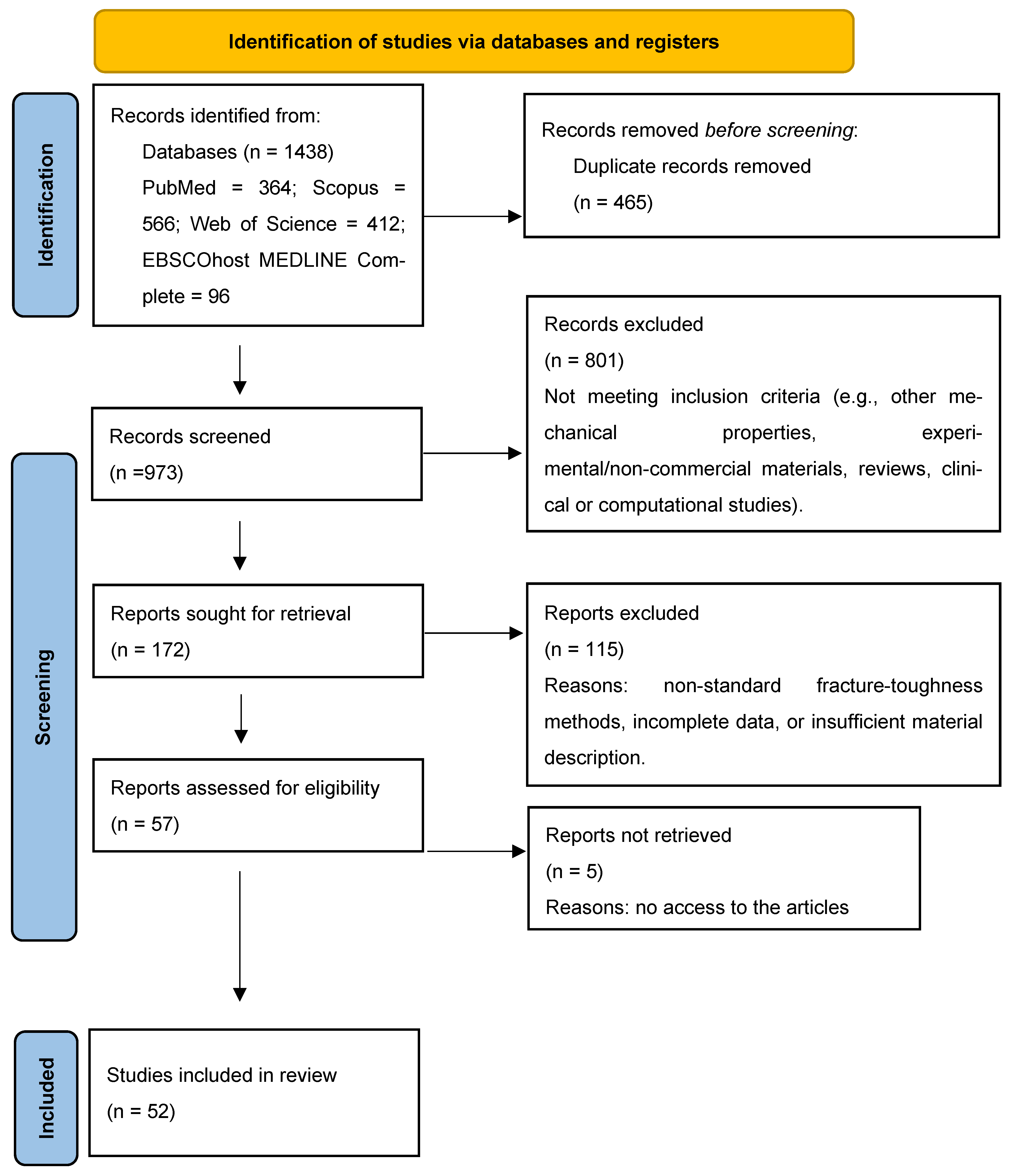
Figure 1. PRISMA flow diagram of the screening and selection process of this study.
Applsci 15 12308 g001
Figure 2. Frequency of CAD/CAM resin-based materials investigated across the included in vitro studies. Percentages refer to the proportion of studies that evaluated each material (multiple materials could be assessed within the same study). The most frequently examined were Enamic (Vita) in 10 studies (37.1%), Lava Ultimate (3M) in 7 studies (25.9%), Cerasmart (GC) in 7 studies (25.9%), and Brilliant Crios (Coltene) in 3 studies (11.1%).
Applsci 15 12308 g002
Figure 3. Reported ranges of fracture toughness (K_IC) for commonly studied direct composites: Filtek Z250 (0.53–2.31 MPa·m1/2), Filtek Supreme (0.67–1.89 MPa·m1/2), and Tetric EvoCeram (0.48–1.79 MPa·m1/2). Values illustrate the variability across different in vitro studies.
Applsci 15 12308 g003
Figure 4. Distribution of risk-of-bias ratings among the included in vitro studies (n = 52). Most studies were classified as medium risk (79%), with 6% rated low- and 15% rated high-risk according to the adapted QUIN criteria.
Applsci 15 12308 g004
Table 1. Main methodological data and results from included studies.
Author Year of PublicationComposite
Resin
Light Curing UnitCuring ModeIrradiance (mW/cm2)Exposure Time (s)Number of SpecimensMethodologyFracture
Toughness
1. Abdulhameed, 2020 [40]Tetric EvoCeram
Tetric EvoCeram Bulk Fill
Tetric EvoFlow
Tetric EvoFlow Bulk Fill (IVOCLAR/VIVADENT)
IVOCLAR/VIVADENT Bluephase StylePolywave1000108SENB
ASTM
D 5045-14 [41]
0.48 (0.15)
0.53 (0.13)
0.68 (0.7)
0.80 (0.15)
2. Alshabib, 2019 [42]NovaPro Universal, (NANOVA INC.)
NovaPro Flow, (NANOVA INC.),
ever X Posterior, (GC)
Filtek Supreme XTE (3M/ESPE)
Filtek Supreme XTE Flowable (3M/ESPE)
Filtek Bulk fill (3M/ESPE)
Elipar S10NR12006 × 20 overlapping12SENB
British
Standard
12135:2021 [43]
1.23 (0.14)
0.96 (0.09)
2.14 (0.16)
1.37 (0.23)
0.97 (0.08)
1.46 (0.17)
1.45 (0.09)
3. Attik, 2022 [44]everX Flow, (GC)
Filtek Bulk Fill Posterior Restorative
SDR Flow+
Elipar DeepCure SNRNo
data
available
No
data
available
8ISO
20795-1 [45]
2.55
1.87
1.92
4. Bijelic-Donova, 2016a [46]everX Posterior (GC)
G-ænial anterior (GC)
Elipar S10Monowave16005 × 20 overlapping10SENB
ISO
20795-1 [45]
2.6
1.03
5. Bijelic-Donora, 2016b [47]everX Posterior (GC)
G-ænial Anterior (GC)
Filtek Supreme XTE (3M/ESPE)
Filtek Bulk Fill (3M/ESPE)
Elipar S10Monowave16005 × 20 overlapping8SENB
ISO
20795-1 [45]
2.4 (0.5)
0.9 (0.1)
1.1 (0.1)
1.1 (0.1)
6. Bonilla, 2000 [48]Ti-Core (ESSENTIAL DENTAL SYSTEMS)
Fluorocore (CAULK)
Astralis5 (IVOCLAR/VIVADENT)NRNR120NRSENB
ASTM
E399 [49]
1.41 (0.05)
1.66 (0.09)
7. Bonilla, 2003 [50]Aeliteflo (BISCO)
Crystal Essence (CONFI-DENTAL)
Flow-It (JENERIC/PENTRON)
Permaflo (ULTRADENT)
Revolution (KERR)
Tetric Flow (IVOCLAR)
VersaFlo (CENTRIX)
Wave (SDI)
FloRestore (DEN-MAT)
Coltolux 4 (COLTENE)NR65045 incrementally10SENB
ASTM E399-90 [49]
1.39 (0.17)
1.31 (0.19)
1.38 (0.2)
1.65 (0.13)
1.32 (0.18)
1.43 (0.09)
1.26 (0.13)
1.15 (0.1)
1.35 (0.14)
8. Cho, 2009 [51]Filtek Supreme Plus (3M/ESPE)
Tetric EvoCeram (IVOCLAR)
Premise (KERR)
Esthet-X (DENTSPLY)
Demetron
Model VCL 401 (KERR)
NR588No
data
available
10SENB
ASTM
E399 [49]
1.08 (0.17)
1.16 (0.11)
1.25 (0.06)
1.47 (0.08)
9. Choi, 2000 [52]ALERT (JENERIC/PENTRON)
Pyramid-Enamel (BISCO)
Pyrarnid-Dentin (BISCO)
Solitaire (KULZER)
SureFil (CAULK)
Heliomolar (VIVADENT)
ZlOO (3M/ESPE)
Curing unit
Triad 11 (DENTSPLY)
NRNR406SENB
ASTM E399 [49]
1.8
1.2
1.6
0.9
1.4
1.2
1.6
10. Engelhardt, 2016 [53]ENAMEL plus HFO Flow
ENAMEL plus Hri Flow
G-aenial Flo(GC)
G-aenial Universal Flo (GC)
Tetric Evo Flow
x-tra base
SDR
Venus Bulk Fill
Sinfony Enamel (GC)
Elipar S 10NR120040 s
in 2 mm layers
10SENB0.76 (0.05)
1.33 (0.19)
0.64 (0.60)
1.26 (0.18)
0.76 (0.05)
1.34 (0.13)
1.33 (0.28)
1.22 (0.14)
1.00
11. Garoushi, 2013 [54]X-tra base (VOCO)
Venus bulk fill (KULZER)
TetricEvoCeram (3M/ESPE)
SDR (DENTSPLY)
Filtek Bulk Fill (3M/ESPE)
Alert (JENERIC/PENTRON)
SonicFill (KERR)
TC-01 (SPRING HEALTH PRODUCTS)Polywave1100manufacturer recommendations6SENB2.0
2.2
2.2
2.6
1.7
2.9
3.0
12. Garoushi, 2024 [55]Essentia Universal (GC)
Alert (GENERIC PENTRON)
Fibrafill Flow (ADM)
Fibrafill Dentin (ADM)
everX flow (GC)
everX Posterior (GC)
Elipar S10Monowave160020 s in five different parts8SENB
ISO
20795-13 [45]
1.1 (0.1)
1.7 (0.4)
1.1 (0.07)
1.2 (0.1)
2.8 (0.4)
2.6 (0.4)
13. Jun, 2013 [56]Spectrum TPH (DENTPLY)
Arabesk Top (VOCO)
Charisma (KULZER)
Revolution2 (KERR)
Dyract flow (DENTSPLY)
Filtek supreme (3M/ESPE)
Grandio (VOCO)
L.E.Demetron I (KERR)NR700403SENB2.42 (0.48)
2.57 (1.08)
1.30 (0.32)
2.32 (0.07)
1.58 (0.05)
2.33 (0.18)
1.97 (0.34)
14. Ilie, 2012 [57]Venus (KULZER)
Tetric Ceram (IVOCLAR)
Z100 (3M/ESPE)
Clearfil ST (KURARAY)
Charisma (KULZER)
Point 4 (KERR)
Herculite XRV (KERR)
TPH Spectrum (DENTSPLY)
Synergy Duo Shade (COLTENE)
Esthet X (DENTSPLY)
Z250 (3M/ESPE)
Enamel plus (GDF)
Miris (COLTENE)
Filtek Supreme (3M/ESPE)
oven Dentacolor XS (KULZER)PolywaveNR408SENB
ASTM E399 [49]
1.18 (0.21)
1.70 (0.18)
1.81 (0.22)
1.81 (0.44)
1.87 (0.20)
1.89 (0.40)
1.96 (0.25)
2.09 (0.22)
2.12 (0.21)
2.15 (0.25)
2.31 (0.16)
2.40 (0.56)
2.17 (0.33)
1.46 (0.28)
15. Ilie, 2021 [58]Venus
Venus Diamond
Charisma Classic
Charisma Topaz
BluePhase Style (IVOCLAR/VIVADENT)PolywaveNR2015Notchless triangular prism (NTP)1.54
2.01
1.04
0.96
16. Kamourieh,
2024 [19]
ever X Posterior
ever X Flow
Demi Plus
(KERR)
Monowave7009 overlapping
Window
technique
10SENB
ISO
20795 [45]
1.90 (0.19)
2.72 (0.37)
17. Knobloch, 2002 [59]Alert (JENERIC/PENTRON)
SureFil (DENTSPLY)
Solitaire (HERAEUS KULZER)
Heliomolar (IVOCLAR/VIVADENT)
Belleglass (BELLE DE ST.CLAIRE)
NRNRNRManufacturer’s specifications Compact tension specimen
ASTM E399 [49]
1.57
1.25
0.67
0.80
1.27
18. Kim, 2002 [60]Silux Plus (3M/ESPE)
Heliomolar (IVOCLAR/VIVADENT)
Aelitefil (BISCO)
Charisma (KULZER)
Herculite XR (KERR)
TPH (DENTSPLY)
Z-100 (3M/ESPE)
Powerlite 100 (LONE STAR DENTAL CORP)NRNR305SENB
ASTM E399 [49]
0.81 (0.07)
0.84 (0.02)
0.98 (0.03)
0.75 (0.02)
0.85 (0.01)
1.04 (0.04)
0.97 (0.04)
19. Kim, 2002 [61]AElitefil (BISCO)
Charisma (KULZER)
Herculite XR (KERR)
Hipolite (B&P, INCHEON)
TPH (DENTSPLY)
Veridonfil (HYOSUNG)
NRNRNRFive 30 s steps5SENB
ASTM E399 [49]
0,98 (0,03)
0.75 (0.02)
1.02 (0.01)
1.14 (0.07)
1.04 (0.04)
1.08 (0.03)
20. Lassila, 2019 [62]SDR (DENTSPLY)
Filtek Bulk Fill Flowable (3M/ESPE)
Tetric EvoFlow Bulk Fill (IVOCLAR/VIVADENT)
Estelite Bulk Fill Flow (TOKUYAMA)
Short fiber flowable (GC)
Elipar S10 (3M/ESPE)Monowave160020
in five separate
overlapping portions
6SENB
ISO
20795 [45]
1.6 (0.1)
1.2 (0.1)
1.4 (0.2)
1.3 (0.1)
2.8 (0.4)
21. Lassila, 2020 [63]Alert (JENERIC/PENTRON)
NovaPro Flow (NANOVA)
NovaPro Fill (NANOVA)
everX Flow (GC)
everX Posterior (GC)
Elipar TM S10 (3M/ESPE)Monowave1600208SENB
ISO
20795 [45]
1.7 (0.4)
1.6 (0.3)
1.3 (0.2)
2.8 (0.4)
2.6 (0.4)
22. Lien, 2010 [64]Beautifil-II (SHOFU)
Esthet-X (DENTSPLY)
Filtek LS (3M/ESPE)
Filtek Supreme (3M/ESPE)
Filtek Z250 (3M/ESPE)
(Bluephase 16i, IVOCLAR)Polywave16002010SENB0.59
0.58
0.68
0.59
0.67
23. Lin, 2009 [65]Micronew (BISCO)
Renew (BISCO)
Filtek Supreme Plus (3M/ESPE)
BelleGlass HP (SDS-KERR)
Oven
Triad (DENTSPLY)
NRNR120 each sideN/ASENB
ASTM Standard PS070-097 [66]
0.82 (0.35)
1.41 (0.11)
1.40 (0.05)
2.06 (0.08)
24. Lohbauer, 2020 [67]Ceram X mono (DENTSPLY)
Filtek Supreme XTE (3M/ESPE)
Heliomolar (IVOCLAR)
Elipar TrilightNR7502015SENB
ISO 13586 [41]
(ASTM E399) [49]
0.83 (0.03)
1.03 (0.08)
0.74 (0.04)
25. Manhart, 2000 [68]Alert (JENERIC PENTRON)
Surefil (DENTSPLY)
Solitaire (KULZER)
Definite (DEGUSA)
Tetric
Ceram (VIVADENT)
Elipar Highlight (ESPE)NR7004010SENB
ASTM E399 [49]
2.3 (0.2)
2.0 (0.2)
1.4 (0.2)
1.6 (0.3)
2.0 (0.1)
26. Mese, 2016 [69]Estelite
Sigma Quick
(TOKUYAMA)
Esthet X HD
(DENTPLY)
Filtek Supreme
XT (3M/ESPE)
Heliomolar (IVOCLAR/VIVADENT)
RoK (SDI)
Vit-l-escence (ULTRADENT)
Raddi Plus (SDI)Monowave150040 × 3 incrementally6SENB
ASTM D5045 [70]
0.51 (0.15) 0.90 (0.17) 0.91 (0.17)
0.56 (0.07) 1.45 (0.24) 1.34 (0.20)
0.58 (0.05) 1.48 (0.20) 1.45 (0.13)
0.49 (0.06) 1.05 (0.19) 1.13 (0.08)
0.56 (0.06) 1.28 (0.24) 1.16 (0.17)
0.75 (0.08) 1.75 (0.09) 1.48 (0.07)
0.53 (0.10) 1.17 (0.07) 1.08 (0.15)
27. Nakade, 2024 [71]Luxacore Z
Lumiglass DeepCure
SelfComp
NRNRNRNR15SENB
ASTM E399 [49]
0.99
0.82
0.36
28. Ribeiro, 2025 [72]Tetric PowerFill (IVOCLAR)
Tetric EvoCeram (IVOCLAR)
Filtek Supreme (3M/ESPE)
Admira Fusion X-tra (VOCO)
Monet Laser (AMD Lasers)
PowerCure (IVOCLAR)
PinkWave (APEX VISTA)
Laser
Dual-wave
Quad-wave
1
10/3
3
2000–2400
1200/3000
>1515
10ASTM E1820 [73]0.5 (0.06) 0.6 (0.07) 0.6 (0.06) 0.5 (0.08)
0.4 (0.07) 0.6 (0.06) 0.6 (0.05) 0.5 (0.09)
0.4 (0.09) 0.5 (0.05) 0.6 (0.08) 0.6 (0.09)
0.4 (0.04) 0.4 (0.04) 0.5 (0.05) 0.4 (0.04)
29. Scherrer, 2000 [74]Columbus (CENDRES ET METAUX S.A)
Artglass (KULZER)
Targis (IVOCLAR)
UniXst oven, (KULZER)NRNR1806chevron-notched precracked Brazilian
disk test
0.60 (0.09)
0.63 (0.04)
0.48 (0.03)
30. Sochacki, 2022 [75]ceramX
Universal (3M/ESPE)
Esthet X HD (3M/ESPE)
TPH Spectra
HT HV (3M/ESPE))
TPH Spectra
HT LV (3M/ESPE)
Bluephase G2
(IVOCLAR)
Polywave1000manufacturer recommended time12SENB
ASTM D5045 [70]
0.491 (0.04)
0.655 (0.05)
0.721 (0.03)
0.610 (0.06)
0.620 (0.04)
31. St-Georges, 2003 [76]Herculite XRV (KERR)
Revolution formula 2 (KERR)
Spectrum 800 (DENTSPLY)
Elipar Trilight soft start mode (ESPE)
Elipar Trilight regular mode (3M/ESPE)
Virtuosoc (DEN-MAT)
Accucure 3000 (LASERMED)QTH
QTH
QTH
PAC
Argon-ion laser
40
40
40
3/5
15
550
100–850
860
1980
725
NRnon-standard, excluded from pooled interpretation1.40 (0.11)
1.43 (0.11)
1.35 (0.09)
1.44 (0.07)
32. Thadathil Varghese, 2024 [77]ACTIVA Bioactive (PULPDENT)
Fill Up! (COLTENE) Surefil One (DENTSPLY)
Cention N (IVOCLAR)
Stela Automix (SDI)
Stela Capsule (SDI)
Radii plusNR1500NR5SENB
ISO
20795 [45]
1.2
0.8
0.43
1.4
1.0
1.41
33. Thomaidis, 2013 [78]Filtek Z-250 (3M/ESPE)
Filtek Ultimate (3M/ESPE)
Admira (VOCO)
Majesty Posterior (KURARAY)
Radii plus (SDI)Monowave950five slightly overlapping
irradiations (40 s each)
10SENB
ASTM E399 [49]
Brazilian test
1.52 (0.16)/0.63 (0.09)
1.43 (0.11)/0.52 (0.04)
1.15 (0.20)/0.45 (0.10)
1.20 (0.20)/0.58 (0.08)
34. Yang, 2022 [79]Beautifil-Bulk Restorative (SHOFU)
Filtek One Bulk fill (3M/ESPE)
SonicFill 3 (KERR)
Viscalor (VOCO)
Elipar S10NR1200280
seven center-overlapping areas
6SENB
British
Standard 54749:1978
E399 [49]
1.13 (0.04)
1.58 0.20)
1.44 (0.04)
1.38 (0.10)
35. Yap, 2004 [80]Tetric Ceram (VIVADENT)
Z250 (3M/ESPE)
Esthet X (DENTSPLY)
Max (DENTSPLY)NR402top and bottom surfaces of the
specimens were then light polymerized in three overlapping
manufacturer’s curing time
7SENB1.75 (0.23)
1.79 (0.37)
1.92 (0.38)
36. Watanabe 2008 [81]Venus (KULZER)
Filtek z250 (3M/ESPE)
Filtek Supreme (3M/ESPE)
Gradia Direct Anterior (GC)
Durafil (KUZLER)
Point 4 (KERR)
Optilux 501 (KERR)NR5002005Brazilian
Test
0.53 (0.03)
0.48 (0.04)
0.42 (0.01)
0.38 (0.01)
0.37 (0.00)
0.26 (0.02)
CAD CAM
37. Alsarani, 2024 [82]Cerasmart (GC)
Lava Ultimate (3M/ESPE)
Shofu Block HC (SHOFU)
Enamic (VITA)
IPS Emax CAD (IVOCLAR)
NRNRNRNR8SENB1.17 (0.15)
1.24 (0.23
1.20 (0.24)
1.39 (0.29)
1,78 (0,21)
38. Della Bona, 2014 [83]Enamic (VITA)NRNRNRNR7SENB
ASTM C1421-10 [84]
1.09 (0.05)
39. Elraggal, 2022 [85]Enamic; VITA
Grandio blocks; VOCO
IPS Emax CAD (IVOCLAR)
NRNRNRNR10Quantitative
fractographic analysis
0.76 (0.17)
1.21 (0.09)
1.89 (0.23)
40. Goujat, 2018 [86]Cerasmart (GC)
Lava Ultimate (3M/ESPE)
Enamic (VITA)
IPS EmaxCAD
(IVOCLAR)
NRNRNRNR10SEVNB
ISO 6872 [87]
1.2
1.4
1.6
1.8
41. Hampe, 2019 [15]Ambarino High-Class
(CREAMED)
Brilliant Crios (COLTENE)
Cerasmart (GC)
Katana Avencia (KURARAY)
Lava Ultimate (3M/ESPE)
Enamic (VITA)
IPS Emax CAD (IVOCLAR
IPS Empress CAD (IVOCLAR)
NRNRNRNR10SENB
ISO 6872 [87]
1.43 (0.27)
1.41 (0.14)
1.22 (0.20)
1.47 (0.28)
1.29 (0.15)
1.24 (0.18)
2.15 (0.24)
0.84 (0.48)
42. Harada, 2015 [16]Lava Ultimate
Estenia C&B
IPS Emax Press
NRNRNRNR5SENB
ISO 6872 [87]
1.4
1.3
2.3
43. Karaer, 2020 [88]Cerasmart 300 (GC)
Katana Avencia P (KURARAY)
KZR-CAD HR3 Gamma Theta (YAMAKIN)
NRNRNRNR10notchless triangular prism (NTP)5.057
4.193
4.880
44. Ling, 2022 [89]Paradigm MZ100 (3M ESPE),
Lava Ultimate (3M ESPE),
Grandio blocs (VOCO, Germany),
Cerasmart (GC, Japan),
Shofu Block HC (SHOFU)
NRNRNRNR10SENB
ASTM D5045-14 [70]
1.74 (0.15)
1.15 (0.09)
1.73 (0.17)
1.71 (0.21)
1.06 (0.09)
1.38 (0.16)
45. Lucsanszky, 2020 [90]Enamic Universal HT (VITA)
RCB; KZR-CAD
HR2 (YAMAKIN)
CERASMART (GC)
CAMouflageNOW
(GLIDEWELL DENTAL LABORATORIES, USA)
NRNRNRNR25notchless triangular prism (NTP)0.83 (0.16)
0.64 (0.11)
1.37 (0.33)
0.68 (0.11)
1.47 (0.19)
46. Moradi, 2020 [91]HIPC (BREDENT)
Crios (COLTENE)
Gradia (GC)
WoodpeckerNR8004010SENB
ASTM E399 [49]
(the values here represent flexular strength)
16.27
22.44
16.42
47. Nguyen, 2012 [92]Gradia (GC)
Grandio (VOCO)
EsthetX (DENTSPLY)
VitaVM LC (VITA)
Paradigm (3M/ESPE)
Radii, (SDI)
30 min post cure in curing chamber
NR897408notchless
triangular prism (NTP)
1.58 (0.18)
1.17 (0.21)
1.52 (0.10)
0.99 (0.17)
0.78 (0.21)
48. Sonmez, 2018 [93]Lava Ultimate
(3M/ESPE)
Enamic (VITA)
Vita Mark II (VITA)
IPS Empress CAD (IVOCLAR)
IPS Emax CAD (IVOCLAR)
NRNRNRNR10non-standard (excluded)1.29 (0.03)
1.23 (0.02)
2.34(0.04)
1.9 (0.03)
1.67 (0.03)
49. Sulaiman, 2022 [94]Luxacrown ( DMG)
Filtek Supreme Ultra (3M/ESPE)
Enamic, (VITA)
(Elipar DeepCure-S, 3MNR1200NRN/ASENB
ASTM
D5045-14 [70]
1.28(0.5)
1.48 (1.0)
1.45 (0.7)
50. Suzaki, 2020 [95]Trinia (SHOFU)
everX Posterior (GC)
Beauti core flow paste (SHOFU)
PenCure 2000, Kyoto, Japan (MORITA) 200030
both upper and side surfaces
NRNotchless triangular prism9
2
3.5
2.5
51. Swain, 2016 [96]Enamic (72%)
Enamic (64%)
Emax CAD
(IVOCLAR)
NRNRNRNRNo
data
available
SENB1 (00.4)
1.51 (0.11)
2.37 (0.28)
52. Wendler, 2021 [97]Gradio blocks (VOCO)
Lava Ultimate (3M/ESPE)
CeraSmart (GC)
Brilaint Crios (COLTENE)
Gradio SO (VOCO)
Elipar TriLight 3MHalogen700–800NR10compact tension C(T)
ASTM E1820-13 [73]
1.42 (0.07)
1.14 (0.05)
0.99 (0.15)
0.97 (0.12)
Older methodologies (pre-2010) and indentation-based tests are not directly comparable and were excluded from quantitative synthesis. NR = not reported.
Table 2. Summary of testing methods used for fracture toughness evaluation in resin-based dental materials.
Testing MethodStrengthsWeaknessesTypical Outcomes
SENB (single-edge notched beam)Most widely used; standardized; reliable K_IC values; allows cross-study comparison [54,57]Specimen prep demanding; requires precise notch; time-consuming [5]Moderate-to-high K_IC, reproducible values [15,16]
NTP (Notchless Triangular Prism)Simple specimen preparation; less machining required [58]Sensitive to notch geometry; often underestimates K_IC; less standardized [94]Lower K_IC values, more variable [92]
C(T) (Compact Tension)Well-established in fracture mechanics; accurate K_IC for large specimens [72]Requires large specimens; difficult in dental context [97]Values comparable to SENB but fewer studies [69]
Brazilian Disk TestEasy setup; widely known in ceramics testing [81]Low reproducibility; high variability; questionable validity for composites [78]Lower, inconsistent K_IC [78]
Indentation (Vickers Crack Length)Quick, requires minimal equipment; small specimens possible [76]Not standardized; often underestimates toughness; indirect method [93]Lowest reported values, often underestimated [93]
FractographyDirect examination of fracture surfaces; identifies true crack origins [4]Qualitative; not suitable for quantitative comparison across materials [4]Confirmed fracture origins, qualitative insights [4]
Disclaimer/Publisher’s Note: The statements, opinions and data contained in all publications are solely those of the individual author(s) and contributor(s) and not of MDPI and/or the editor(s). MDPI and/or the editor(s) disclaim responsibility for any injury to people or property resulting from any ideas, methods, instructions or products referred to in the content.