Review

Integration and Innovation in Digital Implantology—Part I: Capabilities and Limitations of Contemporary Workflows: A Narrative Review

by
Alexandre Perez
1,* and
Tommaso Lombardi
2
1
Unit of Oral Surgery and Implantology, Division of Oral and Maxillofacial Surgery, Department of Surgery, Faculty of Medicine, University Hospitals of Geneva, University of Geneva, 1205 Geneva, Switzerland
2
Unit of Oral Medicine and Oral Maxillofacial Pathology, Division of Oral and Maxillofacial Surgery, Department of Surgery, Faculty of Medicine, University Hospitals of Geneva, University of Geneva, 1205 Geneva, Switzerland
*
Author to whom correspondence should be addressed.
Appl. Sci. 2025, 15(22), 12214; https://doi.org/10.3390/app152212214
Submission received: 29 September 2025 / Revised: 10 November 2025 / Accepted: 12 November 2025 / Published: 18 November 2025

Abstract

Advances in digital dental technologies have transformed implant therapy from analog, stepwise processes into advanced, data-driven workflows spanning diagnosis, planning, surgery, and prosthetic delivery. Contemporary digital implantology integrates multiple techniques, tools, and multimodal datasets into comprehensive diagnostic models and treatment workflows, enhancing implant placement accuracy, procedural efficiency, patient experience, and interdisciplinary coordination. However, integration remains constrained by fragmented datasets, diverse software platforms, and parallel surgical and prosthetic streams. These interfaces often require manual user intervention to convert, process, and align data, thereby increasing the risk of data loss, artifact generation, misalignment, and error accumulation, which may impact implant and prosthetic restorative outcomes. Similarly, implant and prosthetic planning steps continue to rely on subjective, non-standardized user input, requiring advanced experience and training. This narrative review synthesizes current evidence and technical developments in digital implant dentistry based on literature searches in PubMed, Scopus, and Web of Science, with emphasis on publications from 2010 onward, prioritizing systematic reviews, randomized clinical trials, and technical reports focusing on key technological innovations. It presents the current state of the art in digital implantology and identifies major workflow interfaces that constrain seamless, end-to-end integration. This part I summarizes contemporary tools and approaches in digital implant technology. In contrast, Part II of this series will address the emerging roles of artificial intelligence and robotics in overcoming these limitations and advancing toward fully integrated digital implant prosthodontic workflows. Overall, current digital implant workflows are clinically reliable and are equivalent to, or often superior to, conventional approaches in terms of efficiency and accuracy. Nevertheless, their full potential remains limited by persistent software, data, and process interface barriers.

1. Introduction

Digital technologies have induced a profound and accelerating transformation in dental care provision. The first dental digital workflows were introduced by Moermann and Brandestini as early as the mid-1980s [1]. These proof-of-concept workflows involved the milling of ceramic inlays using direct computer-aided processes, comprising digital scanning, computer-aided design, and chairside manufacturing (CAD/CAM). Even by today’s standards, these early workflows would be considered advanced.
Although conceptually designed as chair-side processes, dental laboratories were the first to adopt such digital workflows for the CAD/CAM fabrication of dental and implant prostheses [2,3]. Widespread chair-side integration only occurred within the last decade. This ongoing transition has led to the coalescence and centralization of workflow stages that were originally performed at separate locations by different professional entities [4]. Increasing spatial integration and task centralization have not only enhanced workflow efficiency but also exposed the limitations and barriers of modern digital workflows, thereby driving further innovation.
In particular, implant dentistry has undergone a marked transition from traditional analog to modern digital techniques. Two decades ago, surgical planning was mainly based on 2D radiographs and extended surgical visual access. Likewise, surgeries were mainly executed using freehand drilling and placement, and followed by analog impression and cast-based prosthetic workflows. After an intermediary phase of mixed digital–analog approaches, recent trends in implant dentistry indicate a shift toward fully digitalized workflows across all treatment stages and modalities [5]. As a result, fully digital protocols combining advanced 3D virtual surgical planning, guided and minimally invasive implant placement, and CAD/CAM fabrication of both provisional and definitive restorations have widely replaced traditional analog techniques [6,7,8,9,10].
Multiple systematic reviews have reported that digital implant technologies may offer potential advantages over conventional analog approaches—particularly regarding implant placement accuracy, chair time, and patient comfort—although individual results remain heterogeneous across study designs and dependent on the exact clinical context [11,12,13,14,15,16]. Several comparative studies and meta-analyses have documented reductions in laboratory and clinical processing times with digital prosthetic workflows, generally showing comparable or improved accuracy. However, the magnitude and type of benefit may vary across indications and system types used [17,18,19,20,21,22,23]. Another advantage of digital technologies, and specifically virtual planning, is the ability to visualize restorative and esthetic outcomes directly. This feature may not only enhance treatment planning and interdisciplinary coordination but has also been associated with improved patient communication and treatment acceptance [24,25,26].
Despite these benefits, digital implant workflows are not without potential limitations. These limitations are, e.g., related to the fact that digital workflows often require specialized knowledge and training, but also careful case selection to ensure expected treatment outcomes. Implant positional accuracy has, e.g., been reported to depend on various factors, such as the type of tissue support or the guide design [27,28]. Long edentulous spans or extensive mucosal coverage, in turn, may reduce intraoral scanning accuracy and generate scanning artifacts, thereby impacting prosthetic planning and delivery in the absence of appropriate clinical countermeasures [25]. Regular calibration of devices, verification of guide seating, the adoption of hybrid digital–analog workflows for complex cases, and the use of software or future AI-based verification tools are therefore required to mitigate potential treatment risks [29]. Consequently, while often perceived as plug-and-play solutions, digital workflows remain situationally error-prone and demand corresponding counter-measures, including critical evaluation, strict procedural control, and operator experience to achieve the desired treatment outcomes [30,31,32].
Ultimately, digital technologies may play a crucial role in integrating implant therapy into comprehensive, multidisciplinary treatment strategies. Comprehensive approaches have recently been presented that, e.g., combine prosthodontic, orthodontic, surgical, and even otolaryngological aspects in unified treatment plans (Figure 1) [33,34,35]. These examples illustrate how digital planning tools can facilitate collaboration among specialists to create cohesive, cross-disciplinary workflows [36].
Despite these advances, the full potential of digital workflows remains hindered by numerous interfaces and barriers that persist across tools, systems, and data types. These barriers must be overcome to realize truly seamless integrated digital workflows [37]. Emerging technologies, such as artificial intelligence (AI) and robotics, are on the verge of converging and integrating individual workflow steps by enabling, e.g., dynamic, real-time data exchange between platforms and planning tools, automating data processing, and autonomously executing planning- but also treatment-related workflow tasks [38,39,40,41,42].
This narrative review is the first part of a two-part series. It aims to synthesize the current capabilities and limitations of digital tools and workflows in modern digital implant dentistry.
By focusing on the surgical and prosthetic integration of these technologies, it provides a structured overview of the individual workflow aspects and the advantages and limitations of contemporary digital implantology.
Specifically, the objectives of this narrative review were to:
(1)
summarize the current applications and performance of key digital technologies in implant prosthodontics workflows;
(2)
assess their practical benefits and constraints relative to conventional methods; and
(3)
identify the critical technical, procedural, and data interfaces that still limit seamless digital integration.
The second part of this series will explore how emerging technologies such as machine learning (ML), artificial intelligence (AI), and robotics may reduce or overcome these barriers, towards the realization of fully integrated, end-to-end digital workflows [42].

2. Methods

This review was conducted as a narrative synthesis of the literature focusing on digital workflows in implant dentistry. The synthesis of the contemporary literature was not performed according to strict systematic review guidelines and schemes, while aiming to provide an integrative, descriptive, and comprehensive overview of the individual technologies, applications, and their capabilities and limitations in current clinical practice.
Relevant peer-reviewed publications were identified through searches of PubMed, Scopus, and Web of Science using combinations of key terms such as “digital implantology,” “guided surgery,” “intraoral scanning,” “CAD/CAM,” “digital smile design,” “virtual articulator,” and “workflow integration.” Priority was given to literature published from 2010 onward in English, with a strong focus on reviews—specifically systematic reviews—and randomized controlled trials, prospective cohort studies, and case presentations illustrating specific technical developments.
References cited in key review articles were further screened to capture additional relevant sources. This synthesis of current concepts and innovations was performed without adopting a systematic appraisal of study quality and without formal inclusion/exclusion criteria, dual-review procedures, or risk-of-bias assessment, but with the intention to provide a comprehensive and neutral overview of the current techniques and their advantages and limitations. The narrative structure was organized thematically around the principal components of the digital implantology workflow—diagnostic, surgical, and prosthetic—with particular attention to the interfaces linking these domains and to the individual features that enable seamless end-to-end workflows.

3. Foundations of Digital Implantology

Digital technologies have become deeply embedded in implant dentistry, supporting both surgical and restorative procedures through increasingly integrated data-driven workflows [13,43]. These technologies span key treatment steps from implant planning and placement to functional and esthetic prosthetic design to manufacturing and delivery.
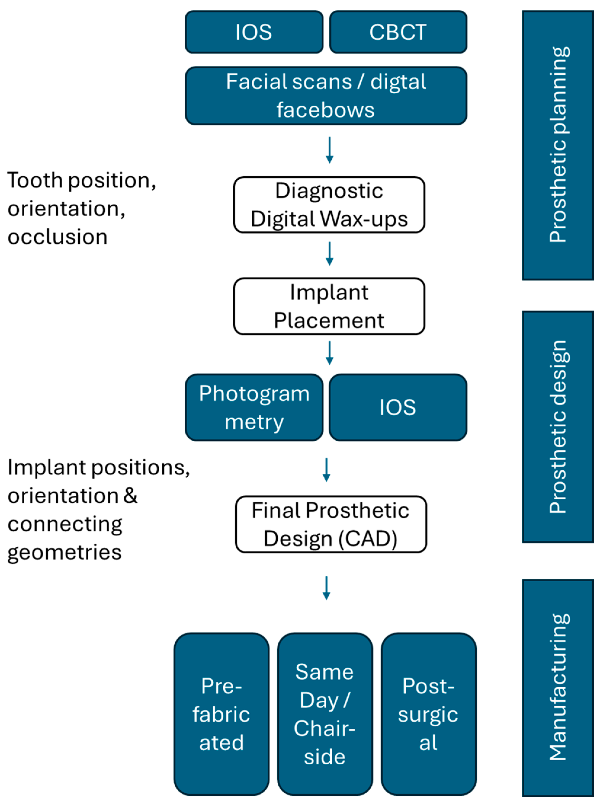
Advanced diagnostic imaging, virtual treatment planning, and computer-assisted surgery now enable highly individualized and prosthetically driven implant placement [44]. Moreover, the integration of facially driven digital smile design and virtual occlusion-based planning underscores the shift toward comprehensive treatment strategies that harmonize function and patient-specific esthetic objectives [5,36]. This chapter outlines the state of the art in digital implantology by delineating the foundational components of surgical, prosthetic, and esthetic workflows, and by critically appraising their capabilities and limitations in contemporary clinical practice (Figure 2).

3.1. Current State of Guided Implant Surgery

Optimal implant positioning, driven by restorative considerations, remains the cornerstone of implant survival and success in modern restorative implant therapy [45,46,47,48]. The demand for accurate implant placement has fueled the development of computer-assisted implant surgery (CAIS).
Early developments in computer-assisted implant surgery (CAIS) can be traced back to the mid-1990s, when Fortin et al. reported the first attempts using CT-derived static guides fabricated from resin splints [49]. Around the same period, pioneering work on dynamic, navigation-based CAIS systems was initiated by a Leuven-based group, with the first reports published by Verstreken et al. [50,51]. These early approaches were subsequently refined into the systematic use of stereolithographic static guides in the early 2000s, laying the groundwork for contemporary static guided CAIS systems [50,52,53].
CAIS is based on integrated digital 3-dimensional anatomic patient models to plan implant positions and their translation using guided surgical protocols. CAIS has been reported to offer potential advantages over conventional non-guided procedures, including improved accuracy, procedural predictability, and efficiency, as well as reductions in surgical time and perioperative morbidity through flapless approaches [8,14,15,49,54,55]. Notably, the extent to which these benefits over conventional freehand approaches may be realized may remain dependent on the clinical indication, case complexity, and operator experience.
The higher accuracy reported for CAIS compared with traditional free-hand (FH) procedures has been associated with an improved ability to mitigate surgical risks and to enhance procedural predictability [56]. Laleman et al. concluded in their systematic review that the incidence of surgical complications associated with CAIS procedures is negligible [57]. Precise control of implant positions, angulations, and depths allows, e.g., accurate management and respect of interimplant and tooth-to-implant distances. At the same time, the remaining intrinsic positional and angular placement inaccuracy must be considered when planning horizontal and vertical safety margins around critical anatomical structures, such as the inferior alveolar nerve or neighboring teeth [11,12,13,58].
Several systematic reviews have repeatedly highlighted the superior accuracy of CAIS compared to FH techniques [16,54,59,60,61,62,63]. Despite heterogeneity related to the reported indication, e.g., presence and location of residual teeth or type of guide support, a recent meta-analysis comparing the accuracy outcomes of flapless CAIS with flapped FH placement reported −3.88 degrees statistically significantly less angular deviation and −0.75 mm reduced apical three-dimensional positional deviation. Furthermore, CAIS demonstrated a −0.28 mm reduced depth deviation and a −0.60 mm reduced coronal three-dimensional bodily deviation with differences remaining below significance [8]. Oliveira et al. have furthermore recently concluded, from their systematic review on the use of static CAIS in full-arch treatments, that the accuracy of guided implant placement was within clinically acceptable levels [56]. Moreover, several authors have highlighted potential advantages of CAIS in advanced workflows, such as the immediate loading of screw-retained full-arch restorations, where achieving an accurate passive fit is essential [27,64,65,66,67,68]. Likewise, Lau and Shirani et al. have concluded that CAIS offers specific advantages over FH placement in terms of accuracy and marginal bone levels in immediate placement procedures [69,70].
By supporting flapless and minimally invasive techniques, CAIS may also directly impact patient-reported outcomes by reducing post-operative discomfort and improving overall treatment acceptance when compared to traditional free-hand and open-flap procedures, as supported by the majority of recent systematic reviews [8,14,15,57,61,68,70,71,72].
Several studies and systematic reviews have also compared CAIS and FH implant placement with respect to workflow efficiency, chair time, and economic factors. Although CAIS has been associated with reduced chair-time compared to FH placement—most notably in flapless and multi-implant settings—this advantage is less evident in single-implant procedures, where the advantages of CAIS over FRH placement may remain less pronounced due to methodological and clinical heterogeneity among studies [8,12,54,61,73,74,75]. Taken together, the marked heterogeneity across study workflows, applied techniques, and clinical indications hinders the establishment of consistent evidence or generalizable conclusions regarding chair time and workflow efficiency [75].
CAIS comprises two main modalities: static CAIS (S-CAIS) and dynamic CAIS (D-CAIS) [59,62,69,74,76,77,78,79,80]. S-CAIS is based on the use of prefabricated surgical guides to direct osteotomy and implant placement [68]. D-CAIS, by contrast, utilizes real-time virtual navigation to guide osteotomy drilling and implant placement and, unlike S-CAIS, retains the flexibility to adjust the procedure intraoperatively if necessary, while omitting the need for surgical guide design and production [80,81]. Its operation relies on real-time optical tracking of the patient’s and surgical instruments’ relative positions [16,58,82].
A wide body of evidence, including systematic reviews, supports the notion that D-CAIS and S-CAIS are comparable in terms of implant placement accuracy, while remaining superior to FH placement [59,62,78,79,83,84,85]. Tang et al. recently emphasized that the adoption of D-CAIS was associated with substantial upfront costs, training, preparatory time, and increased patient exposure to radiation when compared to S-CAIS, while S-CAIS by itself has also been associated with a broad spectrum of potential clinical and procedural risks [29,81]. Finally, and for completeness, Pomares-Puig et al. recently introduced a hybrid workflow that integrates static and dynamic CAIS within a single protocol, referred to as the “double-factor technique” [86,87].
Based on whether surgical guides are used throughout the entire surgical procedure or only at selected stages, i.e., osteotomy preparation or implant placement, S-CAIS can further be classified into fully guided procedures (FG) comprising guided drilling and placement, partially guided procedures (PG), using guidance only during osteotomy preparation, or conventional free-hand (FH) procedures [61]. Both D-CAIS and FG S-CAIS have demonstrated significantly higher positional accuracy relative to the planned implant position compared with FH implant placement [11,76,84,88,89]. Furthermore, Gargallo et al. and Tattan et al. recently reported higher accuracy for fully guided (FG) implant placement compared with partially guided (PG) or freehand (FH) techniques in their systematic reviews. In contrast, other authors found comparable accuracy between FG and PG approaches, while both methods appear to outperform FH placement [16,61,79,88,90]. In quantitative terms, Tattan et al. reported significantly lower mean angular (Mean Difference (MD) = 4.41°, 95% CI 3.99–4.83; p < 0.00001), coronal (MD = 0.65 mm, 95% CI 0.50–0.79; p < 0.00001), and apical (MD = 1.13 mm, 95% CI 0.92–1.34; p < 0.00001) deviations for fully guided (FG) compared with freehand (FH) implant placement, while a smaller but still significant angular advantage (MD = 2.11°) was observed when comparing FG to partially guided (PG) procedures [88]. Notably, specific outcomes of S-CAIS and their advantages over alternative methods may depend on the individual indication and procedural factors, such as guide design, guide support, and guide tissue support type [9,27,91,92,93,94,95,96]. Gingiva- or tooth-supported guides have, e.g., been documented to outperform bone-supported designs in terms of implant placement accuracy [9,12,93,94].
All CAIS procedures rely on virtual 3D patient models for implant planning and, where applicable, for the design and fixation of surgical guides and digital prosthetic waxups [42,44,97]. These models are derived from cone-beam computed tomography (CBCT) in the form of Digital Imaging and Communications in Medicine (DICOM) data, containing voxel-based three-dimensional representations of anatomical structures based on radiodensity [50,97,98]. To enable clinical application, DICOM raw data must be segmented and associated with relevant anatomic tissue types, i.e., bone, teeth, and in some cases soft tissues, as well as existing prosthetic restorations [98,99]. More sophisticated virtual models can be generated by incorporating intraoral surface contour data from intraoral scans (IOS), commonly stored as standard tessellation language (STL) files. CBCT and IOS data can be merged in a process termed registration to yield enhanced and more detailed anatomic patient models [44,97]. The resulting models significantly facilitate the design and fabrication of anatomically adapted prosthetic wax-ups and tooth- and soft-tissue–supported surgical guides [44,100,101].
In conclusion, this overview of the current state of guided implant surgery and its various approaches indicates that CAIS techniques are well established and widely accepted. The accuracy of different CAIS variants is strongly supported by a substantial body of evidence, including detailed analyses across clinical indications and guide-support designs. However, other aspects of CAIS, e.g., patient-reported outcomes and procedural and technical workflow factors, remain less well explored and are only infrequently reported in the literature [68,70,74,102].

3.2. Current State of Implant-Prosthetic Planning and Manufacturing Workflows

The past decade has witnessed a marked evolution in digital implant-prosthetic workflows, with conventional analog methods—such as silicone impressions and stone casts—being increasingly replaced by chairside CAD/CAM systems that utilize intraoral scanners and digital prosthetic planning [17,19,103]. Similarly, virtual articulators, digital photographs, and facial scans have become available as digital alternatives for conventional facebows and mechanical articulators [36]. The advances in digital dental implant technologies have been associated with potentially enhanced precision, reproducibility, and esthetic control while potentially offering shorter treatment timelines and improved patient- and peer-communication [5,17,22,36].
The integration of multiple digital datasets into a unified virtual patient model lies at the core of modern digital prosthetic planning [44,104,105] (Figure 3). IOS has become a cornerstone of the digitalization of oral tissues, offering direct access to highly accurate representations of dental and soft-tissue contours [17,19,106]. Compared with indirect extraoral scanning of conventional impressions, IOS has been shown to provide accuracy comparable to or superior to that of conventional impressions [19,106]. IOS datasets are often combined with CBCT-based 3D skeletal and dental anatomical information and are increasingly complemented by facial scans or digital facebows [36,37,97,105]. The latter integrates important esthetic and occlusal function-related references into the unified patient model, greatly enhancing the prosthetic design process [24,36,104,107]. Recent developments have, e.g., presented advanced digital workflows capable of considering 3-dimensional soft-tissue contours and thicknesses to deliver anatomically individualized and esthetically optimized prosthetic designs [107,108].
Digital diagnostic wax-ups have become a cornerstone of esthetic and functional planning in contemporary digital workflows [36,104,107]. They establish the foundation for prosthetically driven implant positioning and guide the development of the surgical plan [49,54]. Accurate execution of this plan is essential to achieving prosthetic objectives, particularly in cases involving immediate restoration, e.g., with prefabricated prostheses [109]. Following surgery, IOS enables the efficient and patient-friendly capture and registration of implant positions and angulations into the final prosthetic wax-up, supporting the direct chairside or laboratory-based fabrication of both provisional and definitive restorations [17,18,19,20,21,22,23]. To overcome the limitations of IOS related to image stitching in scans of larger edentulous spans, photogrammetry has been proposed as a reliable alternative [17,25,106,110,111].
Advances in CAD/CAM technologies have expanded prosthetic manufacturing options across different clinical scenarios. Prefabricated restorations, designed prior to surgery based on digital wax-ups, IOS, and CBCT, are increasingly used in immediate loading protocols, provided that surgical accuracy is maintained throughout the procedure to achieve an immediate passive fit [109,112,113]. Alternatively, same-day restorations can be produced chairside following implant placement. These options have been extensively described for situations ranging from single-unit crowns to small bridges and full-arch restorations, using on-site milling or 3D printing [24,108,114,115]. Definitive restorations are typically fabricated after osseointegration and occlusal adaptation using monolithic zirconia, lithium disilicate, or hybrid ceramic materials, offering versatile solutions for both esthetic and functional demands [116].

3.3. An Overview of Current Digital Smile Design Workflows

Patient perception and patient-centered outcomes have gained increasing importance in contemporary dentistry, reflecting a broader shift toward individualized treatment objectives that extend beyond purely functional outcomes [117,118]. The ability of modern digital workflows to directly virtualize esthetic outcomes has greatly enhanced esthetic planning and patient communication. According to Coachman, this approach significantly enhances patient engagement and communication, diagnostic understanding, and treatment acceptance, while improving the predictability and quality of treatments with elevated esthetic demands [36]. Recent systematic reviews have repeatedly highlighted increased case acceptance and higher patient satisfaction rates associated with DSD workflows [119,120,121]. More specifically, Luniyal et al. reported, e.g., significantly higher mean satisfaction scores in patients treated using Digital Smile Design (DSD) compared with conventional smile design (85.4 ± 6.2 vs. 79.8 ± 7.1). At the same time, the proportion of cases rated “excellent” for restoration fit, occlusal fit, and esthetics increased from 78% to 92% with the implementation of DSD [122].
Originally introduced by Ackermann and Ackermann, and Coachman, Digital Smile Design (DSD) has emerged as a structured, data-driven approach to integrating facially driven dental esthetics into treatment planning [123,124]. DSD integrates two- and three-dimensional data to visualize and simulate the intended restorative outcome in harmony with the patient’s facial features and emotional expectations, facilitating patient participation and emotional alignment through collaborative visualization [36]. By integrating standardized facial photography, facial and intraoral scans, DSD creates a comprehensive virtual patient model that enables an objective evaluation of esthetic parameters and supports a facially driven prosthetic design based on established esthetic principles [26,44,105,125]. Recent approaches have evolved into 4D technology, incorporating the dynamic analysis of the patient’s smile using real-time digital motion sensors [85,126]. Furthermore, integrating natural tooth and smile libraries with advanced algorithms enables the selection of tooth phenotypes and proportions that best suit the patient’s facial characteristics, yielding more objective, predictable, and patient-specific outcomes than conventional wax-ups [36].
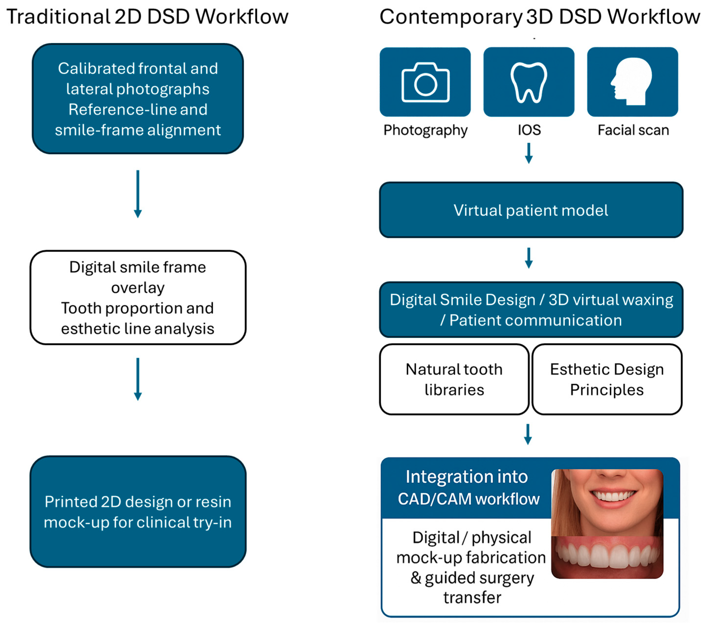
DSD is grounded in well-established esthetic prosthodontic principles, including ideal, natural, and harmonious tooth dimensions, gingival levels, smile and facial symmetry, and the integration of pink and white esthetics [26,127]. White esthetics refer to the shape, size, and proportions of the teeth. In contrast, pink esthetics relate to the contour, volume, and symmetry of the gingiva in relation to dental and facial reference lines. Modern DSD approaches consider these static parameters while accounting for dynamic factors, such as lip mobility and smile dynamics, to achieve more natural and individualized outcomes [125]. Modern DSD software solutions, including DSDApp, 3Shape Smile Design, and exocad Smile Creator, enable the integration of standardized facial photography at rest and in full smile, as well as intraoral and facial scans, into a unified virtual patient model, facilitating facially driven planning and 3D virtual waxing [36,121]. Figure 4 illustrates the transition from traditional two-dimensional, photo-based DSD workflows to fully integrated three-dimensional approaches that combine photographic, intraoral, and facial scan data for comprehensive esthetic visualization and CAD/CAM integration. Furthermore, access to extensive libraries of natural tooth phenotypes and donor smiles, combined with the ability to perform virtual or physical mock-up try-ins, enables efficient validation and refinement of patient-specific designs, thereby enhancing the efficiency, predictability, and reproducibility of esthetic treatment provision [125,128].
DSD has become increasingly integrated into implant-prosthetic workflows, particularly in cases with high esthetic demands. Single-tooth restorations in the esthetic zone have, for example, been realized by accurately aligning the planned tooth position with the patient’s smile line, lips, and facial symmetry [24]. In more complex anterior reconstructions, DSD may facilitate the simultaneous optimization of implant positions and prosthetic and soft-tissue contours, for example, by delivering an anatomically guided interim prosthesis that serves as a scaffold for controlled peri-implant soft-tissue conditioning and emergence profile sculpting [107]. Finally, DSD may greatly enhance full-arch treatments due to the absence of reference teeth. Several advanced workflows integrating DSD with, e.g., prosthetically driven bone reduction and guided implant placement for the delivery of pink-free FP-1 or FP-2 restorations with anatomically and esthetically optimized cervical and coronal aspects have been presented [107,108,129]. Amin et al. have, for example, recently demonstrated the integration of 3D facial scans into DSD-based full-arch planning, thereby improving harmony between dental morphology, smile dynamics, and facial esthetics, while streamlining guided surgery and immediate loading protocols [104].
In addition to improved esthetic predictability, the integration of DSD and, more broadly, digital prosthetic techniques into implant-prosthodontic workflows has demonstrated several practical clinical benefits. Bessadet et al. reported a significant reduction in laboratory working time and costs. In contrast, differences in adjustment time did not reach statistical significance when comparing digital and conventional workflows in a recent systematic review and meta-analysis [130]. Corsalini et al. further observed that digital workflows could deliver prosthetic restorations with significantly superior interproximal and occlusal contacts in a significantly shorter time, while also resulting in higher patient satisfaction compared with conventional approaches [131]. Hashemi et al. showed that both conventional and digital workflows yielded three-unit implant-supported fixed dental prostheses with comparable impression accuracy, framework adaptation, passivity of fit, and esthetics, yet the authors also reported a significantly shorter total working time in the digital group [132]. In an earlier study, Joda et al. quantified total workflow time for single-implant crown design and manufacturing as 185.4 ± 17.9 min for the digital workflow versus 223.0 ± 26.2 min for the conventional pathway (p = 0.0001), corresponding to a 16% overall reduction, including significantly shorter chairside and laboratory phases [23]. In a subsequent investigation, the same authors demonstrated significantly shorter adjustment times (2.2 ± 2.1 min vs. 6.0 ± 3.9 min) when comparing posterior single-implant hybrid-design restorations fabricated via CAD/CAM-based versus conventional workflows [133]. Collectively, these studies indicate that beyond esthetic advantages, digital techniques can substantially enhance multiple practical aspects of the implant-prosthodontic workflow, improving efficiency and reproducibility.

3.4. Virtual Articulators in Functional Digital Prosthodontics

Complementary to DSD, virtual articulators (VAs) are essential tools for ensuring accurate functional integration in contemporary digital prosthodontics [37]. Serving as digital counterparts to traditional mechanical articulators, VAs enable the precise registration of maxillocranial, maxillo-mandibular, and occlusal relationships during prosthetic planning [134]. These parameters play a key role in patient-specific functional analysis and occlusal planning, helping to minimize discrepancies, avoid premature contacts, and reduce the need for subsequent adjustments [134,135].
VAs have become widely available in modern CAD software systems, enhancing the diagnostic and planning capabilities of digital workflows for both prosthetic and surgical decision-making [134]. Two types can be distinguished: completely adjustable, patient-specific VAs and mathematical, average-value-based VAs [37]. The former allows precise replication of patient-specific mandibular dynamics by incorporating a range of static and dynamic parameters, including the Bennett angle, horizontal condylar inclination, vertical dimension of occlusion, virtual facebow, and incisal table inclination [134]. Mathematical VAs, by contrast, use standardized, preset population-averaged values in place of patient-specific values, greatly simplifying their implementation [37]. Mathematical VAs are often sufficiently accurate but may be limited in complex cases, e.g., full-arch rehabilitations or cases in which functional parameters deviate significantly from the statistical norm [136].
Regardless of the articulator type, an initial transfer of the patient’s maxillo-mandibular relationship into the virtual model is necessary. This process establishes the spatial position of the maxilla relative to cranial reference planes, via a facebow transfer, and serves as the basis for accurately determining mandibular positioning and movement relative to the maxilla [134]. Commonly used reference planes to orient the arches include the Frankfort plane and Camper’s plane, which are defined by the porion–orbitale line or the line connecting the tragus to the ala of the nose, respectively. These planes serve as surrogate cranial references for positioning the maxilla within the virtual articulator environment.
Alternatively, individualized reference planes can also be derived from CBCT, cephalometric imaging, or 3D facial-scan data. Recent developments also allow the derivation of functional reference planes from kinematic data obtained via digital axiography, jaw-motion tracking, or stereophotogrammetry, as well as orientation using standardized extraoral photographs or photogrammetric 3D reconstructions [134]. The virtual facebow transfer can thus be accomplished using various methods, depending on the desired level of anatomical or functional precision [37].
Compared to mathematical VAs, implementing fully adjustable VAs is more demanding and typically requires integrating multiple datasets across diverse digital platforms, rendering the associated workflow technically complex and often steep [137]. As a consequence, fully adjustable virtual articulators (VAs) remain particularly indicated for advanced rehabilitations that require the functional reconstruction of occlusion. In contrast, average-value–based VAs are commonly employed when replicating the static relationship between the arches is sufficient for planning the occlusal morphology of prosthetic restorations, while omitting individualized information on the dynamic movements of occlusal surfaces [134,138].
Several studies have compared the performance of average-value and fully adjustable VAs. Lin et al., e.g., compared six VA mounting procedures—including average mounting and average facebow transfer versus fully adjustable methods such as kinematic digital facebow and CBCT-based techniques [139]. The authors found that the former resulted in significantly larger angular deviations, whereas kinematic and facial-scan–based approaches generally demonstrated superior accuracy.
In a subsequent clinical study, the same authors evaluated average-value VAs using arbitrary facebow mounting, compared with fully adjustable models incorporating actual Bonwill triangle and Balkwill angle parameters to simulate changes in occlusal vertical dimension (OVD) [140]. Both methods produced comparable results up to an OVD increase of approximately 6 mm, whereas larger OVD changes required fully adjustable models based on virtual facebows and jaw-tracking data to accurately reproduce the clinical situation.
In addition to the type of VA, the data acquisition method of the VA can be further categorized into direct or indirect methods, based on whether analog steps, e.g., impression taking, stone cast production, and bite registrations are performed or if data acquisition is purely based on direct digital techniques, e.g., IOS, virtual interocclusal records and virtual facebows [134].
Finally, recent developments extend the application of patient-specific VAs within fully integrated ‘4D virtual patient’ workflows. Merli et al., e.g., demonstrated a technique that combined jaw motion tracking, intraoral and 3D facial scans, and CBCT datasets within a CAD environment (Exocad) to simulate dynamic mandibular movements and optimize occlusion, illustrating the growing convergence of VAs with comprehensive digital treatment planning in contemporary implant prosthodontic therapy [141].

4. Integrated Workflows in Contemporary Digital Implantology

4.1. Conceptual Framework—Workflow Digitization vs. Workflow Integration

The digital transformation of implant restorative dentistry has shifted clinical workflows from a series of isolated analog steps to integrated, data-driven treatment protocols [98]. To better illustrate this transformation of workflows and their potential future evolution, it may be useful to distinguish between digitized and integrated workflows. Digitization refers to replacing analog procedures with digital alternatives, such as substituting conventional silicone impressions with IOS [1]. Integration and integrated workflows, by contrast, refer to the orchestration and alignment of datasets, tools, and systems within a workflow to create unified, seamless end-to-end workflows [142]. Notably, these integrated workflows, which enable the near-seamless exchange of information across devices, software systems, and procedural steps at the current time, remain idealized models. Workflow integration may thus be understood as the process of connecting the various digital technologies employed throughout the digital implant-prosthetic workflow—encompassing diagnostics, virtual planning, guided surgery, and prosthetic rehabilitation—into a cohesive, end-to-end treatment continuum (Figure 2).
Central to the realization of integrated workflows is the management of interfaces, which arise wherever datasets, software platforms, or individual procedural techniques are linked into a sequence of interdependent steps and must communicate and exchange information [142]. A potential strategy to reduce the number of such interfaces lies in direct digital workflows, in which anatomical data required for restorative planning are captured directly, as, e.g., realized by IOS instead of indirectly digitizing physical impressions or casts [1,19,37].
Notably, the implementation of direct, integrated workflows is somewhat hindered by the inherent complexity of implant prosthodontic therapy. Specifically, this complexity is primarily linked to the high variability in case-specific anatomic situations, different possible treatment protocols, and multidimensional prosthetic objectives [143,144,145]. In contrast, other disciplines, such as endodontics or periodontology, often allow the application of more standardized procedures. This inherent complexity of implant prosthodontics often necessitates combining multiple tools, techniques, systems, and stakeholders, highlighting the critical importance of seamless communication among these entities to achieve predictable, esthetic, and functional outcomes [144].
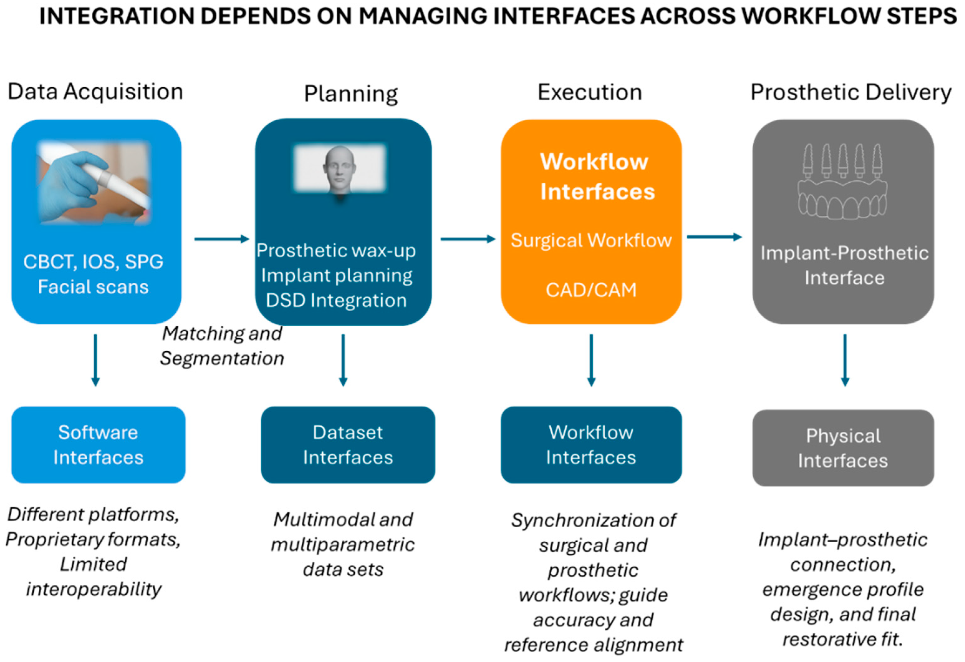
A pivotal step in realizing fully integrated digital implant-prosthodontic workflows involves managing interfaces. These interfaces exist where datasets, platforms, or processes must interconnect within the workflow (Figure 5) [98]. Successful integration depends on resolving these interfaces to ensure accurate, reliable, and reproducible synthesis into diagnostic models and their translation into the final physical restoration while avoiding or minimizing artifact generation and error propagation [142,146].

4.2. Data Integration and Dataset Interfaces

In digital workflows, the integrity and synchronization of datasets remain the foundation for accurate planning and execution; hence, data integration remains the first and most critical layer of interoperability.
Software interfaces remain one of the central barriers to integrated workflows in digital dental implantology and occur when data must be converted or transferred between systems, e.g., importing IOS datasets into CAD software or CBCT into virtual planning tools. Proprietary formats, inconsistent data standards, and differences in data dimensionality often necessitate manual imports, exports, conversions, processing, and extraction, all of which may lead to data loss, artifact generation, and reduced interoperability [37].
Dataset interfaces are often encountered in digital implant workflows, and become, e.g., apparent when combining multimodal and multiparametric datasets such as CBCT, IOS, facial scans, and prosthetic wax-ups into unified virtual patient models [44,100,146]. These datasets often contain overlapping anatomical information, such as bone, dentition, and soft tissues, and require precise segmentation into the tissue types and anatomic structures of interest.
Careful registration of individual data sets using shared anatomic landmarks or fiducial markers is crucial to ensure accurate virtual anatomic planning models [98,147]. Specifically, errors introduced during data set registration, whether due to poor primary data quality or inaccurate registration, may propagate into surgical and restorative planning and execution, underscoring the importance of both adequate data quality and processing [11,44,98]. In fact, segmentation and registration errors have been reported as non-negligible contributors to overall system error, affecting the accuracy of dental implant placement, while remaining influenced by a variety of procedural, operator-related, and anatomical factors [148,149].
The impact of different registration methods and their respective influencing factors on virtual model and dental implant placement accuracy remains an active field of research. Depending on the presence and distribution of residual teeth as landmarks, registration of IOS–derived STL data with CBCT-based DICOM data can, e.g., be performed using various approaches, including fiducial marker–based or markerless techniques [150]. Recent studies have further refined conventional registration approaches through improved alignment algorithms. Comparative analyses have shown that optimized markerless and semi-automated techniques can achieve reproducibility comparable to expert-guided workflows while maintaining full operator control, while generalizability of such results may remain subject to indication and specific clinical scenarios [62,151,152]. Furthermore, a variety of registration algorithms are currently in use—including small-point (SP), large-point (LP), and entire-surface (ES) matching [153]. Collectively, these sources of variability render image segmentation and registration a critical and potentially error-prone data interface within contemporary digital implant–prosthetic workflows [69].
High-quality CBCT and IOS data remain prerequisites for precise data set registration into accurate virtual models. Factors such as local bone density may, however, affect radiographic contrast and potentially image segmentation, while long edentulous spans may result in IOS stitching artifacts that compromise surface scan accuracy [110,111,153]. Metallic or other highly radiopaque restorations (e.g., zirconia) can further degrade CBCT image quality through beam-hardening and streak artifacts [148,150], all of which may impact image segmentation and registration and thus model and ultimately placement accuracy.
Workflow interfaces emerge when, e.g., surgical and prosthetic procedures are conducted in parallel but must remain synchronized. A prosthetic wax-up, for instance, may define both implant positioning, as translated via surgical guides into an implant restoration, and be used in parallel for immediate CAD/CAM of implant prosthetics. Surgical guide accuracy and precise alignment between surgical and prosthetic workflows remain critical to ensure, for example, an immediate passive fit between both restorative elements [108,146,147,154]. Likewise, complex protocols often require additional verification steps and intra-treatment diagnostic scans, e.g., using strategic temporary or artificial reference landmarks as fiducial markers to update virtual patient models and digital wax-ups throughout the workflow [108,147].
Finally, accumulated inaccuracy within the digital workflow may ultimately manifest at the implant-abutment interface, which is potentially the most critical physical interface for long-term treatment success [154,155,156,157]. Specifically, software, datasets, and workflow interfaces may each introduce potential inaccuracies and become clinically relevant at the physical implant–abutment connection [98,142,155]. High-quality primary data, rigorous upstream planning, and seamless integration between digital and clinical workflow components remain essential for reducing physical interface errors.

4.3. Software Interoperability and System Ecosystems

Despite their described clinical and operational advantages, current digital implant-prosthodontic workflows remain limited by several challenges [36,98,142]. Multiple independent systems need to be operated, e.g., to acquire diagnostic data that must be synthesized and adapted according to the individual workflow stages. These processes require specialized training, technical expertise, and rigorous quality control [98].
Closed systems may streamline digital workflows—such as CAD/CAM processes—by enabling multiple tasks to be performed within a single software ecosystem [37,158]. These systems reduce the need for file conversions, minimize platform interface complexity, and may lower the risk of technical errors [5,37]. However, they often create software silos, limiting interoperability and restricting cross-platform collaboration [37]. In contrast, open systems offer broader compatibility with third-party software platforms and treatment units, but typically require file conversions between the different standard formats, e.g., STL, PLY, OBJ, and DICOM, which may introduce data loss, artifacts, or misalignment [37].
In recent years, hybrid or semi-open systems have emerged that combine the integration advantages of closed architectures with the interoperability of open ones. These systems typically rely on application programming interfaces (APIs)—software interfaces that enable data exchange between different programs or devices within a standardized communication framework [134,159,160]. Standardized or publicly documented APIs (e.g., based on REST or JSON protocols) increasingly allow CAD, CAM, and implant planning software from different vendors to exchange 3D geometry, metadata, and patient information directly, without manual file conversion. Examples include STL or PLY mesh data transfer through open connectors (e.g., Exocad, 3Shape) or DICOM- and FHIR-based APIs that enable integration of CBCT datasets and patient metadata across platforms. Such developments are paving the way for more interoperable “hybrid” digital ecosystems, where proprietary modules remain protected yet communicate securely through standardized data interfaces [134,159,160].
As further emphasized by Flügge et al., the functional scope of current software platforms remains highly heterogeneous. Depending on the system used, options for a virtual set-up, integration of a virtual articulator, and visualization of a prosthetic design may be limited or absent. Moreover, the range of implant libraries supported by different programs varies substantially, creating a mismatch between the implant systems used clinically and those digitally available within each software environment [159,160].
Consequently, the choice of software often depends not only on the desired level of integration or openness but also on the compatibility with the clinician’s preferred implant system and prosthetic components. These discrepancies highlight the continuing need for broader standardization and unified data exchange protocols to ensure consistent planning capabilities across platforms.

4.4. Human–Machine Interaction and Workflow Feedback Loops

While automation and software integration have advanced rapidly, human–machine interaction remains central to clinical execution. A noteworthy development in contemporary digital workflows is the integration of feedback loops, in which intra-operative or post-surgical diagnostic data—particularly information about the post-implant placement situation—are collected to dynamically update the virtual plan. Such procedures are most commonly applied in advanced workflows involving immediate restoration, where it is often necessary to adjust the digital provisional wax-up after implant placement to reflect the actual implant positions and ensure an adequate prosthetic design and optimal passive fit, especially in multi-implant or full-arch restorations [109,154].
Several fully digital workflows have been described for the immediate full-arch restoration of partially or fully edentulous patients [66]. These approaches are considered technically demanding, as edentulous arches often lack distinctive landmarks for accurate scan registration—unless strategic extraction protocols are used, such as in cases transitioning from a failing dentition [18,111,161,162,163]. Direct digital workflow strategies for completely edentulous situations have been presented, e.g., using artificial reference markers (e.g., split surgical guides), auxiliary scanning guides, temporary mucosal markers, or pins serving as fiducial landmarks for registering post-placement scans with the provisional prosthetic models [108,147,152,164,165,166,167,168]. Although feedback loops are now incorporated into many digital implant workflows, they remain particularly critical when implant positions deviate from the original plan or when immediate restoration protocols are employed, while remaining technically complex and potentially error-prone.
In summary, the following key findings and areas of uncertainty anchor the above framework in current evidence:
  • Segmentation/registration errors are measurable contributors to overall deviation in CAIS, with performance varying based on primary data quality, registration algorithms, presence and type of fiducial markers, and operator factors [148,149,150,153].
  • Guide support & design influence implant placement accuracy: tooth- or mucosa-supported guides generally outperform bone-supported designs in most reports, though indication-specific results vary [58,93,94,100].
  • Dataset interfaces (CBCT + IOS + facial scans): multimodal registration improves planning fidelity but introduces additional potential error sources; metallic artifacts and long edentulous spans remain recurring sources of potential inaccuracies [98,110,111,148,149,150].
  • Open vs. closed ecosystems: studies comparing planning platforms document heterogeneous feature sets and library mismatches between clinical implant systems and their digital availability, supporting the interoperability concerns outlined above [159,160].
  • Efficiency evidence is context-dependent: time savings are most consistent in multi-implant/flapless settings; single-unit scenarios show equivocal results due to methodological and clinical heterogeneity [8,12,54,61,73,74,75].
  • Contradictory or neutral findings exist (e.g., nonsignificant differences in adjustment time; comparable impression/framework outcomes in some comparisons), underscoring the need for case-selection and workflow-specific appraisal [130,131,132].

4.5. Future Directions—AI and Robotics as Integrative Enablers

Emerging technologies such as artificial intelligence (AI), machine learning (ML), and robotics may mitigate many interface-related barriers towards integration by improving data quality control, automating registration and verification, and enabling adaptive execution. AI-driven algorithms have the potential to enhance segmentation accuracy and efficiency, automate error detection, and provide real-time feedback during clinical workflows. At the same time, robotics and computer-assisted systems may strengthen the link between virtual planning and surgical implementation through precision-guided interventions. Together, these advances could substantially reduce inter-system but also human–machine interface complexity and help close existing gaps in digital workflow integration. A detailed and methodical appraisal of these technologies falls outside the scope of Part I and will be addressed in Part II of this review, which focuses on their evidentiary status, clinical use cases, and current limitations [142].

5. Conclusions

Individual digital implant prosthodontic techniques have rapidly evolved into a comprehensive set of reliable and sophisticated tools that span the entire treatment workflow, comprising digitally guided, prosthetically driven planning, guided surgery, and chairside CAD/CAM prosthetic fabrication. Advanced digital workflows can now synthesize complex anatomical datasets from intraoral, CBCT, and facial scans into comprehensive, patient-specific models that guide both surgical procedures and prosthetic fabrication. These workflows enable facially and prosthetically driven treatment approaches with promising potential for enhancing accuracy, efficiency, and patient-centeredness relative to traditional workflows. Comparative evidence remains heterogeneous, and specific advantages of digital over conventional analog techniques remain, however, indication and workflow-dependent.
At present, progress is constrained by several interrelated limitations, including (i) incomplete software interoperability and fragmentation across platforms, (ii) variability and potential inaccuracy in data registration and segmentation, and (iii) challenges in synchronizing surgical and prosthetic workflow stages. Possible roles for AI and robotics are noted conceptually here, but their systematic evaluation is reserved for Part II.
Despite significant technological progress and a high level of maturity, current direct digital workflows remain limited by the need to combine various tools, navigate across different software platforms, manage heterogeneous data formats, and perform intermediate manual data conversions or planning steps. These persistent interfaces require substantial operator input and manual handling, introducing risks of data loss, artifacts, misalignment, and cumulative errors [169]. Closed ecosystems may reduce such barriers and improve workflow efficiency, but they may also limit interoperability. In contrast, open architectures offer greater flexibility and cross-platform compatibility; however, they typically require additional file conversions and verification steps to maintain data integrity and ensure output quality. Independent of the exact types and methods used, the current state of digital workflows remains characterized by multiple interfaces among tools, techniques, and datasets that constrain the realization of fully integrated, seamless, end-to-end workflows.
From a clinical perspective, the establishment of standardized and validated data-exchange and registration protocols—particularly for DICOM–STL conversions and multimodal dataset alignment—represents a practical and necessary step toward ensuring reproducibility, cross-platform compatibility, and workflow reliability and efficiency in daily practice.
Realizing the full potential of digitalization in implant prosthodontics will depend primarily on improving interface management and standardization, including facilitating data exchange, conversion, and verification processes, to ensure a reliable and seamless information flow across the diagnostic, planning, surgical, and prosthetic stages, towards fully integrated, end-to-end digital implant prosthodontic workflows. This review consolidates the current state of digital implant workflows and identifies interface-related barriers as key limiting factors, thereby laying the groundwork for future research into AI- and robotics-based integration.

Author Contributions

Conceptualization, methodology, investigation, writing—original draft preparation, and writing—review and editing, A.P.; supervision and critical revision of the manuscript, T.L. All authors have read and agreed to the published version of the manuscript.

Funding

This research received no external funding.

Institutional Review Board Statement

Not applicable.

Informed Consent Statement

Not applicable.

Data Availability Statement

No new data were created or analyzed in this study. Data sharing is not applicable.

Conflicts of Interest

The authors declare no conflicts of interest.

Abbreviations

The following abbreviations are used in this manuscript:
AIArtificial Intelligence
CAISComputer-Assisted Implant Surgery
CBCTCone-Beam Computed Tomography
CADComputer-Aided Design
CAMComputer-Aided Manufacturing
D-CAISDynamic Computer-Assisted Implant Surgery
DSDDigital Smile Design
DICOMDigital Imaging and Communications in Medicine
FHFree-hand
FGFully Guided
IOSIntraoral Scanner/Intraoral Scanning
MLMachine Learning
OBJObject File Format
PGPartially Guided
PLYPolygon File Format
S-CAISStatic Computer-Assisted Implant Surgery
STLStandard Tessellation Language
VAVirtual Articulator

References

  1. Moörmann, W.H. The Evolution of the CEREC System. J. Am. Dent. Assoc. 2006, 137, 7S–13S. [Google Scholar] [CrossRef]
  2. Tran, D.; Nesbit, M.; Petridis, H. Survey of UK Dentists Regarding the Use of CAD/CAM Technology. Br. Dent. J. 2016, 221, 639–644. [Google Scholar] [CrossRef]
  3. Blackwell, E.; Nesbit, M.; Petridis, H. Survey on the Use of CAD-CAM Technology by UK and Irish Dental Technicians. Br. Dent. J. 2017, 222, 689–693. [Google Scholar] [CrossRef] [PubMed]
  4. Al-Hassiny, A.; Végh, D.; Bányai, D.; Végh, Á.; Géczi, Z.; Borbély, J.; Hermann, P.; Hegedüs, T. User Experience of Intraoral Scanners in Dentistry: Transnational Questionnaire Study. Int. Dent. J. 2023, 73, 754–759. [Google Scholar] [CrossRef]
  5. Joda, T.; Zarone, F.; Ferrari, M. The Complete Digital Workflow in Fixed Prosthodontics: A Systematic Review. BMC Oral Health 2017, 17, 124. [Google Scholar] [CrossRef] [PubMed]
  6. Xiang, B.; Yu, J.; Lu, J.; Yan, Z. Comparisons between Digital-Guided and Non-Digital Protocol in Implant Planning, Placement, and Restorations: A Systematic Review and Meta-Analysis of Randomized Controlled Trials. J. Evid. Based Dent. Pr. 2023, 23, 101919. [Google Scholar] [CrossRef] [PubMed]
  7. Scolozzi, P.; Michelini, F.; Crottaz, C.; Perez, A. Computer-Aided Design and Computer-Aided Modeling (CAD/CAM) for Guiding Dental Implant Surgery: Personal Reflection Based on 10 Years of Real-Life Experience. J. Pers. Med. 2023, 13, 129. [Google Scholar] [CrossRef]
  8. Romandini, M.; Ruales-Carrera, E.; Sadilina, S.; Hämmerle, C.H.F.; Sanz, M. Minimal Invasiveness at Dental Implant Placement: A Systematic Review with Meta-analyses on Flapless Fully Guided Surgery. Periodontol. 2000 2023, 91, 89–112. [Google Scholar] [CrossRef]
  9. Raico Gallardo, Y.N.; Da Silva-Olivio, I.R.T.; Mukai, E.; Morimoto, S.; Sesma, N.; Cordaro, L. Accuracy Comparison of Guided Surgery for Dental Implants According to the Tissue of Support: A Systematic Review and Meta-analysis. Clin. Oral Implant. Res. 2017, 28, 602–612. [Google Scholar] [CrossRef]
  10. Chen, P.; Nikoyan, L. Guided Implant Surgery: A Technique Whose Time Has Come. Dent. Clin. N. Am. 2021, 65, 67–80. [Google Scholar] [CrossRef]
  11. Tahmaseb, A.; Wu, V.; Wismeijer, D.; Coucke, W.; Evans, C. The Accuracy of Static Computer-Aided Implant Surgery: A Systematic Review and Meta-Analysis. Clin. Oral Implant. Res. 2018, 29, 416–435. [Google Scholar] [CrossRef] [PubMed]
  12. Schneider, D.; Marquardt, P.; Zwahlen, M.; Jung, R.E. A Systematic Review on the Accuracy and the Clinical Outcome of Computer-Guided Template-Based Implant Dentistry. Clin. Oral Implant. Res. 2009, 20, 73–86. [Google Scholar] [CrossRef] [PubMed]
  13. Vercruyssen, M.; Laleman, I.; Jacobs, R.; Quirynen, M. Computer-supported Implant Planning and Guided Surgery: A Narrative Review. Clin. Oral Implant. Res. 2015, 26, 69–76. [Google Scholar] [CrossRef]
  14. Nkenke, E.; Eitner, S.; Radespiel-Tröger, M.; Vairaktaris, E.; Neukam, F.W.; Fenner, M. Patient-centred Outcomes Comparing Transmucosal Implant Placement with an Open Approach in the Maxilla: A Prospective, Non-randomized Pilot Study. Clin. Oral Implant. Res. 2007, 18, 197–203. [Google Scholar] [CrossRef] [PubMed]
  15. Fortin, T.; Bosson, J.L.; Isidori, M.; Blanchet, E. Effect of Flapless Surgery on Pain Experienced in Implant Placement Using an Image-Guided System. Int. J. Oral Maxillofac. Implant. 2006, 21, 298–304. [Google Scholar]
  16. Gargallo-Albiol, J.; Barootchi, S.; Salomó-Coll, O.; Wang, H. Advantages and Disadvantages of Implant Navigation Surgery. A Systematic Review. Ann. Anat.—Anat. Anz. 2019, 225, 1–10. [Google Scholar] [CrossRef]
  17. Rutkūnas, V.; Auškalnis, L.; Pletkus, J. Intraoral Scanners in Implant Prosthodontics. A Narrative Review. J. Dent. 2024, 148, 105152. [Google Scholar] [CrossRef]
  18. Papaspyridakos, P.; Vazouras, K.; Chen, Y.; Kotina, E.; Natto, Z.; Kang, K.; Chochlidakis, K. Digital vs Conventional Implant Impressions: A Systematic Review and Meta-Analysis. J. Prosthodont. 2020, 29, 660–678. [Google Scholar] [CrossRef]
  19. Michelinakis, G.; Apostolakis, D.; Kamposiora, P.; Papavasiliou, G.; Özcan, M. The Direct Digital Workflow in Fixed Implant Prosthodontics: A Narrative Review. BMC Oral Health 2021, 21, 37–61. [Google Scholar] [CrossRef]
  20. Lee, S.J.; Gallucci, G.O. Digital vs. Conventional Implant Impressions: Efficiency Outcomes. Clin. Oral Implant. Res. 2013, 24, 111–115. [Google Scholar] [CrossRef]
  21. Delize, V.; Bouhy, A.; Lambert, F.; Lamy, M. Intrasubject Comparison of Digital vs. Conventional Workflow for Screw-retained Single-implant Crowns: Prosthodontic and Patient-centered Outcomes. Clin. Oral Implant. Res. 2019, 30, 892–902. [Google Scholar] [CrossRef] [PubMed]
  22. De Angelis, P.; Manicone, P.F.; De Angelis, S.; Grippaudo, C.; Gasparini, G.; Liguori, M.G.; Camodeca, F.; Piccirillo, G.B.; Desantis, V.; D’Amato, G.; et al. Patient and Operator Centered Outcomes in Implant Dentistry: Comparison between Fully Digital and Conventional Workflow for Single Crown and Three-Unit Fixed-Bridge. Materials 2020, 13, 2781. [Google Scholar] [CrossRef] [PubMed]
  23. Joda, T.; Brägger, U. Time-Efficiency Analysis Comparing Digital and Conventional Workflows for Implant Crowns: A Prospective Clinical Crossover Trial. Int. J. Oral Maxillofac. Implant. 2015, 30, 1047–1053. [Google Scholar] [CrossRef] [PubMed]
  24. Pozzi, A.; Arcuri, L.; Moy, P.K. The Smiling Scan Technique: Facially Driven Guided Surgery and Prosthetics. J. Prosthodont. Res. 2018, 62, 514–517. [Google Scholar] [CrossRef]
  25. Pozzi, A.; Arcuri, L.; Carosi, P.; Laureti, A.; Londono, J.; Wang, H. Photogrammetry Versus Intraoral Scanning in Complete-Arch Digital Implant Impression: A Systematic Review and Meta-Analysis. Clin. Implant Dent. Relat. Res. 2025, 27, e70059. [Google Scholar] [CrossRef] [PubMed]
  26. Blatz, M.B.; Chiche, G.; Bahat, O.; Roblee, R.; Coachman, C.; Heymann, H.O. Evolution of Aesthetic Dentistry. J. Dent. Res. 2019, 98, 1294–1304. [Google Scholar] [CrossRef]
  27. Alves Marlière, D.; Demétrio, M.; Picinini, L.; De Oliveira, R.; Chaves Netto, H.D.M. Accuracy of Computer-Guided Surgery for Dental Implant Placement in Fully Edentulous Patients: A Systematic Review. Eur. J. Dent. 2018, 12, 153. [Google Scholar] [CrossRef]
  28. Putra, R.H.; Yoda, N.; Astuti, E.R.; Sasaki, K. The Accuracy of Implant Placement with Computer-Guided Surgery in Partially Edentulous Patients and Possible Influencing Factors: A Systematic Review and Meta-Analysis. J. Prosthodont. Res. 2021, 66, 29–39. [Google Scholar] [CrossRef] [PubMed]
  29. Tatakis, D.N.; Chien, H.-H.; Parashis, A.O. Guided Implant Surgery Risks and Their Prevention. Periodontol. 2000 2019, 81, 194–208. [Google Scholar] [CrossRef]
  30. Fernández-Gil, Á.; Gil, H.; Velasco, M.; Vázquez, J. An In Vitro Model to Evaluate the Accuracy of Guided Implant Placement Based on the Surgeon’s Experience. Int. J. Oral Maxillofac. Implant. 2017, 32, 515–524. [Google Scholar] [CrossRef] [PubMed]
  31. Pettersson, A.; Kero, T.; Söderberg, R.; Näsström, K. Accuracy of Virtually Planned and CAD/CAM-Guided Implant Surgery on Plastic Models. J. Prosthet. Dent. 2014, 112, 1472–1478. [Google Scholar] [CrossRef]
  32. Sun, T.-M.; Lee, H.-E.; Lan, T.-H. The Influence of Dental Experience on a Dental Implant Navigation System. BMC Oral Health 2019, 19, 222. [Google Scholar] [CrossRef] [PubMed]
  33. Kirlys, R.; Nedzinskaitė, R.; Rongo, R.; Severino, M.; Puisys, A.; D’Antò, V. Digital Planning Technique for Surgical Guides for Prosthetic Implants before Orthodontic Treatment. Appl. Sci. 2022, 12, 5566. [Google Scholar] [CrossRef]
  34. Gomes, P.; Karotkin, J.; Alfi, D.; Simmons, J.H.; Scheyer, E.T. Digital Workflow in Interdisciplinary Dentistry with an Airway Analysis: A Case Report with 2-Year Follow-Up. J. Prosthodont. 2025, 34, 80–89. [Google Scholar] [CrossRef]
  35. Bianchi, J.; Mendonca, G.; Gillot, M.; Oh, H.; Park, J.; Turkestani, N.A.; Gurgel, M.; Cevidanes, L. Three-Dimensional Digital Applications for Implant Space Planning in Orthodontics: A Narrative Review. J. World Fed. Orthod. 2022, 11, 207–215. [Google Scholar] [CrossRef]
  36. Coachman, C.; Sesma, N.; Blatz, M.B. The Complete Digital Workflow in Interdisciplinary Dentistry. Int. J. Esthet. Dent. 2021, 16, 34–49. [Google Scholar]
  37. Watanabe, H.; Fellows, C.; An, H. Digital Technologies for Restorative Dentistry. Dent. Clin. N. Am. 2022, 66, 567–590. [Google Scholar] [CrossRef]
  38. Shirani, M. Trends and Classification of Artificial Intelligence Models Utilized in Dentistry: A Bibliometric Study. Cureus 2025, 17, e81836. [Google Scholar] [CrossRef]
  39. Samaranayake, L.; Tuygunov, N.; Schwendicke, F.; Osathanon, T.; Khurshid, Z.; Boymuradov, S.A.; Cahyanto, A. The Transformative Role of Artificial Intelligence in Dentistry: A Comprehensive Overview. Part 1: Fundamentals of AI, and Its Contemporary Applications in Dentistry. Int. Dent. J. 2025, 75, 383–396. [Google Scholar] [CrossRef]
  40. Salvi, S.; Vu, G.; Gurupur, V.; King, C. Digital Convergence in Dental Informatics: A Structured Narrative Review of Artificial Intelligence, Internet of Things, Digital Twins, and Large Language Models with Security, Privacy, and Ethical Perspectives. Electronics 2025, 14, 3278. [Google Scholar] [CrossRef]
  41. Najeeb, M.; Islam, S. Artificial Intelligence (AI) in Restorative Dentistry: Current Trends and Future Prospects. BMC Oral Health 2025, 25, 592. [Google Scholar] [CrossRef]
  42. Wang, J.; Wang, B.; Liu, Y.Y.; Luo, Y.L.; Wu, Y.Y.; Xiang, L.; Yang, X.M.; Qu, Y.L.; Tian, T.R.; Man, Y. Recent Advances in Digital Technology in Implant Dentistry. J. Dent. Res. 2024, 103, 787–799. [Google Scholar] [CrossRef]
  43. Wismeijer, D.; Joda, T.; Flügge, T.; Fokas, G.; Tahmaseb, A.; Bechelli, D.; Bohner, L.; Bornstein, M.; Burgoyne, A.; Caram, S.; et al. Group 5 ITI Consensus Report: Digital Technologies. Clin. Oral Implant. Res. 2018, 29, 436–442. [Google Scholar] [CrossRef]
  44. Joda, T.; Gallucci, G.O. The Virtual Patient in Dental Medicine. Clin. Oral Implant. Res. 2015, 26, 725–726. [Google Scholar] [CrossRef]
  45. Chen, S.T.; Buser, D. Esthetic Outcomes Following Immediate and Early Implant Placement in the Anterior Maxilla-a Systematic Review. Int. J. Oral Maxillofac. Implant. 2014, 29, 186–215. [Google Scholar] [CrossRef]
  46. Albrektsson, T.; Zarb, G.; Worthington, P.; Eriksson, A.R. The Long-Term Efficacy of Currently Used Dental Implants: A Review and Proposed Criteria of Success. Int. J. Oral Maxillofac. Implant. 1986, 1, 11–25. [Google Scholar]
  47. Misch, C.E.; Perel, M.L.; Wang, H.-L.; Sammartino, G.; Galindo-Moreno, P.; Trisi, P.; Steigmann, M.; Rebaudi, A.; Palti, A.; Pikos, M.A.; et al. Implant Success, Survival, and Failure: The International Congress of Oral Implantologists (ICOI) Pisa Consensus Conference. Implant. Dent. 2008, 17, 5–15. [Google Scholar] [CrossRef]
  48. Buser, D.; Mericske-Stern, R.; Bernard, J.P.; Behneke, A.; Behneke, N.; Hirt, H.P.; Belser, U.C.; Lang, N.P. Long-Term Evaluation of Non-Submerged ITI Implants. Part 1: 8-Year Life Table Analysis of a Prospective Multi-Center Study with 2359 Implants. Clin Oral Implant. Res. 1997, 8, 161–172. [Google Scholar] [CrossRef]
  49. Fortin, T.; Coudert, J.L.; Champleboux, G.; Sautot, P.; Lavallée, S. Computer-Assisted Dental Implant Surgery Using Computed Tomography. J. Image Guid. Surg. 1995, 1, 53–58. [Google Scholar] [CrossRef]
  50. Verstreken, K.; Van Cleynenbreugel, J.; Marchal, G.; Naert, I.; Suetens, P.; van Steenberghe, D. Computer-Assisted Planning of Oral Implant Surgery: A Three-Dimensional Approach. Int. J. Oral Maxillofac. Implant. 1996, 11, 806–810. [Google Scholar]
  51. Verstreken, K.; Van Cleynenbreugel, J.; Marchal, G.; van Steenberghe, D.; Suetens, P. Computer-Assisted Planning of Oral Implant Surgery. An Approach Using Virtual Reality. Stud. Health Technol. Inf. 1996, 29, 423–434. [Google Scholar]
  52. van Steenberghe, D.; Naert, I.; Andersson, M.; Brajnovic, I.; Van Cleynenbreugel, J.; Suetens, P. A Custom Template and Definitive Prosthesis Allowing Immediate Implant Loading in the Maxilla: A Clinical Report. Int. J. Oral Maxillofac. Implant. 2002, 17, 663–670. [Google Scholar]
  53. Vrielinck, L.; Politis, C.; Schepers, S.; Pauwels, M.; Naert, I. Image-Based Planning and Clinical Validation of Zygoma and Pterygoid Implant Placement in Patients with Severe Bone Atrophy Using Customized Drill Guides. Preliminary Results from a Prospective Clinical Follow-up Study. Int. J. Oral Maxillofac. Surg. 2003, 32, 7–14. [Google Scholar] [CrossRef]
  54. Araujo-Corchado, E.; Pardal-Peláez, B. Computer-Guided Surgery for Dental Implant Placement: A Systematic Review. Prosthesis 2022, 4, 540–553. [Google Scholar] [CrossRef]
  55. Nomiyama, L.M.; Matumoto, E.K.; Corrêa, M.G.; Cirano, F.R.; Ribeiro, F.V.; Pimentel, S.P.; Casati, M.Z. Comparison between Flapless-Guided and Conventional Surgery for Implant Placement: A 12-Month Randomized Clinical Trial. Clin. Oral Investig. 2023, 27, 1665–1679. [Google Scholar] [CrossRef]
  56. Oliveira Dos Santos, T.; Lidani, R.; Pauletto, P.; Sabatini, G.; Awais, R.; Dutra, V.; Mezzomo, L.A. Accuracy of Static Computer-Assisted Implant Surgery in Full- Arches: A Systematic Review of Clinical Studies with Meta- Analysis. Int. J. Oral Maxillofac. Implant. 2025, 0, 1–35. [Google Scholar] [CrossRef]
  57. Laleman, I.; Bernard, L.; Vercruyssen, M.; Jacobs, R.; Bornstein, M.M.; Quirynen, M. Guided Implant Surgery in the Edentulous Maxilla: A Systematic Review. Int. J. Oral Maxillofac. Implant. 2016, 31, s103–s117. [Google Scholar] [CrossRef]
  58. Tahmaseb, A.; Wismeijer, D.; Coucke, W.; Derksen, W. Computer Technology Applications in Surgical Implant Dentistry: A Systematic Review. Int. J. Oral Maxillofac. Implant. 2014, 29, 25–42. [Google Scholar] [CrossRef]
  59. Castro, F.; Pereira, P.; Falcão-Costa, C.; Falcão, A.; Fernandes, J.C.H.; Fernandes, G.V.O.; Rios, J.-V. Comparison of the Accuracy/Precision among Guided (Static), Manual, and Dynamic Navigation in Dental Implant Surgery: A Systematic Review and Meta-Analysis. Oral Maxillofac. Surg. 2025, 29, 170. [Google Scholar] [CrossRef] [PubMed]
  60. Dutta, D.; Mohanty, A.K.; Raut, A.; Pal, D.; Mahato, M.; Jain, A.; Sarkar, D.F. Comparison of Accuracy of Dental Implant Placement Between Freehand and Guided Implant Surgery-A Systematic Review and Meta-Analysis. J. Maxillofac. Oral Surg. 2025, 24, 865–876. [Google Scholar] [CrossRef]
  61. Gargallo-Albiol, J.; Barootchi, S.; Marqués-Guasch, J.; Wang, H.-L. Fully Guided Versus Half-Guided and Freehand Implant Placement: Systematic Review and Meta-Analysis. Int. J. Oral Maxillofac. Implant. 2020, 35, 1159–1169. [Google Scholar] [CrossRef]
  62. Jorba-García, A.; González-Barnadas, A.; Camps-Font, O.; Figueiredo, R.; Valmaseda-Castellón, E. Accuracy Assessment of Dynamic Computer–Aided Implant Placement: A Systematic Review and Meta-Analysis. Clin. Oral Investig. 2021, 25, 2479–2494. [Google Scholar] [CrossRef] [PubMed]
  63. Matvijenko, K.; Borusevičius, R. Comparison of Dynamic Navigation Systems in Dental Implantology: A Systematic Literature Review of in Vitro Studies. Int. J. Oral Maxillofac. Surg. 2025, 54, 647–656. [Google Scholar] [CrossRef]
  64. Chen, S.T.; Buser, D.; Sculean, A.; Belser, U.C. Complications and Treatment Errors in Implant Positioning in the Aesthetic Zone: Diagnosis and Possible Solutions. Periodontol. 2000 2023, 92, 220–234. [Google Scholar] [CrossRef] [PubMed]
  65. Wittneben, J.; Joda, T.; Weber, H.; Brägger, U. Screw Retained vs. Cement Retained Implant-supported Fixed Dental Prosthesis. Periodontol. 2000 2017, 73, 141–151. [Google Scholar] [CrossRef]
  66. Morton, D.; Gallucci, G.; Lin, W.-S.; Pjetursson, B.; Polido, W.; Roehling, S.; Sailer, I.; Aghaloo, T.; Albera, H.; Bohner, L.; et al. Group 2 ITI Consensus Report: Prosthodontics and Implant Dentistry. Clin. Oral Implant. Res. 2018, 29 (Suppl. S16), 215–223. [Google Scholar] [CrossRef]
  67. Orentlicher, G.; Horowitz, A.; Kobren, L. Computer-Guided Dental Implant Treatment of Complete Arch Restoration of Edentulous and Terminal Dentition Patients. Oral Maxillofac. Surg. Clin. N. Am. 2019, 31, 399–426. [Google Scholar] [CrossRef]
  68. Joda, T.; Derksen, W.; Wittneben, J.G.; Kuehl, S. Static Computer-aided Implant Surgery (s-CAIS) Analysing Patient-reported Outcome Measures (PROMs), Economics and Surgical Complications: A Systematic Review. Clin. Oral Implant. Res. 2018, 29, 359–373. [Google Scholar] [CrossRef]
  69. Lau, K.; Fung, T.K.C.; Ho, D.K.L.; Pelekos, G.; Fok, M.R. Accuracy of Static and Dynamic Computer-Aided Implant Surgery for Immediate Implant Placement: A Systematic Review and Meta-Analysis. J. Prosthodont. Res. 2025. [Google Scholar] [CrossRef]
  70. Shirani, M.; Emami, M. Comparison Between Computer-Assisted and Free-Hand Dental Implant Placement: A Systematic Review and Meta-Analysis. Int. J. Oral Maxillofac. Implant. 2025, 0, 1–26. [Google Scholar] [CrossRef]
  71. Arisan, V.; Karabuda, C.Z.; Ozdemir, T. Implant Surgery Using Bone- and Mucosa-Supported Stereolithographic Guides in Totally Edentulous Jaws: Surgical and Post-Operative Outcomes of Computer-Aided vs. Standard Techniques. Clin. Oral Implant. Res. 2010, 21, 980–988. [Google Scholar] [CrossRef]
  72. García-Valdez, D.; Velasco-Ortega, E.; Ortiz-Garcia, I.; Monsalve-Guil, L.; López-López, J.; Núñez-Márquez, E.; Matos-Garrido, N.; Jiménez-Guerra, Á.; Moreno-Muñoz, J.; Rondón-Romero, J.L. Impact of Guided Implant Dentistry on Patient Quality of Life, Satisfaction, and Psychological Well-Being: A Systematic Review. J. Clin. Med. 2025, 14, 6638. [Google Scholar] [CrossRef]
  73. Cristache, C.M.; Burlibasa, M.; Tudor, I.; Totu, E.E.; Di Francesco, F.; Moraru, L. Accuracy, Labor-Time and Patient-Reported Outcomes with Partially versus Fully Digital Workflow for Flapless Guided Dental Implants Insertion—A Randomized Clinical Trial with One-Year Follow-Up. J. Clin. Med. 2021, 10, 1102. [Google Scholar] [CrossRef]
  74. Sadilina, S.; Vietor, K.; Doliveux, R.; Siu, A.; Chen, Z.; Al-Nawas, B.; Mattheos, N.; Pozzi, A. Beyond Accuracy: Clinical Outcomes of Computer Assisted Implant Surgery. Clin. Exp. Dent. Res. 2025, 11, e70129. [Google Scholar] [CrossRef]
  75. Graf, T.; Keul, C.; Wismeijer, D.; Güth, J.F. Time and Costs Related to Computer-assisted versus Non-computer-assisted Implant Planning and Surgery. A Systematic Review. Clin. Oral Implant. Res. 2021, 32, 303–317. [Google Scholar] [CrossRef]
  76. Younis, H.; Lv, C.; Xu, B.; Zhou, H.; Du, L.; Liao, L.; Zhao, N.; Long, W.; Elayah, S.A.; Chang, X.; et al. Accuracy of Dynamic Navigation Compared to Static Surgical Guides and the Freehand Approach in Implant Placement: A Prospective Clinical Study. Head Face Med. 2024, 20, 30. [Google Scholar] [CrossRef]
  77. Khaohoen, A.; Powcharoen, W.; Sornsuwan, T.; Chaijareenont, P.; Rungsiyakull, C.; Rungsiyakull, P. Accuracy of Implant Placement with Computer-Aided Static, Dynamic, and Robot-Assisted Surgery: A Systematic Review and Meta-Analysis of Clinical Trials. BMC Oral Health 2024, 24, 359. [Google Scholar] [CrossRef]
  78. Werny, J.G.; Frank, K.; Fan, S.; Sagheb, K.; Al-Nawas, B.; Narh, C.T.; Schiegnitz, E. Freehand vs. Computer-Aided Implant Surgery: A Systematic Review and Meta-Analysis—Part 1: Accuracy of Planned and Placed Implant Position. Int. J. Implant Dent. 2025, 11, 35. [Google Scholar] [CrossRef]
  79. Kang, S.; Hou, Y.; Cao, J.; Li, S.; Xue, P.; Jiang, Y. Comparison of Implantation Accuracy Among Different Navigated Approaches: A Systematic Review and Network Meta-Analysis. Int. J. Oral Maxillofac. Implant. 2024, 39, 455–467. [Google Scholar] [CrossRef]
  80. Marques-Guasch, J.; Bofarull-Ballús, A.; Giralt-Hernando, M.; Hernández-Alfaro, F.; Gargallo-Albiol, J. Dynamic Implant Surgery—An Accurate Alternative to Stereolithographic Guides—Systematic Review and Meta-Analysis. Dent. J. 2023, 11, 150. [Google Scholar] [CrossRef]
  81. Tang, W.L.; Chao, X.Y.; Ye, Z.; Liu, M.W.; Jiang, H. The Use of Dynamic Navigation Systems as a Component of Digital Dentistry. J. Dent. Res. 2024, 103, 119–128. [Google Scholar] [CrossRef] [PubMed]
  82. Tallarico, M.; Esposito, M.; Xhanari, E.; Caneva, M.; Meloni, S.M. Computer-Guided vs Freehand Placement of Immediately Loaded Dental Implants: 5-Year Post- Loading Results of a Randomised Controlled Trial. Eur. J. Oral Implant. 2018, 11, 203–213. [Google Scholar]
  83. Aghaloo, T.; Hadaya, D.; Schoenbaum, T.R.; Pratt, L.; Favagehi, M. Guided and Navigation Implant Surgery: A Systematic Review. Int. J. Oral Maxillofac. Implant. 2023, 38, 7–15. [Google Scholar] [CrossRef] [PubMed]
  84. Pellegrino, G.; Ferri, A.; Del Fabbro, M.; Prati, C.; Gandolfi, M.G.; Marchetti, C. Dynamic Navigation in Implant Dentistry: A Systematic Review and Meta-Analysis. Int. J. Oral Maxillofac. Implant. 2021, 36, e121–e140. [Google Scholar] [CrossRef]
  85. Yue, Z.; Luo, Z.; Hou, J.; Zhang, H. Application of 3D Digital Smile Design Based on Virtual Articulation Analysis in Esthetic Dentistry: A Technique. J. Prosthet. Dent. 2025, 133, 24–30. [Google Scholar] [CrossRef]
  86. Pomares-Puig, C.; Sánchez-Garcés, M.A.; Jorba-García, A. Dynamic and Static Computer-Guided Surgery Using the Double-Factor Technique for Completely Edentulous Patients: A Dental Technique. J. Prosthet. Dent. 2022, 128, 852–857. [Google Scholar] [CrossRef] [PubMed]
  87. Pomares-Puig, C.; Sánchez-Garcés, M.A.; Jorba-García, A. Dynamic and Static Computer-Assisted Implant Surgery for Completely Edentulous Patients. A Proof of a Concept. J. Dent. 2023, 130, 104443. [Google Scholar] [CrossRef]
  88. Tattan, M.; Chambrone, L.; González-Martín, O.; Avila-Ortiz, G. Static Computer-aided, Partially Guided, and Free-handed Implant Placement: A Systematic Review and Meta-analysis of Randomized Controlled Trials. Clin. Oral Implant. Res. 2020, 31, 889–916. [Google Scholar] [CrossRef] [PubMed]
  89. Wei, S.; Zhu, Y.; Wei, J.; Zhang, C.; Shi, J.; Lai, H. Accuracy of Dynamic Navigation in Implant Surgery: A Systematic Review and Meta-analysis. Clin. Oral Implant. Res. 2021, 32, 383–393. [Google Scholar] [CrossRef]
  90. Siqueira, R.; Chen, Z.; Galli, M.; Saleh, I.; Wang, H.; Chan, H. Does a Fully Digital Workflow Improve the Accuracy of Computer-assisted Implant Surgery in Partially Edentulous Patients? A Systematic Review of Clinical Trials. Clin. Implant Dent. Relat. Res. 2020, 22, 660–671. [Google Scholar] [CrossRef] [PubMed]
  91. Balaguer-Martí, J.C.; Canet-López, Á.; Peñarrocha-Diago, M.; Romeo-Rubio, M.; Peñarrocha-Diago, M.; García-Mira, B. Influence of Splint Support on the Precision of Static Totally Guided Dental Implant Surgery: A Systematic Review and Network Meta-Analysis. Int. J. Oral Maxillofac. Implant. 2023, 38, 157–168. [Google Scholar] [CrossRef]
  92. Derksen, W.; Wismeijer, D.; Flügge, T.; Hassan, B.; Tahmaseb, A. The Accuracy of Computer-guided Implant Surgery with Tooth-supported, Digitally Designed Drill Guides Based on CBCT and Intraoral Scanning. A Prospective Cohort Study. Clin. Oral Implant. Res. 2019, 30, 1005–1015. [Google Scholar] [CrossRef]
  93. Geng, W.; Liu, C.; Su, Y.; Li, J.; Zhou, Y. Accuracy of Different Types of Computer-Aided Design/Computer-Aided Manufacturing Surgical Guides for Dental Implant Placement. J. Oral Maxillofac. Surg. 2009, 67, 394–401. [Google Scholar]
  94. Ozan, O.; Turkyilmaz, I.; Ersoy, A.E.; McGlumphy, E.A.; Rosenstiel, S.F. Clinical Accuracy of 3 Different Types of Computed Tomography-Derived Stereolithographic Surgical Guides in Implant Placement. J. Oral Maxillofac. Surg. 2009, 67, 394–401. [Google Scholar] [CrossRef] [PubMed]
  95. Shi, Y.; Wang, J.; Ma, C.; Shen, J.; Dong, X.; Lin, D. A Systematic Review of the Accuracy of Digital Surgical Guides for Dental Implantation. Int. J. Implant Dent. 2023, 9, 38. [Google Scholar] [CrossRef] [PubMed]
  96. Zhou, W.; Liu, Z.; Song, L.; Kuo, C.-L.; Shafer, D.M. Clinical Factors Affecting the Accuracy of Guided Implant Surgery-A Systematic Review and Meta-Analysis. J. Evid. Based Dent. Pr. 2018, 18, 28–40. [Google Scholar] [CrossRef]
  97. Mangano, C.; Luongo, F.; Migliario, M.; Mortellaro, C.; Mangano, F.G. Combining Intraoral Scans, Cone Beam Computed Tomography and Face Scans: The Virtual Patient. J. Craniofacial Surg. 2018, 29, 2241–2246. [Google Scholar] [CrossRef]
  98. Schubert, O.; Schweiger, J.; Stimmelmayr, M.; Nold, E.; Güth, J.-F. Digital Implant Planning and Guided Implant Surgery—Workflow and Reliability. Br. Dent. J. 2019, 226, 101–108. [Google Scholar] [CrossRef]
  99. Cui, X.; Reason, T.; Pardi, V.; Wu, Q.; Martinez Luna, A.A. CBCT Analysis of Crestal Soft Tissue Thickness before Implant Placement and Its Relationship with Cortical Bone Thickness. BMC Oral Health 2022, 22, 593. [Google Scholar] [CrossRef]
  100. Carosi, P.; Ferrigno, N.; De Renzi, G.; Laureti, M. Digital Workflow to Merge an Intraoral Scan and CBCT of Edentulous Maxilla: A Technical Report. J. Prosthodont. 2020, 29, 730–732. [Google Scholar] [CrossRef]
  101. Lanis, A.; Alvarez del Canto, O. The Combination of Digital Surface Scanners and Cone Beam Computed Tomography Technology for Guided Implant Surgery Using 3Shape Implant Studio Software: A Case History Report. Int. J. Prosthodont. 2015, 28, 169–178. [Google Scholar] [CrossRef]
  102. Yeo, X.H.; Uei, L.J.; Yi, M.; Kungsadalpipob, K.; Subbalehka, K.; Al-Nawas, B.; Mattheos, N. Computer-Assisted Implant Surgery: Patients’ Experience and Perspectives. Clin. Exp. Dent. Res. 2025, 11, e70143. [Google Scholar] [CrossRef] [PubMed]
  103. Kafedzhieva, A.; Vlahova, A.; Chuchulska, B. Digital Technologies in Implantology: A Narrative Review. Bioengineering 2025, 12, 927. [Google Scholar] [CrossRef]
  104. Amin, S.A.; Hann, S.; Elsheikh, A.K.; Boltchi, F.; Zandinejad, A. A Complete Digital Approach for Facially Generated Full Arch Diagnostic Wax up, Guided Surgery, and Implant-supported Interim Prosthesis by Integrating 3D Facial Scanning, Intraoral Scan and CBCT. J. Prosthodont. 2023, 32, 90–93. [Google Scholar] [CrossRef]
  105. Joda, T.; Gallucci, G.O.; Wismeijer, D.; Zitzmann, N.U. Augmented and Virtual Reality in Dental Medicine: A Systematic Review. Comput. Biol. Med. 2019, 108, 93–100. [Google Scholar] [CrossRef]
  106. Shah, N.; Thakur, M.; Gill, S.; Shetty, O.; Alqahtani, N.M.; Al-Qarni, M.A.; Alqahtani, S.M.; Elagib, M.F.A.; Chaturvedi, S. Validation of Digital Impressions’ Accuracy Obtained Using Intraoral and Extraoral Scanners: A Systematic Review. J. Clin. Med. 2023, 12, 5833. [Google Scholar] [CrossRef] [PubMed]
  107. Pozzi, A.; Arcuri, L.; Block, M.S.; Moy, P.K. Digital Assisted Soft Tissue Sculpturing (DASS) Technique for Immediate Loading Pink Free Complete Arch Implant Prosthesis. J. Prosthodont. Res. 2021, 65, 119–124. [Google Scholar] [CrossRef]
  108. Sobczak, B.; Majewski, P. An Integrated Fully Digital Prosthetic Workflow for the Immediate Full-Arch Restoration of Edentulous Patients—A Case Report. Int. J. Environ. Res. Public Health 2022, 19, 4126–4137. [Google Scholar] [CrossRef]
  109. Makarov, N.; Pompa, G.; Papi, P. Computer-Assisted Implant Placement and Full-Arch Immediate Loading with Digitally Prefabricated Provisional Prostheses without Cast: A Prospective Pilot Cohort Study. Int. J. Implant Dent. 2021, 7, 80. [Google Scholar] [CrossRef]
  110. Gan, N.; Xiong, Y.; Jiao, T. Accuracy of Intraoral Digital Impressions for Whole Upper Jaws, Including Full Dentitions and Palatal Soft Tissues. PLoS ONE 2016, 11, e0158800. [Google Scholar] [CrossRef] [PubMed]
  111. Wulfman, C.; Naveau, A.; Rignon-Bret, C. Digital Scanning for Complete-Arch Implant-Supported Restorations: A Systematic Review. J. Prosthet. Dent. 2020, 124, 161–167. [Google Scholar] [CrossRef]
  112. Pozzi, A.; Carosi, P.; Laureti, A.; Mattheos, N.; Pimkhaokham, A.; Chow, J.; Arcuri, L. Accuracy of Navigation Guided Implant Surgery for Immediate Loading Complete Arch Restorations: Prospective Clinical Trial. Clin. Implant Dent. Relat. Res. 2024, 26, 954–971. [Google Scholar] [CrossRef] [PubMed]
  113. Pozzi, A.; Hansson, L.; Carosi, P.; Arcuri, L. Dynamic Navigation Guided Surgery and Prosthetics for Immediate Loading of Complete-arch Restoration. J. Esthet. Restor. Dent. 2021, 33, 224–236. [Google Scholar] [CrossRef] [PubMed]
  114. Pariente, L.; Dada, K.; Linder, S.; Dard, M. Immediate Implant Placement in the Esthetic Zone Using a Novel Tapered Implant Design and a Digital Integrated Workflow: A Case Series. Int. J. Periodontics Restor. Dent. 2023, 43, 578–587. [Google Scholar] [CrossRef] [PubMed]
  115. Sobczak, B.; Majewski, P.; Egorenkov, E. Survival and Success of 3D-Printed Versus Milled Immediate Provisional Full-Arch Restorations: A Retrospective Analysis. Clin. Implant Dent. Relat. Res. 2025, 27, e13418. [Google Scholar] [CrossRef]
  116. Blatz, M.B.; Conejo, J. The Current State of Chairside Digital Dentistry and Materials. Dent. Clin. N. Am. 2019, 63, 175–197. [Google Scholar] [CrossRef]
  117. Wittneben, J.; Wismeijer, D.; Brägger, U.; Joda, T.; Abou-Ayash, S. Patient-reported Outcome Measures Focusing on Aesthetics of Implant- and Tooth-supported Fixed Dental Prostheses: A Systematic Review and Meta-analysis. Clin. Oral Implant. Res. 2018, 29, 224–240. [Google Scholar] [CrossRef]
  118. De Bruyn, H.; Raes, S.; Matthys, C.; Cosyn, J. The Current Use of Patient-Centered/Reported Outcomes in Implant Dentistry: A Systematic Review. Clin. Oral Implant. Res. 2015, 26, 45–56. [Google Scholar] [CrossRef]
  119. Alharkan, H.M. Integrating Digital Smile Design into Restorative Dentistry: A Narrative Review of the Applications and Benefits. Saudi Dent. J. 2024, 36, 561–567. [Google Scholar] [CrossRef]
  120. Jain, A.; Bhushan, P.; Mahato, M.; Solanki, B.B.; Dutta, D.; Hota, S.; Raut, A.; Mohanty, A.K. The Recent Use, Patient Satisfaction, and Advancement in Digital Smile Designing: A Systematic Review. Cureus 2024, 16, e62459. [Google Scholar] [CrossRef]
  121. Thomas, P.A.; Krishnamoorthi, D.; Mohan, J.; Raju, R.; Rajajayam, S.; Venkatesan, S. Digital Smile Design. J. Pharm. Bioallied Sci. 2022, 14, S43–S49. [Google Scholar] [CrossRef] [PubMed]
  122. Luniyal, C.; Shukla, A.K.; Priyadarshi, M.; Ahmed, F.; Kumari, N.; Bankoti, P.; Makkad, R.S. Assessment of Patient Satisfaction and Treatment Outcomes in Digital Smile Design vs. Conventional Smile Design: A Randomized Controlled Trial. J. Pharm. Bioallied Sci. 2024, 16, S669–S671. [Google Scholar] [CrossRef]
  123. Ackerman, M.B.; Ackerman, J.L. Smile Analysis and Design in the Digital Era. J. Clin. Orthod. 2002, 36, 221–236. [Google Scholar] [PubMed]
  124. Coachman, C.; Paravina, R.D. Digitally Enhanced Esthetic Dentistry—From Treatment Planning to Quality Control. J. Esthet. Restor. Dent. 2016, 28, S3–S4. [Google Scholar] [CrossRef]
  125. Coachman, C.; Georg, R.; Bohner, L.; Rigo, L.C.; Sesma, N. Chairside 3D Digital Design and Trial Restoration Workflow. J. Prosthet. Dent. 2020, 124, 514–520. [Google Scholar] [CrossRef]
  126. Bassett, J.; Kois, J.C. Creating a Hybrid Smile Design Workflow: The Analog Brain Drives the Digital Technology. J. Esthet. Restor. Dent. 2023, 35, 773–786. [Google Scholar] [CrossRef]
  127. Alikhasi, M.; Yousefi, P.; Afrashtehfar, K.I. Smile Design. Dent. Clin. N. Am. 2022, 66, 477–487. [Google Scholar] [CrossRef]
  128. Santi, M.R.; Nastri, V.H.T.; Lins, R.B.E. Advanced Digital Planning Approach Using a 3D-Printed Mock-up. A Case Report. Int. J. Esthet. Dent. 2024, 19, 186–194. [Google Scholar]
  129. Salama, M.A.; Pozzi, A.; Clark, W.A.; Tadros, M.; Hansson, L.; Adar, P. The “Scalloped Guide”: A Proof-of-Concept Technique for a Digitally Streamlined, Pink-Free Full-Arch Implant Protocol. Int. J. Periodontics Restor. Dent. 2018, 38, 791–798. [Google Scholar] [CrossRef]
  130. Bessadet, M.; Auduc, C.; Drancourt, N.; Nicolas, E.; El Osta, N. Comparative Analyses of Time Efficiency and Cost in Fabricating Fixed Implant-Supported Prostheses in Digital, Hybrid, and Conventional Workflows: A Systematic Review and Meta-Analysis. J. Prosthet. Dent. 2025, 133, 689–712. [Google Scholar] [CrossRef]
  131. Corsalini, M.; Barile, G.; Ranieri, F.; Morea, E.; Corsalini, T.; Capodiferro, S.; Palumbo, R.R. Comparison between Conventional and Digital Workflow in Implant Prosthetic Rehabilitation: A Randomized Controlled Trial. J. Funct. Biomater. 2024, 15, 149. [Google Scholar] [CrossRef]
  132. Hashemi, A.M.; Hashemi, H.M.; Siadat, H.; Shamshiri, A.; Afrashtehfar, K.I.; Alikhasi, M. Fully Digital versus Conventional Workflows for Fabricating Posterior Three-Unit Implant-Supported Reconstructions: A Prospective Crossover Clinical Trial. Int. J. Environ. Res. Public Health 2022, 19, 11456. [Google Scholar] [CrossRef]
  133. Joda, T.; Katsoulis, J.; Brägger, U. Clinical Fitting and Adjustment Time for Implant-Supported Crowns Comparing Digital and Conventional Workflows. Clin. Implant Dent. Relat. Res. 2016, 18, 946–954. [Google Scholar] [CrossRef]
  134. Lepidi, L.; Galli, M.; Mastrangelo, F.; Venezia, P.; Joda, T.; Wang, H.; Li, J. Virtual Articulators and Virtual Mounting Procedures: Where Do We Stand? J. Prosthodont. 2021, 30, 24–35. [Google Scholar] [CrossRef]
  135. Chirimar, V.; Trivedi, A.; Saxena, D. Virtual Articulator and Virtual Facebow an Aid in Complete Digital Workflow: A Review Article. IP Ann. Prosthodont. Restor. Dent. 2024, 10, 173–178. [Google Scholar] [CrossRef]
  136. Hsu, M.R.; Driscoll, C.F.; Romberg, E.; Masri, R. Accuracy of Dynamic Virtual Articulation: Trueness and Precision. J. Prosthodont. 2019, 28, 436–443. [Google Scholar] [CrossRef]
  137. Al Hamad, K.Q.; Ayyad, J.Q.; Al-Rashdan, B.A.; Al Quran, F.A. Trueness and Precision of Facial Scan and Virtual Patient Representation Workflow. J. Prosthodont. 2025, 34, 703–711. [Google Scholar] [CrossRef]
  138. Doshi, K.N.; Sathe, S.; Dubey, S.A.; Bhoyar, A.; Dhamande, M.; Jaiswal, T. A Comprehensive Review on Virtual Articulators. Cureus 2024, 16, e52554. [Google Scholar] [CrossRef]
  139. Lin, H.; Pan, Y.; Wei, X.; Wang, Y.; Yu, H.; Cheng, H. Comparison of the Performance of Various Virtual Articulator Mounting Procedures: A Self-Controlled Clinical Study. Clin. Oral Investig. 2023, 27, 4017–4028. [Google Scholar] [CrossRef]
  140. Lin, Y.-C.; Scialabba, R.; Lee, J.D.; Sun, J.; Lee, S.J. Assessment of Occlusal Vertical Dimension Change in Mechanical and Virtual Articulation: A Pilot Study. Dent. J. 2022, 10, 212. [Google Scholar] [CrossRef]
  141. Merli, M.; Aquilanti, L.; Casucci, A.; Nieri, M.; Mariotti, G.; Merli, M.; Rappelli, G. A Personalized 4D Workflow for the Manufacturing of Functional and Removable Esthetic Devices: A Technical Report. J. Esthet. Restor. Dent. 2025, 37, 1645–1654. [Google Scholar] [CrossRef]
  142. Elgarba, B.M.; Fontenele, R.C.; Tarce, M.; Jacobs, R. Artificial Intelligence Serving Pre-Surgical Digital Implant Planning: A Scoping Review. J. Dent. 2024, 143, 104862. [Google Scholar] [CrossRef]
  143. Zitzmann, N.U.; Krastl, G.; Hecker, H.; Walter, C.; Waltimo, T.; Weiger, R. Strategic Considerations in Treatment Planning: Deciding When to Treat, Extract, or Replace a Questionable Tooth. J. Prosthet. Dent. 2010, 104, 80–91. [Google Scholar] [CrossRef]
  144. Pjetursson, B.E.; Lang, N.P. Prosthetic Treatment Planning on the Basis of Scientific Evidence. J. Oral Rehabil. 2008, 35, 72–79. [Google Scholar] [CrossRef]
  145. France, K.; Sollecito, T.P. How Evidence-Based Dentistry Has Shaped the Practice of Oral Medicine. Dent. Clin. N. Am. 2019, 63, 83–95. [Google Scholar] [CrossRef] [PubMed]
  146. Auduc, C.; Douillard, T.; Nicolas, E.; El Osta, N. Fully Digital Workflow in Full-Arch Implant Rehabilitation: A Descriptive Methodological Review. Prosthesis 2025, 7, 85. [Google Scholar] [CrossRef]
  147. Papaspyridakos, P.; Chen, Y.; Gonzalez-Gusmao, I.; Att, W. Complete Digital Workflow in Prosthesis Prototype Fabrication for Complete-Arch Implant Rehabilitation: A Technique. J. Prosthet. Dent. 2019, 122, 189–192. [Google Scholar] [CrossRef] [PubMed]
  148. Flügge, T.; Derksen, W.; Te Poel, J.; Hassan, B.; Nelson, K.; Wismeijer, D. Registration of Cone Beam Computed Tomography Data and Intraoral Surface Scans—A Prerequisite for Guided Implant Surgery with CAD/CAM Drilling Guides. Clin. Oral Implant. Res. 2017, 28, 1113–1118. [Google Scholar] [CrossRef] [PubMed]
  149. Marquez Bautista, N.; Meniz-García, C.; López-Carriches, C.; Sánchez-Labrador, L.; Cortés-Bretón Brinkmann, J.; Madrigal Martínez-Pereda, C. Accuracy of Different Systems of Guided Implant Surgery and Methods for Quantification: A Systematic Review. Appl. Sci. 2024, 14, 11479. [Google Scholar] [CrossRef]
  150. Biun, J.; Dudhia, R.; Arora, H. The In-vitro Accuracy of Fiducial Marker-based versus Markerless Registration of an Intraoral Scan with a Cone-beam Computed Tomography Scan in the Presence of Restoration Artifact. Clin. Oral Implant. Res. 2023, 34, 1257–1266. [Google Scholar] [CrossRef]
  151. Preda, F.; Nogueira-Reis, F.; Stanciu, E.M.; Smolders, A.; Jacobs, R.; Shaheen, E. Validation of Automated Registration of Intraoral Scan onto Cone Beam Computed Tomography for an Efficient Digital Dental Workflow. J. Dent. 2024, 149, 105282. [Google Scholar] [CrossRef] [PubMed]
  152. Ruiz-Romero, V.; Jorba-Garcia, A.; Camps-Font, O.; Figueiredo, R.; Valmaseda-Castellón, E. Accuracy of Dynamic Computer-Assisted Implant Surgery in Fully Edentulous Patients: An in Vitro Study. J. Dent. 2024, 149, 105290. [Google Scholar] [CrossRef]
  153. Woo, H.-W.; Mai, H.-N.; Lee, D.-H. Comparison of the Accuracy of Image Registration Methods for Merging Optical Scan and Radiographic Data in Edentulous Jaws. J. Prosthodont. 2020, 29, 707–711. [Google Scholar] [CrossRef] [PubMed]
  154. Mukhopadhyay, P. The Passive Fit Concept-A Review of Methods to Achieve and Evaluate in Multiple Unit Implant Supported Screw Retained Prosthesis. J. Dent. Oral Sci. 2021, 3, 1–7. [Google Scholar] [CrossRef]
  155. Pan, Y.; Tsoi, J.K.H.; Lam, W.Y.H.; Zhao, K.; Pow, E.H.N. The Cumulative Effect of Error in the Digital Workflow for Complete-Arch Implant-Supported Frameworks: An in Vitro Study. Clin. Oral Implant. Res. 2022, 33, 886–899. [Google Scholar] [CrossRef]
  156. Rutkunas, V.; Dirse, J.; Kules, D.; Simonaitis, T. Misfit Simulation on Implant Prostheses with Different Combinations of Engaging and Nonengaging Titanium Bases. Part 1: Stereomicroscopic Assessment of the Active and Passive Fit. J. Prosthet. Dent. 2023, 129, 589–596. [Google Scholar] [CrossRef] [PubMed]
  157. Katsoulis, J.; Takeichi, T.; Gaviria, A.S.; Peter, L.; Katsoulis, K. Misfit of Implant Prostheses and Its Impact on Clinical Outcomes. Definition, Assessment and a Systematic Review of the Literature. Eur. J. Oral Implantol. 2017, 10, 121–138. [Google Scholar]
  158. Shely, A.; Nissan, J.; Rosner, O.; Zenziper, E.; Lugassy, D.; Abidulkrem, K.; Ben-Izhack, G. The Impact of Open versus Closed Computer-Aided Design/Computer-Aided Manufacturing Systems on the Marginal Gap of Zirconia-Reinforced Lithium Silicate Single Crowns Evaluated by Scanning Electron Microscopy: A Comparative In Vitro Study. J. Funct. Biomater. 2024, 15, 130. [Google Scholar] [CrossRef]
  159. Flügge, T.; Kramer, J.; Nelson, K.; Nahles, S.; Kernen, F. Digital Implantology—A Review of Virtual Planning Software for Guided Implant Surgery. Part II: Prosthetic Set-up and Virtual Implant Planning. BMC Oral Health 2022, 22, 23. [Google Scholar] [CrossRef]
  160. Kernen, F.; Kramer, J.; Wanner, L.; Wismeijer, D.; Nelson, K.; Flügge, T. A Review of Virtual Planning Software for Guided Implant Surgery - Data Import and Visualization, Drill Guide Design and Manufacturing. BMC Oral Health 2020, 20, 251. [Google Scholar] [CrossRef]
  161. Dada, K.; Pariente, L.; Daas, M. Strategic Extraction Protocol: Use of an Image-Fusion Stereolithographic Guide for Immediate Implant Placement. J. Prosthet. Dent. 2016, 116, 652–656. [Google Scholar] [CrossRef]
  162. Koch, G.K.; Gallucci, G.O.; Lee, S.J. Accuracy in the Digital Workflow: From Data Acquisition to the Digitally Milled Cast. J. Prosthet. Dent. 2016, 115, 749–754. [Google Scholar] [CrossRef] [PubMed]
  163. Rutkūnas, V.; Gečiauskaitė, A.; Jegelevičius, D.; Vaitiekūnas, M. Accuracy of Digital Implant Impressions with Intraoral Scanners. A Systematic Review. Eur. J. Oral Implant. 2017, 10 (Suppl. 1), 101–120. [Google Scholar]
  164. Roberts, M.; Shull, F.; Schiner, B. Maxillary Full-Arch Reconstruction Using a Sequenced Digital Workflow. J. Esthet. Restor. Dent. 2020, 32, 336–356. [Google Scholar] [CrossRef]
  165. Espona, J.; Roig, E.; Ali, A.; Roig, M. Immediately Loaded Interim Complete-Arch Implant-Supported Fixed Dental Prostheses Fabricated with a Completely Digital Workflow: A Clinical Technique. J. Prosthet. Dent. 2020, 124, 423–427. [Google Scholar] [CrossRef]
  166. Michelinakis, G.; Nikolidakis, D. Using the Surgical Guide for Impression-Free Digital Bite Registration in the Edentulous Maxilla—A Technical Note. Int. J. Implant Dent. 2019, 5, 19. [Google Scholar] [CrossRef]
  167. Moura, G.F.; Siqueira, R.; Meirelles, L.; Maska, B.; Wang, H.-L.; Mendonça, G. Denture Scanning Technique for Computer-Guided Implant-Supported Restoration Treatment of Edentulous Patients. J. Prosthet. Dent. 2020, 125, 726–731. [Google Scholar] [CrossRef] [PubMed]
  168. Lepidi, L.; Galli, M.; Grammatica, A.; Joda, T.; Wang, H.-L.; Li, J. Indirect Digital Workflow for Virtual Cross-Mounting of Fixed Implant-Supported Prostheses to Create a 3D Virtual Patient. J. Prosthodont. 2021, 30, 177–182. [Google Scholar] [CrossRef]
  169. Chackartchi, T.; Romanos, G.E.; Parkanyi, L.; Schwarz, F.; Sculean, A. Reducing Errors in Guided Implant Surgery to Optimize Treatment Outcomes. Periodontol. 2000 2022, 88, 64–72. [Google Scholar] [CrossRef]
Figure 1. Digitalization is fostering the convergence of disciplines and tools across dentistry, a trend expected to further accelerate with artificial intelligence and robotics.
Applsci 15 12214 g001
Figure 2. Schematic overview of the individual steps and tools adopted in contemporary advanced digital implantology from data acquisition and treatment planning to surgery and prosthetic delivery.
Applsci 15 12214 g002
Figure 3. Digital implant-prosthetic workflow incorporating IOS, CBCT, facial scans, and digital facebows to enable precise planning, guided implant placement, and efficient CAD/CAM-based prosthetic fabrication.
Applsci 15 12214 g003
Figure 4. Comparison between traditional 2D and contemporary 3D Digital Smile Design (DSD) workflows. The 2D workflow illustrates a conventional photo-based process using calibrated photographs and digital smile-frame analysis for esthetic visualization and mock-up fabrication. In contrast, the 3D workflow integrates photography, intraoral, and facial scans into a virtual patient model, enabling three-dimensional esthetic planning, CAD/CAM mock-up production, and guided-surgery transfer.
Applsci 15 12214 g004
Figure 5. Interfaces as critical junctions in digital implant-prosthetic workflows. Integration requires managing software interfaces between platforms, dataset interfaces during merging (e.g., CBCT, IOS, and facial scans), workflow interfaces between prosthetic and surgical procedures, and physical interfaces when translating virtual designs into restorations.
Applsci 15 12214 g005
Disclaimer/Publisher’s Note: The statements, opinions and data contained in all publications are solely those of the individual author(s) and contributor(s) and not of MDPI and/or the editor(s). MDPI and/or the editor(s) disclaim responsibility for any injury to people or property resulting from any ideas, methods, instructions or products referred to in the content.