Systematic Review

Influence of Local and Systemic Antibiotics in Non-Surgical Peri-Implantitis Treatment: A Systematic Review and Meta-Analysis Update

by
Madalena Meném
1,
Catarina Estácio
1,
Paulo Mascarenhas
1,2,* and
Alexandre Santos
1,2
1
Clinical Research Unit (CRU), Periodontology Department, Egas Moniz School of Health & Science, Caparica, 2829-511 Almada, Portugal
2
Egas Moniz Center for Interdisciplinary Research (CiiEM), Egas Moniz School of Health & Science, Caparica, 2829-511 Almada, Portugal
*
Author to whom correspondence should be addressed.
Appl. Sci. 2025, 15(21), 11422; https://doi.org/10.3390/app152111422
Submission received: 2 August 2025 / Revised: 20 October 2025 / Accepted: 21 October 2025 / Published: 25 October 2025
(This article belongs to the Section Applied Biosciences and Bioengineering)

Abstract

Background: Adjunctive antibiotics are frequently used alongside mechanical debridement (MD) for peri-implantitis, yet their additional clinical benefit remains uncertain. Objective: To systematically assess whether adding local or systemic antibiotics to non-surgical MD improves clinical outcomes in peri-implantitis. Methods: The review protocol was registered in PROSPERO (CRD42022380401). We included randomised controlled trials (RCTs) involving peri-implantitis patients treated with MD plus local or systemic antibiotics, compared to MD alone, with at least 3 months of follow-up. Searches were conducted in PubMed, Cochrane Library, LILACS, Web of Science, and Embase up to 9 April 2025. Eleven RCTs (634 patients) were included in the qualitative synthesis. The Cochrane RoB 2.0 tool evaluated the risk of bias. Random-effects meta-analyses of data from 10 studies, adjusting results to an equivalent 6-month follow-up time-frame, assessed treatment efficacy based on changes in probing pocket depth (PPD) and bleeding on probing (BoP), the primary outcomes. Meta-regressions examined the influence of mean patient age and implant-to-patient ratio on adjusted outcomes. Results: Systemic antibiotics resulted in generally greater PPD reduction and BoP reduction over MD alone or plus chlorhexidine, with the greatest benefits observed in amoxicillin-based multi-agent regimens and longer follow-up duration. Comparatively, local antimicrobial adjuncts performed less effectively on PPD reduction. No implant losses were reported, and adverse events were rare. Limitations: Some included trials had a high risk of bias and considerable heterogeneity. Follow-up was limited to the short term, and definitions of clinical “success” varied across studies. Conclusions: Adjunctive systemic antibiotics, particularly amoxicillin-based combinations, substantially improve short-term clinical outcomes of non-surgical peri-implantitis treatment compared to MD alone. Nevertheless, given the variability in study quality and potential risks associated with antibiotic use, their application should be judicious. Further long-term RCTs are warranted to confirm sustained efficacy and safety.

1. Introduction

1.1. Clinical Relevance of Dental Implants and the Emergence of Peri-Implantitis

Dental implants are an important and well-established therapeutic solution for addressing tooth loss in various clinical settings [1]. Thanks to advances in research and technology, dental implants have become a reliable procedure, guided by a careful assessment of individual and anatomical factors [1]. However, the landscape is shifting as evidence highlights the increasing prevalence of peri-implant inflammatory conditions, which frequently affect the adjacent soft and hard tissues. These inflammatory conditions can have severe consequences, including implant loss. Accordingly, there is a growing focus on the prevention and effective management of peri-implant diseases, with these strategies increasingly being integrated into contemporary dental rehabilitation protocols [2].
Inflammatory conditions affecting the peri-implant tissues, known as peri-implant diseases, result from peri-implant biofilm induction [3]. Two distinctive pathological entities exist: peri-implant mucositis and peri-implantitis. The current understanding defines peri-implant mucositis as “an inflammatory lesion of the peri-implant mucosa, in the absence of continuing marginal bone loss” [4]. Clinical presentation is predominantly characterised by bleeding on probing (BoP). Additional inflammatory clinical manifestations may include erythema, swelling and/or suppuration, while increased probing pocket depth (PPD) is commonly observed in peri-implant mucositis cases due to oedematous changes or reduced probing resistance [5]. The primary aetiology of peri-implant mucositis involves disrupted host-microbial homeostasis at the implant-mucosa interface, representing a reversible pathological state when evaluated indirectly through host biomarker assessment [4]. Contributing factors linked to peri-implant mucositis development and progression encompass biofilm accumulation, smoking habits and radiation therapy exposure [5]. Current evidence supports the premise that peri-implant mucositis responds to treatment and can be effectively managed through meticulous peri-implant biofilm control. Nevertheless, when left untreated, progression to peri-implantitis occurs, as contemporary understanding indicates that peri-implant mucositis consistently precedes peri-implantitis development [4].
The term ‘peri-implantitis’ (also spelt ‘periimplantitis’) was introduced more than twenty years ago to describe an infectious pathological condition affecting the tissues surrounding dental implants [6,7]. In 1993, the First European Workshop on Periodontology reached a consensus on a definition of peri-implantitis, describing it as primarily a destructive inflammatory process that occurs in the tissues around a functioning osseointegrated implant. This pathological process is characterised by the formation of peri-implant pockets and progressive loss of supporting bone around the implant [8].
By definition, a diagnosis of peri-implantitis assumes that the initial healing phase was successful and that osseointegration occurred. Therefore, it is essential to distinguish bone loss resulting from physiological remodelling associated with implant integration from bone loss caused by inflammatory processes [6].
It is important to acknowledge that a certain amount of bone loss is expected and considered normal. According to established success criteria, an implant should exhibit no more than 0.2 mm of marginal bone loss annually following the first year of placement. However, the amount of crestal bone surrounding the implant is critical in determining the overall success of dental implants [9].
The contemporary definition established peri-implantitis as a “peri-implant biofilm-associated pathological condition, occurring in tissues around dental implants, and characterised by inflammation in the peri-implant mucosa and subsequent progressive loss of supporting bone” [5]. From a clinical perspective, sites affected by peri-implantitis present with inflammation, BoP and/or suppuration, increased PPD and/or recession of the mucosal margin, alongside radiographic bone loss when compared with previous examinations [5].
The accumulation of peri-implant plaque biofilm represents the principal aetiological factor underlying peri-implantitis development and progression. These biofilms develop on the rigid, non-shedding surfaces of implants and implant-supported restorations, analogous to dental plaque biofilm formation on natural teeth [10]. Several significant risk factors have been established, encompassing a history of severe periodontitis, inadequate plaque control and absence of regular supportive peri-implant care. Smoking and diabetes present less definite evidence, as do local factors, including submucosal cement remnants following prosthetic restoration of the implant, or implant positioning that restricts access for proper oral hygiene and maintenance procedures. Additional factors, including the lack of peri-implant keratinised mucosa, occlusal overload, titanium particle presence within peri-implant tissues, bone compression necrosis, overheating, micromotion or biocorrosion, have been suggested as potential risk factors for peri-implant disease initiation and/or progression, though additional research is necessary to establish their definitive roles [11].
Peri-implant diseases, particularly peri-implantitis, constitute an expanding public health concern given their elevated prevalence and associated sequelae (implant and implant-supported prosthesis loss), encompassing considerable dental care expenditures [3].
Human observational investigators reveal elevated risk of incident peri-implantitis among patients exhibiting poor biofilm control and/or non-compliance with maintenance care protocols, supported by intervention studies employing anti-infective approaches [5].

1.2. Current Therapeutic Challenges and Approaches

Considering the evidence presented previously and the growing number of implants being used in routine clinical practice, it is expected that peri-implantitis cases will rise. This anticipated increase highlights the need for a consistent and reliable therapeutic approach. Despite ongoing research, peri-implantitis and its aetiological factors remain incompletely understood. Therefore, clinical decision-making regarding its management and treatment must be grounded in a rational, evidence-based framework [12].
Most documented therapeutic strategies for peri-implantitis are adapted from periodontitis treatment protocols. This is due to the similarity in the mechanisms of bacterial colonisation on dental and implant surfaces; microbial biofilms are a well-established contributor to the development of peri-implant inflammation.
Management of peri-implantitis may involve both conservative (non-surgical) and surgical interventions [2]. However, the therapeutic process should begin with infection control measures. Decontaminating the implant surface is more challenging than decontaminating natural teeth, primarily due to the roughness of implant surfaces, which facilitates bacterial adherence and colonisation [13].
A wide range of treatment modalities has been proposed, including scaling the implant surface using plastic or titanium curettes, laser therapy, applying low-abrasive powders, and using local or systemic antibiotics or antimicrobial photodynamic therapy [14,15,16,17,18,19,20,21,22].
Nevertheless, the current evidence is still incomplete to provide clinicians with clear, definitive guidance on the optimal treatment approach for effective treatment of peri-implantitis [23].

1.3. Role of Antibiotics and Study Objective

Mombelli and Lang were among the first to propose the use of antibiotics as adjunctive therapy for peri-implantitis in 1992, demonstrating that reducing subgingival bacterial mass and suppressing anaerobic pathogens could yield immediate clinical improvements, with clinical success attributed to temporary relief from massive bacterial load, allowing host response mechanisms to recover rather than the elimination of a single pathogen. However, they noted that several of these parameters tended to shift back towards pretreatment values [24]. Since then, multiple studies have explored the potential benefits of incorporating systemic or local antibiotics into conventional non-surgical treatment protocols. However, this approach raises a significant clinical dilemma. While antibiotics have undoubtedly improved morbidity and mortality worldwide, their early success is being undermined by antimicrobial resistance. This phenomenon now claims an estimated 70,000 lives globally each year and threatens the effectiveness of healthcare as pathogens become multidrug-resistant. [25]. Antibiotic resistance—defined as reduced efficacy of an antibiotic against a pathogenic strain at a minimum inhibitory concentration—is particularly concerning in dentistry, where 66% of prescribed antibiotics are not clinically indicated [25]. Prior systematic reviews have yielded conflicting results—some reporting modest benefits, others finding no significant advantage—and often combined heterogeneous interventions, which limited the ability to isolate the specific effect of antibiotics. Considering newly published randomised controlled trials (RCTs) and the methodological limitations of earlier reviews, a more focused and updated analysis is warranted [24,25,26,27]. The present systematic review and meta-analysis aims to reassess the efficacy of adjunctive local and systemic antibiotics in non-surgical peri-implantitis management, incorporating the most recent evidence and applying a standardised methodological framework.

2. Materials and Methods

2.1. Search Strategy

The protocol for this review was registered with PROSPERO under the number CRD42022380401. This systematic review follows the PRISMA 2020 (Preferred Reporting Items for Systematic Reviews and Meta-Analyses) guidelines [28].
The PICO question for this systematic review was: The PICO question was: In adult patients with peri-implantitis (Population), does adding local or systemic antibiotics to non-surgical mechanical debridement compared to debridement alone (Intervention/Control), improve clinical outcomes in the non-surgical treatment of peri-implantitis (Outcome)?
The search algorithm was formulated as follows: (antibiotic OR “non-surgical treatment” OR “antibiotic agent” OR “non-surgical therapy”) AND (peri-implantitis OR “peri implantitis” OR periimplantitis), and was applied to PubMed, the Cochrane Library, LILACS, Web of Science, and Embase. Searches covered all years up to 9 April 2025 (no start date restriction was applied).

2.2. Inclusion and Exclusion Criteria

Studies were eligible for inclusion if they met the following criteria: (i) randomised controlled trials (RCTs); (ii) included patients with at least one dental implant diagnosed with peri-implantitis, based on the 2017 classification system [11]; (iii) assessed non-surgical debridement in at least one treatment group, either alone or combined with local or systemic antibiotics; (iv) reported a minimum follow-up period of three months; and (v) were published in English. Studies published before 2017 were eligible if they applied diagnostic criteria consistent with the 2017 classification.
Studies were excluded if they failed to report any data on the predefined clinical outcomes (e.g., probing depth, bleeding on probing, clinical attachment level) or did not control for known confounding factors affecting treatment response, such as smoking and alcohol consumption.

2.3. Study Selection

The search results were imported into the Rayyan platform (Qatar Computing Research Institute, Doha, Qatar, https://new.rayyan.ai/). Following the removal of duplicates, two independent reviewers (M.M. and C.E.) undertook the preliminary screening of the remaining articles’ titles and abstracts. Thereafter, the same reviewers conducted the final study selection by independently reviewing the full texts of the articles under the pre-established criteria. Any discrepancies between the two reviewers were resolved through discussion with a third reviewer (A.S. or P.M.).

2.4. Data Extraction

Two reviewers independently extracted data (M.M. and C.E.), and a third party verified for consistency (P.M.). Data extracted from each selected study included: authors and year of publication; follow-up duration; type of antibiotic therapy; treatment success and any reported adverse effects; classification system; number of subjects (and implants, if specified); study location; and reported clinical measures. All data were recorded in a predetermined data extraction table.

2.5. Risk of Bias Assessment

Two reviewers (M.M. and P.M.) independently evaluated the risk of bias in the included trials using the Cochrane Collaboration’s RoB 2.0 tool, categorising each study as having high risk, some concerns, or low risk of bias. The evaluation encompassed five domains: bias arising from the randomisation process; deviations from intended interventions; missing outcome data; outcome measurement; and selection of the reported result. The impact of risk of bias on the evidence synthesis was considered qualitatively during the interpretation of results. Studies were not excluded based on risk of bias, but results were examined for robustness concerning study quality.

2.6. Statistical Analysis

All quantitative analyses were conducted in R (version 4.3.1), using the ‘metafor’ and ‘meta’ packages. The primary outcome variables were reductions in PPD and BoP. These were expressed as differences between follow-up and baseline, with corresponding 95% confidence intervals (CIs). A series of random-effects meta-analyses were conducted to assess the primary outcomes, with the results standardised for a six-month follow-up period. Furthermore, as PPD change is known to depend on initial pocket depth, PPD reduction was also adjusted for baseline PPD by including it as a covariate in the meta-analysis model. Outcome data were extracted at the time point closest to six months. The generation of forest plots was undertaken for the purpose of visualising the pooled effects of the sorted treatment and treatment type.
Statistical heterogeneity was assessed using the I2 statistic. The I2 statistic was derived as the ratio of the between-study variance component (t2) to the total variance (t2 + σ2), where σ represents the average within-study sampling variance. This computation was performed across all included effect sizes while accounting for the correlation structure among outcomes.
To investigate sources of heterogeneity, multivariate meta-regression analyses were also conducted, using the data associated with the individual treatment arms rather than pooled results. These regressions were likewise standardised to a six-month follow-up. The following covariates were included as moderators, based on clinical relevance and data availability: mean patient age and implant-to-patient ratio. The statistical significance of moderator effects in the meta-regression models was assessed using the Wald test.

3. Results

3.1. Search Results

A flowchart illustrating the search strategy is provided (Figure 1). The search yielded 532 potential articles. After removing duplicates (n = 68), 464 articles remained. Title/abstract screening excluded 437, leaving 27 articles for full-text review, of which 16 did not meet the criteria. Ultimately, 11 studies were included in the review [29,30,31,32,33,34,35,36,37,38,39].

3.2. Study Characteristics and Results of Individual Studies

This systematic review included 11 RCTs published from 2004 to 2024, encompassing 634 patients. Follow-up duration ranged from 3 to 12 months across studies. All studies conducted mechanical debridement (MD) with comprehensive plaque-control instructions before treatment initiation. Supplementary Table S1 provides an overview of outcomes, while the main findings across all trials are summarised in Table 1.
Five studies evaluated local antimicrobial adjuncts: single-dose topical doxycycline [31], one-time 1 mg minocycline [29,37,38], or combined minocycline plus metronidazole (0.5 g weekly for 3 weeks) or minocycline alone (0.5 g once weekly for 3 weeks), depending on the assigned group [34].
Six studies examined systemic antibiotic regimens with diverse protocols, including metronidazole 400 mg + amoxicillin 500 mg (TID for 14 days) [39], metronidazole 500 mg + amoxicillin 500 mg (TID for 7 days) [32], metronidazole 250 mg + amoxicillin 375 mg (TID for 7 days) [36]; metronidazole 250 mg alone (TID for 7 days) [30]; metronidazole 500 mg alone (TID for 7 days) [33], or amoxicillin–clavulanic acid (875/125 mg) or clindamycin (600 mg) combined with serratiopeptidase (5 mg), (BID for 15 days) [35].

3.3. Primary Clinical Outcomes

Ten of the 11 RCTs met criteria for meta-analyses [29,30,31,32,33,34,35,36,38,39], representing 511 patients and 681 implants. To be considered eligible, it was necessary for each study to report the outcome and the associated uncertainty, whilst also being independent from all others. It was observed that the data from Renvert et al. (2006) [37] demonstrated parallels with those of Renvert et al. (2008) [38]. This was most likely due to the latter being an update of the former, and therefore, the two studies were not considered to be entirely independent, therefore Renvert et al. (2006) [37] was excluded, and the most recent study [38] was selected for meta-analysis. Further, in Alhumaidan et al. (2022) [29], only the non-smokers’ arm data were used for meta-analysis. Table S2 shows the details about all the data included in the meta-analysis and meta-regressions. Four comparative multivariate meta-analyses were conducted. The initial two meta-analyses investigated the impact of treatment and its modality on the mitigation of PPD (mm), while the subsequent two examined the effect on reducing BoP (%). In addition to the PPD and BoP outcomes, other variables (e.g., radiographic bone level changes and implant success and losses) were collected when available. However, due to the sparsity of the data, these were analysed descriptively.
The selected trials evaluated both systemic antibiotic regimens, including amoxicillin (AMX), amoxicillin with clavulanic acid (AMC), clindamycin (CD), metronidazole (MTZ), and combinations such as AMC or CD with the anti-inflammatory enzyme serratiopeptidase, and locally delivered antibiotics or antiseptics, such as doxycycline (DOX), chlorhexidine (CHX), minocycline (MIN), minocycline hydrochloride (MH), and minocycline microspheres (MINm), used as adjuncts to MD.

3.3.1. Probing Pocket Depth Reduction

All studies reported PPD reductions, with values ranging from 0.00 ± 0.50 mm [37] to 3.82 ± 1.20 mm (mean ± standard deviation) [35]. The forest plot graph of the adjusted treatment effects at 6-month follow-up is shown in Figure 2.
Adjusted treatment effects on PPD reduction at 6-month follow-up consistently demonstrated efficacy across all interventions. Combination therapies generally outperformed monotherapies and MD alone. MD + AMC or CD + serratiopeptidase achieved the largest reduction (3.57 mm; 95% CI: 2.06–5.06). Systemic regimens combining AMX and MTZ, with or without AMC or CD, demonstrated greater PPD reductions than MD alone, supporting a moderate-to-high certainty of benefit. These results suggest that adjunctive systemic antibiotic use contributes substantially to treatment outcomes. Among local therapies, DOX emerges as an effective exception, with a notable effect size comparable to systemic regimens, suggesting moderate certainty evidence due to its consistency and plausible mechanism of local delivery. MD alone produced a modest but significant reduction (1.37 mm; 95% CI: 0.71–2.04), serving as an essential comparator to quantify the added value of adjunctive agents. The modest effect seen with CHX, either alone or in combination, was either small or non-significant.
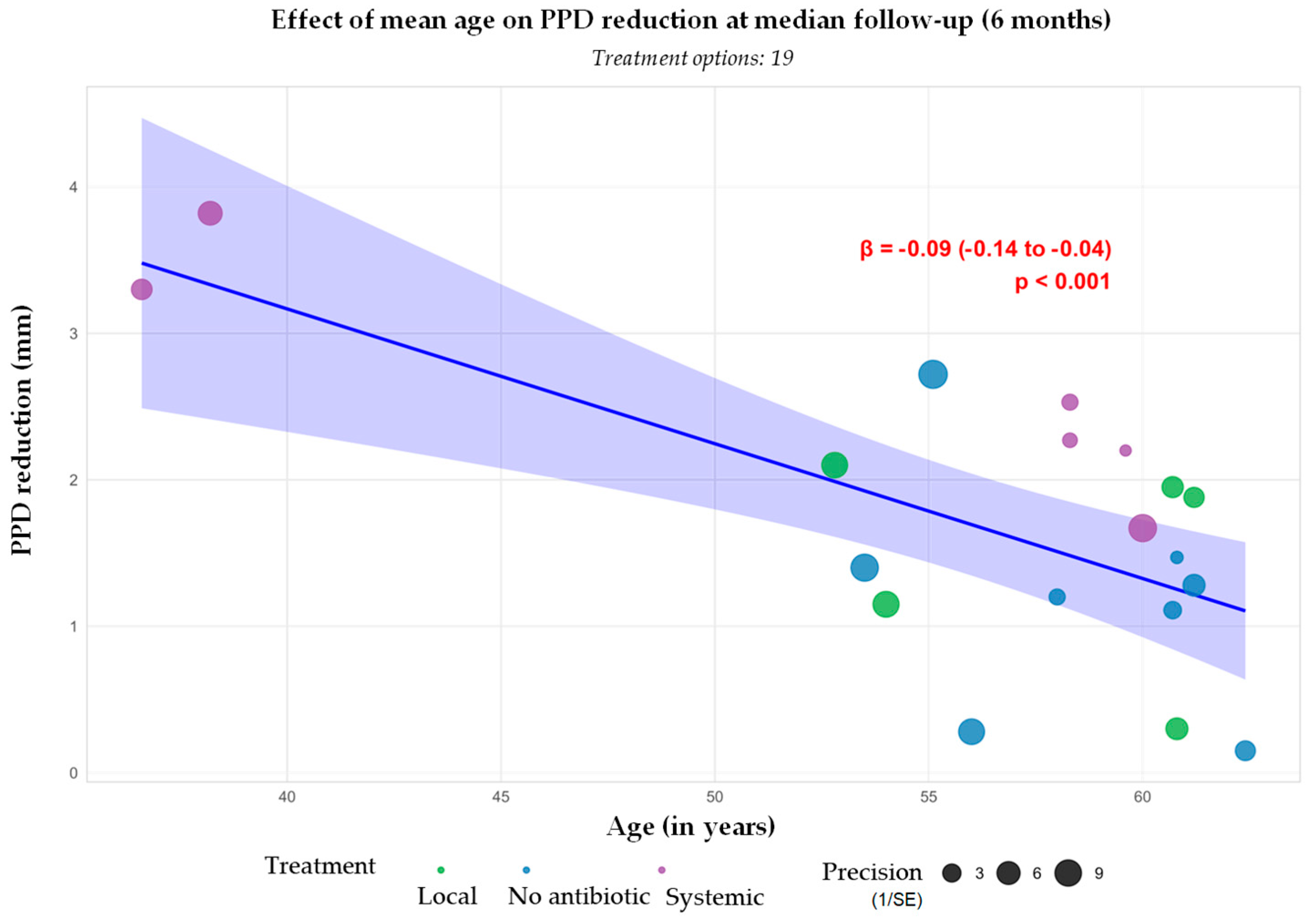
Meta-regression analysis revealed a statistically significant inverse association between mean patient age and PPD reduction (β = −0.09; 95% CI: −0.14 to −0.04; p < 0.001), as the regression line depicts a gradual downward trend, with estimated reductions declining from ≈3.5 mm at age 40 to ≈1.7 mm at age 60 (Figure 3). This age-related difference of nearly 1.8 mm is both statistically and clinically relevant. The visual weighting of each data point, indicated by bubble size, reflects the relative precision of the studies, reinforcing the strength and consistency of the observed trend across varying sample sizes. These findings highlight age as a potential effect modifier in treatment response and suggest the need for age-stratified approaches in both clinical trial design and individual patient management.
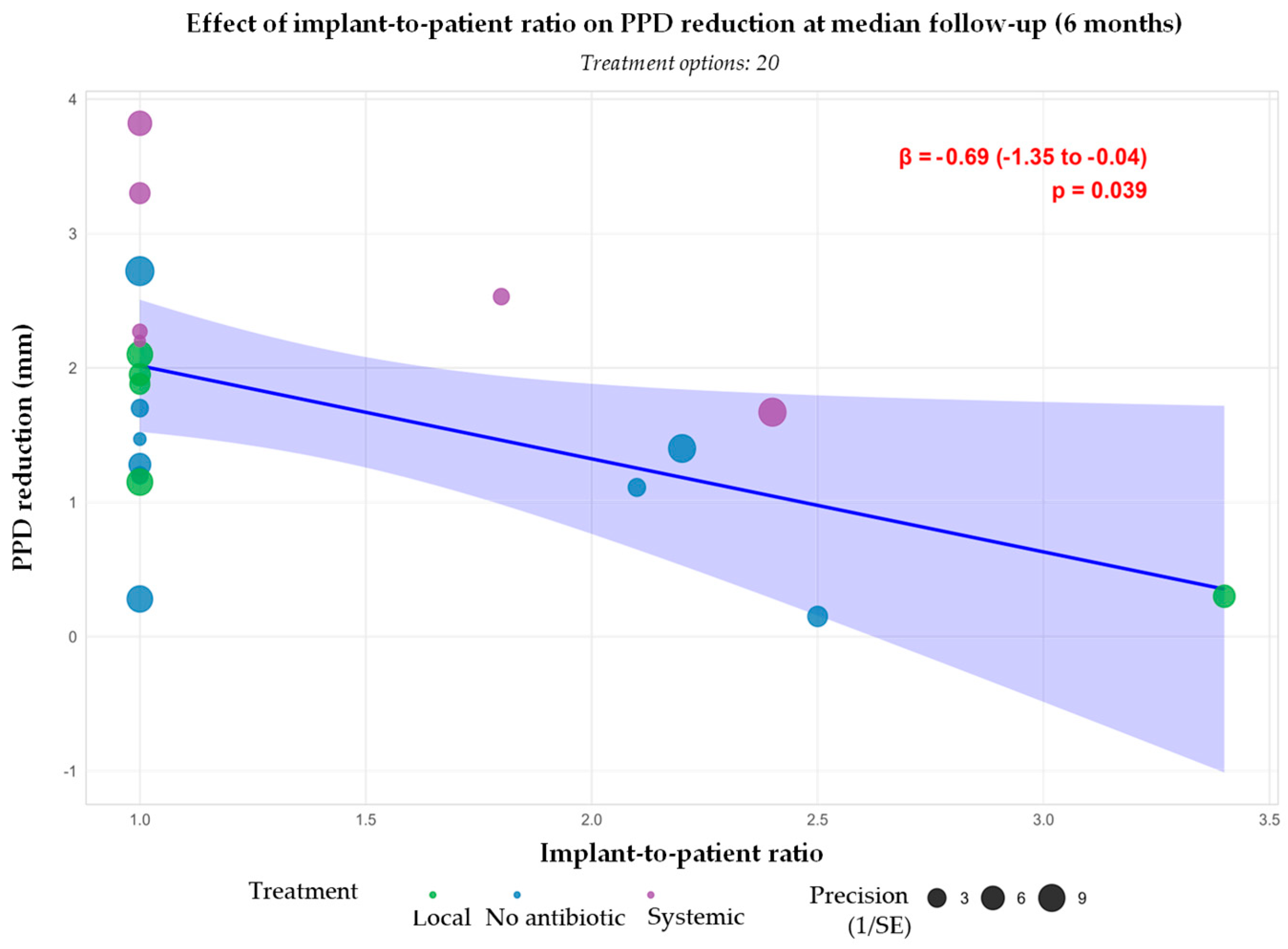
As shown in Figure 4, the meta-regression suggests a significant inverse association between implant-to-patient ratio and PPD reduction, with a higher implant-to-patient ratio associated with decreased PPD reduction (β = −0.69; 95% CI: −1.35 to −0.04; p = 0.039). Clinically, this corresponds to a decrease in PPD reduction from ≈2.0 mm at a balanced implant-to-patient ratio (1 implant/patient) to ≈0.3 mm at a higher implant-to-patient ratio (3.5 implants/patient). This trend may indicate that patients with more implants experience a less pronounced clinical benefit from non-surgical therapy. Nevertheless, this observed association should be interpreted with caution. The analysis is based on aggregated study-level data, which limits the ability to fully account for confounding factors. While the direction and strength of the effect are consistent with a plausible biological gradient, the underlying studies were not designed to compare different implant-to-patient ratios directly.
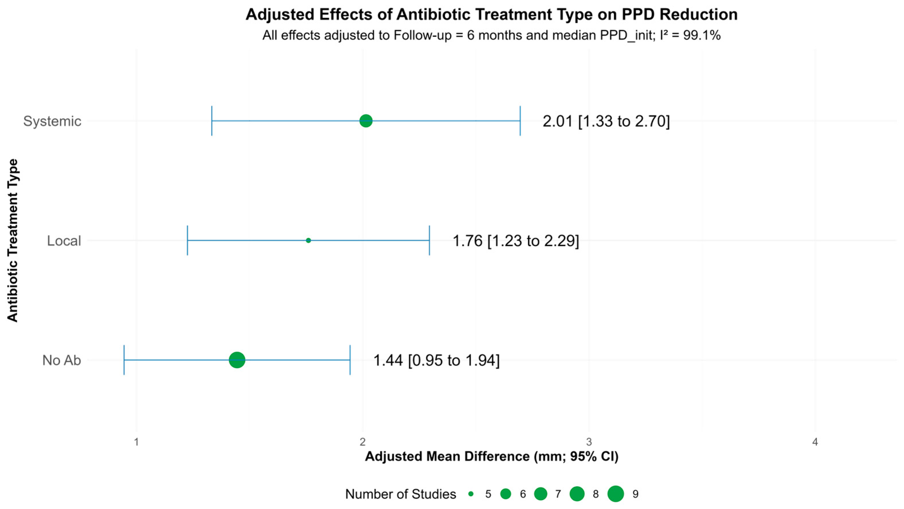
On average, systemic antibiotics demonstrated the greatest PPD reduction (≈2.0 mm), followed by local antibiotics, with the antibiotic absent group (MD ± CHX) showing lower reductions (≈1.4 mm) (Figure 5). While there is visible overlap in the confidence intervals across treatment types, indicating uncertainty, the trend favours the use of systemic antibiotics for possible enhanced clinical benefit in the short term.

3.3.2. Bleeding on Probing Reduction

Eight studies reported BoP reductions, with test arm reductions ranging from 19.1 ± 31.7% [30] to 69.4 ± 26.4% [35] (Table S1). As previously mentioned, Renvert et al. (2006) [37] was excluded from the meta-analysis. Since Liñares et al. (2024) [34] lacked BoP-associated uncertainty, this resulted in a total of six studies contributing to the BoP meta-analysis results. Figure 6 shows the forest plot graph for adjusted treatment effects at the six-month follow-up.
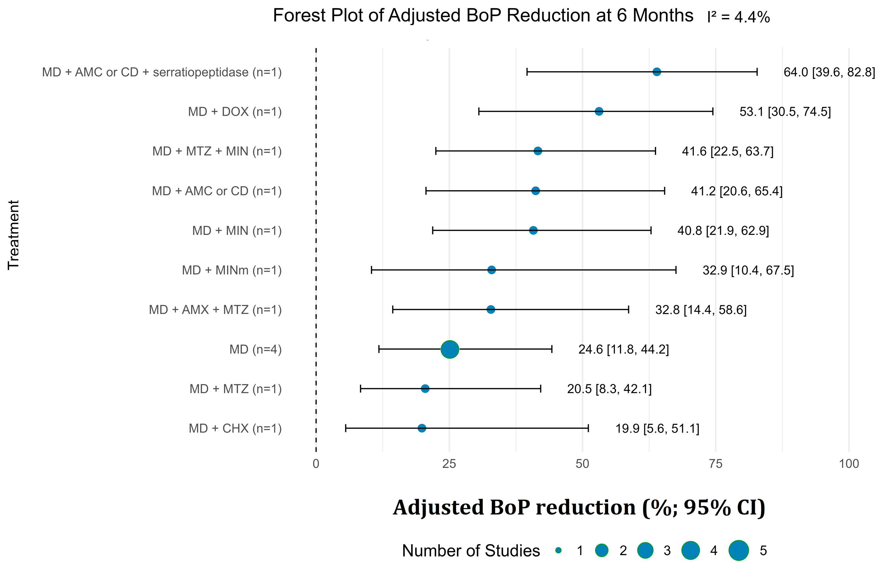
MD alone achieved 24.6% BoP reduction (95% CI: 11.8–42.4%). Adjunctive antimicrobials with MD enhanced outcomes: MD + AMC or CD + serratiopeptidase yielded the highest reduction (64.0%; 95% CI: 39.6–82.8%), followed by MD + DOX (53.1%; 95% CI: 30.5–74.5%), MD + MTZ + MIN (41.6%; 95% CI: 22.5–63.7%), MD + AMC or CD (41.2%; 95% CI: 20.6% to 65.4%), and MD + MIN or MINm produced improvements in the range of approximately 33% to 40%. Although these findings suggest that adjunctive antimicrobial therapy enhances BoP reduction relative to MD alone, several comparisons are based on single study estimates with wide confidence intervals. These limitations, alongside potential variability in study populations and intervention protocols, reduce the certainty of the evidence. Nonetheless, the direction and consistency of effects, reflected by low between-study heterogeneity (I2 = 4.4%), support a likely additive benefit of antimicrobial adjuncts, particularly those involving broad-spectrum agents active against anaerobic biofilms.
Meta-regression analysis illustrated in Figure 7 did not identify a significant association between mean patient age and BoP reduction at 6 months (β = −0.77; 95% CI: −1.97 to 0.43; p = 0.207). Although the trend suggests a potential decrease in BoP reduction with increasing age, the wide confidence interval crossing the null and the non-significant p-value indicate that this relationship is not robust. The precision of the included studies was variable, as reflected by bubble sizes, and the overall number of treatment arms was limited (n = 9), further restricting the strength of inference. These findings suggest that, within the available evidence, age does not appear to be a strong or consistent effect modifier of clinical response in terms of BoP reduction.
The meta-regression shown in Figure 8 did not reveal a significant association between implant-to-patient ratio and BoP reduction (β = −7.04; 95% CI: −27.39 to 13.31; p = 0.498). Although the regression line suggests a modest negative slope—indicating a potential decrease in BoP reduction with increasing implant load per patient—this trend is not supported by evidence, as the wide confidence interval crosses the null and encompasses both clinically relevant benefits and harms. The lack of significance and the high degree of uncertainty likely reflect the small number of treatment arms (n = 10) and variation in study precision.
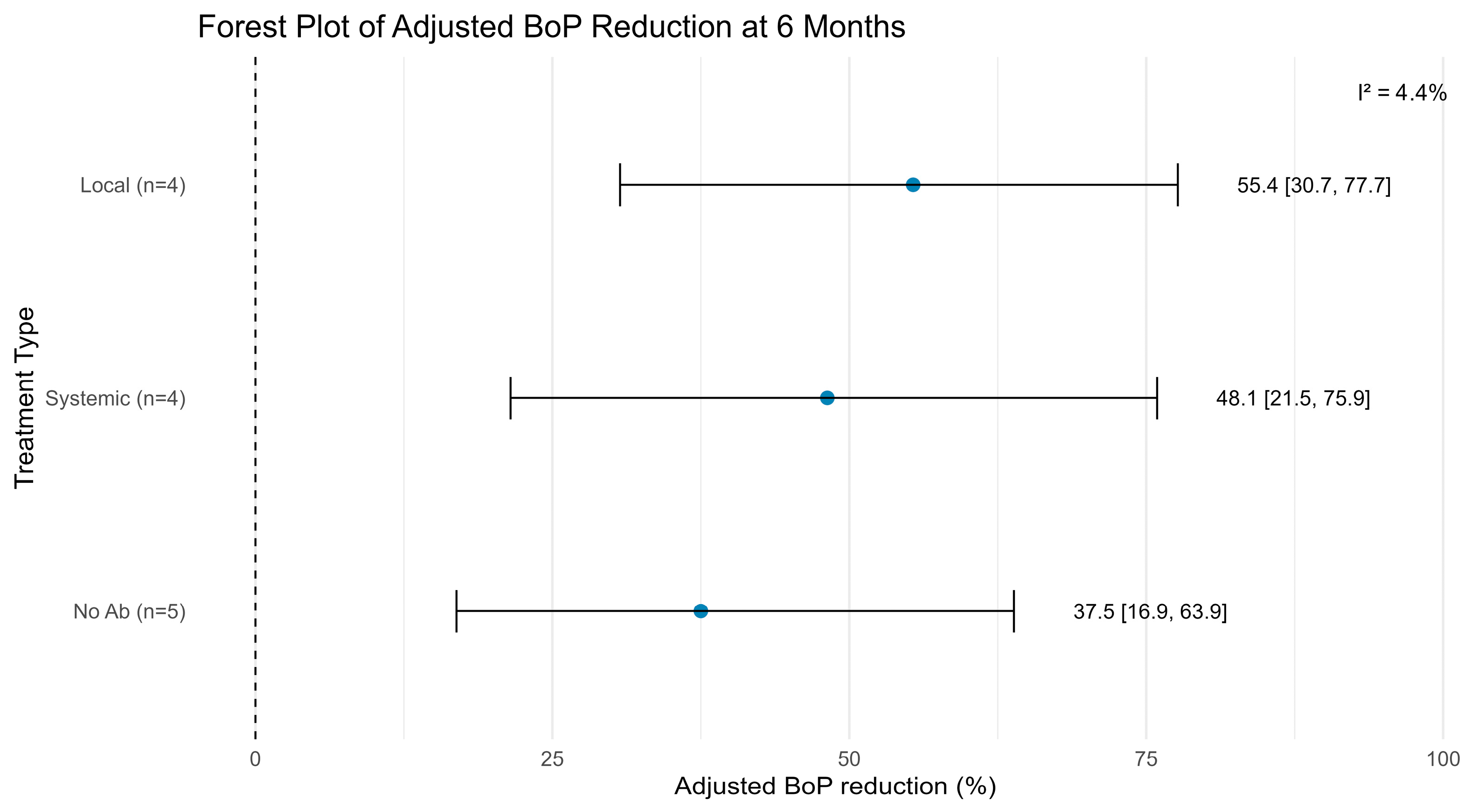
As can be seen in Figure 9, local antibiotics achieved the greatest mean reduction in BoP, followed by systemic antibiotics and then no antibiotics. Overlapping confidence intervals indicate uncertainty regarding the superiority of local versus systemic approaches. The consistency in direction of effect across studies, particularly within the antibiotic groups, supports the likelihood of a true treatment-related benefit. However, given the moderate between-arm variability and the limited number of studies per group, caution is warranted in interpreting the magnitude of difference between treatment types.
This finding appears paradoxical compared to the greater absolute PPD reductions observed in deeper pockets, suggesting that while patients with severe initial disease may achieve larger absolute improvements, they are less likely to reach predefined success criteria. This relationship likely reflects the challenge of achieving complete resolution in severely compromised sites, where substantial improvements may still fall short of clinical success thresholds.

3.4. Secondary Outcomes and Success Rates

Only three studies provided depth of bone lesion (DBL), with reductions up to 1.96 mm [35]. Five studies assessed treatment success, with none achieving 100% success rates; however, definitions varied across studies (Table S1). Notably, no implant losses occurred during follow-up periods. Adverse events data were sparse and could not be pooled.

3.5. Risk of Bias Results

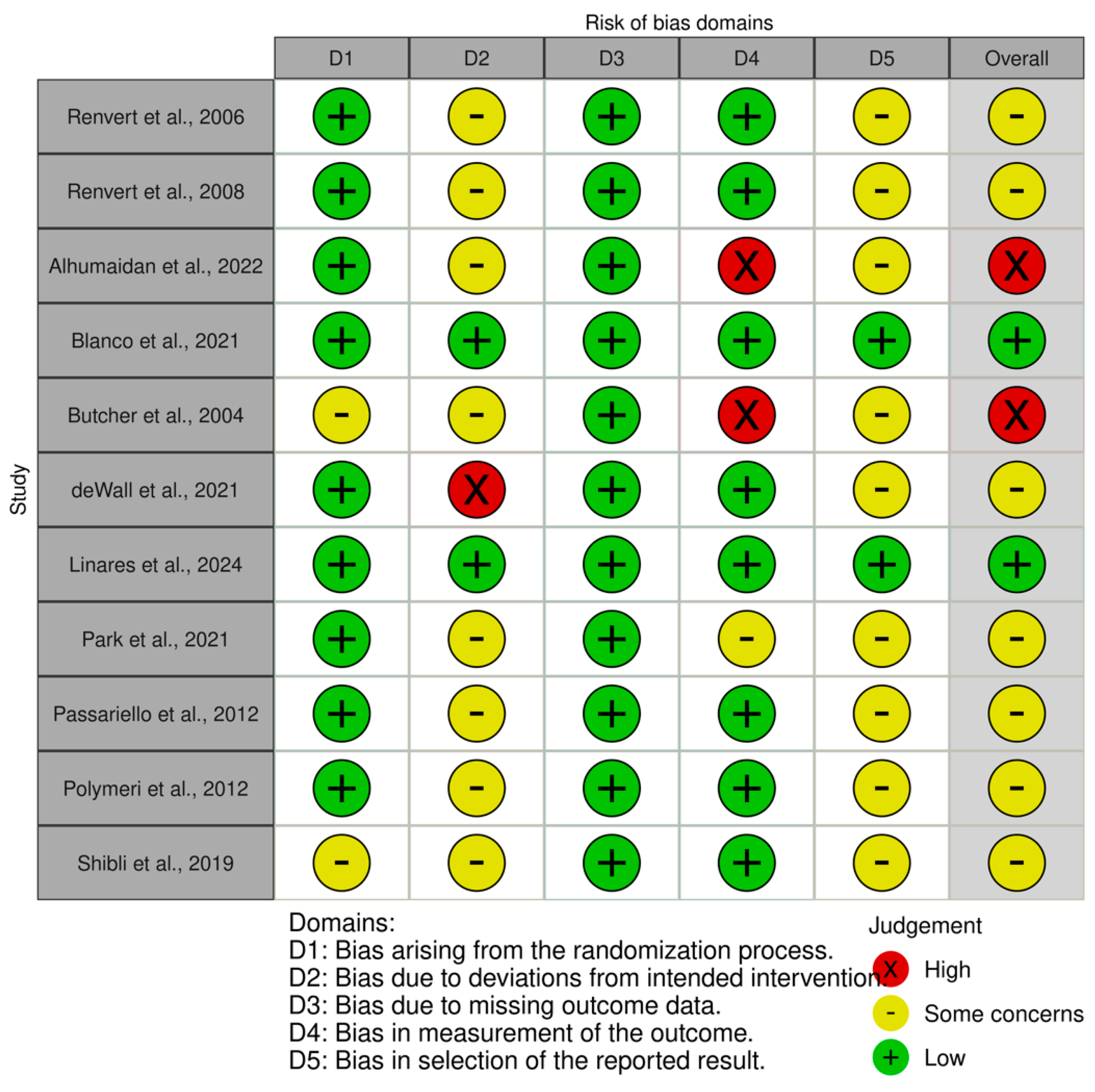
Among the 11 included studies, 3 were considered as having domains with a high risk of bias [29,31,32], 7 were considered as having some concerns overall [32,34,35,36,37,38,39], and 2 were rated as having a low risk of bias [30,33]. The results were presented in two ways: traffic light plot (Figure 10) and summary plot (Figure 11).

3.6. Summary of Evidence

Across 11 RCTs, MD alone produced modest but statistically significant clinical improvements, whereas adjunctive therapies—particularly systemic antibiotics—resulted in substantially greater reductions in PPD and BoP, as well as higher overall success rates. Systemic antibiotic regimens, especially amoxicillin-based combinations with serratiopeptidase, demonstrated the most consistent and pronounced effects, with a mean PPD reduction of 3.8 mm (95% CI: 2.09–5.55) and BoP reduction of 69% (95% CI: 37.7–101.1). The addition of proteolytic enzymes to systemic antibiotics provided the greatest clinical benefit, whereas locally administered antimicrobials conferred only limited adjunctive effects. Trials with longer follow-up durations (6–12 months) generally reported more favourable outcomes, and treatment response appeared to be influenced by patient-related factors such as smoking status and implant duration.
Despite improvements in surrogate clinical parameters, complete disease resolution (as variably defined across studies) was not consistently enhanced by adjunctive therapy. Meta-regression analyses revealed that younger patients and those presenting with deeper baseline pockets achieved significantly greater reductions in PPD, while individuals with a higher number of implants per patient exhibited smaller improvements. Although greater baseline disease severity was associated with larger absolute PPD reductions, these patients were less likely to achieve complete resolution. None of these factors significantly influenced BoP reduction or overall treatment success.

4. Discussion

This systematic review included 11 randomised controlled trials (RCTs) involving 634 patients, all evaluating non-surgical treatment of peri-implantitis. Unlike earlier reviews that grouped diverse adjunctive therapies, our analysis focused exclusively on antibiotics and introduced key methodological enhancements. These included the addition of several new RCTs published since 2022 (e.g., Liñares et al., 2024 [33]), standardisation to a 6-month follow-up, and meta-regressions exploring patient- and treatment-related modifiers—approaches not employed in prior analyses.
This focused strategy enabled a more refined understanding of treatment efficacy and potential effect modifiers such as age. Our findings align with previous systematic reviews [17,40], confirming that adjunctive systemic antibiotics lead to significantly greater short-term reductions in PPD and BoP compared to MD alone. In contrast, local antimicrobial adjuncts demonstrated more limited benefit, particularly for PPD reduction, corroborating earlier results for both peri-implantitis and peri-implant mucositis [17,41,42]. While Barbato et al. (2023) [17] also found systemic antibiotics effective for PPD reduction, a 2022 review by Grusovin et al. [43] paradoxically reported greater effects with local antibiotics. This discrepancy may be due to the latter’s smaller sample sizes, shorter follow-up periods, and differing definitions of clinical success. Our updated analysis, with a larger evidence base, supports the greater efficacy of systemic antibiotics, particularly amoxicillin-based regimens for PPD reduction.
While our review demonstrates the efficacy of antimicrobial adjuncts, it is essential to contextualise these findings within the broader spectrum of non-surgical peri-implantitis management. Several protocols have been described in the literature; however, there is no universal consensus on the most efficient approach [44,45]. Non-surgical therapy is fundamentally based on mechanical debridement and local disinfection, though the implant’s rough surface structure may provide bacteria with areas inaccessible to conventional mechanical removal [45]. Despite this challenge, alternative strategies merit consideration.
Crespi et al. (2019) [45] demonstrated that mechanical removal of bacterial biofilm, chemical detoxification of implant surfaces with chlorhexidine and tetracycline irrigation, and maintenance of granulation tissues in pockets provided better clinical outcomes than mechanical debridement alone over 36 months. Their findings suggest that surgical removal of granulation tissue may eliminate pluripotent stem cells that could potentially contribute to tissue healing. Notably, their protocol achieved stable outcomes without systemic antibiotics, emphasising that local adjunctive delivery of antiseptic/antibiotic irrigations can be effective when combined with meticulous mechanical debridement [45].
Beyond pharmacological interventions, patient compliance and behavioural modifications play critical roles in peri-implant disease management. Cosola et al. (2022) [46] demonstrated that full-mouth decontamination paired with hypochlorite-based home brushing (PerioTabs®) significantly improved clinical parameters around dental implants, with peri-implant mucositis stabilised without adjunctive surgery after motivation, domestic treatment, and supportive non-surgical therapy. Their results indicated that all clinical parameters (modified plaque index and modified bleeding index) significantly improved after only 1 week of domestic care, with continued improvement following non-surgical periodontal therapy [46]. This underscores that resolution of peri-implant inflammation is not solely dependent on antibiotics but can be achieved through behaviourally oriented interventions and chemical plaque control.
These studies collectively demonstrate that optimal outcomes in peri-implantitis management may require a combination of mechanical, pharmacological, and patient-driven strategies rather than reliance on any single intervention. The choice of treatment should be individualised based on disease severity, patient factors, and the broader context of antimicrobial stewardship.
A consistent observation across included studies was the absence of implant loss during the 3–12-month follow-up periods, which may reflect favourable short-term outcomes. Nonetheless, implant survival alone does not equate to peri-implant health. Clinical success should also consider inflammation resolution, pocket depth, BoP, and bone stability. Therefore, the lack of implant loss must be interpreted cautiously and within a broader clinical context.
Reductions in PPD were observed in all studies following non-surgical debridement, reinforcing its baseline effectiveness. However, variability in baseline disease severity, operator technique, and outcome definitions likely contributed to the observed heterogeneity.
Meta-analyses revealed that systemic antibiotic adjuncts resulted in an average of 1.0–1.5 mm greater PPD reduction compared to MD alone, with BoP reduced by an additional 20–30 percentage points. Notably, combinations such as MD with amoxicillin and clavulanic acid (or clindamycin and serratiopeptidase) produced the most pronounced improvements, approximately 3.8 mm reduction in PPD (95% CI: 2.09 to 5.55) and a 69% absolute decrease in BoP (95% CI: 37.7 to 101.1). These findings highlight the added clinical benefit of systemic adjunctive therapy.
Meta-regression analyses also suggested that younger patients experienced significantly greater PPD reductions, while those with a higher implant-to-patient ratio showed smaller improvements. These factors did not appear to significantly influence BoP reduction. No consistent differences were observed between antibiotic types, though the strongest effects were seen with amoxicillin-based combinations.
Despite these promising results, the routine use of systemic antibiotics remains controversial due to concerns about adverse effects and the emergence of antimicrobial resistance [17]. The overuse of antibiotics poses a significant global public health risk, particularly through the development of antibiotic resistance [47]. Moreover, systemic antibiotic regimens have demonstrated long-lasting effects on the intestinal microbiome, including an increase in genes associated with antimicrobial resistance [47]. This underscores a tension between statistical significance and clinical prudence that extends beyond individual patient outcomes to encompass broader public health implications. Any benefit versus harm analysis must therefore consider not only the individual patient but also the collective impact of antibiotic use on antimicrobial stewardship and resistance patterns [47]. Although efficacy is supported by pooled data, most authors were cautious in recommending widespread use, reflecting awareness of broader issues such as antimicrobial stewardship, patient safety, and resistance patterns.
The findings of Crespi et al. (2019) [45] and Cosola et al. (2022) [46] reinforce that effective peri-implantitis management can be achieved through alternative approaches that minimise or avoid systemic antibiotic use. Given that bacteria on implant surfaces may express more virulent and resistant profiles in ecological niches inaccessible to conventional removal, local antiseptic delivery combined with patient education and behavioural modification may offer a more sustainable approach for selected cases [45,46].
Ultimately, while both local and systemic antibiotics may serve as useful adjuncts in peri-implantitis therapy, achieving long-term success remains challenging. Prudent antibiotic use, individual patient assessment, consideration of alternative mechanical and behavioural strategies, and an improved understanding of subgingival microbiota are essential for responsible clinical decision-making.
The certainty of the evidence is limited by heterogeneity in treatment protocols, outcome definitions, and study quality. Few trials evaluated specific antibiotic regimens in sufficient detail to support robust subgroup analysis. Additionally, short follow-up periods and methodological limitations restrict the ability to assess long-term efficacy. Inconsistent definitions of treatment success further limit comparability across studies.
Importantly, adverse effects and resistance risks were not systematically addressed in the included trials. Consequently, despite favourable short-term outcomes, the overall certainty of the evidence is moderate, and findings should be interpreted with caution. Further high-quality, long-term RCTs are needed to confirm sustained benefits and inform clinical practice, particularly studies that compare antibiotic adjuncts with alternative mechanical and behavioural approaches.

5. Conclusions

This systematic review and meta-analysis found that adjunctive systemic antibiotics significantly improved short-term clinical outcomes of non-surgical peri-implantitis treatment compared with mechanical debridement alone. Patients receiving antibiotics, particularly amoxicillin-based regimens, demonstrated greater reductions in peri-implant probing depth and bleeding on probing, suggesting improved resolution of inflammation. These findings support the potential benefit of systemic antibiotic therapy as an adjunct to mechanical debridement in the management of peri-implantitis.
In summary, adjunctive systemic antibiotics (particularly amoxicillin-based) provide a clear short-term benefit in peri-implantitis non-surgical therapy compared to debridement alone. However, considering rising antimicrobial resistance and the global imperative for antibiotic stewardship, clinicians should balance these benefits with potential risks (adverse effects and resistance) and exercise prudence in routine use. Treatment decisions should be individualised based on disease severity, patient-specific risk factors, previous treatment responses, and local resistance patterns. Long-term efficacy and safety remain to be confirmed; thus, further well-designed RCTs with extended follow-up are needed. Future studies should also standardise outcome definitions (especially clinical success) to allow better comparison and application of findings and should include comparative evaluations of antibiotic versus non-antibiotic approaches to guide evidence-based, individualised clinical decision-making.

Supplementary Materials

The following supporting information can be downloaded at: https://www.mdpi.com/article/10.3390/app152111422/s1, Table S1. Qualitative summary data from selected articles (to be continued). Table S2. Summary data from selected articles for Meta-analysis.

Author Contributions

Conceptualization, M.M., A.S., C.E. and P.M.; methodology, M.M., A.S. and P.M.; software, M.M. and P.M.; validation, M.M., A.S. and P.M.; formal analysis, M.M. and P.M.; investigation, M.M.; data curation, M.M. and P.M.; writing—original draft preparation, M.M., A.S., C.E. and P.M.; writing—review and editing, M.M., A.S. and P.M.; visualisation, M.M.; supervision, A.S. and P.M.; project administration, M.M., A.S. and P.M.; funding acquisition, M.M., A.S. and P.M. All authors have read and agreed to the published version of the manuscript.

Funding

The review did not receive any specific external funding.

Data Availability Statement

Not applicable.

Acknowledgments

The authors thank FCT/MCTES for the financial support to CiiEM (10.54499/UIDB/04585/2020) through national funds.

Conflicts of Interest

The authors declare no conflicts of interest.

Glossary

AMXAmoxicillin
AMCAmoxicillin–clavulanic acid
BIDBis in die (twice daily)
BoPBleeding on probing
CDClindamycin
CHXChlorhexidine
CIConfidence interval
CRPC-reactive protein
DBLDepth of bone lesion
DOXDoxycycline
IL-6Interleukin-6
I2I-squared statistic
MDMechanical debridement
MHMinocycline hydrochloride
MINMinocycline
MINmMinocycline microspheres
MTZMetronidazole
PPDProbing pocket depth
PRISMA 2020Preferred Reporting Items for Systematic Reviews and Meta-Analyses 2020
PROSPEROInternational Prospective Register of Systematic Reviews
RCTRandomised controlled trial
RoB 2.0Cochrane Risk of Bias 2.0 tool
SEStandard error
SerratiopeptidaseProteolytic enzyme with anti-inflammatory activity
TIDTer in die (three times daily)
TNF-αTumour necrosis factor-alpha
t2Between-study variance
βRegression coefficient
p-valueProbability value

References

  1. Simonis, P.; Dufour, T.; Tenenbaum, H. Long-term implant survival and success: A 10–16-year follow-up of non-submerged dental implants. Clin. Oral Implants Res. 2010, 21, 772–777. [Google Scholar] [CrossRef]
  2. Smeets, R.; Henningsen, A.; Jung, O.; Heiland, M.; Hammächer, C.; Stein, J.M. Definition, etiology, prevention and treatment of peri-implantitis—a review. Head Face Med. 2014, 10, 34. [Google Scholar] [CrossRef]
  3. Herrera, D.; Berglundh, T.; Schwarz, F.; Chapple, I.; Jepsen, S.; Sculean, A.; Kebschull, M.; Papapanou, P.N.; Tonetti, M.S.; Sanz, M.; et al. Prevention and treatment of peri-implant diseases—The EFP S3 level clinical practice guideline. J. Clin. Periodontol. 2023, 50 (Suppl. S26), 4–76. [Google Scholar] [CrossRef]
  4. Heitz-Mayfield, L.J.A.; Salvi, G.E. Peri-implant mucositis. J. Clin. Periodontol. 2018, 45 (Suppl. S20), S237–S245. [Google Scholar] [CrossRef]
  5. Berglundh, T.; Armitage, G.; Araujo, M.G.; Avila-Ortiz, G.; Blanco, J.; Camargo, P.M.; Chen, S.; Cochran, D.; Derks, J.; Figuero, E.; et al. Peri-implant diseases and conditions: Consensus report of workgroup 4 of the 2017 World Workshop on the Classification of Periodontal and Peri-Implant Diseases and Conditions. J. Clin. Periodontol. 2018, 45 (Suppl. S20), S286–S291. [Google Scholar] [CrossRef] [PubMed]
  6. Mombelli, A.; Van Oosten, M.A.C.; Schürch, E.; Lang, N.P. The microbiota associated with successful or failing osseointegrated titanium implants. Oral Microbiol. Immunol. 1987, 2, 145–151. [Google Scholar] [CrossRef] [PubMed]
  7. Levignac, J. [Periimplantation osteolysis- periimplantosis—periimplantitis]. Rev. Fr. Odontostomatol. 1965, 12, 1251–1260. [Google Scholar] [PubMed]
  8. Albrektsson, T.; Isidor, F. Consensus Report of Session IV. In Proceedings of the First European Workshp on Periodontology; Lang, N.P., Karring, T., Eds.; Quintessence Publishing: London, UK, 1994; pp. 365–369. [Google Scholar]
  9. Uppala, S.; Parihar, A.; Modipalle, V.; Manual, L.; Oommen, V.; Karadiguddi, P.; Gupta, P. Crestal bone loss around dental implants after implantation of Tricalcium phosphate and Platelet- Rich Plasma: A comparative study. J. Fam. Med. Prim. Care 2020, 9, 229. [Google Scholar] [CrossRef]
  10. Bermejo, P.; Sánchez, M.C.; Llama-Palacios, A.; Figuero, E.; Herrera, D.; Sanz Alonso, M. Biofilm formation on dental implants with different surface micro-topography: An in vitro study. Clin. Oral Implants Res. 2019, 30, 725–734. [Google Scholar] [CrossRef]
  11. Schwarz, F.; Derks, J.; Monje, A.; Wang, H. Peri-implantitis. J. Clin. Periodontol. 2018, 45 (Suppl. S20), S246–S266. [Google Scholar] [CrossRef]
  12. Prathapachandran, J.; Suresh, N. Management of peri-implantitis. Dent. Res. J. 2012, 9, 516. [Google Scholar] [CrossRef] [PubMed]
  13. Mellado-Valero, A.; Buitrago-Vera, P.; Sola-Ruiz, M.; Ferrer-Garcia, J. Decontamination of dental implant surface in peri-implantitis treatment: A literature review. Med. Oral Patol. Oral Cirugia Bucal 2013, 18, e869–e876. [Google Scholar] [CrossRef]
  14. Renvert, S.; Samuelsson, E.; Lindahl, C.; Persson, G.R. Mechanical non-surgical treatment of peri-implantitis: A double-blind randomized longitudinal clinical study. I: Clinical results. J. Clin. Periodontol. 2009, 36, 604–609. [Google Scholar] [CrossRef]
  15. Schwarz, F.; Sculean, A.; Rothamel, D.; Schwenzer, K.; Georg, T.; Becker, J. Clinical evaluation of an Er:YAG laser for nonsurgical treatment of peri-implantitis: A pilot study. Clin. Oral Implants Res. 2005, 16, 44–52. [Google Scholar] [CrossRef]
  16. Sahm, N.; Becker, J.; Santel, T.; Schwarz, F. Non-surgical treatment of peri-implantitis using an air-abrasive device or mechanical debridement and local application of chlorhexidine: A prospective, randomized, controlled clinical study: Non-surgical therapy of peri-implantitis. J. Clin. Periodontol. 2011, 38, 872–878. [Google Scholar] [CrossRef]
  17. Barbato, L.; Cavalcanti, R.; Rupe, C.; Scartabelli, D.; Serni, L.; Chambrone, L.; Cairo, F. Clinical efficacy of adjunctive methods for the non-surgical treatment of peri-implantitis: A systematic review and meta-analysis. BMC Oral Health 2023, 23, 375. [Google Scholar] [CrossRef] [PubMed]
  18. Barootchi, S.; Wang, H.-L. Peri-implant diseases: Current understanding and management. Int. J. Oral Implantol. Berl. Ger. 2021, 14, 263–282. [Google Scholar]
  19. Heitz-Mayfield, L.J.A.; Salvi, G.E.; Botticelli, D.; Mombelli, A.; Faddy, M.; Lang, N.P.; On Behalf of the Implant Complication Research Group (ICRG). Anti-infective treatment of peri-implant mucositis: A randomised controlled clinical trial: Treatment of peri-implant mucositis. Clin. Oral Implants Res. 2011, 22, 237–241. [Google Scholar] [CrossRef]
  20. Wong, R.L.; Hiyari, S.; Yaghsezian, A.; Davar, M.; Lin, Y.-L.; Galvan, M.; Tetradis, S.; Camargo, P.M.; Pirih, F.Q. Comparing the Healing Potential of Late-Stage Periodontitis and Peri-Implantitis. J. Oral Implantol. 2017, 43, 437–445. [Google Scholar] [CrossRef]
  21. Schwarz, F.; Nuesry, E.; Bieling, K.; Herten, M.; Becker, J. Influence of an Erbium, Chromium-Doped Yttrium, Scandium, Gallium, and Garnet (Er,Cr:YSGG) Laser on the Reestablishment of the Biocompatibility of Contaminated Titanium Implant Surfaces. J. Periodontol. 2006, 77, 1820–1827. [Google Scholar] [CrossRef] [PubMed]
  22. Romanos, G.E.; Nentwig, G.H. Regenerative therapy of deep peri-implant infrabony defects after CO2 laser implant surface decontamination. Int. J. Periodontics Restorative Dent. 2008, 28, 245–255. [Google Scholar] [PubMed]
  23. Faggion, C.M.; Listl, S.; Frühauf, N.; Chang, H.; Tu, Y. A systematic review and Bayesian network meta-analysis of randomized clinical trials on non-surgical treatments for peri-implantitis. J. Clin. Periodontol. 2014, 41, 1015–1025. [Google Scholar] [CrossRef] [PubMed]
  24. Mombelli, A.; Lang, N.P. Antimicrobial treatment of peri-implant infections. Clin. Oral Implants Res. 1992, 3, 162–168. [Google Scholar] [CrossRef]
  25. Sukumar, S.; Martin, F.; Hughes, T.; Adler, C. Think before you prescribe: How dentistry contributes to antibiotic resistance. Aust. Dent. J. 2020, 65, 21–29. [Google Scholar] [CrossRef]
  26. van Winkelhoff, A.J. Antibiotics in the treatment of peri-implantitis. Eur. J. Oral Implantol. 2012, 5, S43–S50. [Google Scholar]
  27. Busa, A.; Parrini, S.; Chisci, G.; Pozzi, T.; Burgassi, S.; Capuano, A. Local Versus Systemic Antibiotics Effectiveness: A Comparative Study of Postoperative Oral Disability in Lower Third Molar Surgery. J. Craniofac. Surg. 2014, 25, 708–709. [Google Scholar] [CrossRef] [PubMed]
  28. Page, M.J.; McKenzie, J.E.; Bossuyt, P.M.; Boutron, I.; Hoffmann, T.C.; Mulrow, C.D.; Shamseer, L.; Tetzlaff, J.M.; Akl, E.A.; Brennan, S.E.; et al. The PRISMA 2020 statement: An updated guideline for reporting systematic reviews. BMJ 2021, 372, n71. [Google Scholar] [CrossRef]
  29. Alhumaidan, A.A.; Alrabiah, M.; Al-Aali, K.A.; Javed, F.; Vohra, F.; Abduljabbar, T. Efficacy of adjunct subgingival minocycline delivery for treatment of peri-implantitis in moderate cigarette smokers. Eur. Rev. Med. Pharmacol. Sci. 2022, 26, 5698–5705. [Google Scholar] [CrossRef]
  30. Blanco, C.; Pico, A.; Dopico, J.; Gándara, P.; Blanco, J.; Liñares, A. Adjunctive benefits of systemic metronidazole on non-surgical treatment of peri-implantitis. A randomized placebo-controlled clinical trial. J. Clin. Periodontol. 2021, 49, 15–27. [Google Scholar] [CrossRef]
  31. Büchter, A.; Meyer, U.; Kruse-Lösler, B.; Joos, U.; Kleinheinz, J. Sustained release of doxycycline for the treatment of peri-implantitis: Randomised controlled trial. Br. J. Oral Maxillofac. Surg. 2004, 42, 439–444. [Google Scholar] [CrossRef]
  32. De Waal, Y.C.M.; Vangsted, T.E.; Van Winkelhoff, A.J. Systemic antibiotic therapy as an adjunct to non-surgical peri-implantitis treatment: A single-blind RCT. J. Clin. Periodontol. 2021, 48, 996–1006. [Google Scholar] [CrossRef]
  33. Liñares, A.; Dopico, J.; Blanco, C.; Pico, A.; Sobrino, T.; Blanco, J.; Leira, Y. The systemic impact of non-surgical treatment of peri-implantitis with or without adjunctive systemic metronidazole: Secondary analysis of a randomized clinical trial. Clin. Oral Implants Res. 2024, 35, 1519–1530. [Google Scholar] [CrossRef] [PubMed]
  34. Park, S.; Song, Y.W.; Cha, J.; Lee, J.; Kim, Y.; Shin, H.; Lee, D.; Lee, J.H.; Kim, C. Adjunctive use of metronidazole-minocycline ointment in the nonsurgical treatment of peri-implantitis: A multicenter randomized controlled trial. Clin. Implant Dent. Relat. Res. 2021, 23, 543–554. [Google Scholar] [CrossRef] [PubMed]
  35. Passariello, C.; Lucchese, A.; Pera, F.; Gigola, P. Clinical, Microbiological and Inflammatory Evidence of the Efficacy of Combination Therapy Including Serratiopeptidase in the Treatment of Periimplantitis. Eur. J. Inflamm. 2012, 10, 463–472. [Google Scholar] [CrossRef]
  36. Polymeri, A.; Van Der Horst, J.; Anssari Moin, D.; Wismeijer, D.; Loos, B.G.; Laine, M.L. Non-surgical peri-implantitis treatment with or without systemic antibiotics: A randomized controlled clinical trial. Clin. Oral Implants Res. 2022, 33, 548–557. [Google Scholar] [CrossRef]
  37. Renvert, S.; Lessem, J.; Dahlén, G.; Lindahl, C.; Svensson, M. Topical minocycline microspheres versus topical chlorhexidine gel as an adjunct to mechanical debridement of incipient peri-implant infections: A randomized clinical trial. J. Clin. Periodontol. 2006, 33, 362–369. [Google Scholar] [CrossRef]
  38. Renvert, S.; Lessem, J.; Dahlén, G.; Renvert, H.; Lindahl, C. Mechanical and Repeated Antimicrobial Therapy Using a Local Drug Delivery System in the Treatment of Peri-Implantitis: A Randomized Clinical Trial. J. Periodontol. 2008, 79, 836–844. [Google Scholar] [CrossRef]
  39. Shibli, J.A.; Ferrari, D.S.; Siroma, R.S.; Figueiredo, L.C.D.; Faveri, M.D.; Feres, M. Microbiological and clinical effects of adjunctive systemic metronidazole and amoxicillin in the non-surgical treatment of peri-implantitis: 1 year follow-up. Braz. Oral Res. 2019, 33 (Suppl. S1), e080. [Google Scholar] [CrossRef]
  40. Ramanauskaite, A.; Fretwurst, T.; Schwarz, F. Efficacy of alternative or adjunctive measures to conventional non-surgical and surgical treatment of peri-implant mucositis and peri-implantitis: A systematic review and meta-analysis. Int. J. Implant Dent. 2021, 7, 112. [Google Scholar] [CrossRef]
  41. Toledano-Osorio, M.; Vallecillo, C.; Toledano, R.; Aguilera, F.S.; Osorio, M.T.; Muñoz-Soto, E.; García-Godoy, F.; Vallecillo-Rivas, M. A Systematic Review and Meta-Analysis of Systemic Antibiotic Therapy in the Treatment of Peri-Implantitis. Int. J. Environ. Res. Public. Health 2022, 19, 6502. [Google Scholar] [CrossRef]
  42. Barootchi, S.; Ravidà, A.; Tavelli, L.; Wang, H.-L. Nonsurgical treatment for peri-implant mucositis: A systematic review and meta-analysis. Int. J. Oral Implantol. Berl. Ger. 2020, 13, 123–139. [Google Scholar]
  43. Grusovin, M.G.; Pispero, A.; Del Fabbro, M.; Sangiorgi, M.; Simion, M.; Stefanini, M.; Varoni, E.M. Antibiotics as Adjunctive Therapy in the Non-Surgical Treatment of Peri-Implantitis: A Systematic Review and Meta-Analysis. Antibiotics 2022, 11, 1766. [Google Scholar] [CrossRef]
  44. Esposito, M.; Grusovin, M.G.; Worthington, H.V. Treatment of peri-implantitis: What interventions are effective? A Cochrane systematic review. Eur. J. Oral Implantol. 2012, 5, S21–S41. [Google Scholar] [PubMed]
  45. Crespi, R.; Marconcini, S.; Crespi, G.; Giammarinaro, E.; Menchini Fabris, G.B.; Barone, A.; Covani, U. Nonsurgical Treatment of Peri-implantitis Without Eliminating Granulation Tissue: A 3-Year Study. Implant Dent. 2019, 28, 4–10. [Google Scholar] [CrossRef]
  46. Cosola, S.; Oldoini, G.; Giammarinaro, E.; Covani, U.; Genovesi, A.; Marconcini, S. The effectiveness of the information-motivation model and domestic brushing with a hypochlorite-based formula on peri-implant mucositis: A randomized clinical study. Clin. Exp. Dent. Res. 2022, 8, 350–358. [Google Scholar] [CrossRef]
  47. Sanz, M.; Herrera, D.; Kebschull, M.; Chapple, I.; Jepsen, S.; Berglundh, T.; Sculean, A.; Tonetti, M.S.; EFP Workshop Participants and Methodological Consultants. Treatment of stage I–III periodontitis—The EFP S3 level clinical practice guideline. J. Clin. Periodontol. 2020, 47 (Suppl. S22), 4–60. [Google Scholar] [CrossRef] [PubMed]
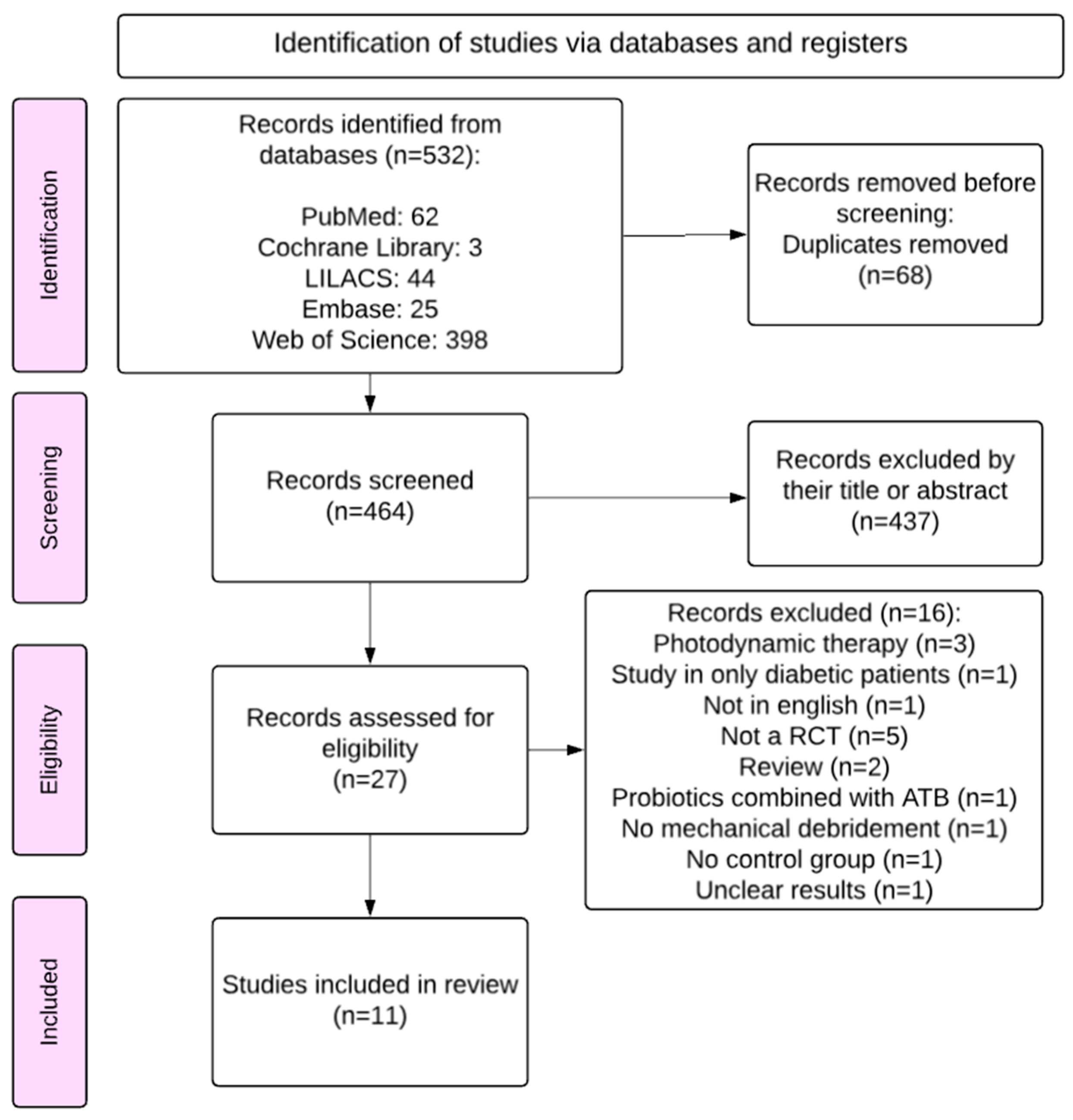
Figure 1. Flowchart diagram showing the search strategy (adapted from the PRISMA flow diagram).
Applsci 15 11422 g001
Figure 2. Forest plot graph of treatment effects on adjusted PPD reduction (mm) for follow-up at 6 months and median initial PPD. Overall, 10 studies contributed to the meta-analysis results [29,30,31,32,33,34,35,36,38,39]. MD = mechanical debridement; DOX = doxycycline; CHX = chlorhexidine; MIN = minocycline; MINm = minocycline microspheres; AMC = amoxicillin/clavulanic acid; CD = clindamycin; MTZ = metronidazole; AMX = amoxicillin; MH = minocycline hydrochloride; PPD = probing pocket depth; CI = confidence interval.
Applsci 15 11422 g002
Figure 3. Meta-regression of the effect of mean age on PPD reduction. β corresponds to the coefficient of the meta-regression, and p represents the p-value of the Wald test. PPD = probing pocket depth. The size of the coloured points indicates the associated level of precision, as shown by the black dots (1/SE); SE means standard error; SE = standard error.
Applsci 15 11422 g003
Figure 4. Meta-regression of the effect of the implant-to-patient ratio on PPD reduction. β corresponds to the coefficient of the meta-regression, and p represents the p-value of the Wald test. The size of the coloured points indicates the associated level of precision, as shown by the black dots (1/SE); SE means standard error; SE means standard error.
Applsci 15 11422 g004
Figure 5. Forest plot graph of treatment type on adjusted PPD reduction (mm) for follow-up at 6 months and median initial PPD. Overall, 10 studies contributed to the meta-analysis results [29,30,31,32,33,34,35,36,38,39]. Ab = Antibiotic; CI = confidence interval.
Applsci 15 11422 g005
Figure 6. Forest plot graph of adjusted treatment effects on BoP reduction (%) for follow-up at 6 months. CI is confidence interval. Overall, 6 studies contributed to the meta-analysis results [30,31,34,35,38,39]. MD = mechanical debridement; DOX = doxycycline; CHX = chlorhexidine; MIN = minocycline; MINm = minocycline microspheres; AMC = amoxicillin/clavulanic acid; CD = clindamycin; MTZ = metronidazole; AMX = amoxicillin; BoP = Bleeding on probing.
Applsci 15 11422 g006
Figure 7. Meta-regression of the effect of mean age on BoP reduction (%). β corresponds to the coefficient of the meta-regression, and p represents the p-value of the Wald test. The size of the coloured points indicates the associated level of precision, as shown by the black dots (1/SE); SE means standard error.
Applsci 15 11422 g007
Figure 8. Meta-regression of the effect of implant-to-patient ratio on the BoP reduction (%). β corresponds to the coefficient of the meta-regression, and p represents the p-value of the Wald test. The size of the coloured points indicates the associated level of precision, as shown by the black dots (1/SE); SE means standard error.
Applsci 15 11422 g008
Figure 9. Forest plot graph of adjusted treatment effects on BoP reduction (%) for follow-up at 6 months. CI is confidence interval. Ab = Antibiotic; CI = confidence interval.
Applsci 15 11422 g009
Figure 10. Traffic light plot of the risk of bias (Cochrane RoB 2.0) analysis of the selected studies [29,30,31,32,33,34,35,36,37,38,39].
Applsci 15 11422 g010
Figure 11. Summary plot of the risk of bias (Cochrane RoB 2.0) analysis.
Applsci 15 11422 g011
Table 1. Summary data from selected articles.
AuthorsTreatment
Patients (Implants)
LocationFollow-Up
(Months)
Type of
Treatment
Evaluated MeasuresConclusions
ControlTest
Büchter et al., 2004 [31]MD
14
MD + DOX
14
Germany4LocalPPD
reduction (mm), BoP reduction (%)
Adjunctive local antimicrobial treatment gave good results in the short term, with a reduction in pocket probing depth and probing attachment levels.
Renvert et al., 2006 [37]MD + CHX
14
MD + MINm
16
Sweden12LocalPPD
reduction (mm), BoP reduction (%)
The use of adjunctive minocycline microspheres resulted in improvements in both probing depths and bleeding scores.
Renvert et al., 2008 [38]MD + CHX
15 (38)
MD + MINm
17 (57)
Sweden12LocalPPD
reduction (mm), BoP reduction (%)
Adjunctive use of minocycline microspheres is beneficial in the treatment of peri-implant lesions, but the treatment may have to be repeated.
Passarielo et al., 2012 [35]MD + AMC or CD
64 (64)
MD + AMC or CD
+
serratiopeptidase/
64 (64)
Italy6SystemicPPD
reduction (mm), BoP reduction (%), DBL (mm)
Combined systemic administration of antibiotics and serratiopeptidase associated with MD significantly enhances success rates.
Shibli et al., 2019 [39]MD
20
MD + AMX + MTZ
20
Brazil12SystemicPPD
reduction (mm), BoP reduction (%)
The results of this study do not support the adjunctive use of systemic MTZ + AMX to the non-surgical treatment of peri-implantitis.
De Waal et al., 2021 [32]MD + CHX
29 (64)
MD + AMX + MTZ
28 (68)
Netherlands3SystemicPPD
reduction (mm), DBL (mm)
Adjunctive systemic antibiotic therapy of AMX and MTZ does not improve clinical and microbiological outcomes and should not be recommended.
Park et al., 2021 [34]MD
37 (37)
MD + MTZ + MIN
38 (38)
China3LocalPPD
reduction (mm), BoP reduction (%)
Local adjunctive use of minocycline, either with or without the addition of metronidazole, results in significantly higher treatment success rates compared to non-surgical treatment. The combination of metronidazole might enhance the PPD reduction in pockets ≥8 mm.
MD + MIN
39 (39)
Blanco et al., 2021 [30]MD
16 (34)
MD + MTZ
16 (28)
Spain12SystemicPPD
reduction (mm), BoP reduction (%)
The adjunctive use of systemic metronidazole as an adjunct to non-surgical treatment resulted in additional improvements in clinical, radiographic and microbiologic parameters.
Polymeri et al., 2022 [36]MD + CHX
19
MD + CHX + MTZ + AMX
18
Netherlands3SystemicPPD
reduction (mm), DBL (mm)
No clinical benefit from the adjunctive use of systemic AMX and MTZ in the non-surgical treatment of peri-implantitis, suggesting that the routine use of systemic antibiotics is not recommended.
Alhumaidan et al., 2022 [29]MD
(current smokers)
12 (12)
MD + MH
(current smokers)
12 (12)
United States of America6LocalPPD
reduction (mm)
A single subgingival delivery of minocycline hydrochloride (MH) is as effective as non-surgical MD alone.
MD
(non-smokers)
12 (12)
MD + MH
(non-smokers)
12 (12)
Liñares et al., 2024 [33]MD
11
MD + MTZ
10
Spain6SystemicPPD
reduction (mm), BoP reduction (%)
Systemic anti-inflammatory impact with reduction in peripheral levels of CRP, IL-6 and TNF-α was observed regardless of the use of systemic metronidazole.
MD = mechanical debridement; DOX = doxycycline; CHX = chlorhexidine; MIN = minocycline; MINm = minocycline microspheres; AMC = amoxicillin/clavulanic acid; CD = clindamycin; MTZ = metronidazole; AMX = amoxicillin; MH = minocycline hydrochloride; PPD = probing pocket depth; BoP = bleeding on probing; DBL = depth of bone lesion; IL-6 = interleukin-6; TNF-α = tumour necrosis factor-alpha; CRP = C-reactive protein.
Disclaimer/Publisher’s Note: The statements, opinions and data contained in all publications are solely those of the individual author(s) and contributor(s) and not of MDPI and/or the editor(s). MDPI and/or the editor(s) disclaim responsibility for any injury to people or property resulting from any ideas, methods, instructions or products referred to in the content.