Article

Effects of a Pneumatic External Abdominal Pressure Assist Belt on Trunk and Lower Limb Muscle Activity and Joint Kinematics During Lifting Tasks

1
Department of Physical Therapy, Faculty of Rehabilitation, Reiwa Health Sciences University, 2-1-12 Wajirogaoka, Higashi-ku, Fukuoka 811-0213, Japan
2
Department of Information, Artificial Intelligence and Data Science, Faculty of Engineering, Daiichi Institute of Technology, 1-10-2 Kokubuchuo, Kirishima City 899-4395, Japan
*
Author to whom correspondence should be addressed.
Appl. Sci. 2025, 15(20), 10897; https://doi.org/10.3390/app152010897
Submission received: 29 August 2025 / Revised: 3 October 2025 / Accepted: 8 October 2025 / Published: 10 October 2025
(This article belongs to the Special Issue Human Biomechanics and EMG Signal Processing)

Abstract

Manual lifting is a major risk factor for low back pain, with intra-abdominal pressure playing a key role in spinal stability. This study investigated the effects of a pneumatic external abdominal pressure assist belt on trunk and lower limb muscle activity and joint kinematics during lifting. Twenty-four healthy adults performed lifting tasks under four external abdominal pressure assist conditions. Trunk and lower limb muscle activities were measured using surface electromyography. Sagittal limb angles were assessed using 3D motion analysis. Peak and mean muscle activities (%MVIC) and joint angles were analyzed with repeated-measures ANOVA or Friedman tests. Peak muscle activity significantly decreased in the internal oblique, erector spinae, and biceps femoris (all p < 0.05), while increases were observed in the multifidus, rectus femoris, and vastus lateralis (all p < 0.05). Mean amplitude analysis showed reduced internal oblique activity (p < 0.001) and significant increases in rectus femoris and multifidus (p < 0.05). Hip and knee flexion angles were significantly greater under assisted conditions (p = 0.002), indicating a shift toward squat-type lifting. The pneumatic external belt redistributed peak loads from the back to the knee extensors, reduced internal oblique activity, and modestly increased multifidus activation. It also induced greater hip and knee flexion, suggesting a shift toward squat-type lifting. These effects were statistically significant but small, indicating limited practical relevance.

1. Introduction

Heavy occupational loads, frequent lifting tasks, and repetitive manual handling in daily life and the workplace are major risk factors for work-related low back pain [1]. Lifting movements, when performed improperly or repeatedly, impose excessive stress on the lumbar region and are recognized as one of the primary causes of low back pain [2]. Lumbar stability during such tasks is primarily supported by the trunk muscles, with intra-abdominal pressure (IAP) receiving particular attention as a physiological mechanism that stabilizes the spine and reduces lumbar load [3,4]. IAP is generated through the coordinated activity of the trunk muscles and has been shown to enhance spinal stability without increasing intervertebral compression forces [5]. Biomechanical model analysis reports that doubling IAP increases spinal stability by an average of 1.8 times [6]. Furthermore, the transversus abdominis (TrA) and multifidus (MF) muscles are known to activate in anticipatory feedforward control, preceding limb movements and postural changes, thereby contributing to the maintenance of lumbar stability [7,8].
Numerous studies have suggested that orthotic support for IAP and trunk stabilization can enhance lumbar stability. The application of external abdominal belts has been reported to increase IAP, reinforce lumbar stability, and potentially reduce shear forces [9]. X-ray studies have demonstrated that support belts reduce lumbar compression and shear deformation during lifting tasks [10]. Non-elastic belts have also been shown to increase IAP, thereby assisting trunk muscles during high-load postural changes and reducing spinal loads [11]. These belts and braces enhance trunk stability by physically compressing the abdomen; however, issues such as positional shifting, contact point pain during prolonged use, and discomfort from rubbing during movement have been reported [12,13]. Research on novel belt mechanisms designed for comfort and adaptability to movement remains limited. Therefore, this study hypothesizes that applying air pressure to the abdominal region, like the mechanism of a blood pressure cuff, could allow an air-pressure belt to provide effective external abdominal pressure (AP) assistance. Such a device would be expected to simultaneously support trunk stability and improve comfort. By inflating and deflating internal air chambers, the belt uniformly presses against the abdominal wall, thereby reducing localized pressure pain and the risk of circulatory impairment caused by constriction. Moreover, because air conforms dynamically to movement, the belt should maintain trunk support during actions such as bending and lifting, while minimizing discomfort and improving adaptability to trunk motion.
This study aimed to apply an external AP assist belt utilizing air pressure to lifting tasks and to clarify its effects on trunk and lower limb muscle activity as well as joint kinematics during movement. In doing so, the study provides novel electromyographic and kinematic insights that could not be obtained with conventional compression belts.

2. Materials and Methods

2.1. Participants

The sample size was determined a priori using G*Power (G*Power version 3.1.9.7, Heinrich-Heine-University Düsseldorf, Düsseldorf, Germany) [14]. A previous lifting study reported a moderate effect size (Cohen’s r = 0.324, corresponding to f approximately 0.34 when converted for ANOVA designs) for trunk muscle activation using a repeated-measures design [15]. Based on this and to ensure adequate power, a conservative medium effect size (f = 0.25) was assumed with α = 0.05, power (1 − β) = 0.80, correlation among repeated measures = 0.5, and nonsphericity correction ε = 1, yielding an estimated required sample size of 20–25 participants. Twenty-four healthy adults (12 males and 12 females) were recruited from the university community through flyers and word-of-mouth. Inclusion criteria were: age ≥ 18 years, no history of lower limb or trunk fracture or surgery within the past 6 months, and no known neurological disorders. All participants were healthy university students (mean age 21 years) with no current low back or lower-limb pain, and none reported previous chronic low back pain or lower-limb disorders. Participants’ physical characteristics included AP, dominant hand grip strength, dominant leg knee extension strength, lower leg length, and knee extension torque. These measurements were performed following standardized protocols; however, details of the measurement methods are omitted as they are not directly relevant to the primary objectives of this study, except for AP. All participants provided written informed consent prior to participation, and the study was approved by the institutional ethics committee. The study was approved by the institutional ethics committee of our university (Approval No. R5-002) and conducted in accordance with the Declaration of Helsinki. The purpose and procedures of the study were explained verbally, and written informed consent was obtained from all participants prior to enrollment.

2.2. Experimental Conditions

Participants performed object-lifting tasks using an abdominal-trunk training device (RECORE®, Sigmax, Tokyo, Japan) as external AP assistance. This device consists of a 15 cm wide cuff belt wrapped around the trunk, allowing pressurization and depressurization by air injection (Figure 1). This device can measure AP exerted against the constricting force of the cuff belt on the abdomen. The AP value (kPa) was calculated as the difference between the maximum AP exerted (Pmax) and the baseline pressure (Pbase) (AP = Pmax − Pbase) [16]. The reliability of this device has been demonstrated previously, showing intraclass correlation coefficient values of 0.975–0.983 and significant correlations with surface electromyography activity [17]. Each participant’s maximum AP was measured twice in a preliminary test, and the higher value was adopted to calculate the gender-specific averages (male: 15.1 kPa, female: 11.5 kPa). These average values were then used as the reference to set the belt pressure at 70% (Assist 70%, A70), 40% (Assist 40%, A40), or 0% (Assist 0%, A0) of the maximum, in addition to the non-assist condition (NA). A0 was intended to test the effect of wearing the belt alone, whereas A40 and A70 were intended to assess the effect of external AP assistance.
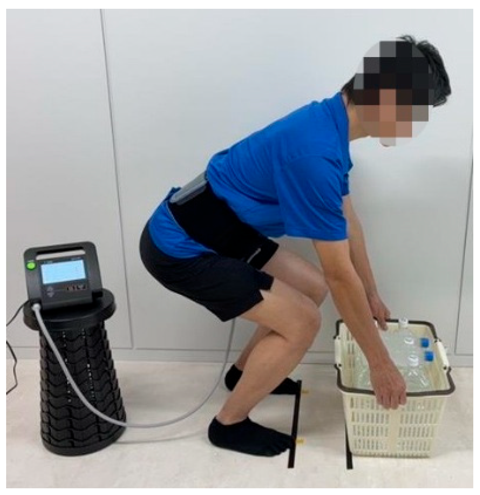
Under each condition, the participants used a basket placed on the floor, which was loaded with plastic bottles filled with water corresponding to 20% of their individual body weight. The load setting was based on previous studies and was chosen to simulate daily lifting tasks while avoiding excessive stress on the lower back [18]. The lifting style was instructed as a “freestyle,” in which participants lifted the load in the most natural manner without excessive knee flexion or extension (Figure 2). The stance width was set approximately to shoulder width, and the positions of both great toes were marked on the floor to ensure reproducibility. The distance between the basket and the toes was standardized to 15 cm. The lifting task started from a crouched position while holding the basket, and after a signal, participants were instructed to complete the lift within 5 s in synchronization with a metronome set at 60 bpm. For each condition, participants performed two practice trials, followed by five main trials with at least 30 s of rest between each. The order of conditions was randomized using a computer-generated randomization table.

2.3. Muscle Activity

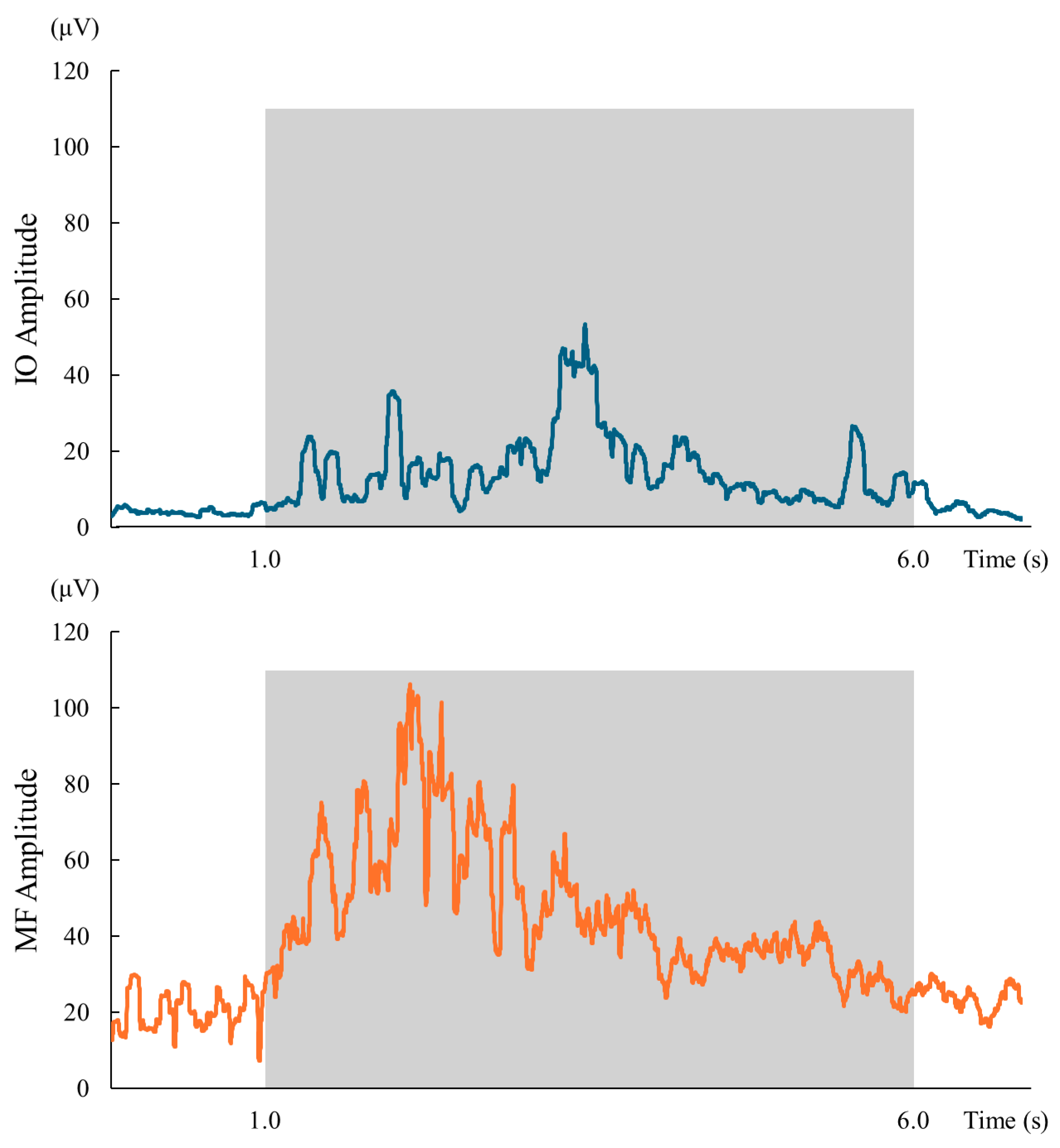
Muscle activity during the lifting tasks was recorded using wireless surface electromyography (sEMG) (Ultium EMG, NORAXON Inc., Scottsdale, AZ, USA) from the following muscles on the dominant side: external oblique (EO), rectus abdominis (RA), internal oblique (IO), erector spinae (ES), MF, rectus femoris (RF), vastus lateralis (VL), and biceps femoris (BF). Electrode placement followed the SENIAM guidelines, with the skin shaved and cleansed with alcohol as necessary, the skin impedance reduced before electrode attachment, and the electrodes placed along the muscle fiber direction with an inter-electrode distance of 2 cm [19,20]. For muscles not specifically described in the SENIAM guidelines, the following placements were adopted according to established recommendations [21,22]: EO, midway between the iliac crest and the lower ribs, aligned with the muscle fibers; RA, 3 cm lateral to the umbilicus at the level of the umbilicus; and IO, 2 cm medial and inferior to the anterior superior iliac spine, following the fiber direction. sEMG signals were sampled at 2000 Hz, band-pass filtered at 10–500 Hz, and full-wave rectified. The mean amplitude during the 5 s analysis period was calculated. In addition, the peak muscle activity during the task was extracted and compared across conditions. Both mean and peak sEMG values were normalized to the maximal voluntary isometric contraction (MVIC) and expressed as %MVIC for comparison across conditions. MVIC was obtained in standardized manual muscle testing positions according to Kendall [23]: EO/IO, resisted trunk rotation in supine with hips and knees flexed to 90°; RA, resisted trunk flexion in supine with knees flexed; ES/MF, prone trunk extension with stabilization of the lower limbs; RF/VL, seated knee extension at 90° knee flexion; and BF, prone knee flexion at 30° knee flexion. Signal acquisition and processing were performed using the myoMUSCLE software module (NORAXON Inc., Scottsdale, AZ, USA). Although the cuff belt occasionally overlapped with some abdominal electrodes, the inflatable chamber distributed pressure uniformly across the abdominal wall, thereby minimizing localized compression. All sEMG recordings were visually inspected, and any trials showing baseline instability or movement artifacts were excluded prior to analysis. Peak values were chosen to capture transient changes in muscle activation, while mean amplitudes were analyzed to provide information on sustained activation levels. To enhance methodological transparency, representative full-wave rectified sEMG signals of the IO and MF under the A40 condition are presented in Figure 3. The shaded area indicates the 1–6 s analysis segment used for calculating mean amplitude and extracting peak values.

2.4. Joint Angle

Sagittal plane angles of the hip, knee, and ankle joints were measured using a markerless three-dimensional motion analysis system (MyoMotion, Noraxon Inc., Scottsdale, AZ, USA). The maximum flexion angle during the lifting task was used for analysis. Prior to measurement, calibration was performed in a static standing position to establish the reference posture. The sampling frequency was set at 100 Hz. Inertial sensors were attached at seven anatomical locations: the pelvis, bilateral thighs, bilateral shanks, and bilateral feet. MyoMotion has been reported to show good agreement with an optical motion capture system for joint angle measurement, with intraclass correlation coefficients greater than 0.75 for sagittal plane angles and root mean square error within ±5 [24].

2.5. Specifications of the Abdominal Pressure Assist Device

The RECORE® device used as an AP assistance system consists of a 15 cm wide cuff belt with an air pressure chamber on the anterior side, designed to apply uniform compression across the entire abdomen. The center of the belt was positioned 2.5 cm below the umbilicus and fitted so as not to interfere with movement. Pressurization was performed at the end of expiration, and air was injected until the preset pressure level was reached. The end-expiratory phase was used as the reference point to ensure stable AP. In the A0 condition, the cuff belt was worn without pressure. All fitting and pressure-setting procedures were consistently performed by an experienced physical therapist to ensure reproducibility between measurements.

2.6. Statistical Analysis

We performed statistical analyses using Modified R Commander Ver 4.5.0 (https://www.r-project.org/). The normality of each variable was assessed using the Shapiro–Wilk test. When normality was confirmed, repeated measures analysis of variance (ANOVA) was performed, and post hoc comparisons were conducted using t-tests with Shaffer’s correction. When normality was not confirmed, the Friedman test was applied, and post hoc comparisons were performed using pairwise Wilcoxon signed-rank tests with Holm correction. Effect sizes were calculated as generalized eta-squared (G·η2) for repeated measures ANOVA and Kendall’s coefficient of concordance (Kendall’s W) for the Friedman test. The interpretation of effect sizes was based on established criteria [25]: for G·η2, small = 0.02, medium = 0.13, and large = 0.26; for Kendall’s W, small = 0.10, medium = 0.30, and large = 0.50. The significance level was set at p < 0.05 for all analyses.

3. Results

Participant characteristics are presented in Table 1. Comparisons of peak muscle activity and joint angles under different levels of external AP assistance are shown in Table 2. Since the muscle activity of the RA was below approximately 5%MVIC, it was excluded from subsequent analyses [26]. For peak muscle activity, significant reductions were observed in the IO (p < 0.001, W = 0.344, moderate), ES (p < 0.001, G·η2 = 0.028, small), and BF (p = 0.041, G·η2 = 0.005, very small) under AP-assisted conditions. In contrast, significant increases were found in the MF (p = 0.005, G·η2 = 0.007, very small), RF (p < 0.001, G·η2 = 0.032, small), and VL (p < 0.001, G·η2 = 0.020, small). Regarding joint kinematics, hip flexion angles were significantly greater with AP assistance (p = 0.002, G·η2 = 0.027, small), as were knee flexion angles (p = 0.002, G·η2 = 0.022, small), while ankle dorsiflexion did not differ significantly among conditions (ns, p = 0.231, G·η2 = 0.007, very small). The 3D motion analysis data, visualized as skeletal representations, confirmed that the peak hip and knee flexion occurred immediately after movement onset. The results of mean amplitude muscle activity are shown in Table 3. A significant decrease was found in IO activity (p < 0.001, W = 0.285, moderate), while MF (p = 0.040, G·η2 = 0.010, very small) and RF (p = 0.011, W = 0.160, small) showed significant increases under AP-assisted conditions. However, no significant differences were observed in the EO, ES, VL, or BF (all ns, very small effect sizes).

4. Discussion

In this study, we examined the effects of an external AP assistance belt using peak muscle activity, mean amplitude over 5 s, and sagittal lower-limb joint angles during the lifting task. Regarding peak muscle activity, ES activity was higher in NA compared with A0, A40, and A70, whereas RF and VL activities were higher in the assisted conditions (A40, A70, and partly A0) than in NA, indicating a redistribution of peak load to the knee extensors. BF activity was also higher in NA compared with A40 and A70. These findings suggest that external AP assistance may modify the distribution of instantaneous peak loads during lifting, reducing relative dependence on trunk extensor peaks while allocating load to the knee extensors. In terms of mean amplitude, IO activity was lower in A40 compared with NA and A0, whereas MF showed only a small main effect, and the significance did not survive post hoc comparisons. No significant differences were observed in the mean amplitude of ES and VL. In contrast, RF activity showed a higher mean amplitude in A70 compared with NA. Furthermore, for joint kinematics in the initial phase of lifting, A70, A40, and A0 demonstrated greater hip and knee flexion angles than NA, suggesting an early shift toward a squat-type lifting strategy.
The suppression of IO activity by AP assistance is consistent with previous reports showing that belt use during squat lifting increases IAP and slightly decreases IO activity [27]. This suggests that external AP assistance may serve as a supplementary role for the IO. Furthermore, in the present study, peak muscle activity revealed a significant reduction in ES and a significant increase in deep muscle MF. These findings are consistent with previous reports indicating that lumbar orthosis use alters the activation pattern of paraspinal muscles [28] and that MF functions synergistically with the TrA to enhance lumbar stability [29]. Thus, external AP assistance may support IAP while promoting recruitment of deep stabilizing muscles. However, although the IO showed a moderate effect size, most of the other observed differences were associated with small or very small effect sizes. This indicates that, while statistical differences were detected, the practical impact of AP assistance on muscle activity during lifting was limited. In other words, the necessity of assistance may be heightened at moments of peak demand but less relevant during other phases of the movement. In this study, the lifting load was set at 20% of body weight. Previous research has reported that increasing load is significantly associated with greater activation of prime movers [30]. The higher the load conditions become, the more pronounced the differences between multiple muscles may become. Moreover, the absence of significant differences between A70 and A40 may reflect inhibition of muscle contraction caused by excessive compression. Indeed, previous studies have shown that external compression exceeding approximately 16 kPa on the upper arm decreases both MVC and EMG activity [31]. Excessive tension in the A70 may have suppressed trunk muscle activity. Taken together, these results suggest that approximately 40% AP assistance may be optimal for lifting tasks involving trunk flexion. In the future, pneumatic belts equipped with mechanisms that can automatically adjust assistance in response to task demands may be effective. Such systems would be particularly beneficial at moments when peak muscle activity occurs.
This study suggests that external AP assistance may induce a lifting pattern closer to the squat type from the very beginning of the movement. In manual lifting, squat-type lifting with greater hip and knee flexion is recommended to reduce lumbar load, and avoidance of forward trunk inclination is also emphasized in instruction as a preventive strategy against low back pain [32,33]. Trunk-forward lifting has been reported to bring the thorax closer to the horizontal position, increase lumbar lordosis, and approach knee extension, thereby imposing excessive stress on the lumbar tissues [34]. In contrast, squat-type lifting positions the thorax more upright, suppresses lumbar curvature, and increases knee flexion, reducing the risk of excessive strain or injury to the lumbar tissues [35,36]. However, squat-type lifting also increases peak knee joint power, and with repeated movements, quadriceps fatigue may lead to a shift toward trunk-forward postures [37,38]. In the present study, hip and knee flexion significantly increased under assisted conditions, shifting the pattern toward squat-type lifting. Correspondingly, peak muscle activity increased in the RF and VL, while it decreased in the BF. These changes, however, were generally small in magnitude and less evident in the mean amplitude of muscle activity throughout the task. These findings suggest that external AP assistance supports lumbar stability and suppresses trunk inclination at the onset of lifting, thereby reducing lumbar load while promoting a squat-type lifting strategy. At the same time, no substantial increase in quadriceps activity was observed across the whole movement, indicating that the risk of excessive lower-limb fatigue associated with deep squat-type lifting is likely minimal. Thus, external AP assistance may represent an effective means of enhancing lumbar stability, reducing spinal load, and supporting safe and efficient lifting performance.
The clinical significance of this study lies in the possibility that external AP assistance may reduce lumbar stress by supporting IO activity and enhancing MF activation, thereby promoting a lower-limb-dominant movement strategy. In patients with low back pain, delayed anticipatory contraction of deep trunk muscles has been observed during the initial phase of movement, which is suggested to be associated with lumbar instability [39]. Moreover, changes in motor control strategies themselves have been reported in individuals with low back pain [40], indicating that assistance for trunk stability may also contribute to motor relearning. The pneumatic design of the external AP assistance belt allows it to follow postures and movements with minimal risk of displacement or discomfort at contact areas. The present study demonstrated that the device reduced peak ES activity while suggesting a possible facilitation of activation in the MF region. However, given the limitations of sEMG in detecting deep muscles and the small effect sizes observed, these findings should be interpreted with caution, and their practical relevance remains limited. Nevertheless, external AP assistance may contribute to trunk control by redistributing muscle activity. Thus, its role should currently be regarded as providing modest biomechanical support rather than a clinically significant intervention.
This study has several limitations. First, the participants were healthy adults, and the generalizability of the findings to clinical and occupational populations, such as individuals with low back pain or manual workers, is limited. Second, the lifting load was set at 20% of body weight, and thus, changes in muscle activity and joint kinematics under higher load conditions could not be evaluated. Third, muscle activity was assessed using sEMG, which does not allow direct evaluation of deep trunk muscles such as the TrA. Similarly, MF activity was measured with surface electrodes, which are susceptible to crosstalk from adjacent muscles. Therefore, the present results should be interpreted cautiously as indirect indicators of MF activation rather than direct measurement. Finally, the observed effect sizes were mostly small, except for a moderate effect size in the IO, indicating that the actual magnitude of changes was limited. Therefore, the belt’s contribution to lifting mechanics should be considered minor, and any potential cumulative effects over repetitive tasks remain speculative. Future studies should investigate its applicability under higher load conditions, during extended tasks, and in populations with a history of low back pain or in occupational settings.

5. Conclusions

The application of a pneumatic external AP assistance belt was shown to support IO activity and may facilitate activation in the MF region during lifting tasks. Peak muscle activity results suggested a corrective effect on instantaneous load peaks in the ES and knee extensor muscles. Furthermore, increased hip and knee flexion angles during the initial phase of lifting indicated a shift toward a safer squat-type lifting strategy. Overall, although a moderate effect size was observed for the IO, most effects were small, and their practical impact should be considered limited. Pneumatic AP assistance may offer modest biomechanical support for redistributing load and promoting safer lifting strategies, but its clinical relevance remains to be established through further studies.

Author Contributions

Conceptualization, Y.N.; Data curation, Y.N. and Y.T.; Formal analysis, Y.N. and Y.T.; Investigation, Y.N. and Y.T.; Methodology, Y.N. and Y.T.; Project administration, Y.N.; Writing—original draft, Y.N. All authors have read and agreed to the published version of the manuscript.

Funding

This work was supported by JSPS KAKENHI (Grant-in-Aid for Scientific Research [C]), Grant Number 24K14419.

Institutional Review Board Statement

The study was conducted in accordance with the Declaration of Helsinki and approved by the Ethics Committee of Daiichi Institute of Technology (protocol code R5-002, 19 October 2023).

Informed Consent Statement

Informed consent was obtained from all subjects involved in the study. Written informed consent has been obtained from the participants to publish this paper.

Data Availability Statement

The data set collected and analyzed in this study is available from the corresponding author upon request for reasonable reasons.

Acknowledgments

The authors would like to thank all participants for their involvement in this study. We also extend our gratitude to Naoki Nakayama for his assistance.

Conflicts of Interest

The authors declare no conflicts of interest.

References

  1. Heneweer, H.; Staes, F.; Aufdemkampe, G.; van Rijn, M.; Vanhees, L. Physical activity and low back pain: A systematic review of recent literature. Eur. Spine J. 2011, 20, 826–845. [Google Scholar] [CrossRef] [PubMed]
  2. Marras, W.S.; Lavender, S.A.; Leurgans, S.E.; Rajulu, S.L.; Allread, W.G.; Fathallah, F.A.; Ferguson, S.A. The role of dynamic three-dimensional trunk motion in occupationally-related low back disorders. The effects of workplace factors, trunk position, and trunk motion characteristics on risk of injury. Spine 1993, 18, 617–628. [Google Scholar] [CrossRef] [PubMed]
  3. Cholewicki, J.; Juluru, K.; McGill, S.M. Intra-abdominal pressure mechanism for stabilizing the lumbar spine. J. Biomech. 1999, 32, 13–17. [Google Scholar] [CrossRef] [PubMed]
  4. Hodges, P.W.; Eriksson, A.E.; Shirley, D.; Gandevia, S.C. Intra-abdominal pressure increases stiffness of the lumbar spine. J. Biomech. 2005, 38, 1873–1880. [Google Scholar] [CrossRef]
  5. Bojairami, I.E.; Driscoll, M. Coordination Between Trunk Muscles, Thoracolumbar Fascia, and Intra-Abdominal Pressure Toward Static Spine Stability. Spine 2022, 47, E423–E431. [Google Scholar] [CrossRef]
  6. Stokes, I.A.; Gardner-Morse, M.G.; Henry, S.M. Abdominal muscle activation increases lumbar spinal stability: Analysis of contributions of different muscle groups. Clin. Biomech. 2011, 26, 797–803. [Google Scholar] [CrossRef]
  7. Hodges, P.W.; Richardson, C.A. Feedforward contraction of transversus abdominis is not influenced by the direction of arm movement. Exp. Brain Res. 1997, 114, 362–370. [Google Scholar] [CrossRef]
  8. Yamane, M.; Aoki, M.; Sasaki, Y.; Hayashi, T. Feedforward coactivation of trunk muscles during rapid shoulder movements. JSES Int. 2022, 6, 660–668. [Google Scholar] [CrossRef]
  9. Cholewicki, J.; Juluru, K.; Radebold, A.; Panjabi, M.M.; McGill, S.M. Lumbar spine stability can be augmented with an abdominal belt and/or increased intra-abdominal pressure. Eur. Spine J. 1999, 8, 388–395. [Google Scholar] [CrossRef]
  10. DeBeliso, M.; Sevene, T.; Adams, K.J.; Harris, C.; Climstein, M.; Berning, J.M. The Effects of a Back-Belt on Lumbar Disc Deformation During Stoop Type Lifting. Sci. Eng. Investig. 2013, 16, 21613-01. [Google Scholar]
  11. Bai, J.; Hua, A.; Weng, D.; Wang, N.; Wang, J. Effects of non-extensible lumbar belts on static and dynamic postural stability. BMC Musculoskelet. Disord. 2023, 24, 362. [Google Scholar] [CrossRef]
  12. Van Duijvenbode, I.C.; Jellema, P.; van Poppel, M.N.; van Tulder, M.W. Lumbar supports for prevention and treatment of low back pain. Cochrane Database Syst. Rev. 2008, 2008, CD001823. [Google Scholar] [CrossRef]
  13. Chen, Z.; Zhang, M.; Zhang, X.; Liu, C.; Wang, H. Understanding the User Experience of Lumbar-Support-Assistive Devices in Lower-Back Pain Patients: Design Recommendations. Appl. Sci. 2024, 14, 7495. [Google Scholar] [CrossRef]
  14. Faul, F.; Erdfelder, E.; Lang, A.G.; Buchner, A. G*Power 3: A flexible statistical power analysis program for the social, behavioral, and biomedical sciences. Behav. Res. Methods 2007, 39, 175–191. [Google Scholar] [CrossRef]
  15. Walter, T.; Stutzig, N.; Siebert, T. Active exoskeleton reduces erector spinae muscle activity during lifting. Front. Bioeng. Biotechnol. 2023, 11, 1143926. [Google Scholar] [CrossRef] [PubMed]
  16. Kato, S.; Murakami, H.; Demura, S.; Yoshioka, K.; Shinmura, K.; Yokogawa, N.; Igarashi, T.; Yonezawa, N.; Shimizu, T.; Tsuchiya, H. Abdominal trunk muscle weakness and its association with chronic low back pain and risk of falling in older women. BMC Musculoskelet. Disord. 2019, 20, 273. [Google Scholar] [CrossRef] [PubMed]
  17. Nakai, Y.; Usumoto, Y.; Takeshita, Y. The Effects of Regional Muscle Strength and Mass on Standing Long Jump Performance. Muscles 2024, 3, 60–70. [Google Scholar] [CrossRef] [PubMed]
  18. Alemi, M.M.; Geissinger, J.; Simon, A.A.; Chang, S.E.; Asbeck, A.T. A passive exoskeleton reduces peak and mean EMG during symmetric and asymmetric lifting. J. Electromyogr. Kinesiol. 2019, 47, 25–34. [Google Scholar] [CrossRef]
  19. De Luca, C.J. The Use of Surface Electromyography in Biomechanics. J. Appl. Biomech. 1997, 13, 135–163. [Google Scholar] [CrossRef]
  20. Hermens, H.J.; Freriks, B.; Disselhorst-Klug, C.; Rau, G. Development of recommendations for SEMG sensors and sensor placement procedures. J. Electromyogr. Kinesiol. 2000, 10, 361–374. [Google Scholar] [CrossRef]
  21. Cram, J.R.; Kasman, G.S.; Holtz, J. Introduction to Surface Electromyography; Aspen Publishers: New York, NY, USA, 1998. [Google Scholar]
  22. Criswell, E.; Cram, J.R. Cram’s Introduction to Surface Electromyography, 2nd ed.; Jones and Bartlett: Boston, MA, USA, 2011; p. 21. 412p. [Google Scholar]
  23. Kendall, F.P. Muscles: Testing and Function with Posture and Pain, 5th ed.; Lippincott Williams & Wilkins: Ambler, PA, USA, 2005. [Google Scholar]
  24. Prisco, G.; Pirozzi, M.A.; Santone, A.; Esposito, F.; Cesarelli, M.; Amato, F.; Donisi, L. Validity of Wearable Inertial Sensors for Gait Analysis: A Systematic Review. Diagnostics 2024, 15, 36. [Google Scholar] [CrossRef]
  25. Cohen, J. Statistical Power Analysis for the Behavioral Sciences; Lawrence Erlbaum Associates: New York, NY, USA, 1988. [Google Scholar]
  26. Tateuchi, H.; Taniguchi, M.; Mori, N.; Ichihashi, N. Balance of hip and trunk muscle activity is associated with increased anterior pelvic tilt during prone hip extension. J. Electromyogr. Kinesiol. 2012, 22, 391–397. [Google Scholar] [CrossRef]
  27. McGill, S.M.; Norman, R.W.; Sharratt, M.T. The effect of an abdominal belt on trunk muscle activity and intra-abdominal pressure during squat lifts. Ergonomics 1990, 33, 147–160. [Google Scholar] [CrossRef]
  28. Cholewicki, J.; Reeves, N.P.; Everding, V.Q.; Morrisette, D.C. Lumbosacral orthoses reduce trunk muscle activity in a postural control task. J. Biomech. 2007, 40, 1731–1736. [Google Scholar] [CrossRef] [PubMed]
  29. MacDonald, D.A.; Moseley, G.L.; Hodges, P.W. The lumbar multifidus: Does the evidence support clinical beliefs? Man. Ther. 2006, 11, 254–263. [Google Scholar] [CrossRef] [PubMed]
  30. Van den Tillaar, R.; Andersen, V.; Saeterbakken, A.H. Comparison of muscle activation and kinematics during free-weight back squats with different loads. PLoS ONE 2019, 14, e0217044. [Google Scholar] [CrossRef] [PubMed]
  31. Yasuda, T.; Brechue, W.F.; Fujita, T.; Sato, Y.; Abe, T. Muscle activation during low-intensity muscle contractions with varying levels of external limb compression. J. Sports Sci. Med. 2008, 7, 467–474. [Google Scholar] [PubMed]
  32. Caneiro, J.P.; O’Sullivan, P.; Smith, A.; Ovrebekk, I.R.; Tozer, L.; Williams, M.; Teng, M.L.W.; Lipp, O.V. Physiotherapists implicitly evaluate bending and lifting with a round back as dangerous. Musculoskelet. Sci. Pract. 2019, 39, 107–114. [Google Scholar] [CrossRef]
  33. Nolan, D.; O’Sullivan, K.; Stephenson, J.; O’Sullivan, P.; Lucock, M. How do manual handling advisors and physiotherapists construct their back beliefs, and do safe lifting posture beliefs influence them? Musculoskelet. Sci. Pract. 2019, 39, 101–106. [Google Scholar] [CrossRef]
  34. McGill, S.M. The biomechanics of low back injury: Implications on current practice in industry and the clinic. J. Biomech. 1997, 30, 465–475. [Google Scholar] [CrossRef]
  35. McGill, S.M.; Hughson, R.L.; Parks, K. Changes in lumbar lordosis modify the role of the extensor muscles. Clin. Biomech. 2000, 15, 777–780. [Google Scholar] [CrossRef]
  36. Gagnon, D.; Plamondon, A.; Larivière, C. A comparison of lumbar spine and muscle loading between male and female workers during box transfers. J. Biomech. 2018, 81, 76–85. [Google Scholar] [CrossRef]
  37. Trafimow, J.H.; Schipplein, O.D.; Novak, G.J.; Andersson, G.B. The effects of quadriceps fatigue on the technique of lifting. Spine 1993, 18, 364–367. [Google Scholar] [CrossRef]
  38. Saraceni, N.; Campbell, A.; Kent, P.; Ng, L.; Straker, L.; O’Sullivan, P. Exploring lumbar and lower limb kinematics and kinetics for evidence that lifting technique is associated with LBP. PLoS ONE 2021, 16, e0254241. [Google Scholar] [CrossRef]
  39. Cholewicki, J.; Panjabi, M.M.; Khachatryan, A. Stabilizing function of trunk flexor-extensor muscles around a neutral spine posture. Spine 1997, 22, 2207–2212. [Google Scholar] [CrossRef]
  40. Van Dieën, J.H.; Reeves, N.P.; Kawchuk, G.; van Dillen, L.R.; Hodges, P.W. Analysis of Motor Control in Patients With Low Back Pain: A Key to Personalized Care? J. Orthop. Sports Phys. Ther. 2019, 49, 380–388. [Google Scholar] [CrossRef]
Figure 1. Participants wear the RECORE® device as an external abdominal pressure assist belt.
Applsci 15 10897 g001
Figure 2. Freestyle lifting task with external abdominal pressure assistance.
Applsci 15 10897 g002
Figure 3. Representative full-wave rectified sEMG signals of the internal oblique (IO, upper) and multifidus (MF, lower) during lifting under the A40 condition. The shaded area indicates the 1–6 s analysis window used for calculating mean amplitude and peak muscle activity.
Applsci 15 10897 g003
Table 1. Characteristics and measurements of participants (n = 24, mean ± Standard Deviation).
VariableTotal (n = 24)Male (n = 12)Female (n = 12)
Age (years)20.8 ± 2.220.7 ± 0.621.0 ± 3.0
Height (cm)166.1 ± 6.1170.2 ± 4.2162.1 ± 4.8
Body weight (kg)57.2 ± 7.760.6 ± 7.853.7 ± 5.9
Abdominal pressure (kPa)13.3 ± 4.515.1 ± 3.511.5 ± 4.6
Handgrip strength (kg)33.5 ± 6.837.9 ± 5.929.1 ± 4.3
Knee extensor strength (N)460.1 ± 101.8496.2 ± 94.8424.0 ± 95.6
Lower leg length (cm)37.3 ± 2.337.5 ± 1.637.2 ± 2.8
Knee extension torque (Nm)172.3 ± 40.3186.2 ± 36.7158.4 ± 38.9
Table 2. Peak sEMG amplitudes (%MVIC) and joint angles (°) during lifting under four external abdominal pressure assist conditions.
MuscleA70A40A0NApEffect SizePost-Hoc Comparisons
EO17.1 ± 7.218.4 ± 10.219.2 ± 10.918.1 ± 8.90.2080.007 (very small)
RA4.2 (2.6–4.9)4.2 (2.6–4.9)4.2 (2.6–4.9)4.2 (2.6–4.9)0.6370.024 (very small)
IO22.7 (16.2–36.1)22.7 (16.2–36.1)22.7 (16.2–36.1)22.7 (16.2–36.1)<0.0010.344 (moderate)A40 < A0 ***, A40 < NA ***
ES43.7 ± 15.943.0 ± 14.546.2 ± 16.649.7 ± 16.6<0.0010.028 (small)NA > A0 **, NA > A40 **, NA > A70 ***; A0 > A40 *, A0 > A70 *
MF44.5 ± 21.145.0 ± 20.742.4 ± 18.141.0 ± 18.80.0050.007 (very small)A40 > A0 *, A40 > NA **
RF9.1 ± 5.38.4 ± 4.38.1 ± 4.56.8 ± 3.90.0010.032 (small)A0 > NA **, A40 > NA ***, A70 > NA ***
VL28.5 ± 12.429.6 ± 14.526.9 ± 13.024.8 ± 11.50.0010.020 (small)A40 > NA **, A70 > NA **, A40 > A0 *
BF27.5 ± 12.327.4 ± 12.827.7 ± 12.229.5 ± 13.10.0410.005 (very small)NA > A40 *, NA > A70 **
Degree
Hip flexion113.1 ± 12.6113.0 ± 12.3111.8 ± 12.5108.1 ± 12.50.0020.027 (small)A70 > NA *, A40 > NA *, A0 > NA *
Knee flexion85.2 ± 16.384.3 ± 15.482.8 ± 16.279.1 ± 15.60.0020.022 (small)A70 > NA **, A40 > NA **, A0 > NA *
Ankle dorsiflexion22.9 ± 8.923.0 ± 8.822.7 ± 8.721.3 ± 8.60.2310.007 (very small)
Values are mean ± Standard Deviation or median (Interquartile Range). Abbreviations: EO, external oblique; RA, rectus abdominis; IO, internal oblique; ES, erector spinae; MF, multifidus; RF, rectus femoris; VL, vastus lateralis; BF, biceps femoris. Conditions: A70 = Assist 70%; A40 = Assist 40%; A0 = Assist 0%; NA = non-Assist. Significance levels: * p < 0.05, ** p < 0.01, *** p < 0.001.
Table 3. Average sEMG amplitudes (%MVIC) during lifting under four external abdominal pressure assist conditions.
MuscleA70A40A0NApEffect SizePost-Hoc Comparisons
EO12.7 ± 5.512.8 ± 6.613.8 ± 8.013.2 ± 6.70.3090.004 (very small)
RA3.4 (2.7–4.4)2.8 (1.9–4.0)2.7 (1.7–4.2)2.7 (1.8–3.6)0.0010.322 (moderate)A70 > NA **, A70 > A40 **, A70 > n ***
IO15.8 (12.5–22.6)16.3 (11.9–28.9)17.9 (12.8–32.1)20.2 (15.3–28.4)0.0010.285 (moderate)A40 < NA ***, A0 < NA**, A70 < NA *
ES42.8 ± 16.743.1 ± 15.244.5 ± 16.442.4 ± 16.30.2410.003 (very small)
MF39.3 ± 19.139.9 ± 20.237.8 ± 18.535.1 ± 17.50.0400.010 (very small)(no pairwise survived correction)
RF4.9 (3.5–6.8)4.9 (4.0–6.3)4.8 (3.2–7.8)4.6 (3.2–5.7)0.0110.160 (small)A70 > NA *, A40 > NA *
VL18.6 ± 9.318.2 ± 8.519.2 ± 9.417.5 ± 8.30.2540.005 (very small)
BF29.2 (19.6–43.2)31.4 (23.1–38.9)31.9 (19.0–40.7)32.9 (20.1–40.4)0.7590.016 (very small)
Values are mean ± Standard Deviation or median (Interquartile Range). Abbreviations: EO, external oblique; RA, rectus abdominis; IO, internal oblique; ES, erector spinae; MF, multifidus; RF, rectus femoris; VL, vastus lateralis; BF, biceps femoris. Conditions: A70 = Assist 70%; A40 = Assist 40%; A0 = Assist 0%; NA = non-Assist. Significance levels: * p < 0.05, ** p < 0.01, *** p < 0.001.
Disclaimer/Publisher’s Note: The statements, opinions and data contained in all publications are solely those of the individual author(s) and contributor(s) and not of MDPI and/or the editor(s). MDPI and/or the editor(s) disclaim responsibility for any injury to people or property resulting from any ideas, methods, instructions or products referred to in the content.